relevance of fucose-rich extracellular polysaccharides produced … · cally planted in modified...
TRANSCRIPT

Relevance of Fucose-Rich Extracellular Polysaccharides Produced byRhizobium sullae Strains Nodulating Hedysarum coronariumL. Legumes
Razika Gharzouli,a Marie-Anne Carpéné,b François Couderc,b Ammar Benguedouar,a Véréna Poinsotb
Department of Animal Biology and Department of Biochemistry and Microbiology, Faculty for Biology, University of Mentouri, Constantine, Algeriaa; Laboratoire desIMRCP, UMR5623 Université Paul Sabatier, Toulouse, Franceb
Specific and complex interactions between soil bacteria, known as rhizobia, and their leguminous host plants result in the devel-opment of root nodules. This process implies a complex dialogue between the partners. Rhizobia synthesize different classes ofpolysaccharides: exopolysaccharides (EPS), Kdo-rich capsular polysaccharides, lipopolysaccharides, and cyclic �-(1,2)-glucans.These polymers are actors of a successful symbiosis with legumes. We focus here on studying the EPS produced by Rhizobiumsullae bacteria that nodulate Hedysarum coronarium L., largely distributed in Algeria. We describe the influence of the carbonsource on the production and on the composition of EPS produced by R. sullae A6 and RHF strains. High-molecular-weight EPSpreserve the bacteria from desiccation. The structural characterization of the EPS produced by R. sullae strains has been per-formed through sugar analysis by gas chromatography-mass spectrometry. The low-molecular-weight EPS of one strain (RHF)has been totally elucidated using nuclear magnetic resonance and quantitative time-of-flight tandem mass spectrometry analy-ses. An unusual fucose-rich EPS has been characterized. The presence of this deoxy sugar seems to be related to nodulationcapacity.
The Hedysarum genus is composed of a great number of forageleguminous plant species, distributed throughout Europe, Af-
rica, Asia, and North America (1). Hedysarum coronarium L.(sulla, French honey-suckle, Spanish sainfoin, and Spanish espar-cet) is a member of the Leguminosae family originating and grow-ing in the Mediterranean basin. It has been established as a foragecrop in several countries (2). These plants play a significant role inmaintaining the productivity in agriculture thanks to its ability tofix nitrogen and to grow satisfactorily over a wide range of soilconditions. Moreover, it tolerates drought and coastal conditions(3). Leguminous plants are able to enter in symbiosis with bacteriabelonging to the rhizobia family. The latter reduce atmosphericnitrogen into ammonium, which is directly usable by the plants(4, 5). Rhizobium sullae is the specific bacterial partner of H. coro-narium L (1, 2). In the symbiotic association of H. coronarium L.with R. sullae there is a clear specificity between the plant ecotypeand the corresponding Rhizobium strain (6). The establishing of asymbiotic nitrogen fixation is important for the plant especiallywhen the plant soil is starved in nutrients. This process occurs inthe roots of the leguminous plants within specialized organs callednodules. The formation of nodules is engaged in the earliest stepsof the process (7).
The Rhizobium-legume symbiosis requires specific chemicalsignaling between the symbiotic partners (4, 8). In addition to theflavonoids and Nod factors which initiate the symbiotic program,exopolysaccharides (EPS), lipopolysaccharides (LPS), Kdo-richcapsular polysaccharides (KPS), and cyclic �-(1,2)-glucans playessential roles in the formation of the infection thread and in nod-ule development (9). Nevertheless, the precise functions of thesecomplex carbohydrates are still being investigated.
Rhizobial EPS are species-specific heteropolysaccharides com-posed of common sugars that can be substituted with noncarbo-hydrate residues, such as pyruvyl, succinyl, or acetyl groups (10,11). The relationship between nodulation ability and rhizobial
EPS production, in culture, has been reported (12, 13). Produc-tion of EPS is influenced by several growth factors, such as carbonand nitrogen sources. These factors also influence the structureand rheological properties of EPS (14, 15).
The rheological properties and peripheral localization of EPSsuggest that they protect bacteria against environmental stress andprovide a first contact between bacteria and the plant surface (9,16), contributing to the intrinsic roles of bacterial EPS (17).
However, the production and structure of EPS are essential ina successful nodulation of host plants for the formation of inde-terminate nodules (18, 19). H. coronarium L. is a host plant thatforms indeterminate nodules. This type of nodule is mostly cylin-drical or exhibits an elongated shape with a persistent apical mer-istem producing a gradient of developmental zones (20). For ex-ample, the Sinorhizobium meliloti EPS have to be succinylated tobe active on Medicago truncatula.
Orgambide et al. (21) began with studying the glycoconjugatesand lipids of R. hedysari IS123. Navarini et al. (22) highlighted theinfluence of carbon source in EPS production in R. hedysariHCNT isolated from H. coronarium L. The objective of the presentstudy was to determine the composition and size of the EPS pro-duced by R. sullae grown in the presence of different carbonsource, to study their symbiotic activities on the plant H. coro-narium L.
Finally, we attempted to establish a relationship between the
Received 21 September 2012 Accepted 19 November 2012
Published ahead of print 26 November 2012
Address correspondence to Véréna Poinsot, [email protected], or RazikaGharzouli, [email protected].
Copyright © 2013, American Society for Microbiology. All Rights Reserved.
doi:10.1128/AEM.02903-12
1764 aem.asm.org Applied and Environmental Microbiology p. 1764–1776 March 2013 Volume 79 Number 6
on May 13, 2020 by guest
http://aem.asm
.org/D
ownloaded from

EPS and the ability of strains to nodulate the host plant and toresist drought conditions. Considering the singular compositionof these EPS, one structure has been totally elucidated (R. sullaestrain RHF grown on mannitol).
Structural determination occurred through nuclear magneticresonance (NMR), electrospray ionization-mass spectrometry(ESI-MS), and gas chromatography-mass spectrometry (GC-MS)analyses. It revealed the production of fucose-rich (ca. 30%) EPSthat have not been previously reported for the Rhizobiaceae fam-ily. The relationship between the high fucose content of low-mo-lecular-weight (LMW) EPS and the nodulation efficiency ofcorresponding strains is discussed here. The production level ofhigh-molecular-weight (HMW) EPS is the key component of thebacterial resistance to drought conditions.
MATERIALS AND METHODSMedia and bacterial growth. The bacterial strains used in the presentstudy belong to two varieties of R. sullae: R. sullae A6 and R. sullae RHF.The strains were collected in Constantine (Algeria) and in Pisa (Italy),respectively. Rhizobia from sulla are related to Rhizobium etli, Rhizobiumleguminosarum, and Sinorhizobium meliloti (23). Strains were grown onthe yeast extract mannitol agar (YMA) solid medium (mannitol, 10 g/li-ter; KH2PO4, 0.5 g/liter; MgSO4·7H2O, 0.2 g/liter; NaCl, 0.1 g/liter; yeastextract, 0.5 g/liter; agar, 15 g/liter) at 28°C for 24 h. The other carbonsources are introduced at 10 g/liter instead of mannitol.
Production, extraction, and purification of EPS. The production ofEPS by strain was tested on solid medium YMA in which mannitol wasreplaced by other sugars (sucrose, glucose, and sorbitol) to estimate theinfluence of carbon source on EPS production. Petri dishes were incu-bated at 28°C for 5 days. Mucoid colonies were scraped with a sterilizedspatula and then resuspended in sterile KCl (0.85%) (24).
Bacterial cells were separated from their EPS by centrifugation (12,800 �g for 30 min at 4°C). The supernatant containing the EPS was vacuumfiltered through 45-�m-pore-size membranes to eliminate cells and largecellular fragments. EPS secreted in the supernatant are not retained by the0.45-�m-pore-size filter. The HMW fraction of bacterial EPS was precip-itated with 3 volumes of 95% cold ethanol. The supernatant was used toprecipitate, by the addition of 7 volumes of ethanol, the LMW EPS frac-tion. The fraction was collected by centrifugation. The fractions of EPSwere then lyophilized.
Colorimetric assay. EPS were quantified by the Dreywood anthrone-sulfuric acid method (25) and the Blumenkrantz et al. phenol-sulfuricacid method (26) to estimate the amount of neutral and acid sugar con-tained in each sample.
Relative viscosity of EPS. Solutions of EPS were prepared in distilledwater, at a final concentration 0.1 g/liter, and they were left under stirring(20 rpm) overnight at 25°C. Solutions of EPS were analyzed with a vis-cometer (Scott Geräte AVS 310) at 25°C with a 5520/II column (diameter,2 mm; length, 4 cm). The results are based on the determination of therelative viscosity calculated as follows:
�rel ��
�0�
t�
t0�0
where �rel is the relative viscosity, � is the viscosity of the EPS solution, �0
is the viscosity of solvent (water), t and t0 represent the time for the ball tocross the capillary for the EPS solution and the solvent, respectively, and �and �0 are their densities.
Recovery of strains after desiccation. After growth on an agar me-dium supplemented at 1% with different sugars (mannitol, sucrose, glu-cose, or sorbitol), mucoid colonies were scraped. Then, 0.1-g portions ofthe colonies were left to dry on a sterile cellulose filter membrane (65 �m,47 mm; Millipore, France) at 35°C in a laminar flow hood until the bac-teria were completely dried. The time required for drying varied from astrain to another. The filter was cut, and the part that contained all of the
dried cells was inoculated into 150 ml of yeast extract mannitol base(YMB; YMA without agar). The flasks were strongly shaken and thenincubated at 28°C. The growth rate of bacteria was estimated by measur-ing the optical density at 600 nm (OD600) for different incubation periods.
Glycosyl composition analyses. The EPS of each strain were hydro-lyzed in 2 M trifluoroacetic acid (TFA) at 110°C for 2 h. The TFA wasremoved by repeated evaporation with isopropanol. The acidic monosac-charides were methylated by diazomethane in ether. The derivation wasperformed by silylation, with 20 �l of pyridine, 100 �l of TMCS-HMDSmixture (trimethylchlorosilane and 1,1,3,3,3-hexamethyldisilazane) at70°C (27). Trimethylsilyl glycoside derivatives of various monosaccharidestandards were also prepared and analyzed by GC-MS for comparison tothe sample peaks on chromatograms. The response factors of the differentsugar types were determined after derivatization of the standards. Thesespecific response coefficients were then applied to quantify each mono-saccharide family.
GC-MS analyses of the EPS were performed with 6890N GC interfacedwith 5973 MSD by using a HP5MS capillary column (25-m length,0.25-mm external diameter and 0.25-�m internal diameter). The ovenprogram was 70 to 300°C over 57 min. The scanning mass range was fromm/z 50 atomic mass units (amu) to m/z 650 amu. The on-column injectedvolume was 0.1 �l.
Size exclusion chromatography (SEC). The LMW EPS were dissolvedin Milli-Q water at a final concentration of 5 mg/ml, and they were filteredthrough a 0.22-�m-pore-size membrane. The EPS fractions were sepa-rated on a size exclusion column (TSK-Gel G2000SWxl; 7.8-mm diame-ter, 30-cm height), which is able to separate globular proteins from 5 to150 kDa. Detection was carried out by an evaporative detector by diffu-sion of light (ELS; Waters 2420). The 20 �l of the 5-mg/ml sample injectedwas eluted by ammonium acetate (20 mM) with a 0.8-ml/min fixed flow,and the time of analysis was 30 min. Detector was set at 20 lb/in2 and100°C.
DOC-PAGE. Desoxycholic acid-polyacrylamide gel electrophoresis(DOC-PAGE) analyses were performed on a 15% acrylamide-bisacrylamideresolving gel, with a 4% acrylamide-bisacrylamide stacking gel. The 30%acrylamide-bisacrylamide (19:1) solution was obtained from Sigma-Al-drich (Steinheim, Germany). For the migration, a generator from Phar-macia Biotech (Freiburg, Germany) was used at 20 mA for 1.5 h. Aprestained protein ladder from Fermentas (Villebon-sur-Yvette, France)was used to estimate the size range of the EPS.
Structural analysis of EPS using NMR. Nuclear magnetic resonance(1H NMR) spectroscopy was performed on a Brücker AMX500 spectrom-eter (Wissenbourg, France). The samples were dissolved in D2O (100%).NMR spectra were recorded at 303 K at 500 MHz (1H) and 125.75 MHz(13C) using a cryoprobe. Chemical shifts are indicated in ppm, and thenumber of accumulated scans was 512. Correlation-observed spectros-copy (COSY) allowed to assign chemical shifts of proton of each residue.The two-dimensional (2D) heteronuclear one-bond proton-carbon cor-relation experiment was registered in the 1H-detection mode via single-quantum coherence (HSQC), and 32 scans were accumulated. Analysis inheteronuclear multiple bond correlation mode (HMBC) was used to de-termine the sequence of residues of the repeating unit.
ESI/time-of-flight MS. Fractions of R. sullae LMW EPS were analyzedin the negative-ion mode on a QqTof system (Ultima; Waters) (the EPSused were from R. sullae RHF cultivated in the presence of mannitol). Thesettings were as follows: probe, 3 kV; cone, 50 V; Rf lens, 25 V; and colli-sion, 10 V (up to 25 V for MS/MS). Samples were dissolved at a concen-tration of 2 mg ml�1 in water-methanol (1:1) with 0.1% ammonia. Theinjection was carried out with a flow of 7 �l min�1.
Plant assays. Seeds of H. coronarium L. were surface sterilized andgerminated as described by Vincent (28). Germinated seeds were asepti-cally planted in modified Leonard’s bottle-jar containing the sand-ver-miculite mixture watered with a Fahraeus nutrition solution (29). Tenplants were then inoculated with 2 ml of exponentially grown rhizobia(OD600 of �1), cultivated in the presence of different carbon sources
Fucose-Rich EPS Produced by R. sullae
March 2013 Volume 79 Number 6 aem.asm.org 1765
on May 13, 2020 by guest
http://aem.asm
.org/D
ownloaded from

(mannitol, glucose, sucrose, or sorbitol) at 28°C. The pots were placed inthe culture room at room temperature (the temperature varied from 16 at21°C during the day and from 8 to 17°C during the night) for 10 weeks.The chamber was equipped with a system that ensured a discontinuouslight illumination (16-h day and 8-h night).
RESULTSProduction of EPS. We studied the EPS produced by two strainsof R. sullae—A6 and RHF—that are specific endosymbionts of H.coronarium L. These strains are pure, as well as phenotypically andgenotypically characterized, and their taxonomic position was de-termined. ARDRA, LMM RNA profiles, and DNA-DNA hybrid-ization showed genotypic similarity of 100% between the twostrains (1). The study of phenotypic characters carried out byStruffi et al. (6) reveals minor differences between the two RHFand A6 strains. The degree of NaCl tolerance is of 1% for strain A6and 0.5% for strain RHF, and the nitrite reductase activity is lowfor strain RHF and absent in strain A6. Strain RHF is more resis-tant to phages than strain A6, in particular for f123c, FHC, F100C,f19a, f44a, and f44c1. There was also a difference in the plasmidprofiles of the two strains.
The production of EPS was affected by the carbon source (Ta-ble 1), even if the strain growth remained similar. However, thegrowth on glucose was quite variable. The A6 strain recorded itsmaximum EPS production in the presence of sorbitol (11.1 mg/g).In contrast, the RHF strain produced a maximum EPS in the pres-ence of glucose (20.5 mg/g). The proportions of HMW and LMWEPS produced also varied, depending on the carbon source. Forexample, the LMW/HMW ratio for the same strain (A6) variedfrom 7/3 (sorbitol) to 3/7 (mannitol). EPS production has beenevaluated by weighing the alcoholic precipitate, after lyophiliza-tion. The measured weights have been confirmed through color-imetric assays (29).
Relative viscosity of EPS. The HMW EPS, due to their abilityto make intra- and intermolecular hydrogen bonds and their lowsolubilities, tend to be more viscous than the LMW ones. Theviscosities measured here vary in accordance with the EPS size,which is dependent on the carbon source (Table 2). The viscositymeasured at 0.1 g/liter for the two classes of EPS ranged for bothstrains from 0.96 to 1.11 for the HMW EPS and from 0.89 to 0.98for the LMW EPS.
Recovery of strains after desiccation. The growth kineticsmeasured after the reculture of desiccated strains (Fig. 1) indi-
cated that the RHF strain is on average more resistant to drought.Actually, the latency time was reduced, and the stationary phasereached in �20 h. For the two strains, the totality of the desiccatedspot has been inoculated into the medium. The presented curvesresult from three replicates. After drying, sucrose enabled the twostrains to resume growth with a higher speed, among the foursugars tested. High concentrations of EPS in the culture mediumresulted in increased turbidity; however, this was observable onlyafter 40 h of growth, corresponding to 18 h of stationary phase.Therefore, the growth kinetics measured at 600 nm over the first24 h were not affected by this artifact.
Glycosyl composition analyses. The identification of sugars iscarried out using GC-MS by comparing the retention times andEI mass spectra to those of standards (our own and those of theNIST database). To ensure the retention time attribution, thechromatograms obtained for the polysaccharide samples are su-perimposed with those of the standards. The EPS of R. sullae aremade up mainly of glucose (Glc), galactose (Gal), and fucose(Fuc). Their proportions depend on the carbon source used forthe strain growth (Table 3). We observed that the compositions ofHMW and LMW EPS differ, indicating that there are two differentpopulations of polysaccharide and not simply different polymer-ization degrees for only one. Galacturonic acid (GalA) propor-
TABLE 1 Effect of carbon source on the production of R. sullae EPS
Strain and carbon source
Mean production of EPS(mg/g of bacteria) SD
LMW EPS HMW EPS Total EPS
R. sullae A6Mannitol 2.7 0.9 4.8 1.0 7.5 2.0Sucrose 3.9 1.6 6.7 2.8 10.6 0.6Glucose 6.7 5.0 4.0 2.9 10.7 1.1Sorbitol 7.5 3.5 11.0
R. sullae RHFMannitol 6.6 2.0 4.8 2.0 11.6 1.3Sucrose 7.0 0.9 6.7 2.4 13.7 1.0Glucose 15.0 6.0 5.6 0.8 20.6 2.6Sorbitol 1.8 0.6 1.6 1.4 3.4 1.0
TABLE 2 Viscosities of R. sullae EPS depending on the carbon source
Strain and carbon source
Relative viscosity
HMW EPS LMW EPS
R. sullae A6Mannitol 1.110 0.910Sucrose 1.030 0.935Glucose 1.070 0.952Sorbitol 1.100 0.890
R. sullae RHFMannitol 1.090 0.937Sucrose 1.030 0.984Glucose 1.110 0.942Sorbitol 0.961 0.915
FIG 1 Growth kinetics of recovery after desiccation of the two R. sullae strainsA6 (white) and RHF (black) and their four sources of carbon (mannitol, tri-angles; sucrose, circles; glucose, squares; sorbitol, diamonds).
Gharzouli et al.
1766 aem.asm.org Applied and Environmental Microbiology
on May 13, 2020 by guest
http://aem.asm
.org/D
ownloaded from

tions determined by GC-MS are less precisely presented in Table 3because the silylation of this uronic acid leads to underestimatedand variable results.
Even if the purification is performed through a two-step pre-cipitation, the produced fractions appear to be clean. Actually,GC-MS analyses of derivatized sugars before and after hydrolysisrevealed neither amino acids, nor ribose (Table 3), nor lipids (Fig.2). Moreover, ESI-MS analyses performed in the two negative andpositive ionization modes exhibited only saccharides in the m/z700 to 2,500 domain. In the higher mass range, the combinedproblems of bad ionization potential and less efficient solubiliza-tion of big carbohydrates could explain the absence in our spectraof the heavier saccharides. To confirm this hypothesis, the effi-ciency of the precipitation has been controlled through SEC andDOC-PAGE analysis. These studies indicated that in the HMWdomain, no EPS exceeded 150 kDa and that in the LMW domainthe sizes ranged effectively from 1 to 60 kDa.
NMR spectrometric experiments. NMR analysis allowed us todetermine the configuration of carbohydrates and to assess mostof the linkages in the polysaccharide. The 1H and 13C chemicalshifts for each unit of the polysaccharide are given in Table 4.
The 500-MHz spectrum of R. sullae LMW EPS (RHF cultivatedin the presence of mannitol) showed the presence of a hexasaccha-ride repeating unit containing Fuc, Glc, Gal, and GalA in a 2:2:1:1ratio. One can observe a slight difference between this composi-tion and the one presented in Table 3. This is due to the fact that inNMR we observed a single sample (the GC-MS analysis of thissample fit perfectly [Fig. 2]), whereas Table 3 presents the meancomposition values over three cultures. It also revealed the pres-ence of the O-acetyl, pyruvyl, and succinyl groups. In order to
achieve the best resolution, the NMR study was performed on a10-volume ethanol-precipitated fraction. This contains oligosac-charides, resulting in fewer molecular interactions and thereforebeing more soluble. Among the different EPS tested, those ofstrain RHF cultivated in the presence of mannitol exhibitedsharper NMR peaks (Fig. 3). These analyses allowed us to deter-mine the types of linkages between each saccharide residue. Thisinterpretation is complicated by the fact that at least five com-pounds are present as a mixture in the NMR probe. Systemati-cally, cross peaks could be observed in the COSY experimentsbetween the anomeric positions (H-1) and the neighbor H-2, fol-lowed by H-3. These 1H signals could then be correlated with the13C signals by studying the HSQC map (Fig. 4). For the attributionof the 1H and 13C chemical shifts of positions 4 to 6, interpretationof the HMBC correlations was necessary (Fig. 5). The determinedchemical shifts were compared to those in the literature (24, 30–36). To establish the EPS sequence, HMBC correlations were ob-served between the 13C of acetylated fucose (� 103.7 ppm) and1H-3 Fuc (� 4.23 ppm). � and � configurations were determinedthrough the COSY 1H/1H coupling constants (3JH1,H2). The pres-ence of three substitutions on the sugar backbone could be ob-served first on the 1H spectrum in the aliphatic CH2/CH3 region(succinate [Succ] CH2, 2.08 ppm; pyruvate [Pyr] CH3, 1.37 ppm;acetate [Ac] CH3, 1.86 ppm), as well as the CH3 of the desoxysugars (CH3; 1.05, 1.19, and 1.25 ppm). A strong HMBC signal wasfound for the pyruvyl between 13C (� 100.1 ppm) and 1H (� 3.82,3.88, and 3.70 ppm) corresponding respectively to the two H-6 and toH-3 or H-4 of the hexose. Attribution of the chemical shifts corre-sponding to the quaternary carbons (C and COOH) of the substitu-ents was performed using the HMBC data.
TABLE 3 Monosaccharide composition of R. sullae EPS as determined by GC-MS
EPS and strain Carbon source
Monosaccharide composition (%)a
Glc Gal Fuc Man GalAb Rha Rib
LMW EPSR. sullae A6 Mannitol 32 1 30.5 2.5 37 4 �1 �1 �1 ND
Sucrose 16.5 3.5 32.5 7.5 51 11 �1 �1 ND NDSorbitol 60.5 10.5 22.5 5.5 �1 8.5 8.5 �1 8.5 0.5 ND
R. sullae RHF Mannitol 38 5 30.5 3.5 30.5 2.5 �1 �1 �1 NDSucrose 33.5 16.5 29 4 37.5 12.5 �1 �1 �1 NDSorbitol 37 12 34 3 27.5 9.5 �1 �1 ND ND
HMW EPSR. sullae A6 Mannitol 45 5 32.5 7.5 10 10 ND �10 �1 ND
Sucrose 30 10 36.5 3.5 17 16 3.5 3.5 �10 �1 NDSorbitol 41.5 8.5 25 8 ND 8.5 8.5 �10 17 0 ND
R. sullae RHF Mannitol 28.5 14.5 28 0 14 14 28.5 28.5 ND ND NDSucrose 46 21 28.5 3.5 12.5 12.5 �1 �10 ND NDSorbitol 33 0 33 0 33 0 ND �1 ND ND
Total EPSR. sullae A6 Mannitol 42.7 6.5 33.4 1.0 19.9 13.5 �1 �10 �1 ND
Sucrose 24.7 6.7 34.9 2.0 30.4 17.0 2.3 1.5 �10 �1 NDSorbitol 54.4 9.5 23.3 1.2 �1 8.5 0 �10 11.2 4.2 ND
R. sullae RHF Mannitol 34.1 4.7 29.5 1.2 23.6 8.2 12.3 12.3 �1 �1 NDSucrose 39.5 6.3 28.8 0.3 25.5 12.5 �1 �10 �1 NDSorbitol 35.5 2.0 33.6 0.5 29.5 2.7 �1 �1 ND ND
a Glc, glucose; Gal, galactose; Fuc, fucose; Man, mannose; GalA, galacturonic acid; Rha, rhamnose; Rib, ribose; ND, not detected. When glucose was used as a carbon source, thecomposition was quite variable. Values are expressed as means standard deviations where applicable.b GalA does not demonstrate a good response factor when derivatized with TMS.
Fucose-Rich EPS Produced by R. sullae
March 2013 Volume 79 Number 6 aem.asm.org 1767
on May 13, 2020 by guest
http://aem.asm
.org/D
ownloaded from

Only one signal can be observed for the pyruvylated hexose,indicating that pyruvylation is complete, as confirmed by the MSdata (see Materials and Methods). Since acetylation is partial onthe position 4 of the fucose, two types of signals can be clearlyobserved for the same sugar. Similarly, a partial succinylationcould be observed.
ESI-QqTof MS/MS analyses. We studied using MS the samesamples as for the NMR experiments. The results presented
here are for the RHF strain cultivated on mannitol. The m/z1,041.31 ion corresponds to the molecular ion [M-H]�. Thismass corresponds to a hexasaccharide carrying a pyruvylgroup. This polymer can be substituted by the O-acetyl (m/z1,083.4 ion) and/or succinyl (m/z 1141.4 ion) (Fig. 6A). Thefragmentation of ion [M-H]� at m/z 1,041.41 was analyzed byMS/MS experiments. The spectra showed B and Y fragmenta-tion patterns.
FIG 2 Chromatograms from GC-MS analyses of the monosaccharide composition of standard silylated monosaccharides and of RHF/mannitol hydrolyzed andsilylated HMW and LMW EPS.
Gharzouli et al.
1768 aem.asm.org Applied and Environmental Microbiology
on May 13, 2020 by guest
http://aem.asm
.org/D
ownloaded from

The atomic mass unit difference of 162.1 represents one hexose(Glc or Gal). Such a neutral loss corresponds to sugar eliminationfrom one extremity of the saccharide. Since the charge state isconserved, the elimination occurs on the opposite side. The mo-lecular mass of the oligosaccharide corresponds to a hexamerformed by two deoxyhexoses (dHex), two hexoses (Hex), a uronicacid (HexA), and a pyruvyl-hexose (PyrHex), as confirmed by thefragmentation pattern (Fig. 1B). Actually, in the low-mass do-main, m/z 145, 161, 176, and 249 ions could be observed corre-sponding, respectively, to dHex, Hex, HexA, and PyrHex losses.The fragment m/z 733 corresponds to the hexasaccharide havinglost a hexose-deoxyhexose disaccharide (Fig. 6B).
The fragment at m/z 587 is consistent with the HexA-Hex-HexPyr entity. Since the fragment m/z 337 is systematically pres-ent (as well as the corresponding neutral loss of 338 amu) andsince the neutral loss of HexA could not be observed, it is reason-able to hypothesize that the hexasaccharide is under a completelylinear form.
In conclusion, this experiment performed with the LMW EPSof R. sullae RHF grown on mannitol showed the presence of a
hexasaccharide always carrying a pyruvate and bearing partially anadditional succinyl and/or acetyl group. This saccharide containsthree Hex, two dHex, and one HexA (Fig. 6C). The sequence de-duced from the MS/MS analyses, as previously shown using NMR(see 3.5), indicated that the polar components (Succ and Pyr) arelocated on one side of the molecule and that more hydrophobicparts (Fuc and Ac) are located on the other side.
Nodulation tests. H. coronarium L. (sulla) plants which wereinoculated with R. sullae A6 and R. sullae RHF were tall and green.The production of EPS (see Materials and Methods) showed thatthe carbon source used by the bacteria can influence the produc-tion rate of EPS. To investigate whether the quantities or quality ofEPS produced have an effect on the number and size of nodulesformed, the germinated seeds were inoculated with 2 ml of bacte-ria in the presence of the different carbon sources (mannitol, su-crose, glucose, or sorbitol according to the method of Navarini etal. [22]). The maximal number of nodules was recorded for theplants inoculated by R. sullae A6 cultivated in the presence ofsucrose (10 nodules) (Fig. 7), and about 6 nodules were obtainedin the presence of glucose. In the presence of sorbitol, the minimalnumber of nodules was obtained with R. sullae RHF (1 nodule).
DISCUSSIONEPS production depends on the carbon source. Bacteria belong-ing to the Rhizobium genus produce important amounts of surfacepolysaccharides. We have shown that R. sullae specific partner ofH. coronarium L., can produce different EPS present in differentmass distribution. The HMW and LMW fractions differ in saccha-ride composition. The amount of EPS is significantly influencedby the carbon source used to cultivate the strain.
Navarini et al. (22) observed high secretion of EPS when R.hedysarum HCNT1 strain are grown on glucose and sucrose. Forthe Rhizobium strain D110 isolated from Dalbergia lanceolaria,mannitol was the most suitable promoter, followed by glucose andgalactose (13).
EPS synthesis is a genetically controlled process. Genes direct-ing and regulating the biosynthesis of EPS (the exo/exs or pss gene)form large clusters located either on the genome or on megaplas-mids (11). The biosynthesis of EPS in Rhizobium is influenced byvarious environmental parameters. The existence of a very com-
FIG 3 1H NMR spectra of RHF/mannitol LMW EPS (well resolved) andA6/sucrose LMW EPS (less resolved).
TABLE 4 Chemical shift data for glycosyl residues of LMW EPS produced by R. sullae RHF grown on mannitol as determined by two-dimensionalNMR in D2O at 500 MHz with the use of a cryoprobea
Designation Glycosyl residue
Assignments (�, ppm)b
H-1/C-1 H-2/C-2 H-3/C-3 H-4/C-4 H-5/C-5 H-6/C-6
A ¡4�Gal1¡ 5.41/99.1 3.84/68.1 4.13/66.5 3.92/71.8* 3.72/62.5 3.82; 3.77/60.6¡4�Gal(6 Succ)1¡ 3.92; 3.79/64.8
B ¡3�Fuc1¡ 5.33/96.7 4.85/70.8 4.12/71.0* 4.04/68.5* 4.38/67.2 1.22/14.5C ¡3�Fuc1¡ 5.30/98.1 5.02/69.9 4.23/76.5 4.20/69.1 4.43/66.7 1.07/14.7D �(4.6 Pyr)Glc1¡ 5.09/92.1 3.54/72.1 3.71/79.8 3.39/75.9 3.64/69.5 3.91; 3.82/67.7E ¡4�GalA1¡ 4.88/100.8 3.86/80.0 3.90/78.4 3.73/76.5* 3.68/76.8 175.0F ¡3�Fuc(4 Ac)1¡ 4.57/103.7 3.31/72.9 3.69/76.2 3.78/71.5 4.06/71.0 1.27/19.7G ¡3�Glc 4.53/96.0 3.25/74.1 3.51/82.6 3.42/67.7 3.67/61.5* 3.70; 3.64/60.6Succ 2.08/20.0 173.0 183.0Pyr 1.37/24.8 100.1 176.0Ac 1.86/22.4 181.1a Succ, succinate; Pyr, pyruvate; Ac, acetate. All other abbreviations are as defined in Table 3, footnote a.b *, assignments that could be exchanged with one other. Assignments indicated in italics were determined by COSY 1H/1H and HSQC 13C/1H.
Fucose-Rich EPS Produced by R. sullae
March 2013 Volume 79 Number 6 aem.asm.org 1769
on May 13, 2020 by guest
http://aem.asm
.org/D
ownloaded from

plex transcriptional and posttranslational process has been re-ported (37).
Various factors have been shown to regulate both the quantityand structure of EPS; these factors include osmolarity, nitrogenstarvation, phosphate limitation, and growth conditions (38, 39).In S. meliloti, EPS I and EPS II exhibit parallel biosynthesis. Amultitude of genes regulate their biosynthesis. The transforma-tion of glucose-6-phosphate uridyl-diphosphate (UDP-Glc) andUDP-galactose (UDP-Gal) is at the base of the biosynthesis of allpolysaccharides. Some groups of genes, such as exo and wgg genes,are expressed to build EPS, while others regulate positively ornegatively the biosynthesis of EPS I and EPS II, respectively.HMW EPS I are obtained through ExoQ. The succinoglycans ofLMW EPS I are obtained either directly or by degradation of theHMW EPS I via ExoK and ExoH proteins. This pathway is re-pressed in conditions of nitrogen deficiency and high osmolarity.The LMW EPS I can also be exported by the proteins ExoP andExoT (9, 11, 39, 40). exoR is expressed to inhibit the biosynthesis ofsuccinoglycans (EPS I) (41), whereas exoS stimulates this biosyn-thesis under high osmolarity (for example, in a highly saline envi-ronment). WggR (or MucS) proteins, as well as ExpR, increase thesynthesis of galactoglucans (EPS II). The MucR protein is a keyregulator that promotes the biosynthesis of the EPS I at the ex-pense of the EPS II biosynthesis. The latter can be produced inanother way when media are phosphate starved (40). In S. meliloti,the SyrM protein is also involved in regulating the relative ratio ofLMW and HMW proteins from EPS I (42). We observed on R.sullae important effects on EPS production/g of bacteria and onthe HMW/LMW proportion, as well as on the carbohydrate com-position, according to the carbon source.
EPS composition depends on the carbon source. R. sullae EPSare mainly constituted by neutral sugars (D-glucose, D-galactose,L-fucose, D-mannose, and sometime L-rhamnose). EPS glycosyllinkages are mainly 1,3- or 1,4- in � or � associations (11). It isimportant to note that the presence of fucose is rare for bacterialEPS. Pawlicki-Jullian et al. (43) found that fucose presents 40% of
the EPS of Enterobacter ludwigii, 35% of the EPS of Klebsiella ter-rigena, 30% of the EPS in Raoultella terrigena, and between 10%and 35% of the EPS in Raoultella terrigena. The presence of fucoseand, more specifically, its important ratio reported here indicatethat we are in the presence of a novel type of rhizobial EPS. For theRHF strain, the carbon source influences the composition of EPSand more precisely their fucose content, varying by 30% where theA6 strain fucose content varies by 100%.
Fraysse et al. (9) reported in their review that the EPS mono-saccharide composition may depend on the carbon source usedfor growth. For example, when grown on arabinose, gluconate, ormannitol, Bradyrhizobium japonicum 2143 exhibits the EPS com-position described earlier (Man-Glc-Gal-GalA [1:2:1:1]) but,when cultured on malate, the EPS became extremely enriched inGal (novel EPS) (12). Modification of EPS has been describedfrequently as a response to different environmental and physio-logical living conditions of bacteria (44).
In the present study, the presence of EPS enriched in deoxysugars is highlighted. Its composition and production depend onthe carbon sources of the bacteria. The apparition of mannose-rich HMW EPS occurs when the strain is grown on mannitol, andthe synthesis of rhamnose instead of fucose happens when thestrain is cultivated on sorbitol. This could indicate that direct im-portation processes exist in the strains studied here and that thesesugars are not strictly used as carbon sources for energy.
Viscosity of EPS and their role in resistance to drought con-ditions. The influence of the size and composition of EPS onviscosity is already well documented and depends on several fac-tors, such as the composition of the culture medium, the type ofstrain, and the growth conditions (pH, concentration of oxy-gen, and amount of agitation) (45). The purpose here was to tryto find out whether the composition or the size (both influencethe viscosity) is the primary parameter determining the symbi-otic activity.
The intrinsic viscosity is a function of the molecular weight andthe hydrodynamic radius of the polymer. Therefore, the viscosity
FIG 4 13C/1H HSQC NMR correlation map of RHF/mannitol LMW EPS. The annotated correlation peaks correspond to the attributions found in Table 4.
Gharzouli et al.
1770 aem.asm.org Applied and Environmental Microbiology
on May 13, 2020 by guest
http://aem.asm
.org/D
ownloaded from

is higher for a higher molecular weight and for a more rigid chain(14), when the composition is constant.
Skorupska et al. (11) showed that EPS I and II produced by S.meliloti are secreted in two major fractions, reflecting differentdegrees of subunit polymerization: the HMW fraction, consistingof hundreds to thousands of repeating units (polymers of 106 to107 Da), and the LMW fraction, which represents monomers,dimers, and trimers in the case of EPS I and oligomers (24, 30) inthe case of EPS II. The average molecular mass of the LMW EPSwas determined here by SEC. The sizes ranged from 1,000 to60,000 Da. The HMW EPS are �150,000 Da but are mostly �106
Da (based on DOC-PAGE analyses).The relative viscosity of the R. sullae LMW EPS was never �1 at
a concentration of 0.1 g/liter (Table 2) but was near 1 when thedistribution in molecular mass increased to 60 kDa. Two excep-tions should be noted: R. sullae A6 grown on sorbitol had a viscos-ity of 0.9 even if its size is close to 60 kDa, and R. sullae RHF grownon glucose exceeded 0.9, with a size of �10 kDa. These findingscan be explained by important composition changes.
H. coronarium (sulla) is one legume known for its broad toler-ance to various environmental stresses and its ability to thrivewithout signs of chlorosis in desert soils. Sulla is well exploited as
a forage crop due to its pronounced drought resistance and itsstrong tolerance to alkaline soils (46). This uncommon behavior,associated with the singular EPS composition of its specific hostobserved here, allowed us to hypothesize that the two could becorrelated. Therefore, we studied the resistance of the strains todrought and their ability to resume proliferation after drying. AsEPS can protect the bacteria against abiotic stress (17), we wereinterested in establishing a relationship between their growth re-sumption after desiccation, viscosity, composition, and produc-tion.
The presence of deoxyhexoses in EPS could suggest that theviscosity of the EPS may be more important (47, 48). The LMWEPS fraction follows in general this rule.
In contrast, the viscosity of HMW EPS does not appear to beinfluenced by the deoxyhexose content (Fig. 2). So, the viscosityappears to depend primarily on the size of the EPS, and then onthe deoxyhexose ratio.
The adaptation of bacteria to environmental conditions (des-iccation, temperature, pressure, and salinity) has been reported(49). We observed that when a strain produced a great amount ofHMW EPS, its resistance to drought was important (Fig. 8). Ac-tually, the better survival explains the better restart of strain RHF
FIG 5 13C/1H HMBC NMR correlation map of RHF/mannitol LMW EPS. The annotated correlation peaks correspond to the attributions found inTable 4.
Fucose-Rich EPS Produced by R. sullae
March 2013 Volume 79 Number 6 aem.asm.org 1771
on May 13, 2020 by guest
http://aem.asm
.org/D
ownloaded from

1772 aem.asm.org Applied and Environmental Microbiology
on May 13, 2020 by guest
http://aem.asm
.org/D
ownloaded from

in liquid media. Even if less noticeable, the same relationship forthe A6 strain could be demonstrated. The HMW EPS are viscousand allow a better restart. When HMW EPS are produced by thebacteria, their envelope presents better gelification triggering andenhanced resistance to drought.
Structure of R. sullae LMW EPS. A large diversity in EPSchemical structure can be found within the Rhizobium genus, con-sidering the sugar composition and linkage in the single subunit,the repeating unit size, and the degree of polymerization, as well asthe noncarbohydrate decoration (11, 50).
EPS are species-specific complex carbohydrate polymers.However, the structures of EPS produced by Rhizobium and bymany other Proteobacteria consist of large heteropolymers formedby repeating the subunit structure (40).
In the present study, our attention has focused on fucose-richEPS and especially on LMW EPS-producing strains exhibiting im-portant biological activity. ESI-MS analysis of the LMW EPS of R.sullae RHF grown in mannitol (similar to R. sullae A6 grown onsucrose) showed that the subunit of this polymer consists of ahexamer (Fig. 6). The results of the QqTof MS/MS showed thatthe repeating unit of LMW EPS is composed of two deoxy sugars,three hexoses, and an uronic acid. This hexamer is pyruvylatedand can carry a supplementary O-succinyl or O-acetyl group.
1H NMR has been performed on the LMW EPS of strain A6grown in sucrose (with the strongest effect on nodulation), onstrain RHF grown with mannitol exhibiting the same HMW/LMW ratio as strain A6 in sucrose, and on strains A6 and RHFgrown in sorbitol for their opposite production yields. Their re-spective 1H NMR spectra indicated a high similarity in carbohy-drate signals but a slight variation in the succinylation ratio. Sincepolysaccharides are difficult to solubilize at the molecular level, weinvestigated using NMR the most resolved NMR signal corre-sponding to the LMW EPS from strain RHF grown on mannitol(Fig. 3).
The combination of NMR and QqTof MS data confirms thestructure of repeating unit of RHF grown on mannitol. It consistsof two fucoses, two glucoses, one galactose, and a galacturonic
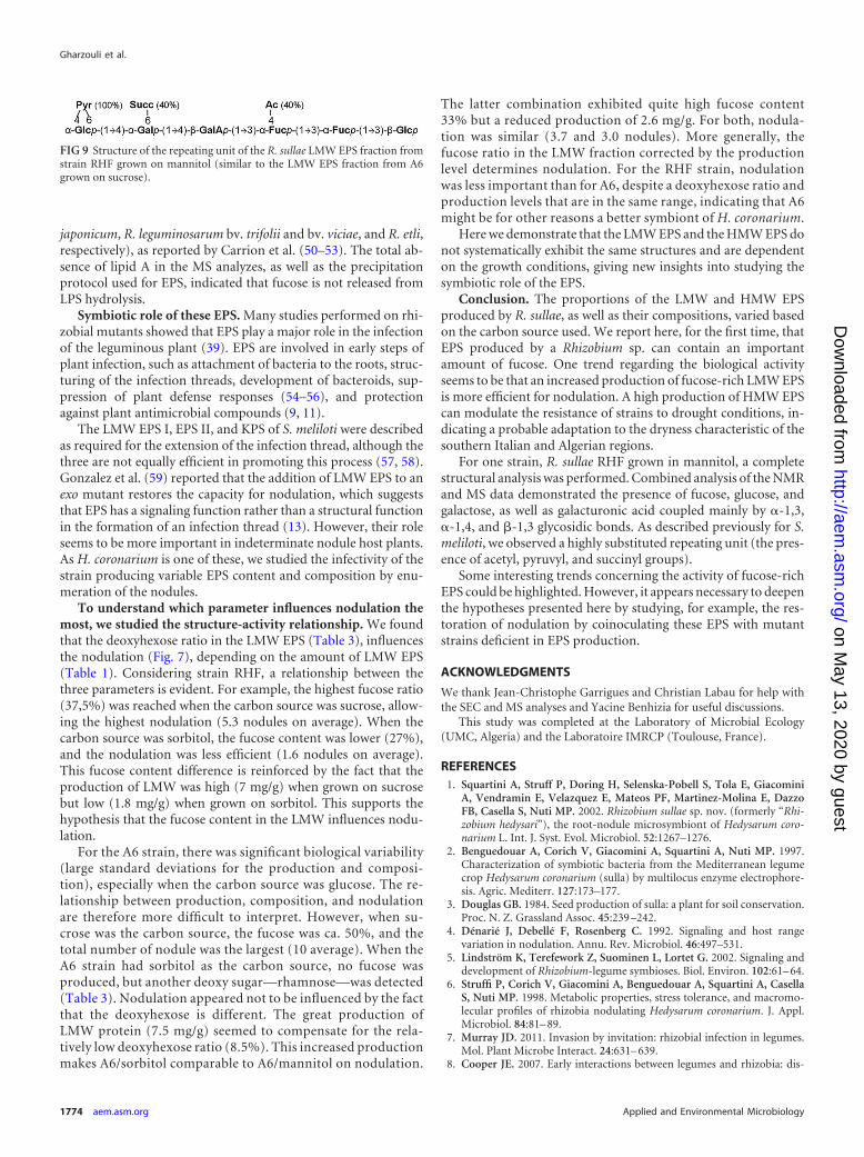
acid, coupled by �-1,3, �-1,4, and �-1,3 glycosidic bonds. Therepeating unit is always substituted by a pyruvyl carried by glu-cose. The combination of the two analytical techniques makes itpossible to determine that ca. 40% of the molecules are succiny-lated on the galactose and that 40% of all the molecules also bearan additional acetyl group on a fucose residue (Fig. 9). Finally,NMR, especially HMBC, was useful for determining the type ofjunction but, due to the numerous 1H and 13C overlappings, onlyESI-MS determined the sequence, highlighting the importance ofcombining NMR and MS methods.
Glucose, galactose, and glucuronic acid are commonly foundin the EPS of different rhizobial species such as S. meliloti, R.leguminosarum bv. viciae, R. leguminosarum bv. trifolii, M. loti,and A. radiobacter (9, 40, 45). The presence of mannose and ga-lacturonic acid characterize the EPS of B. japonicum. Rhamnoseand galacturonic acid are found in Sinorhizobium fredii HH303(9), and the rhamnose-glucuronic acid characterizes the repeatingunit of the nodular EPS of Bradyrhizobium japonicum and Brady-rhizobium elkanii (9).
However, it is uncommon to find as many deoxy sugars as aredescribed here in rhizobial EPS. Moreover, this is the first timethat fucose was found in the structure of EPS of bacteria belongingto the Rhizobium genus. This sugar, rare in the EPS of bacteria, isusually found in Enterobacter EPS (43) or in rhizobial LPS (e.g., B.
FIG 6 Negative-mode ESI-MS results obtained with a QqTof system for LMW EPS fraction from strain A6 grown on sucrose. (A) MS spectrum of R. sullaeLMW EPS fraction; (B) MS/MS spectrum of ion [M-H]� m/z 1,041.4; (C) MS/MS spectra of the observed structural variations—nude, acetylated, orsuccinylated.
FIG 7 Nodulation abilities of the two R. sullae strains grown on differentcarbon sources.
FIG 8 Relationship between the production of HMW EPS of the two strains ofR. sullae grown on different carbon sources (A) and the ability of the twostrains grown on different carbon sources to resume proliferation in a liquidmedium after desiccation (B).
Fucose-Rich EPS Produced by R. sullae
March 2013 Volume 79 Number 6 aem.asm.org 1773
on May 13, 2020 by guest
http://aem.asm
.org/D
ownloaded from
japonicum, R. leguminosarum bv. trifolii and bv. viciae, and R. etli,respectively), as reported by Carrion et al. (50–53). The total ab-sence of lipid A in the MS analyzes, as well as the precipitationprotocol used for EPS, indicated that fucose is not released fromLPS hydrolysis.
Symbiotic role of these EPS. Many studies performed on rhi-zobial mutants showed that EPS play a major role in the infectionof the leguminous plant (39). EPS are involved in early steps ofplant infection, such as attachment of bacteria to the roots, struc-turing of the infection threads, development of bacteroids, sup-pression of plant defense responses (54–56), and protectionagainst plant antimicrobial compounds (9, 11).
The LMW EPS I, EPS II, and KPS of S. meliloti were describedas required for the extension of the infection thread, although thethree are not equally efficient in promoting this process (57, 58).Gonzalez et al. (59) reported that the addition of LMW EPS to anexo mutant restores the capacity for nodulation, which suggeststhat EPS has a signaling function rather than a structural functionin the formation of an infection thread (13). However, their roleseems to be more important in indeterminate nodule host plants.As H. coronarium is one of these, we studied the infectivity of thestrain producing variable EPS content and composition by enu-meration of the nodules.
To understand which parameter influences nodulation themost, we studied the structure-activity relationship. We foundthat the deoxyhexose ratio in the LMW EPS (Table 3), influencesthe nodulation (Fig. 7), depending on the amount of LMW EPS(Table 1). Considering strain RHF, a relationship between thethree parameters is evident. For example, the highest fucose ratio(37,5%) was reached when the carbon source was sucrose, allow-ing the highest nodulation (5.3 nodules on average). When thecarbon source was sorbitol, the fucose content was lower (27%),and the nodulation was less efficient (1.6 nodules on average).This fucose content difference is reinforced by the fact that theproduction of LMW was high (7 mg/g) when grown on sucrosebut low (1.8 mg/g) when grown on sorbitol. This supports thehypothesis that the fucose content in the LMW influences nodu-lation.
For the A6 strain, there was significant biological variability(large standard deviations for the production and composi-tion), especially when the carbon source was glucose. The re-lationship between production, composition, and nodulationare therefore more difficult to interpret. However, when su-crose was the carbon source, the fucose was ca. 50%, and thetotal number of nodule was the largest (10 average). When theA6 strain had sorbitol as the carbon source, no fucose wasproduced, but another deoxy sugar—rhamnose—was detected(Table 3). Nodulation appeared not to be influenced by the factthat the deoxyhexose is different. The great production ofLMW protein (7.5 mg/g) seemed to compensate for the rela-tively low deoxyhexose ratio (8.5%). This increased productionmakes A6/sorbitol comparable to A6/mannitol on nodulation.
The latter combination exhibited quite high fucose content33% but a reduced production of 2.6 mg/g. For both, nodula-tion was similar (3.7 and 3.0 nodules). More generally, thefucose ratio in the LMW fraction corrected by the productionlevel determines nodulation. For the RHF strain, nodulationwas less important than for A6, despite a deoxyhexose ratio andproduction levels that are in the same range, indicating that A6might be for other reasons a better symbiont of H. coronarium.
Here we demonstrate that the LMW EPS and the HMW EPS donot systematically exhibit the same structures and are dependenton the growth conditions, giving new insights into studying thesymbiotic role of the EPS.
Conclusion. The proportions of the LMW and HMW EPSproduced by R. sullae, as well as their compositions, varied basedon the carbon source used. We report here, for the first time, thatEPS produced by a Rhizobium sp. can contain an importantamount of fucose. One trend regarding the biological activityseems to be that an increased production of fucose-rich LMW EPSis more efficient for nodulation. A high production of HMW EPScan modulate the resistance of strains to drought conditions, in-dicating a probable adaptation to the dryness characteristic of thesouthern Italian and Algerian regions.
For one strain, R. sullae RHF grown in mannitol, a completestructural analysis was performed. Combined analysis of the NMRand MS data demonstrated the presence of fucose, glucose, andgalactose, as well as galacturonic acid coupled mainly by �-1,3,�-1,4, and �-1,3 glycosidic bonds. As described previously for S.meliloti, we observed a highly substituted repeating unit (the pres-ence of acetyl, pyruvyl, and succinyl groups).
Some interesting trends concerning the activity of fucose-richEPS could be highlighted. However, it appears necessary to deepenthe hypotheses presented here by studying, for example, the res-toration of nodulation by coinoculating these EPS with mutantstrains deficient in EPS production.
ACKNOWLEDGMENTS
We thank Jean-Christophe Garrigues and Christian Labau for help withthe SEC and MS analyses and Yacine Benhizia for useful discussions.
This study was completed at the Laboratory of Microbial Ecology(UMC, Algeria) and the Laboratoire IMRCP (Toulouse, France).
REFERENCES1. Squartini A, Struff P, Doring H, Selenska-Pobell S, Tola E, Giacomini
A, Vendramin E, Velazquez E, Mateos PF, Martinez-Molina E, DazzoFB, Casella S, Nuti MP. 2002. Rhizobium sullae sp. nov. (formerly “Rhi-zobium hedysari”), the root-nodule microsymbiont of Hedysarum coro-narium L. Int. J. Syst. Evol. Microbiol. 52:1267–1276.
2. Benguedouar A, Corich V, Giacomini A, Squartini A, Nuti MP. 1997.Characterization of symbiotic bacteria from the Mediterranean legumecrop Hedysarum coronarium (sulla) by multilocus enzyme electrophore-sis. Agric. Mediterr. 127:173–177.
3. Douglas GB. 1984. Seed production of sulla: a plant for soil conservation.Proc. N. Z. Grassland Assoc. 45:239 –242.
4. Dénarié J, Debellé F, Rosenberg C. 1992. Signaling and host rangevariation in nodulation. Annu. Rev. Microbiol. 46:497–531.
5. Lindström K, Terefework Z, Suominen L, Lortet G. 2002. Signaling anddevelopment of Rhizobium-legume symbioses. Biol. Environ. 102:61– 64.
6. Struffi P, Corich V, Giacomini A, Benguedouar A, Squartini A, CasellaS, Nuti MP. 1998. Metabolic properties, stress tolerance, and macromo-lecular profiles of rhizobia nodulating Hedysarum coronarium. J. Appl.Microbiol. 84:81– 89.
7. Murray JD. 2011. Invasion by invitation: rhizobial infection in legumes.Mol. Plant Microbe Interact. 24:631– 639.
8. Cooper JE. 2007. Early interactions between legumes and rhizobia: dis-
FIG 9 Structure of the repeating unit of the R. sullae LMW EPS fraction fromstrain RHF grown on mannitol (similar to the LMW EPS fraction from A6grown on sucrose).
Gharzouli et al.
1774 aem.asm.org Applied and Environmental Microbiology
on May 13, 2020 by guest
http://aem.asm
.org/D
ownloaded from

closing complexity in a molecular dialogue. J. Appl. Microbiol. 103:1355–1365.
9. Fraysse N, Couderc F, Poinsot V. 2003. Surface polysaccharide involve-ment in establishing the Rhizobium-legume symbiosis. Eur. J. Biol. Chem.270:1365–1380.
10. González JE, York GM, Walker GC. 1996. Rhizobium meliloti exopoly-saccharides: synthesis and symbiotic function. Gene 179:141–146.
11. Skorupska A, Janczarek M, Marczak M, Mazur A, Krol J. 2006. Rhizo-bial exopolysaccharides: genetic control and symbiotic functions. Microb.Cell Fact. 5:7.
12. Karr DB, Liang RT, Reuhs BL, Emerich DW. 2000. Altered exopolysac-charides of Bradyrhizobium japonicum mutants correlate with impairedsoybean lectin binding, but not with effective nodule formation. Planta211:218 –226.
13. Ghosh AC, Ghosh S, Basu PS. 2005. Production of extracellular polysac-charides by a Rhizobium species from root nodules of the leguminous treeDalbergia lanceolaria. Eng. Life Sci. 5:378 –382.
14. Ibarburu I, Soria-Díaz ME, Rodríguez-Carvajal MA, Velasco SE,Tejero-Mateo P, Gil-Serrano AM, Irastorza A, Dueñas MT. 2007.Growth and exopolysaccharide (EPS) production by Oenococcus oeni I4and structural characterization of their EPSs. J. Appl. Microbiol. 103:477–486.
15. Staudt AK, Wolfe LG, Shrout JD. 2012. Variations in exopolysaccharideproduction by Rhizobium tropici. Arch. Microbiol. 194:197–206.
16. D’Haeze W, Glushka J, De Rycke R, Holsters M, Carlson RW. 2004.Structural characterization of extracellular polysaccharides of Azorhizo-bium caulinodans and importance for nodule initiation on Sesbania ros-trata. Mol. Microbiol. 52:485–500.
17. Potts M. 1994. Desiccation tolerance of prokaryotes. Commun. Micro-biol. Rev. 58:755– 805.
18. Laus MC, Logman TJ, Van Brussel AA, Carlson RW, Azadi P, Gao MY,Kijne JW. 2004. Involvement of exo5 in production of surface polysac-charides in Rhizobium leguminosarum and its role in nodulation of Viciasativa subsp. nigra. J. Bacteriol. 186:6617– 6625.
19. Janczarek M, Skorupska A. 2003. Exopolysaccharide synthesis in Rhizo-bium leguminosarum bv. trifolii is related to various metabolic pathways.Res. Microbiol. 154:433– 442.
20. Patriarca EJ, Tate R, Iaccarino M. 2002. Key role of bacterial NH4
metabolism in rhizobium-plant symbiosis. Microbiol. Mol. Biol. Rev. 66:203–222.
21. Orgambide GG, Philip-Hollingsworth S, Tola E, Cedergren RA, Squar-tini A, Dazzo FB, Hollingsworth RI, Nuti MP. 1996. Glycoconjugate andlipid components of Rhizobium “hedysari” IS123, a root nodule symbiontof the stress-tolerant legume, Hedysarium coronarium. Can. J. Microbiol.42:340 –345.
22. Navarini L, Stredansky M, Matulova M, Bertocchi C. 1997. Productionand characterization of an exopolysaccharide from Rhizobium hedysariHCNT 1. Biotechnol. Lett. 19:1231–1234.
23. Evguenieva-Hackenberg E, Selenska-Pobell S. 1995. Variability of the5=-end of the large subunit rDNA and presence of a new short class ofrRNA in Rhizobiaceae. Lett. Appl. Microbiol. 21:402– 405.
24. Kaci Y, Heyraud A, Barakat M, Heulin T. 2005. Isolation and identifi-cation of an EPS-producing Rhizobium strain from arid soil (Algeria):characterization of its EPS and the effect of inoculation on wheat rhizo-sphere soil structure. Res. Microbiol. 152:522–531.
25. Dreywood R. 1946. Qualitative test for carbohydrates. Ind. Eng. Chem.Res. 18:499.
26. Blumenkrantz N, Asboe-Hansen G. 1973. New method for quantitativedetermination of uronic acids. Anal. Biochem. 54:484 – 489.
27. Rojas-Escudero E, Alarcón-Jiménez AL, Elizalde-Galván P, Rojo-Callejas F. 2004. Optimization of carbohydrate silylation for gas chroma-tography. J. Chromatogr. A 1027:117–120.
28. Vincent JM. 1970. A manual for the practical study of the root-nodulebacteria. IBP handbook 15. Blackwell Scientific Publishers, Oxford, Eng-land.
29. Fahraeus G. 1957. The infection of clover root hairs by nodule bacteriastudied by a simple glass slide technique. J. Gen. Microbiol. 16:374 –381.
30. Ruas-Madiedo P, De Los Reyes-Gavilan CG. 2005. Methods for thescreening, isolation, and characterization of exopolysaccharides producedby lactic acid bacteria. J. Dairy Sci. 88:843– 856.
31. Evans LR, Linker A, Impallomeni G. 2000. Structure of succinoglycanfrom an infectious strain of Agrobacterium radiobacter. Int. J. Biol. Mac-romol. 27:319 –326.
32. Plock A, Beyer G, Hiller K, Gründemann E, Krause E, Nimtz M, WrayV. 2001. Application of MS and NMR to the structure elucidation ofcomplex sugar moieties of natural products: exemplified by the steroidalsaponin from Yucca filamentosa L. Phytochemistry 57:489 – 496.
33. Cescutti P, Kallioinen A, Impallomeni G, Toffanin R, Pollesello P,Laisola M, Eerikaïinen T. 2005. Structure of the exopolysaccharide pro-duced by Enterobacter amnigenus. Carbohydr. Res. 340:439 – 447.
34. Perry MB, MacLean LL, Patrauchan MA, Vinogradov E. 2007. Thestructure of the exocellular polysaccharide produced by Rhodococcus sp.RHA1. Carbohydr. Res. 342:2223–2229.
35. Ihara H, Hanashima S, Okada T, Ito R, Yamaguchi Y, Taniguchi N,Ikeda Y. 2010. Fucosylation of chitooligosaccharides by human �1,6-fucosyltransferase requires a nonreducing terminal chitotriose unit as aminimal structure. Glycobiology 20:1021–1033.
36. MacLean LL, Vinogradov E, Pagotto F, Farber JM, Perry MB. 2010. Thestructure of the O-antigen of Cronobacter sakazakii HPB 2855 isolate in-volved in a neonatal infection. Carbohydr. Res. 345:1932–1937.
37. Zhao L, Chen Y, Ren S, Han Y, Cheng H. 2010. Studies of chemicalstructure and antitumor activity of an exopolysaccharide from Rhizobiumsp. N613. Carbohydr. Res. 345:637– 643.
38. Becker A, Pühler A. 1998. Production of exopolysaccharides in Rhizobi-aceae, p 97–118. In Spaink HP, Kondorosi A, Hooykaas PJJ (ed), TheRhizobiaceae. Kluwer Academic Publishers, Dordrecht, Netherlands.
39. Colpin DL, Cook D. 1990. Molecular genetics of extracellular polysac-charide biosynthesis in vascular phytopathogenic bacteria. Mol. Plant Mi-crobe Interact. 3:271–279.
40. Spaink HP. 2000. Root nodulation and infection factors produced byrhizobial bacteria. Annu. Rev. Microbiol. 54:257–288.
41. Janczarek M. 2011. Environmental signals and regulatory pathways thatinfluence exopolysaccharide production in rhizobia. Int. J. Mol. Sci. 12:7898 –7933.
42. Glenn SA, Gurich N, Feeney MA, Gonzalez JE. 2007. The ExpR/Sinquorum-sensing system controls succinoglycan production in Sinorhizo-bium meliloti. J. Bacteriol. 189:7077–7088.
43. Pawlicki-Jullian N, Courtois B, Pillon M, Lesur D. 2010. Exopolysac-charide production by nitrogen-fixing bacteria within nodules of Medi-cago plants exposed to chronic radiation in the Chernobyl exclusion zone.Res. Microbiol. 161:101–108.
44. Lloret J, Wullf BH, Rubio JM, Dowine JA, Bonilla I, Rivila R. 1998.Exopolysaccharide II production is regulated by salt in the halotolerantstrain Rhizobium meliloti EFB1. Appl. Environ. Microbiol. 64:1024 –1028.
45. Duta FP, De França FP, De Almeida Lopes LM. 2006. Optimization ofculture conditions for exopolysaccharides production in Rhizobium sp.using the response surface method. Electron. J. Biotechnol. 9:391–399.
46. Tola E, Henriquez-Sabà JL, Polone E, Dazzo FB, Concheri G, Casella S,Squartini A. 2009. Shovel roots: a unique stress-avoiding developmentalstrategy of the legume plant Hedysarum coronarium L. Plant Soil 322:25–37.
47. Yemmas L, Soetaert W, Vandamme EJ. 2003. Enzymatic conversion ofthe clavan exopolysaccharide by Streptomyces sp. YSDL-20. Commun. Ag-ric. Appl. Biol. Sci. 68:327–330.
48. Meyer W, Luz S, Schnapper A. 2009. Lectin histochemical aspects ofmucus function in the oesophagus of the reticulated python (Python re-ticulatus). Anat. Histol. Embryol. 38:316 –318.
49. Poli A, Kazak H, Gurleyendag B, Tommonaro G, Pieretti G, ToksoyOner E, Nicolaus B. 2009. High level synthesis of levan by a novel Halo-monas species growing on defined media. Carbohydr. Polymers 78:651–657.
50. Carrion M, Bhat UR, Reuhs B, Carlson RW. 1990. Isolation and char-acterization of the lipopolysaccharides from Bradyrhizobium japonicum. J.Bacteriol. 172:1725–1731.
51. Dazzo FB, Truchet GL, Hollingsworth RI, Hrabak EM, Pankratz HS,Philip-Hollingsworth S, Salzwedel JL, Chapman K, Appenzeller L,Squartini A. 1991. Rhizobium lipopolysaccharide modulates infectionthread development in white clover root hairs. J. Bacteriol. 173:5371–5384.
52. Zhang Y, Hollingsworth RI, Priefer UB. 1992. Characterization of struc-tural defects in the lipopolysaccharides of symbiotically impaired Rhizo-bium leguminosarum biovar viciae VF-39 mutants. Carbohydr. Res. 231:261–271.
53. Noel KD, Box JM, Bonne VJ. 2004. 2-O-methylation of fucosyl residuesof a rhizobial lipopolysaccharide is increased in response to host exudate
Fucose-Rich EPS Produced by R. sullae
March 2013 Volume 79 Number 6 aem.asm.org 1775
on May 13, 2020 by guest
http://aem.asm
.org/D
ownloaded from

and is eliminated in a symbiotically defective mutant. Appl. Environ. Mi-crobiol. 70:1537–1544.
54. Niehaus K, Kapp D, Pühler A. 1993. Plant defense and delayed infectionof alfalfa pseudonodules induced by an exopolysaccharide (EPSI)-deficient Rhizobium meliloti mutant. Planta 190:415– 425.
55. Niehaus K, Baier R, Kohring B, Flashl E, Pühler A. 1997. Symbioticsuppression of the Medicago sativa plant defence system by Rhizobiummeliloti oligosaccharides, p 110 –114. In Legoki A, Bothe H, Pühler A (ed),Biological fixation of nitrogen for ecology and sustainable agriculture.Springer-Verlag, Heidelberg, Germany.
56. van Workum WAT, van Slageren S, van Brussel AAN, Kijne JW. 1998.Role of exopolysaccharides of Rhizobium leguminosarum bv. viciae as host
plant-specific molecules required for infection thread formation duringnodulation of Vicia sativa. Mol. Plant Microbe Interact. 11:1233–1241.
57. Doherty D, Leigh JA, Glazebrook J, Walker GC. 1988. Rhizobiummeliloti mutants that overproduce the R. meliloti acidic calcofluor-bindingexopolysaccharide. J. Bacteriol. 170:4249 – 4256.
58. Brewin NJ. 1998. Tissue and cell invasion by Rhizobium: the structureand development of infection threads and symbiosomes, p 417– 429. InSpaink HP, Kondorosi A, Hooykaas JJ (ed), The Rhizobiaceae. KluwerAcademic Publishers, Dordrecht, Netherlands.
59. Gonzalez JE, Reuhs B, Walker GC. 1996. Low molecular weight EPS II ofRhizobium meliloti promotes nodule invasion of alfalfa. Proc. Natl. Acad.Sci. U. S. A. 93:8636 – 8641.
Gharzouli et al.
1776 aem.asm.org Applied and Environmental Microbiology
on May 13, 2020 by guest
http://aem.asm
.org/D
ownloaded from