relationshipsbetweenslhmotifsfromdifferentglycoside...
TRANSCRIPT
Biologia, Bratislava, 60/Suppl. 16: 115—121, 2005 115
Relationships between SLH motifs from different glycosidehydrolase families
Richard Zona & Štefan Janeček*
Institute of Molecular Biology, Slovak Academy of Sciences, Dúbravská cesta 21, SK-84551 Bratislava, Slovakia; phone:++ 421 2 5930 7420, fax: ++ 421 2 5930 7416, e-mail: [email protected]
Abstract: Many glycoside hydrolases (GH) are very large proteins consisting of catalytic and non-catalytic domains. Withregard to the non-catalytic domains, much research has been performed on the carbohydrate-binding modules (CBM),whereas substantially less attention has been paid to the surface layer homology (SLH) domain. The SLH sequences areinvolved in the attachment of proteins to the underlying cell wall. SLH domains are made of one to three repeats of 50amino acids among which ten to fifteen residues are conserved. Three amylopullulanases from the α-amylase family GH-13contain the SLH motifs; each in three copies. Within the CAZy classification, in addition to the α-amylase family GH-13,the typical SLH motifs are present in six other GH families: GH-5, GH-10, GH-16, GH-26, GH-28 and GH-73. Moreover,longer repeated domains which display some resemblance to SLH motifs have been identified in families GH-15 and GH-57.These so-called SLH motif-bearing domains contain two and a half typical SLH motifs. Based on the present sequencecomparison data, a short sequence fingerprint, localized in the middle of the SLH motif, constitutes a novel third conservedregion in glycoside hydrolase-associated SLH motifs. The evolutionary tree illustrates the relationships among the individualcopies of the SLH motifs as well as between the typical SLH motifs and the longer SLH motif-bearing domains. It has beenconcluded that the evolutionary relationships of the SLH motifs reflect more taxonomy than the enzyme specificity of thecatalytic domain to which they are linked.
Key words: SLH motif, glycoside hydrolase, alpha-amylase family.
Abbreviations: CBM, carbohydrate-binding module; GH, glycoside hydrolase; SLH, surface layer homology.
Introduction
Surface layers (S-layers) from Bacteria and Archaeaare built from protein molecules arrayed in a two-dimensional lattice, forming the outermost cell walllayer in many prokaryotes (Engelhardt & Peters,1998). At the time of the discovery of the S-layers thesequence comparison of S-layers from distantly relatedbacteria did not reveal strong similarities. Nevertheless,one exceptional similarity was identified between the S-layer sequence, i.e. the N-terminal region of about 200amino acid residues, of Thermoanaerobacter kivui andthe N-terminal part of the middle wall protein of Bre-vibacillus brevis (Peters et al., 1989). This similaritywas later shown to be a widely conserved motif amongbacterial surface proteins and named as the S-layer ho-mology (SLH) domain (Lupas et al., 1994). It was pro-posed to function as a peptidoglycan-binding structureof proteins to the underlying cell wall (Lupas et al.,1994). Later, SLH domains were shown to be both nec-
essary and sufficient to bind cell walls (Lemaire et al.,1995; Mesnage et al., 1999).At present the proteins possessing an SLH motif
are divided into three groups (Engelhardt & Pe-ters, 1998): (i) group I – S-layer proteins; (ii) group II– extracellular enzymes and proteins mostly involved inpolysaccharide degradation; and (iii) group III – outermembrane proteins (Omps), also including some hypo-thetical proteins. The SLH motifs are located eitherat the N- or C-terminal end of the protein and theSLH domain consists of one to three SLH motifs (Lu-pas et al., 1994; Engelhardt & Peters, 1998;Mes-nage et al., 2000). A typical SLH motif is a segmentof ∼40–50 amino acids with 10–15 conserved residues,the C-terminus being the best conserved (Lupas et al.,1994; Engelhardt & Peters, 1998). According tothe Pfam database (Bateman et al., 2002) the SLHmodule constitutes the family PF00395.Since the entire sequences of the individual groups
of the S-layer proteins do not share common similari-
* Corresponding author
116 R. Zona & Š. Janeček
(A) SLH motifs:
GH-5 Q59154_ANATHa 576_FEDIN----FENSLYDVIDKLYSKGIIKGISVFKYLPDKNITRAEFA 618 Q59154_ANATHb 636_FSDVKS--GNWYSD--VVYTAYKNKLFEIKENK-FFPENILKREEAV 677 Q59154_ANATHc 700_IADEKLINPQYRES---VKLAIKLGIVDLYSDGTFEPNKSVSRGEVA 743 Q8RLT7_CLOCE 809_FSDVHK--KDS--YYNPVGIAKALGITNGVGHNKFNPNKAISREDML 851 Q59290_CLOJO 809_FSDVNK--KGS--YYNSVGIAKALGITSGVGNNKFNPNKAISREDML 851 P19424_BACS6a 41_FSDVKK--TSWSFP--YIKDLYEQEVITGTSATTFSPTDSVTRAQFT 83 P19424_BACS6b 101_FKDRK----NWAYK--EIQAAYEAGIVTGKTNGEFAPNENITREQMA 141 P19424_BACS6c 164_YNDSSS-ISTFAQD--AVQKAYVLELMEGNTDGYFQPKRNSTREQSA 207 Q9ZA17_THESAa 913_FTDISS---SWAKN--EIQVLASKNIISGYPDGTFKPDKRITRAEFV 954 Q9ZA17_THESAb 973_FSDVNK--GDW--YYGLVEAAKSTGIASGY-GKQFKPDMQITRQEMM 1014 Q9ZA17_THESAc 1041_FKDGGK-VQNWAKD--AMAIGVSNGLIKGTGDEYLSPDGRATRAQAA 1084 GH-10 Q9F1V3_CLOJOa 928_FKDVKK--DSS--YYASVSAAYQKGIISGYKNGEFKPQAKITRQEAM 970 Q9F1V3_CLOJOb 998_FKDSNK-VANWAKA--SVAACIKEGIISGKSGKMIAPQENITVSQTE 1041 P38535_CLOTMa 908_FNDIKD---NWAKD--VIEVLASRHIVEGMTDTQYEPSKTVTRAEFT 949 P38535_CLOTMb 967_FSDVKN--GDW--YANAIEAAYKAGIIEGD-GKNMRPNDSITREEMT 1008 P38535_CLOTMc 1031_FNDDKS-ISDWAKN--VVANAAKLGIINGEPSNVFAPKGIATRAEAA 1074 P36917_THESAa 1056_FDDIKN---SWAKD--AIEVLASRHIVEGMTDTQYEPNKTVTRAEFT 1097 P36917_THESAb 1115_FSDVNS--GDW--YANAIEAAYKAGIIEGD-GKNARPNDSITREEMT 1156 Q60046_THETUa 1055_FNDIKD---NWAKD--VIEVLASRHIVEGMTDTQYEPNKTVTRAEFT 1096 Q60046_THETUb 1114_FSDVKS--GDW--YANAIEAAYKTGIIEGD-GKNARPNDSITREEMT 1155 Q60046_THETUc 1178_FSDDKS-ISDWARN--VVANAAKLGIVNGEPNNVFAPKGNATRAEAA 1221 Q8GHJ4_PAESWa 1149_FADVQH--VLWAKE--AIEAMAARDIIKGISDESFAPAASITRADFI 1191 Q8GHJ4_PAESWb 1210_FSDVQS--TAY--YAQAVAIAKELGIASGFEDNTFKPGSSISRQDMM 1252 Q8GHJ4_PAESWc 1275_YSDAAS-ISTYAVD--SVTSLVGSGIVNGK-GGKIAPTESLTRAEAA 1317 Q60043_THESJa 1169_FNDIKD---NWPKD--VIEVLASRHIVEGMTDTQYEPNKTVARAEFT 1210 Q60043_THESJb 1228_FSDVKS--GDW--YADAIEAAYKAGIIEGD-GKNARPYDSITREEMT 1269 Q60043_THESJc 1292_FSDDKS-ISDWARN--VVANAAKLGIVNGEPNNVFAPKGNATRAEAA 1335 O52373_CALSRa 1424_YKDVPK--THWAYD--TFKQAVTSGLVVGYNDMTLRPAKNVTLAEAA 1466 O52373_CALSRb 1486_---VP----DWAAS--AIKALLDNEIIAEVDDA----NKPLTRIEAV 1519 O52373_CALSRc 1540_FSDLYE---QSSIDVEYLAKAYKLGIVKGYPDGTFRPQNTVTRAELL 1583 GH-13 P38536_THETUa 1682_FNDIKD---NWAKD--VIEVLASRHIVEGMTDTQYEPNKTVTRAEFT 1723 P38536_THETUb 1741_FSDVKS--GDW--YANAIEAAYKAGIIEGD-GKNARPNDSITREEMT 1782 P38536_THETUc 1805_FSDDKS-ISDWARN--VVANAAKLGIVNGEPNNVFAPKGNATRAEAA 1848 Q9EZZ4_BACSTa 1831_FADIVQ---HWAKP--YIDSLAAKQLVRGVTETAYRPNEPMTRAQFA 1872 Q9EZZ4_BACSTb 1890_FADVKGT--EWFNQHGELAAAVKYGVIQGKTPSTFAPNEPITRAQAA 1934 Q9EZZ4_BACSTc 1962_FRDANQL-PAWSKQ--AIEAIYQAGIVQGHPDGTFAPAGRMTRAEMA 2005 Q45643_BACX6a 1845_FSDIEK---HWAKG--YIETLAAKQLVKGMTETAYRPNEQMTRAQFA 1886 Q45643_BACX6b 1904_FADVKGT--EWFNKNGELAAAVKLGIIQGKTANTFAPNEPITRVQAA 1948 Q45643_BACX6c 1976_FRDAKQL-PTWAKQ--AIEAVYQAGIMQGRDNGSFDPTGHMTRAEMA 2019 GH-16 Q59328_CLOTMa 30_INDIRG---HWAEE--DLNKWMEKGILVGYQDGTIRPDNNITRAEFV 71 Q59328_CLOTMb 88_FADVED--SKW--YSREILKARAAGYIAGYGSNVFKPDNYITRQEAV 130 Q59328_CLOTMc 149_FKDGS-LVKEYAKD--SVSALVEKGYIAGYEDGTFRPDNYITRAETI 192 GH-26 Q9XCV5_CELFIa 696_FSDVPK--GHPYET--EILWLHAQGLDDGYDDGTFRPARQVKRQDVA 738 Q9XCV5_CELFIb 757_FLDVRR--SHPAYT--AIEWLVAEGLVD--DGRVFLPSAPLDRATAA 797 Q9XCV5_CELFIc 815_FRDVP----TWHRYRTAITWATEVGVVEPVSASTFGVLKAVQRQELA 857 GH-28 Q60045_THETUa 969_FNDIKD---NWAKD--VIEVLASRHIVEGMTDTQYEPNKTVTRAEFT 1010 Q60045_THETUb 1028_FSDVKS--GDW--YANAIEAAYKAGIIESD-GKNARPNDSITREEMT 1069 Q60045_THETUc 1092_FSDDKS-ISDWARN--VVANAAKLGIVNGEPNNVFAPKGNATRAEAA 1135 GH-73 Q7X0Z0_BACCIa 1760_FDDVPA--GHWAEG--VISKLTSRLMVDGTSETTFEPERVVTRAEFT 1802 Q7X0Z0_BACCIb 1819_FADVKA--GDW--YADAVTAAVEAGIAEGKSAGQFEPQARITREEMV 1861 Q7X0Z0_BACCIc 1884_FTDENQ-ISAWAVE--QVKAAAALQLIQGRAQGKFEPQGTATRAEAV 1927 Conserved regions FxDV GIIxG TRAE
ties, the SLH domain must be regarded as a modularcomponent that was linked to different proteins duringevolution (Engelhardt & Peters, 1998).Many glycoside hydrolases (GHs) contain SLH mo-
tifs (Schwarz et al., 2004). In the frame of the SLHclassification they belong to group II. They are verylarge proteins consisting of catalytic and non-catalyticdomains. With regard to the non-catalytic domains,much research has been performed on the carbohy-drate binding modules (CBMs;Boraston et al., 2004),whereas a substantially less attention has been paidto and/or has been known for the SLH motifs (Bev-eridge et al., 1997). The S-layer protein and the threeglycoside hydrolases of Thermoanaerobacterium ther-mosulfurigenes EM1 (GH-10 xylanase, GH-13 amylop-ullulanase and GH-28 polygalacturonase) were mostdeeply studied (Matuschek et al., 1994; 1996;Brech-tel et al., 1999) with the conclusion that the SLHdomains present in the S-layer and the enzymes areresponsible for the anchoring of both protein typesby binding of the SLH domain to the underlyingpeptidoglycan-containing sacculus (Brechtel et al.,1999). Using the C-terminally truncated forms of thatxylanase (i.e. by removing the SLH motifs), Brechtel& Bahl (1999) demonstrated that multiple SLH mo-
tifs are necessary for the xylanase attachment to thecell wall.Three amylopullulanases from the α-amylase fam-
ily, i.e. the clan GH-H (MacGregor et al., 2001) con-tain the SLH modules; each in three copies. There areabout 30 different enzyme specificities in the α-amylaseclan GH-H (Janecek, 2002; Svensson et al., 2002;MacGregor, 2005) but the amylopullulanase is theonly one containing the module. Within the all CAZyGH families (Coutinho & Henrissat, 1999), theseSLH modules are present in families GH-5, GH-10, GH-16, GH-26, GH-28, and GH-73 in addition to the α-amylase family GH-13. Moreover the SLH-like motifswere found in two more families, in GH-57 (Erra-Pujada et al., 1999) and GH-15 (Mizuno et al., 2004).These SLH-like sequence segments were first describedin the primary structure of GH-57 amylopullulanasefrom Thermococcus hydrothermalis and defined as thelonger SLH motif-bearing domain containing two and ahalf typical SLH motifs (Erra-Pujada et al., 1999).Similar to the situation in α-amylase family GH-13,in GH-57 only the amylopullulanases appear to con-tain SLH-motif-bearing domains (Zona et al., 2004).Based on the three-dimensional structure of the GH-15glucodextranase, the SLH motif-bearing domain covers
SLH motifs in glycoside hydrolases 117
(B)
SLH motif-bearing domains:
<-------------------- SLH correspondence --------------------->
GH-15 Q9LBQ9_ARTGO 852_ATIAGEVTNPWGGQA-ISHQRVNIYLGKGEGGA------TPGLPGTNINLEH--AWDSVIVTD-GRFD-GAGVYAPDGTRTSAVSLL-AVPEAR-QIVTRVPKAALGGLDPATARMS-VAMFGN 961
GH-57 Q8TZQ1_PYRFU 840_FYFKELGGNPWNGPNGFSLQIIEVYLDFKEGGNTSAIKMFPDGPGANVQLDPEHPWDVAFRIA-GW-DYGNLIVLANGTVYQGEMQISADPTKNAVIVK-LPKKYLSIG-DYGLYAAVLV--GS 957
Q72GM0_THET2 815_FPFKEMT-NPWGAPAGFSHQLLNVYLDFKDGG-----RTDPFAKGAKVAFDPEHPWDLFLKAA-GWPQYGQRVGFPDGTDTADGITVGSNPADKQVIVQ-LDKKHFNPASGQRVCFYVLV--GS 928
Q8ZT36_PYRAE 737_FKVRELGDNPWGGPAGFSLQFFHVYINRGSGS-----R--NDTLGLRVALCRDAAWDVALLIGPGW-SGGNRIVYSDNTYVDDAMSIKVAPN-NTVVAD-VPKRYIGEFNSSWKITVFL---TS 847
Q8NKS8_THELI 835_YYFKDLGDNSWNGPNGFSLQIIEAYFDFKEGGNTSAIKMFPDGPGSNVDLDPEHPWDVALRIA-GW-DYGNIIVLPDGTSYQGEMKISADPVKNAIVVE-VPKKYLEISKDYGLYGAILV--GS 953
Q9Y8I8_THEHYa 839_FYFKDLGGNPWNGPNGFSLQIIEVYLDFKDGGNSSAIKMFPDGPGANVNLDPEHPWDVAFRIA-GW-DYGNLIILPNGTAIQGEMQISADPVKNAIIVK-VPKKYIAINEDYGLWGDVLV--GS 957
Q9Y8I8_THEHYb 1080_FHFKDLGGNPWNGPNGFSLQIIEVYFDFKEGGNVSAIKMFPDGPGSNVRLDPNHPWDLALRIA-GW-DYGNLIILPDGTAYQGEMQISADPVKNAIIVK-VPKKYLNIS-DYGLYTAVIV--GS 1197
Q9V294_PYRABa 839_FYFKELGDNPWNAPYGFSLQIMEVYLDYKEGGNTSAIKMFPDGPGSNVDLDPEHPWDVALRIA-GW-DYGNIIVLANGTTYQGEMKISADPVKNRIIVE-VPKKYLPKVP---EFMAVLV--GS 954
Q9V294_PYRABb 1080_FYFKELGDNPWNAPYGFSLQIIEVYLDFKEGGNTSAIKMFPDGPGSNVDLDPEHPWDVALRIA-GW-DYGNIIVPANGTVYTGEMKISADPIKNAIIVE-VPKKFISLDKNYGLYGAVLV--GS 1198
Q9HLU6_THEAC 1118_FKYAQLW-NIWNGPLGFSNQIINIFLS--NGS-----TSGNTYLGSGPNAESSIPWQKMIYIS-GWATY---VQTLTGTYSNG-ILVSVNLSLGEIYVT-IPIEYLGQN-FLSYRYLIVA--GS 1224
Invariant residues N W S Q G G W G T
(C) SLH motifs:
GH-5 Q59154_ANATHa 576_FEDIN----------FENSLYDVIDKLYSK---G--II-------KGISVFKYLPDKN-ITRAEFA 618 Q59154_ANATHb 636_FSDVKS--------GNWYSD--VVYTAYKN---K--LF-------EIKENK-FFPENI-LKREEAV 677 Q59154_ANATHc 700_IADEKLI------NPQYRES---VKLAIKL---G--IV-------DLYSDGTFEPNKS-VSRGEVA 743 Q8RLT7_CLOCE 809_FSDVHK--------KDS--YYNPVGIAKAL---G--IT-------NGVGHNKFNPNKA-ISREDML 851 Q59290_CLOJO 809_FSDVNK--------KGS--YYNSVGIAKAL---G--IT-------SGVGNNKFNPNKA-ISREDML 851 P19424_BACS6a 41_FSDVKK--------TSWSFP--YIKDLYEQ---E--VI-------TGTSATTFSPTDS-VTRAQFT 83 P19424_BACS6b 101_FKDRK----------NWAYK--EIQAAYEA---G--IV-------TGKTNGEFAPNEN-ITREQMA 141 P19424_BACS6c 164_YNDSSS-------ISTFAQD--AVQKAYVL---E--LM-------EGNTDGYFQPKRN-STREQSA 207 Q9ZA17_THESAa 913_FTDISS---------SWAKN--EIQVLASK---N--II-------SGYPDGTFKPDKR-ITRAEFV 954 Q9ZA17_THESAb 973_FSDVNK--------GDW--YYGLVEAAKST---G--IA-------SGY-GKQFKPDMQ-ITRQEMM 1014 Q9ZA17_THESAc 1041_FKDGGK-------VQNWAKD--AMAIGVSN---G--LI-------KGTGDEYLSPDGR-ATRAQAA 1084 GH-10 Q9F1V3_CLOJOa 928_FKDVKK--------DSS--YYASVSAAYQK---G--II-------SGYKNGEFKPQAK-ITRQEAM 970 Q9F1V3_CLOJOb 998_FKDSNK-------VANWAKA--SVAACIKE---G--II-------SGKSGKMIAPQEN-ITVSQTE 1041 P38535_CLOTMa 908_FNDIKD---------NWAKD--VIEVLASR---H--IV-------EGMTDTQYEPSKT-VTRAEFT 949 P38535_CLOTMb 967_FSDVKN--------GDW--YANAIEAAYKA---G--II-------EGD-GKNMRPNDS-ITREEMT 1008 P38535_CLOTMc 1031_FNDDKS-------ISDWAKN--VVANAAKL---G--II-------NGEPSNVFAPKGI-ATRAEAA 1074 P36917_THESAa 1056_FDDIKN---------SWAKD--AIEVLASR---H--IV-------EGMTDTQYEPNKT-VTRAEFT 1097 P36917_THESAb 1115_FSDVNS--------GDW--YANAIEAAYKA---G--II-------EGD-GKNARPNDS-ITREEMT 1156 Q60046_THETUa 1055_FNDIKD---------NWAKD--VIEVLASR---H--IV-------EGMTDTQYEPNKT-VTRAEFT 1096 Q60046_THETUb 1114_FSDVKS--------GDW--YANAIEAAYKT---G--II-------EGD-GKNARPNDS-ITREEMT 1155 Q60046_THETUc 1178_FSDDKS-------ISDWARN--VVANAAKL---G--IV-------NGEPNNVFAPKGN-ATRAEAA 1221 Q8GHJ4_PAESWa 1149_FADVQH--------VLWAKE--AIEAMAAR---D--II-------KGISDESFAPAAS-ITRADFI 1191 Q8GHJ4_PAESWb 1210_FSDVQS--------TAY--YAQAVAIAKEL---G--IA-------SGFEDNTFKPGSS-ISRQDMM 1252 Q8GHJ4_PAESWc 1275_YSDAAS-------ISTYAVD--SVTSLVGS---G--IV-------NGK-GGKIAPTES-LTRAEAA 1317 Q60043_THESJa 1169_FNDIKD---------NWPKD--VIEVLASR---H--IV-------EGMTDTQYEPNKT-VARAEFT 1210 Q60043_THESJb 1228_FSDVKS--------GDW--YADAIEAAYKA---G--II-------EGD-GKNARPYDS-ITREEMT 1269 Q60043_THESJc 1292_FSDDKS-------ISDWARN--VVANAAKL---G--IV-------NGEPNNVFAPKGN-ATRAEAA 1335 O52373_CALSRa 1424_YKDVPK--------THWAYD--TFKQAVTS---G--LV-------VGYNDMTLRPAKN-VTLAEAA 1466 O52373_CALSRb 1486_---VP----------DWAAS--AIKALLDN---E--II-------AEVDDA----NKP-LTRIEAV 1519 O52373_CALSRc 1540_FSDLYE---------QSSIDVEYLAKAYKL---G--IV-------KGYPDGTFRPQNT-VTRAELL 1583 GH-13 P38536_THETUa 1682_FNDIKD---------NWAKD--VIEVLASR---H--IV-------EGMTDTQYEPNKT-VTRAEFT 1723 P38536_THETUb 1741_FSDVKS--------GDW--YANAIEAAYKA---G--II-------EGD-GKNARPNDS-ITREEMT 1782 P38536_THETUc 1805_FSDDKS-------ISDWARN--VVANAAKL---G--IV-------NGEPNNVFAPKGN-ATRAEAA 1848 Q9EZZ4_BACSTa 1831_FADIVQ---------HWAKP--YIDSLAAK---Q--LV-------RGVTETAYRPNEP-MTRAQFA 1872 Q9EZZ4_BACSTb 1890_FADVKGT--------EWFNQHGELAAAVKY---G--VI-------QGKTPSTFAPNEP-ITRAQAA 1934 Q9EZZ4_BACSTc 1962_FRDANQL-------PAWSKQ--AIEAIYQA---G--IV-------QGHPDGTFAPAGR-MTRAEMA 2005 Q45643_BACX6a 1845_FSDIEK---------HWAKG--YIETLAAK---Q--LV-------KGMTETAYRPNEQ-MTRAQFA 1886 Q45643_BACX6b 1904_FADVKGT--------EWFNKNGELAAAVKL---G--II-------QGKTANTFAPNEP-ITRVQAA 1948 Q45643_BACX6c 1976_FRDAKQL-------PTWAKQ--AIEAVYQA---G--IM-------QGRDNGSFDPTGH-MTRAEMA 2019 GH-16 Q59328_CLOTMa 30_INDIRG---------HWAEE--DLNKWMEK---G--IL-------VGYQDGTIRPDNN-ITRAEFV 71 Q59328_CLOTMb 88_FADVED--------SKW--YSREILKARAA---G--YI-------AGYGSNVFKPDNY-ITRQEAV 130 Q59328_CLOTMc 149_FKDGS-L------VKEYAKD--SVSALVEK---G--YI-------AGYEDGTFRPDNY-ITRAETI 192 GH-26 Q9XCV5_CELFIa 696_FSDVPK--------GHPYET--EILWLHAQ---G--LD-------DGYDDGTFRPARQ-VKRQDVA 738 Q9XCV5_CELFIb 757_FLDVRR--------SHPAYT--AIEWLVAE---G--LV-------D--DGRVFLPSAP-LDRATAA 797 Q9XCV5_CELFIc 815_FRDVP----------TWHRYRTAITWATEV---G--VV-------EPVSASTFGVLKA-VQRQELA 857 GH-28 Q60045_THETUa 969_FNDIKD---------NWAKD--VIEVLASR---H--IV-------EGMTDTQYEPNKT-VTRAEFT 1010 Q60045_THETUb 1028_FSDVKS--------GDW--YANAIEAAYKA---G--II-------ESD-GKNARPNDS-ITREEMT 1069 Q60045_THETUc 1092_FSDDKS-------ISDWARN--VVANAAKL---G--IV-------NGEPNNVFAPKGN-ATRAEAA 1135 GH-73 Q7X0Z0_BACCIa 1760_FDDVPA--------GHWAEG--VISKLTSR---L--MV-------DGTSETTFEPERV-VTRAEFT 1802 Q7X0Z0_BACCIb 1819_FADVKA--------GDW--YADAVTAAVEA---G--IA-------EGKSAGQFEPQAR-ITREEMV 1861 Q7X0Z0_BACCIc 1884_FTDENQ-------ISAWAVE--QVKAAAAL---Q--LI-------QGRAQGKFEPQGT-ATRAEAV 1927 SLH correspondence from the SLH motif-bearing domains:
GH-15 Q9LBQ9_ARTGO 884_TPGLPGTNINLEH--AWDS---VIVTD-GRFD-GAGVYAPDGTRTSAVSLL-AVPEAR-QIVTRVP 940 GH-57 Q8TZQ1_PYRFU 880_FPDGPGANVQLDPEHPWDV---AFRIA-GW-DYGNLIVLANGTVYQGEMQISADPTKNAVIVK-LP 938 Q72GM0_THET2 848_DPFAKGAKVAFDPEHPWDL---FLKAA-GWPQYGQRVGFPDGTDTADGITVGSNPADKQVIVQ-LD 908 Q8ZT36_PYRAE 770_-NDTLGLRVALCRDAAWDV---ALLIGPGW-SGGNRIVYSDNTYVDDAMSIKVAPN-NTVVAD-VP 828 Q8NKS8_THELI 874_FPDGPGSNVDLDPEHPWDV---ALRIA-GW-DYGNIIVLPDGTSYQGEMKISADPVKNAIVVE-VP 933 Q9Y8I8_THEHYa 878_FPDGPGANVNLDPEHPWDV---AFRIA-GW-DYGNLIILPNGTAIQGEMQISADPVKNAIIVK-VP 937 Q9Y8I8_THEHYb 1119_FPDGPGSNVRLDPNHPWDL---ALRIA-GW-DYGNLIILPDGTAYQGEMQISADPVKNAIIVK-VP 1178 Q9V294_PYRABa 878_FPDGPGSNVDLDPEHPWDV---ALRIA-GW-DYGNIIVLANGTTYQGEMKISADPVKNRIIVE-VP 937 Q9V294_PYRABb 1119_FPDGPGSNVDLDPEHPWDV---ALRIA-GW-DYGNIIVPANGTVYTGEMKISADPIKNAIIVE-VP 1178 Q9HLU6_THEAC 1149_GNTYLGSGPNAESSIPWQK---MIYIS-GWATY---VQTLTGTYSNG-ILVSVNLSLGEIYVT-IP 1205
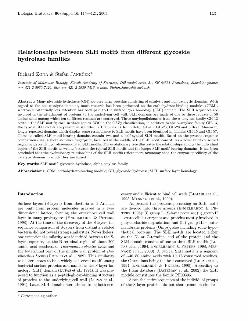
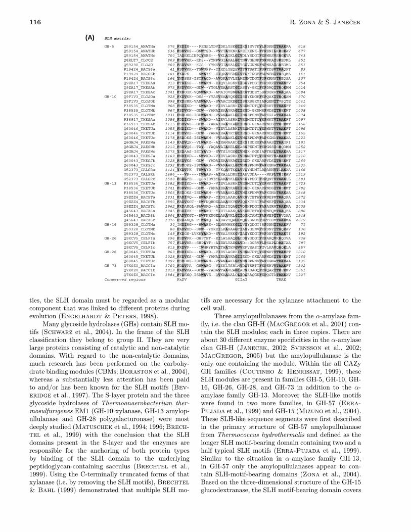
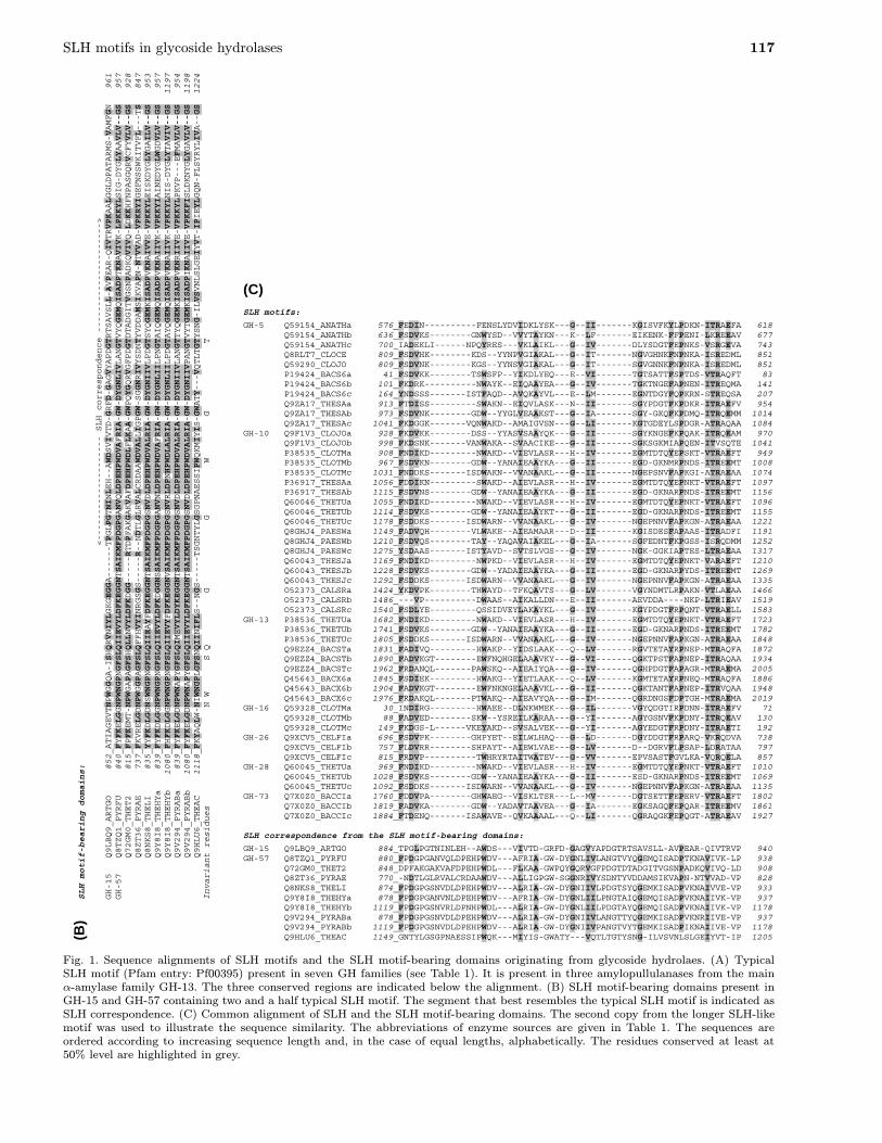
Fig. 1. Sequence alignments of SLH motifs and the SLH motif-bearing domains originating from glycoside hydrolaes. (A) TypicalSLH motif (Pfam entry: Pf00395) present in seven GH families (see Table 1). It is present in three amylopullulanases from the mainα-amylase family GH-13. The three conserved regions are indicated below the alignment. (B) SLH motif-bearing domains present inGH-15 and GH-57 containing two and a half typical SLH motif. The segment that best resembles the typical SLH motif is indicated asSLH correspondence. (C) Common alignment of SLH and the SLH motif-bearing domains. The second copy from the longer SLH-likemotif was used to illustrate the sequence similarity. The abbreviations of enzyme sources are given in Table 1. The sequences areordered according to increasing sequence length and, in the case of equal lengths, alphabetically. The residues conserved at least at50% level are highlighted in grey.
118 R. Zona & Š. Janeček
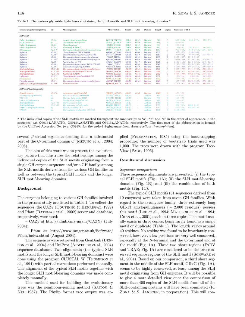
Table 1. The various glycoside hydrolases containing the SLH motifs and SLH motif-bearing domains.a
a The individual copies of the SLH motifs are marked throughout the manuscript as “a”, “b” and “c” in the order of appearance in thesequence, e.g. Q59154 ANATHa, Q59154 ANATHb and Q59154 ANATHc, respectively. The first part of the abbreviation is formedby the UniProt Accession No. (e.g. Q59154 for the endo-1,4-glucanase from Anaerocellum thermophilum).
several β-strand segments forming thus a substantialpart of the C-terminal domain C (Mizuno et al., 2004;2005).The aim of this work was to present the evolution-
ary picture that illustrates the relationships among theindividual copies of the SLH motifs originating from asingle GH enzyme sequence and/or a GH family, amongthe SLH motifs derived from the various GH families aswell as between the typical SLH motifs and the longerSLH motif-bearing domains.
Background
The enzymes belonging to various GH families involvedin the present study are listed in Table 1. To collect thesequences, the CAZy (Coutinho & Henrissat, 1999)and Pfam (Bateman et al., 2002) server and database,respectively, were used:– CAZy at http://afmb.cnrs-mrs.fr/CAZY/ (July
2004);– Pfam at http://www.sanger.ac.uk/Software/
Pfam/index.shtml (August 2004).The sequences were retrieved from GenBank (Ben-
son et al., 2004) and UniProt (Apweiler et al., 2004)sequence databases. Two alignments (the typical SLHmotifs and the longer SLH motif-bearing domains) weredone using the program CLUSTAL W (Thompson etal., 1994) with partial corrections performed manually.The alignment of the typical SLH motifs together withthe longer SLH motif-bearing domains was made com-pletely manually.The method used for building the evolutionary
trees was the neighbour-joining method (Saitou &Nei, 1987). The Phylip format tree output was ap-
plied (Felsenstein, 1985) using the bootstrappingprocedure; the number of bootstrap trials used was1,000. The trees were drawn with the program Tree-View (Page, 1996).
Results and discussion
Sequence comparisonThree sequence alignments are presented: (i) the typi-cal SLH motifs (Fig. 1A); (ii) the SLH motif-bearingdomains (Fig. 1B); and (iii) the combination of bothmotifs (Fig. 1C).The typical SLH motifs (51 sequences derived from
19 enzymes) were taken from seven GH families. Withregard to the α-amylase family, three extremely longGH-13 amylopullulanases (∼ 2,000 residues) possessthis motif (Lee et al., 1994; Matuschek et al., 1994;Chen et al., 2001); each in three copies. The motif usu-ally exists in three copies, being rarely found as a singlemotif or duplicate (Table 1). The length varies around40 residues. No residue was found to be invariantly con-served, however, a few positions are very well conserved,especially at the N-terminal and the C-terminal end ofthe motif (Fig. 1A). These two short regions (FxDVand TRAE; Fig. 1A) are considered to be the two con-served sequence regions of the SLH motif (Schwarz etal., 2004). Based on our comparison, a third short seg-ment in the middle of the SLH motif, GIIxG (Fig. 1A),seems to be highly conserved, at least among the SLHmotif originating from GH enzymes. It will be possibleto give a more detailed view once the comparison ofmore than 400 copies of the SLH motifs from all of theSLH-containing proteins will have been completed (R.Zona & S. Janecek, in preparation). This will con-
SLH motifs in glycoside hydrolases 119
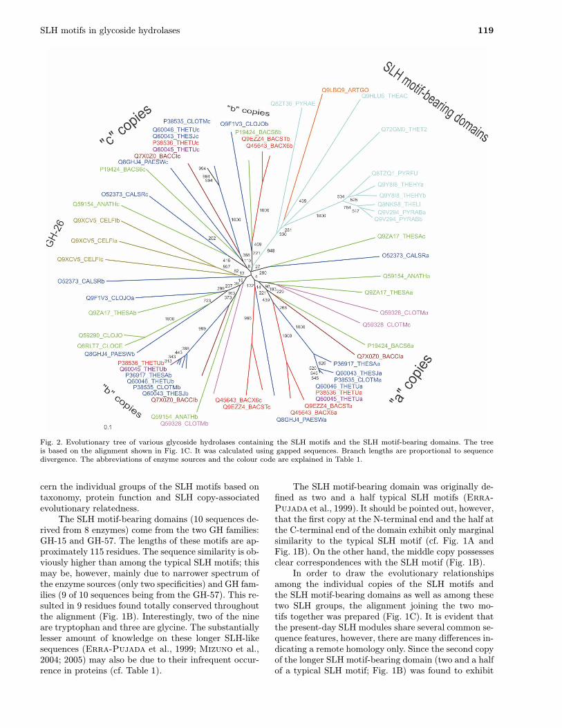
Fig. 2. Evolutionary tree of various glycoside hydrolases containing the SLH motifs and the SLH motif-bearing domains. The treeis based on the alignment shown in Fig. 1C. It was calculated using gapped sequences. Branch lengths are proportional to sequencedivergence. The abbreviations of enzyme sources and the colour code are explained in Table 1.
cern the individual groups of the SLH motifs based ontaxonomy, protein function and SLH copy-associatedevolutionary relatedness.The SLH motif-bearing domains (10 sequences de-
rived from 8 enzymes) come from the two GH families:GH-15 and GH-57. The lengths of these motifs are ap-proximately 115 residues. The sequence similarity is ob-viously higher than among the typical SLH motifs; thismay be, however, mainly due to narrower spectrum ofthe enzyme sources (only two specificities) and GH fam-ilies (9 of 10 sequences being from the GH-57). This re-sulted in 9 residues found totally conserved throughoutthe alignment (Fig. 1B). Interestingly, two of the nineare tryptophan and three are glycine. The substantiallylesser amount of knowledge on these longer SLH-likesequences (Erra-Pujada et al., 1999; Mizuno et al.,2004; 2005) may also be due to their infrequent occur-rence in proteins (cf. Table 1).
The SLH motif-bearing domain was originally de-fined as two and a half typical SLH motifs (Erra-Pujada et al., 1999). It should be pointed out, however,that the first copy at the N-terminal end and the half atthe C-terminal end of the domain exhibit only marginalsimilarity to the typical SLH motif (cf. Fig. 1A andFig. 1B). On the other hand, the middle copy possessesclear correspondences with the SLH motif (Fig. 1B).In order to draw the evolutionary relationships
among the individual copies of the SLH motifs andthe SLH motif-bearing domains as well as among thesetwo SLH groups, the alignment joining the two mo-tifs together was prepared (Fig. 1C). It is evident thatthe present-day SLH modules share several common se-quence features, however, there are many differences in-dicating a remote homology only. Since the second copyof the longer SLH motif-bearing domain (two and a halfof a typical SLH motif; Fig. 1B) was found to exhibit
120 R. Zona & Š. Janeček
the highest similarity to the typical SLH motif, it wastaken to show the correspondences in Figure 1C.Of the two well-accepted conserved sequence re-
gions that are best conserved among the SLH motifs(Schwarz et al., 2004) only the first segment (FxDV)has its clear counterpart in the SLH motif-bearing do-mains (Fig. 1C). The second segment (TRAE) cannotbe identified. It is worth mentioning that the third con-served segment proposed here (GIIxG; Fig. 1C) can bepresent in the SLH motif-bearing domain, although itis necessary to insert a few gaps to achieve the cor-respondences. The insertions, however, may reflect theabove-mentioned remote homology.
Evolutionary relationshipsThe evolutionary tree common for both the SLH andSLH motif-bearing domains is shown in Figure 2. One ofthe expected results is that the longer SLH-like motifswere not scattered among the typical SLH motifs, i.e.each of the two types keeps its own independence.The position and the branch length of the only rep-
resentative originating from the family GH-15 (Mizunoet al., 2004; 2005) indicate that its similarity to therest of the SLH motif-bearing domains is comparableto those found between the motifs originating from thesame family GH-57. Within the family GH-57 there areonly two amylopullulanases that contain the longer SLHmotifs in two copies: from Thermococcus hydrother-malis and Pyrococcus abyssi (cf. Table 1). The biochem-istry of the former amylopullulanase has been studiedin a detail (Erra-Pujada, 2001;Chang-Pi-Hin et al.,2002) whereas the latter enzyme is a putative proteindeduced from the genome ORF (Cohen et al., 2003).It should be pointed out that the copies (“a” and “b”)in both cases share the same branch (Fig. 2).With regard to the typical (shorter) SLH motifs,
four groups were revealed that can be characterized ascopy-specific groups, i.e. groups containing the samecopies in terms of their appearance in the sequence.The copies marked as “a” and “c”, i.e. the first and thethird copy of the motif, form their own groups, whereasthe copy located in the middle, marked as “b”, formsone larger and one smaller group (Fig. 2). The most im-portant observation is that all these groups are formedregardless the GH family from which the SLH motiforiginates. The only β-mannanase from GH-26 (Stollet al., 1999) should be of interest since it contains allthe three SLH copies that are mutually similar thusforming their own cluster (Fig. 2).It is not easy to hypothesize about the fact why
some amylopullulanases and also some α-D-glucan act-ing enzymes (or members from various GH families; seeTable 1) are preferentially associated with SLH motifs.This fact can be compared with the presence of starch-binding domain mainly of the CBM-20 type in the se-quences of amylolytic enzymes. Only 10% of sequencesof amylases contain that domain (Janecek & Sevcik,1999; Janecek et al., 2003;Rodriguez-Sanoja et al.,
2005). It might be a consequence of some advantageousevolutionary behavior that is still not fully understood.It could be concluded that, in general, the evo-
lutionary relationships of the SLH motifs reflect moretaxonomy than the enzyme specificity of the catalyticdomain to which they are linked. This fact seemsto be a more general feature of non-catalytic mod-ules of glycoside hydrolases since also, e.g., the above-mentioned starch-binding domain of the CBM-20 typeexhibits similar behaviour (Janecek & Sevcik, 1999;Janecek et al., 2003). A more detailed study takinginto account all available SLH motifs, i.e. not onlythose present in glycoside hydrolases studied here, isin progress.
Acknowledgements
This work was financially supported in part by the VEGAgrant number 2/5067/25 from the Slovak Grant Agency forScience.
References
APWEILER, R., BAIROCH, A., WU, C.H., BARKER, W.C.,BOECKMANN, B., FERRO, S., GASTEIGER, E., HUANG, H.,LOPEZ, R., MAGRANE, M., MARTIN, M.J., NATALE, D.A.,O’DONOVAN, C., REDASCHI, N. & YEH, L.S. 2004. UniProt:the Universal Protein knowledgebase. Nucleic Acids Res. 32:D115–D119.
BATEMAN, A., BIRNEY, E., CERRUTI, L., DURBIN, R., ETWIL-LER, L., EDDY, S.R., GRIFFITHS-JONES, S., HOWE, K.L.,MARSHALL, M. & SONNHAMMER, E.L. 2002. The Pfam pro-tein families database. Nucleic Acids Res. 30: 276–280.
BENSON, D.A., KARSCH-MIZRACHI, I., LIPMAN, D.J., OSTELL,J. & WHEELER, D.L. 2004. GenBank: update. Nucleic AcidsRes. 32: D23–D26.
BEVERIDGE, T.J., POUWELS, P.H., SARA, M., KOTIRANTA, A.,LOUNATMAA, K., KARI, K., KEROSUO, E., HAAPASALO, M.,EGELSEER, E.M., SCHOCHER, I., SLEYTR, U.B., MORELLI,L., CALLEGARI, M.L., NOMELLINI, J.F., BINGLE, W.H.,SMIT, J., LEIBOVITZ, E., LEMAIRE, M., MIRAS, I., SALAMI-TOU, S., BEGUIN, P., OHAYON, H., GOUNON, P., MA-TUSCHEK, M. & KOVAL, S.F. 1997. Functions of S-layers.FEMS Microbiol. Rev. 20: 99–149.
BORASTON, A.B., BOLAM, D.N., GILBERT, H.J. & DAVIES, G.J.2004. Carbohydrate-binding modules: fine-tuning polysaccha-ride recognition. Biochem. J. 382: 769–781.
BRECHTEL, E. & BAHL, H. 1999. In Thermoanaerobacteriumthermosulfurigenes EM1 S-layer homology domains do notattach to peptidoglycan. J. Bacteriol. 181: 5017–5023.
BRECHTEL, E., MATUSCHEK, M., HELLBERG, A., EGELSEER,E.M., SCHMID, R. & BAHL, H. 1999. Cell wall of Ther-moanaerobacterium thermosulfurigenes EM1: isolation of itscomponents and attachment of the xylanase XynA. Arch. Mi-crobiol. 171: 159–165.
CHANG-PI-HIN, F., ERRA-PUJADA, M., DAUCHEZ, M., DEBEIRE,P., DUCHIRON, F. &. O’DONOHUE, M.J. 2002. Expressionand characterization of the catalytic domain of an archaealfamily 57 pullulanase type II. Biologia, Bratislava 57 (Suppl.11): 155–162.
CHEN, J.T., CHEN, M.C., CHEN, L.L. & CHU, W.S. 2001. Struc-ture and expression of an amylopullulanase gene from Bacil-lus stearothermophilus TS-23. Biotechnol. Appl. Biochem.33: 189–199.
SLH motifs in glycoside hydrolases 121
COHEN, G.N., BARBE, V., FLAMENT, D., GALPERIN, M.,HEILIG, R., LECOMPTE, O., POCH, O., PRIEUR, D., QUEREL-LOU, J., RIPP, R., THIERRY, J.C., VAN DER OOST, J., WEIS-SENBACH, J., ZIVANOVIC, Y. & FORTERRE, P. 2003. An in-tegrated analysis of the genome of the hyperthermophilic ar-chaeon Pyrococcus abyssi. Mol. Microbiol. 47: 1495–1512.
COUTINHO, P. M. & HENRISSAT, B. 1999. Carbohydrate-ActiveEnzymes server; http://afmb.cnrs-mrs.fr/CAZY/.
ENGELHARDT, H. & PETERS, J. 1998. Structural research onsurface layers: a focus on stability, surface layer homologydomains, and surface layer-cell wall interactions. J. Struct.Biol. 124: 276–302.
ERRA-PUJADA, M., DEBEIRE, P., DUCHIRON, F. & O’DONOHUE,M.J. 1999. The type II pullulanase of Thermococcus hy-drothermalis: molecular characterization of the gene and ex-pression of the catalytic domain. J. Bacteriol. 181: 3284–3287.
ERRA-PUJADA, M., CHANG-PI-HIN, F., DEBEIRE, P., DUCHI-RON, F. & O’DONOHUE, M.J. 2001. Purification and proper-ties of the catalytic domain of the thermostable pullulanasetype II from Thermococcus hydrothermalis. Biotechnol. Lett.23: 1273–1277.
FELSENSTEIN, J. 1985. Confidence limits on phylogenies: an ap-proach using the bootstrap. Evolution 39: 783–791.
JANECEK, S. 2002. How many conserved sequence regions arethere in the α-amylase family? Biologia, Bratislava 57(Suppl. 11): 29–41.
JANECEK, S. & SEVCIK, J. 1999. The evolution of starch-bindingdomain. FEBS Lett. 456: 119–125.
JANECEK, Š., SVENSSON, B. & MACGREGOR, E.A. 2003. Re-lation between domain evolution, specificity, and taxonomyof the α-amylase family members containing a C-terminalstarch-binding domain. Eur. J. Biochem. 270: 635–645.
LEE, S.P., MORIKAWA, M., TAKAGI, M. & IMANAKA, T. 1994.Cloning of the aapT gene and characterization of its product,α-amylase-pullulanase (AapT), from thermophilic and alka-liphilic Bacillus sp. strain XAL601. Appl. Environ. Microbiol.60: 3764–3773.
LEMAIRE, M., OHAYON, H., GOUNON, P., FUJINO, T. & BÉGUIN,P. 1995. OlpB, a new outer layer protein of Clostridium ther-mocellum, and binding of its S-layer-like domains to compo-nents of the cell envelope. J. Bacteriol. 177: 2451–2459.
LUPAS, A., ENGELHARDT, H., PETERS, J., SANTARIUS, U.,VOLKER, S. & BAUMEISTER, W. 1994. Domain structure ofthe Acetogenium kivui surface layer revealed by electron crys-tallography and sequence analysis. J. Bacteriol. 176: 1224–1233.
MACGREGOR, E.A. 2005. An overview of clan GH-H anddistantly-related families. Biologia, Bratislava 60 (Suppl.16): 5–12.
MACGREGOR, E.A., JANECEK, S. & SVENSSON, B. 2001. Re-lationship of sequence and structure to specificity in the α-amylase family of enzymes. Biochim Biophys Acta 1546: 1–20.
MATUSCHEK, M., BURCHHARDT, G., SAHM, K. & BAHL, H.1994. Pullulanase of Thermoanaerobacterium thermosulfu-rigenes EM1 (Clostridium thermosulfurogenes): molecularanalysis of the gene, composite structure of the enzyme, anda common model for its attachment to the cell surface. J.Bacteriol. 176: 3295–3302.
MATUSCHEK, M., SAHM, K., ZIBAT, A. & BAHL, H. 1996. Char-acterization of genes from Thermoanaerobacterium thermo-sulfurigenes EM1 that encode two glycosyl hydrolases withconserved S-layer-like domains. Mol. Gen. Genet. 252: 493–496.
MESNAGE, S., TOSI-COUTURE, E. & FOUET, A. 1999. Produc-tion and cell surface anchoring of functional fusions betweenthe SLH motifs of the Bacillus anthracis S-layer proteins andthe Bacillus subtilis levansucrase. Mol. Microbiol. 31: 927–936.
MESNAGE, S., FONTAINE, T., MIGNOT, T., DELEPIERRE, M.,MOCK, M. & FOUET, A. 2000. Bacterial SLH domain pro-teins are non-covalently anchored to the cell surface via aconserved mechanism involving wall polysaccharide pyruvy-lation. EMBO J. 19: 4473–4484.
MIZUNO, M., TONOZUKA, T., ICHIKAWA, K., KAMITORI, S.,NISHIKAWA, A. & SAKANO, Y. 2005. Three-dimensionalstructure of glucodextranase, a glycoside hydrolase family 15enzyme. Biologia, Bratislava 60 (Suppl. 16): 171–177.
MIZUNO, M., TONOZUKA, T., SUZUKI, S., UOTSU-TOMITA, R.,KAMITORI, S., NISHIKAWA, A. & SAKANO, Y. 2004. Struc-tural insight into substrate specificity and function of glu-codextranase. J. Biol. Chem. 279: 10575–10583.
PAGE, R.D. 1996. TREEVIEW: an application to display phylo-genetic trees on personal computers. Comput. Applic. Biosci.12: 357–358.
PETERS, J., PETERS, M., LOTTSPEICH, F. & BAUMEISTER, W.1989. S-layer protein gene of Acetogenium kivui: cloning andexpression in Escherichia coli and determination of the nu-cleotide sequence. J. Bacteriol. 171: 6307–6315.
RODRIGUEZ-SANOJA, R., OVIEDO, N. & SANCHEZ, S. 2005. Mi-crobial starch-binding domain. Curr. Opin. Microbiol. 8: 260–267.
SAITOU, N. & NEI, M. 1987. The neighbor-joining method: a newmethod for reconstructing phylogenetic trees. Mol. Biol. Evol.4: 406–425.
SCHWARZ, W.H., ZVERLOV, V.V. & BAHL, H. 2004. Extracellularglycosyl hydrolases from clostridia. Adv. Appl. Microbiol. 56:215–261.
STOLL, D., STALBRAND, H. & WARREN, R.A.J. 1999. Mannan-degrading enzymes from Cellulomonas fimi. Appl. Environ.Microbiol. 65: 2598–2605.
SVENSSON, B., JENSEN, M.T., MORI, H., BAK-JENSEN, K.S.,BŘNSAGER, B., NIELSEN, P.K., KRAMHŘFT, B., PRTORIUS-IBBA, M., NŘHR, J., JUGE, N., GREFFE, L., WILLIAMSON,G. & DRIGUEZ, H. 2002. Fascinating facets of function andstructure of amylolytic enzymes of glycoside hydrolase family13. Biologia, Bratislava 57 (Suppl. 11): 5–19.
THOMPSON, J.D., HIGGINS, D.G. & GIBSON, T.J. 1994. CLUS-TAL W: improving the sensitivity of progressive multiple se-quence alignment through sequence weighting, position spe-cific gap penalties and weight matrix choice. Nucleic AcidsRes. 22: 4673–4680.
ZONA, R., CHANG-PI-HIN, F., O’DONOHUE, M.J. & JANECEK,S. 2004. Bioinformatics of the glycoside hydrolase family 57and identification of catalytic residues in amylopullulanasefrom Thermococcus hydrothermalis. Eur. J. Biochem. 271:2863–2872.
Received February 9, 2005Accepted April 1, 2005