relationship between paravertebral muscle twitching and ... · sory-motor nerve innervating the...
TRANSCRIPT

Korean J Pain 2017 October; Vol. 30, No. 4: 296-303pISSN 2005-9159 eISSN 2093-0569https://doi.org/10.3344/kjp.2017.30.4.296
| Original Article |
Relationship between paravertebral muscle twitching and long-term effects of radiofrequency medial
branch neurotomy1Department of Anesthesiology and Pain Medicine, Gangnam Severance Hospital,
Anesthesia and Pain Research Institute, Yonsei University College of Medicine, Seoul, 2Department of Anesthesiology and Pain Medicine, Ajou University School of Medicine, Suwon, Korea
Jae Chul Koh1, Do Hyeong Kim1, Youn Woo Lee1, Jong Bum Choi2, Dong Hun Ha1, and Ji Won An1
Background: To achieve a prolonged therapeutic effect in patients with lumbar facet joint syndrome, radiofrequency medial branch neurotomy (RF-MB) is commonly performed. The purpose of this study was to evaluate the prognostic value of paravertebral muscle twitching when performing RF-MB in patients with lumbar facet joint syndrome.
Methods: We collected and analyzed data from 68 patients with confirmed facet joint syndrome. Sensory stimulation was performed at 50 Hz with a 0.5 V cut-off value. Patients were divided into 3 groups according to the twitching of the paravertebral muscle during 2 Hz motor stimulation: ‘Complete’, when twitching was observed at all needles; ‘Partial’, when twitching was present at 1 or 2 needles; and ‘None’, when no twitching was observed. The relationship between the long-term effects of RF-MB and paravertebral muscle twitching was analyzed.
Results: The mean effect duration of RF-MB was 4.6, 5.8, and 7.0 months in the None, Partial, and Complete groups, respectively (P = 0.47). Although the mean effect duration of RF-MB did not increase significantly in proportion to the paravertebral muscle twitching, the Complete group had prolonged effect duration (> 6 months) than the None group in subgroup analysis. (P = 0.03).
Conclusions: Paravertebral muscle twitching while performing lumbar RF-MB may be a reliable predictor of long-term efficacy when sensory provocation under 0.5 V is achieved. However, further investigation may be necessary for clarifying its clinical significance. (Korean J Pain 2017; 30: 296-303)
Key Words: Ablation technique; Facet joint; Fasciculation; Innervation; Lower back pain; Prognosis; Radio-frequency catheter ablation.
Received April 10, 2017. Revised September 6, 2017. Accepted September 6, 2017. Correspondence to: Ji Won AnDepartment of Anesthesiology and Pain Medicine, Anesthesia and Pain Research Institute, Yonsei University College of Medicine, 50-1 Yonsei-ro, Seodaemun-gu, Seoul 03722, KoreaTel: +82-2-2019-3520, Fax: +82-2-3463-0940, E-mail: [email protected]
This is an open-access article distributed under the terms of the Creative Commons Attribution Non-Commercial License (http:// creativecommons.org/licenses/by-nc/4.0/), which permits unrestricted non-commercial use, distribution, and reproduction in any medium, provided the original work is properly cited.Copyright ⓒ The Korean Pain Society, 2017

Koh, et al / Paravertebral muscle and medial branch neurotomy 297
www.epain.org
Fig. 1. Flow diagram of patient allocation.
INTRODUCTION
Facet joint syndrome is a common cause of lumbar back
pain [1-6]. To diagnose and treat patients with facet joint
syndrome, intra-articular facet joint injections or medial
branch blocks (MBB) are performed [4,5]. These proce-
dures are also performed to predict the efficacy of de-
nervation treatment prior to facet joint denervation [3,6].
Radiofrequency medial branch neurotomy (RF-MB) is
commonly performed in patients with facet joint syndrome
to achieve the prolonged effects of facet joint denervation.
During the RF-MB procedure, the most important ob-
jective means of confirming the safe and precise needle
position is the radiologic finding [7-9]. Nerve stimulation
is also a useful method for detecting whether the electrode
is close to the nerve. However, this method depends on
the patient’s subjective sensation of a 50-Hz stimulus
rather than on an objective measurement. In addition,
when the adjacent peripheral nerves that converge into the
same nerve root are stimulated, similar sensory provoca-
tion may be produced as referred pain [7].
The medial branch nerves in the lumbar spine are sen-
sory nerves that innervate the facet joints of the lumbar
spine. In addition to functioning as sensory nerves, these
nerves also act as motor nerves that innervate the para-
vertebral muscles, including the multifidus, iliocostalis, and
longissimus muscles [8]. When the electrode stimulates the
medial branch to exclude the involvement of the sen-
sory-motor nerve innervating the lower extremity, sensory
provocation and twitching of the paravertebral muscle may
be observed [3,9].
However, to the best of our knowledge, there are no
previous studies that investigate the relationship between
paravertebral muscle twitching and the prognosis of pa-
tients undergoing RF-MB. Therefore, the aim of this study
was to evaluate the prognostic value of paravertebral mus-
cle twitching in patients with lumbar facet syndrome who
had undergone RF-MB.
MATERIALS AND METHODS
This retrospective case-control study was approved by the
Institutional Review Board (Approval No.: 3-2015- 0189) of
Gangnam Severance Hospital, Seoul, Korea, and registered
at clinicaltrials.gov (NCT02580383). We collected data
from 120 patients who were diagnosed with facet joint
syndrome and had undergone lumbar RF-MB.
Patient consent to review their medical records was not
required by the Institutional Review Board of Gangnam
Severance Hospital, because patient identification data
were encoded and scrambled using a restricted computer
to protect the privacy of all subjects.
Patients without 12-month follow-up data, those who
underwent bilateral RF-MB, and those who underwent
spine surgery or other interventional procedures during the
follow-up period were excluded due to the potential effects
of these procedures on pain derived from the lumbar facet
joint.
Fig. 1 shows the flow diagram of patient allocation.
Patients who had other painful conditions before enrollment
were treated with interventional procedures in the lower
back. The diagnosis and determination of target nerves
were made first with physical examinations such as para-
vertebral tenderness or pain at facet loading. The diag-
nosis was confirmed with radiographic evidence of facet
degeneration on Computed Tomography (CT) or Magnetic

298 Korean J Pain Vol. 30, No. 4, 2017
www.epain.org
Resonance Imaging (MRI) [10,11]. Diagnostic MBBs were
performed at two medial branches supplying the target
level. Once the needle position was confirmed, 0.3 ml of
0.5% bupivacaine was injected at each site after negative
aspiration of the blood. The diagnosis was confirmed after
2 or more sequential diagnostic MBBs. When indicated,
patients underwent RF-MB.
RF-MB was performed in patients whose numeric rat-
ing scale (NRS; 0 - no pain, 10 - worst-imaginable pain)
score decreased to less than half of the initial NRS score
after the diagnostic MBB. The procedure was not per-
formed in patients who experienced prolonged or short ef-
fects (> 3 days or < 5 h) after the diagnostic MBB in con-
sideration of the effective duration of local anesthetics [12].
RF-MB was performed with the patient in a prone
position. Before insertion of the RF needle, a 22-gauge
needle was inserted as a guide along the pathway of the
target medial branch, similar to the procedure for the di-
agnostic MBB. After needle placement, the fluoroscopy
views were positioned in the ipsilateral-oblique and cau-
dal-cephalad directions to ensure adequate RF needle
placement. Consequently, a 10 cm long, 10-mm active-tip
curved RF needle with a 20-gauge external diameter was
advanced under fluoroscopic guidance to place the needle
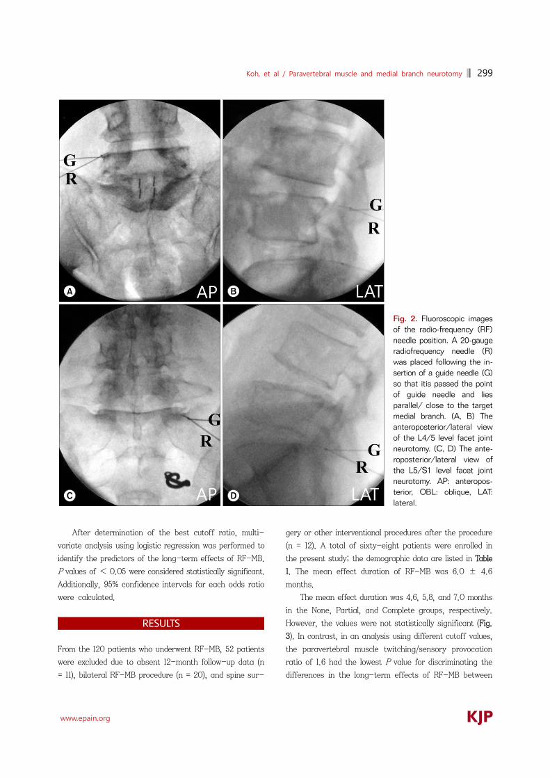
close to and in parallel with the target medial branch. With
the assistance of the prepositioned guide needle, the RF
needle was placed in the groove between the transverse
and superior articular processes, maximizing the contact
area between the active tip and the groove (Fig. 2A and
2B). For the L5 dorsal rami, the needle was advanced
through a groove between the sacral ala and the articular
process (Fig. 2C and 2D). When positioning the needle, the
needle was advanced carefully so that it did not pass the
anterior border of the superior articular process in the lat-
eral fluoroscopic view.
After the needle was in position, the sensory stim-
ulation at a frequency of 50 Hz was provided. The stim-
ulation was started at 0.1 V and slowly increased up to
0.5 V. If provocation was not observed until the cutoff val-
ue of 0.5 V. The needle was repositioned. In sequence, the
stimulation at a frequency of 2 Hz was performed to within
double the voltage level at which sensory provocation or
the cutoff value of 1 V was acquired. When the contraction
of the paravertebral muscle was observed, the voltage level
was recorded. If sensory or motor provocation was noted
in the lower extremities, the needle was repositioned. After
confirming that the needle was in position, 1 ml of 2% me-
pivacaine mixed with 1 mg of dexamethasone was injected
through the guide needle. RF lesioning was performed
twice using an RF generator (Pain Management Generator
230V PMG-230; Baylis Medical, Montreal, Canada), which
was able to maintain an 80°C lesioning temperature for 75
s.
One hundred twenty patients were confirmed with the
facet joint syndrome and enrolled in this study. All the di-
agnostic and therapeutic procedures were performed by
the same physician in this study. After the RF-MB, the
duration of the effective block was also recorded. Patients
were followed up for 12 months after the RF-MB. If the
patient’s maximum numeric pain intensity score decreased
to less than half of the initial pain score, then the proce-
dure was regarded as effective. The patients’ pain intensity
using the NRS (scores ranging from 0 to 10) and the dura-
tion of the effective period were investigated and recorded
for each patient at 3, 6, and 12 months after the proce-
dure. During the follow up period, only conservative oral
medication (nonsteroidal anti-inflammatory drugs) was
prescribed to patients.
The patients were grouped according to the adequacy
of the RF needle position when performing RF-MB as fol-
lows: “Complete,” when paravertebral muscle twitching
was observed at all needles; “Partial,” when twitching was
observed at 1 or 2 of the needles; and “None,” when no
twitching was observed for any of the needles.
1. Statistical analysis
Statistical analyses were performed using Statistical
Package for the Social Sciences (SPSS) version 18.0 soft-
ware (SPSS Inc., Chicago, IL, USA). ANOVA was used for
analysis of the long-term effect of RF-MB.
For a subgroup analysis, regrouping was performed
according to the different voltage level ratios. The ratio
was set by adjusting the criteria of adequate needle posi-
tioning as paravertebral muscle twitching was observed
within different multipliers of the voltage level where sen-
sory provocation was observed (1.0 to 2.0 in 0.1 intervals).
To determine the best cut off ratio for the para-
vertebral twitching/sensory stimulation voltage level, uni-
variate logistic regression was performed for each cutoff
value. The best cutoff ratio was determined as a value that
has the lowest P value for discriminating muscle twitching
on long term effect of RF-MB.
Koh, et al / Paravertebral muscle and medial branch neurotomy 299
www.epain.org
Fig. 2. Fluoroscopic images of the radio-frequency (RF) needle position. A 20-gauge radiofrequency needle (R) was placed following the in-sertion of a guide needle (G)so that itis passed the point of guide needle and lies parallel/ close to the target medial branch. (A, B) The anteroposterior/lateral view of the L4/5 level facet joint neurotomy. (C, D) The ante-roposterior/lateral view of the L5/S1 level facet joint neurotomy. AP: anteropos-terior, OBL: oblique, LAT: lateral.
After determination of the best cutoff ratio, multi-
variate analysis using logistic regression was performed to
identify the predictors of the long-term effects of RF-MB.
P values of < 0.05 were considered statistically significant.
Additionally, 95% confidence intervals for each odds ratio
were calculated.
RESULTS
From the 120 patients who underwent RF-MB, 52 patients
were excluded due to absent 12-month follow-up data (n
= 11), bilateral RF-MB procedure (n = 20), and spine sur-
gery or other interventional procedures after the procedure
(n = 12). A total of sixty-eight patients were enrolled in
the present study; the demographic data are listed in Table
1. The mean effect duration of RF-MB was 6.0 ± 4.6
months.
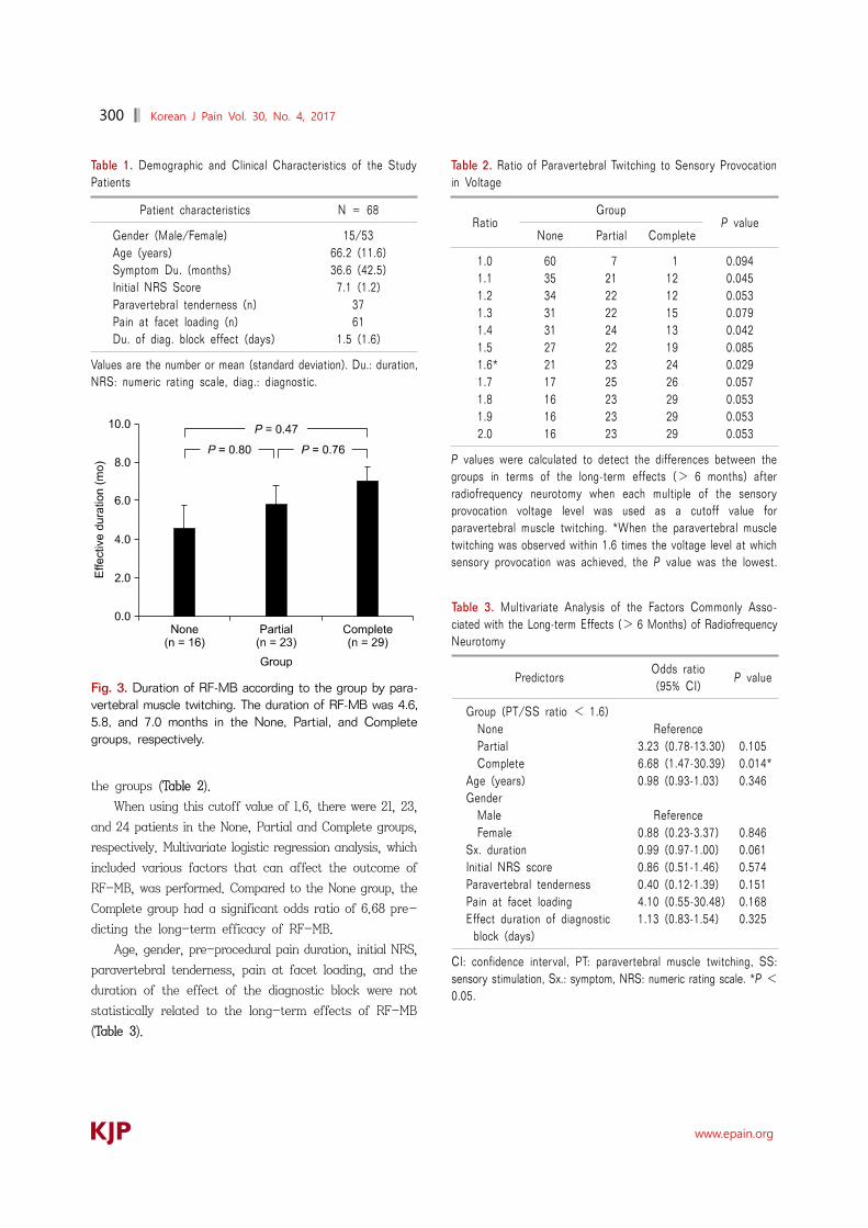
The mean effect duration was 4.6, 5.8, and 7.0 months
in the None, Partial, and Complete groups, respectively.
However, the values were not statistically significant (Fig.
3). In contrast, in an analysis using different cutoff values,
the paravertebral muscle twitching/sensory provocation
ratio of 1.6 had the lowest P value for discriminating the
differences in the long-term effects of RF-MB between
300 Korean J Pain Vol. 30, No. 4, 2017
www.epain.org
Table 1. Demographic and Clinical Characteristics of the Study Patients
Patient characteristics N = 68
Gender (Male/Female) 15/53Age (years) 66.2 (11.6)Symptom Du. (months) 36.6 (42.5)Initial NRS Score 7.1 (1.2)Paravertebral tenderness (n) 37Pain at facet loading (n) 61Du. of diag. block effect (days) 1.5 (1.6)
Values are the number or mean (standard deviation). Du.: duration,NRS: numeric rating scale, diag.: diagnostic.
Fig. 3. Duration of RF-MB according to the group by para-vertebral muscle twitching. The duration of RF-MB was 4.6,5.8, and 7.0 months in the None, Partial, and Complete groups, respectively.
Table 2. Ratio of Paravertebral Twitching to Sensory Provocation in Voltage
RatioGroup
P valueNone Partial Complete
1.0 60 7 1 0.0941.1 35 21 12 0.0451.2 34 22 12 0.0531.3 31 22 15 0.0791.4 31 24 13 0.0421.5 27 22 19 0.0851.6* 21 23 24 0.0291.7 17 25 26 0.0571.8 16 23 29 0.0531.9 16 23 29 0.0532.0 16 23 29 0.053
P values were calculated to detect the differences between the groups in terms of the long-term effects (> 6 months) after radiofrequency neurotomy when each multiple of the sensory provocation voltage level was used as a cutoff value for paravertebral muscle twitching. *When the paravertebral muscle twitching was observed within 1.6 times the voltage level at which sensory provocation was achieved, the P value was the lowest.
Table 3. Multivariate Analysis of the Factors Commonly Asso-ciated with the Long-term Effects (> 6 Months) of RadiofrequencyNeurotomy
PredictorsOdds ratio (95% CI)
P value
Group (PT/SS ratio < 1.6) None Reference Partial 3.23 (0.78-13.30) 0.105 Complete 6.68 (1.47-30.39) 0.014*Age (years) 0.98 (0.93-1.03) 0.346Gender Male Reference Female 0.88 (0.23-3.37) 0.846Sx. duration 0.99 (0.97-1.00) 0.061Initial NRS score 0.86 (0.51-1.46) 0.574Paravertebral tenderness 0.40 (0.12-1.39) 0.151Pain at facet loading 4.10 (0.55-30.48) 0.168Effect duration of diagnostic
block (days)1.13 (0.83-1.54) 0.325
CI: confidence interval, PT: paravertebral muscle twitching, SS: sensory stimulation, Sx.: symptom, NRS: numeric rating scale. *P <0.05.
the groups (Table 2).
When using this cutoff value of 1.6, there were 21, 23,
and 24 patients in the None, Partial and Complete groups,
respectively. Multivariate logistic regression analysis, which
included various factors that can affect the outcome of
RF-MB, was performed. Compared to the None group, the
Complete group had a significant odds ratio of 6.68 pre-
dicting the long-term efficacy of RF-MB.
Age, gender, pre-procedural pain duration, initial NRS,
paravertebral tenderness, pain at facet loading, and the
duration of the effect of the diagnostic block were not
statistically related to the long-term effects of RF-MB
(Table 3).

Koh, et al / Paravertebral muscle and medial branch neurotomy 301
www.epain.org
DISCUSSION
According to the results of the present study, the mean
effect duration of RF-MB did not increase significantly
when more paravertebral muscle twitching was observed
during the procedure. However, when using different cutoff
values, higher probability of a longer effect duration (>
6 months) was expected when paravertebral muscle
twitching was observed at every RF-MB needle with a
statistically significant difference. When we performed the
sensory and motor stimulation at 50 Hz with a 0.5 V cutoff
value and at 2 Hz with a 1 V cutoff value, respectively, the
findings of the paravertebral muscle twitch may have pos-
sibility as a predictive factor in RF-MB.
When performing RF denervation, a spheroid-shaped
lesion is created, with its long axis parallel to the RF
needle. Thus, when the needle position is not close enough
or parallel to the target medial branch, sufficient nerve de-
nervation cannot be achieved [13,14]. In addition to using
correct anatomical landmark based techniques, sensory
stimulation has often been used to determine the proximity
of the RF needle to the target nerve [3,4]. However, using
sensory stimulation as the only method of identifying the
RF needle proximity is unreliable for several reasons. First,
the stimulation of the ligaments, muscles, or periosteum
near the target nerve can produce similar sensory provo-
cation [7]. Sensory conduction block, which occurs when
the nerve is located close to the electrode needle, may also
result in misinterpretations [15].
In addition, demographic, cultural, or psychological
factors decrease the reliability of sensory stimulation [16].
Therefore, the presence of paravertebral muscle twitching
during RF-MB has been used as an adjuvant method, and
positive outcomes have been reported [17].
The method of identifying paravertebral muscle twitch-
ing during RF-MB has several advantages. For instance,
this more objective measure of needle proximity is more
reliable for both patients and clinicians. In addition, this
method could be utilized with inpatients who are sedated,
intellectually handicapped, or unable to communicate. One
report noted that long-lasting effects could be achieved
after confirming denervation at the multifidus muscle fol-
lowing RF-MB by monitoring the action potentials [18].
However, the method of identifying paravertebral mus-
cle twitching also has its disadvantages when used as the
primary means of needle positioning during RF-MB.
Visually identifying paravertebral muscle twitching through
cutaneous skin movement is problematic, as other muscles
or nerves that are located near the target nerve produce
similar twitching when stimulated [8,19]. [For example, the
iliocostalis lumborum or longissimus muscle twitching that
is evoked by the lateral or intermediate branch nerve stim-
ulation mimics the multifidus muscle twitching evoked by
the medial branch nerve stimulation. In addition, observing
the paravertebral muscle twitching may be difficult in pa-
tients with paravertebral muscle atrophy or obesity
[20-22]. Hence, we used the method of identifying the
paravertebral muscle twitching as an adjuvant only when
sensory provocation at < 0.5 V was confirmed [23].
The best cutoff value for sensory provocation during
RF-MB has not been firmly established. However, when
sensory provocation was performed at values of < 0.5 V,
no statistical differences were noted between the voltage
level and therapeutic outcomes. Therefore, a cutoff value
of 0.5 V was used when performing the sensory provoca-
tion in the present study.
There have been several studies investigating the
prognostic factors of positive outcomes after RF-MB. The
results suggested that strict indication criteria and ad-
equate diagnostic blocks appear to be closely related to the
outcome of RF-MB. While the patient’s symptoms and
physical examinations may provide some evidence, studies
suggest that diagnostic blocks are indispensable for mak-
ing a reliable diagnosis of facet joint syndrome. Direct fac-
et joint intra-articular injection and MBB are the two most
commonly used diagnostic blocks for facet joint syndrome,
although research suggests that MBB is more reliable than
facet joint intra-articular injection [24]. However, the
methodologies used by clinicians when performing or in-
terpreting the results of diagnostic MBBs often differ. For
example, Hooten et al. [22] recommended using a com-
parative MBB with different local anesthetics. The volume
of local anesthetics used for diagnostic MBBs also vary,
ranging from 0.3 to 1.0 ml. However, as previous studies
have reported incidences of motor block in outpatient set-
tings [9,25], we used less than 0.5 ml of 0.5% bupivacaine
in this study. Additionally, clinicians use different cutoff
values for determining the level of pain relief after the di-
agnostic block.
Bogduk recommended that pain relief of more than
80% should be used as a cutoff value after the diagnostic
block [25], while Derby et al. [26] recommended a cutoff

302 Korean J Pain Vol. 30, No. 4, 2017
www.epain.org
value of 80% for single MBBs and 70% for double MBBs.
In contrast, Cohen et al. [3] reported no significant bene-
fits when the cutoff values for pain relief after diagnostic
MBBs were higher than 50%.
Furthermore, it was difficult for some patients to ex-
press whether they experienced a decrease in their pain
intensity according to exact values such as 70% or 80%.
In the present study, we used a cutoff value of 50% for
pain relief experienced by the patient after the diagnostic
MBB. Thus, by using a cutoff value that could be assessed
using the word “half,” we attempted to increase patient
compliance and understanding.
In the present study, we also investigated the prog-
nostic value of various demographic factors. Though not
statistically significant, we observed a negative correlation
between the female sex and RF-MB outcome. A higher
proportion of women may experience pain that is > 50%
of their initial pain after RF-MB due to their greater sen-
sitivity to pain relative to men [18]. This might be one of
the reasons that the overall outcome of this study was not
as effective as in the previous literature [9,27]. We also
examined the pain duration before the procedure and the
patients’ baseline NRS scores. No statistically significant
relationships were noted between either the pain duration
or baseline NRS and RF-MB outcome. Furthermore, the
presence of paravertebral tenderness, pain at facet load-
ing, and effective diagnostic block duration were not re-
lated to the RF-MB outcome in the present study. However,
although no statistically significant correlations were ob-
served, the positive or negative directions of the odds ra-
tios for these factors in our study were similar to the val-
ues reported in other studies [28]. Therefore, studies that
include more patients and employ controlled designs may
be able to identify statistically significant results for these
factors.
As mentioned previously, only a few statistically sig-
nificant prognostic factors can be monitored or adjusted
while performing RF-MB. However, the results of the
present study suggest that a better outcome could be an-
ticipated when paravertebral muscle twitching is encoun-
tered during RF-MB in combination with sensory provoca-
tion at < 0.5 V. Therefore, by attempting to achieve both
sensory stimulation and paravertebral muscle twitching
during the procedure, we may be able to increase the ther-
apeutic effects of RF-MB.
In addition, patients with paravertebral muscle twitch-
ing may have preserved muscular structures supporting
the spine, which make conservative rehabilitation therapies
more feasible [21,25]. Therefore, complete paravertebral
muscle twitching may indicate not only the correct RF nee-
dle position but also the amount of paravertebral muscle
strength needed to preserve the vertebral column and fac-
et joint.
The present study has several limitations. This is a
retrospective study and the pre/post-procedural medical or
psychological factors were not controlled. Actually, almost
all patients in this study had chronic degenerative muscu-
loskeletal pain and they had been taking several an-
algesics, which was not changed after the procedure.
However, during the follow up period, the participants were
asked to notify their aggravated or improved facet joint
pain. Moreover, while we regularly monitored the pain pro-
file of each patient after the procedure, bias may have
been introduced as these pain ratings were dependent upon
the patient’s memory regarding their NRS scores.
The best cutoff value of voltage levels used in the sub-
group analysis also might be calculated differently if
RF-MB was performed using different methods. However,
by using the results of this study as reference data, we
expect that further studies might provide more reliable in-
formation for the prediction of prognosis of RF-MB in clin-
ical settings.
In conclusion, paravertebral muscle twitching while
performing lumbar RF-MB may be a reliable predictor of
long-term efficacy when sensory provocation under 0.5 V
is achieved. However, further investigation may be neces-
sary for clarifying its clinical significance.
REFERENCES
1. Beresford ZM, Kendall RW, Willick SE. Lumbar facet syndromes. Curr Sports Med Rep 2010; 9: 50-6.
2. Cavanaugh JM, Ozaktay AC, Yamashita T, Avramov A, Getchell TV, King AI. Mechanisms of low back pain: a neurophysiologic and neuroanatomic study. Clin Orthop Relat Res 1997: 166-80.
3. Cohen SP, Strassels SA, Kurihara C, Griffith SR, Goff B, Guthmiller K, et al. Establishing an optimal "cutoff" threshold for diagnostic lumbar facet blocks: a prospective correlational study. Clin J Pain 2013; 29: 382-91.
4. Lakemeier S, Lind M, Schultz W, Fuchs-Winkelmann S, Timmesfeld N, Foelsch C, et al. A comparison of intraarticular lumbar facet joint steroid injections and lumbar facet joint radiofrequency denervation in the treatment of low back

Koh, et al / Paravertebral muscle and medial branch neurotomy 303
www.epain.org
pain: a randomized, controlled, double-blind trial. Anesth Analg 2013; 117: 228-35.
5. Manchikanti L, Manchikanti KN, Manchukonda R, Cash KA, Damron KS, Pampati V, et al. Evaluation of lumbar facet joint nerve blocks in the management of chronic low back pain: preliminary report of a randomized, double-blind controlled trial: clinical trial NCT00355914. Pain Physician 2007; 10: 425-40.
6. Rocha ID, Cristante AF, Marcon RM, Oliveira RP, Letaif OB, Barros Filho TE. Controlled medial branch anesthetic block in the diagnosis of chronic lumbar facet joint pain: the value of a three-month follow-up. Clinics (Sao Paulo) 2014; 69: 529-34.
7. Shin KM, Choi SE, Yun SH, Lim SY, Jung BH, Lee KH, et al. New more reliable indicator for confirmation of the medial branch in radiofrequency neurotomy. J Korean Pain Soc 2000; 13: 242-6.
8. Bogduk N, Wilson AS, Tynan W. The human lumbar dorsal rami. J Anat 1982; 134: 383-97.
9. Dreyfuss P, Halbrook B, Pauza K, Joshi A, McLarty J, Bogduk N. Efficacy and validity of radiofrequency neurotomy for chronic lumbar zygapophysial joint pain. Spine (Phila Pa 1976) 2000; 25: 1270-7.
10. Zhou X, Liu Y, Zhou S, Fu XX, Yu XL, Fu CL, et al. The correlation between radiographic and pathologic grading of lumbar facet joint degeneration. BMC Med Imaging 2016; 16: 27.
11. Wilde VE, Ford JJ, McMeeken JM. Indicators of lumbar zygapophyseal joint pain: survey of an expert panel with the Delphi technique. Phys Ther 2007; 87: 1348-61.
12. Fenten MG, Schoenmakers KP, Heesterbeek PJ, Scheffer GJ, Stienstra R. Effect of local anesthetic concentration, dose and volume on the duration of single-injection ultrasound- guided axillary brachial plexus block with mepivacaine: a randomized controlled trial. BMC Anesthesiol 2015; 15: 130.
13. Choi EJ, Choi YM, Jang EJ, Kim JY, Kim TK, Kim KH. Neural ablation and regeneration in pain practice. Korean J Pain 2016; 29: 3-11.
14. Kweon TD, Kim JY, Lee HY, Kim MH, Lee YW. Anatomical analysis of medial branches of dorsal rami of cervical nerves for radiofrequency thermocoagulation. Reg Anesth Pain Med 2014; 39: 465-71.
15. Li J, Kong X, Gozani SN, Shi R, Borgens RB. Current- distance relationships for peripheral nerve stimulation locali-zation. Anesth Analg 2011; 112: 236-41.
16. Fillingim RB, King CD, Ribeiro-Dasilva MC, Rahim-Williams B, Riley JL 3rd. Sex, gender, and pain: a review of recent
clinical and experimental findings. J Pain 2009; 10: 447-85. 17. van Kleef M, Barendse GA, Kessels A, Voets HM, Weber WE,
de Lange S. Randomized trial of radiofrequency lumbar facet denervation for chronic low back pain. Spine (Phila Pa 1976) 1999; 24: 1937-42.
18. Kanchiku T, Imajo Y, Suzuki H, Yoshida Y, Nishida N, Taguchi T. Percutaneous radiofrequency facet joint denervation with monitoring of compound muscle action potential of the multifidus muscle group for treating chronic low back pain: a preliminary report. J Spinal Disord Tech 2014; 27: E262-7.
19. Baek SO, Ahn SH, Jones R, Cho HK, Jung GS, Cho YW, et al. Activations of deep lumbar stabilizing muscles by transcutaneous neuromuscular electrical stimulation of lumbar paraspinal regions. Ann Rehabil Med 2014; 38: 506-13.
20. Cohen SP, Strassels SA, Kurihara C, Lesnick IK, Hanling SR, Griffith SR, et al. Does sensory stimulation threshold affect lumbar facet radiofrequency denervation outcomes? A prospective clinical correlational study. Anesth Analg 2011; 113: 1233-41.
21. Wallwork TL, Stanton WR, Freke M, Hides JA. The effect of chronic low back pain on size and contraction of the lumbar multifidus muscle. Man Ther 2009; 14: 496-500.
22. Hooten WM, Martin DP, Huntoon MA. Radiofrequency neurotomy for low back pain: evidence-based procedural guidelines. Pain Med 2005; 6: 129-38.
23. Veizi E, Hayek S. Interventional therapies for chronic low back pain. Neuromodulation 2014; 17 Suppl 2: 31-45.
24. Cohen SP, Moon JY, Brummett CM, White RL, Larkin TM. Medial branch blocks or intra-articular injections as a prognostic tool before lumbar facet radiofrequency dener-vation: a multicenter, case-control study. Reg Anesth Pain Med 2015; 40: 376-83.
25. Bogduk N. Evidence-informed management of chronic low back pain with facet injections and radiofrequency neuro-tomy. Spine J 2008; 8: 56-64.
26. Derby R, Melnik I, Lee JE, Lee SH. Correlation of lumbar medial branch neurotomy results with diagnostic medial branch block cutoff values to optimize therapeutic outcome. Pain Med 2012; 13: 1533-46.
27. MacVicar J, Borowczyk JM, MacVicar AM, Loughnan BM, Bogduk N. Lumbar medial branch radiofrequency neurotomy in New Zealand. Pain Med 2013; 14: 639-45.
28. Holz SC, Sehgal N. What is the correlation between facet joint radiofrequency outcome and response to comparative medial branch blocks? Pain Physician 2016; 19: 163-72.