reintegration of functional movement of the upper quarter
TRANSCRIPT

Reintegration of Functional Movement of the Upper Quarter
Lisa DeStefano, DO
Professor and Chair
Department of Osteopathic Manipulative Medicine
College of Osteopathic Medicine
Michigan State University
A significant problem in studying overuse injuries is that there are multiple interactions among the various risk
factors making it very difficult to determine the etiology of the injury.
Contributions to NMSK
System Function
• Dynamic Stability• Requires optimal local and
global stabilizing system function that ensures vertebral load is optimally distributed to all supportive structures.• “provide sufficient stability to
the spine to match the instantaneously varying stability demands due to changes in spinal posture, and static and dynamic loads”

Thoracic Stability
• Although the literature is not clear clinically, thorax stability is dependent on lumbopelvic stability and upper extremity and neck stability is dependent on thorax stability.

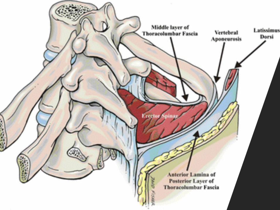
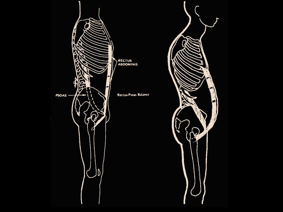
erector spinae muscle transmission of force onto the epaxial fascia/vertebral aponeurosis
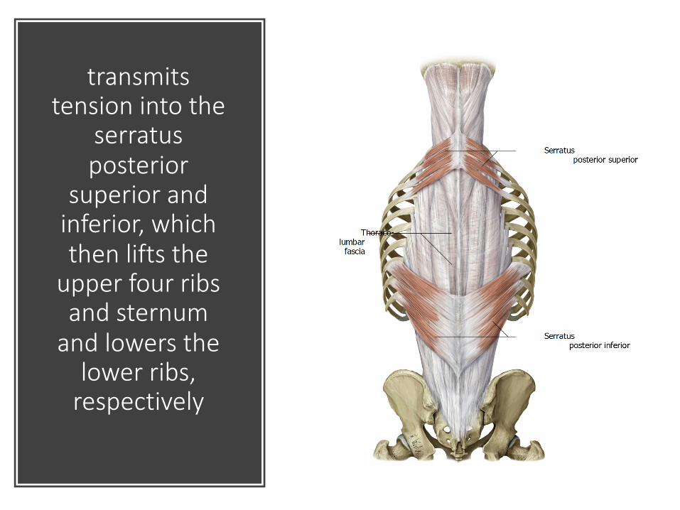
transmits tension into the
serratus posterior
superior and inferior, which then lifts the
upper four ribs and sternum
and lowers the lower ribs,
respectively
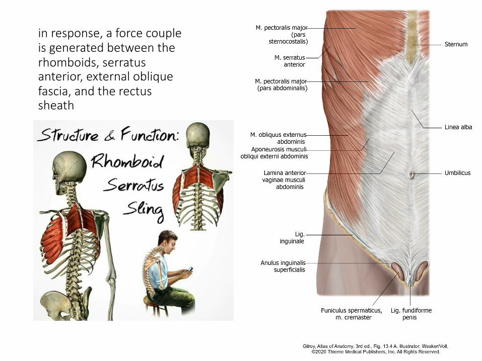
in response, a force couple is generated between the rhomboids, serratus anterior, external oblique fascia, and the rectus sheath
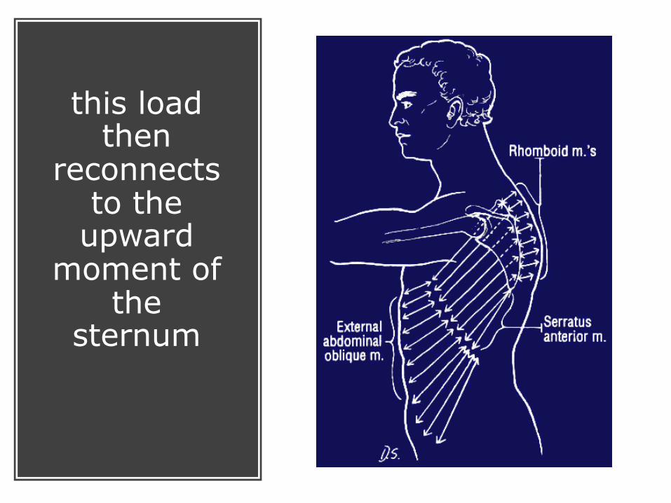
this load then
reconnects to the
upward moment of
the sternum
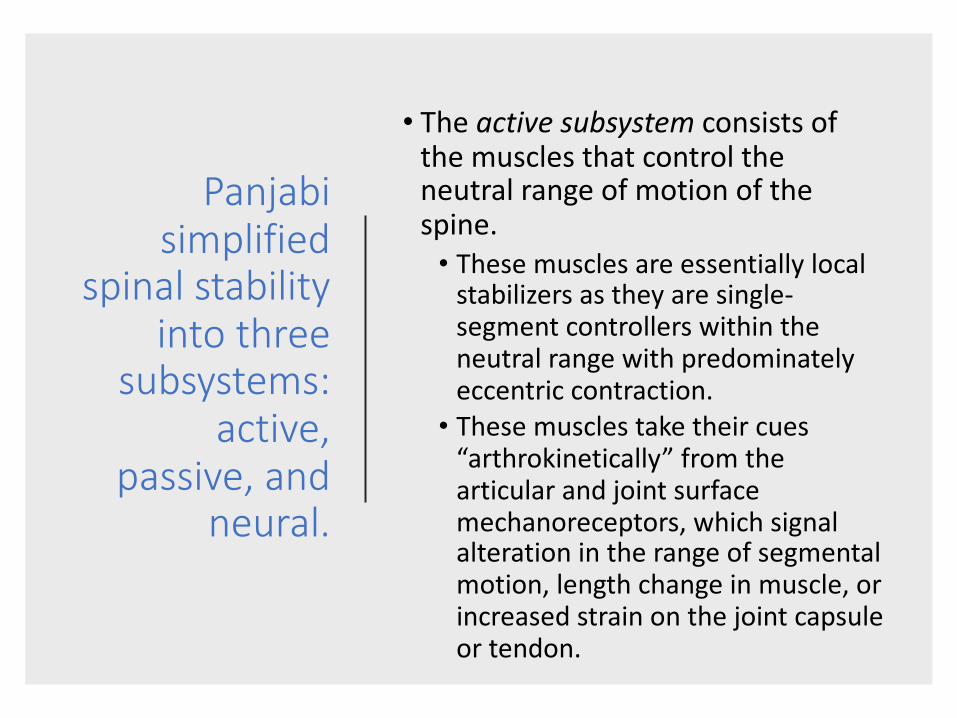
Panjabi simplified
spinal stability into three
subsystems: active,
passive, and neural.
• The active subsystem consists of the muscles that control the neutral range of motion of the spine. • These muscles are essentially local
stabilizers as they are single-segment controllers within the neutral range with predominately eccentric contraction. • These muscles take their cues
“arthrokinetically” from the articular and joint surface mechanoreceptors, which signal alteration in the range of segmental motion, length change in muscle, or increased strain on the joint capsule or tendon.

Panjabi simplified spinal stability into three subsystems: active, passive, and neural.
The passive subsystem consists of bone, the joint capsule, intervertebral disk, aponeuroses, fascia and supporting ligaments.
The neural subsystemconsists of the mechanoreceptors within the passive subsystem and the proprioceptors within the active subsystem, both of which feedback information to the spinal cord as well as to the active subsystem.
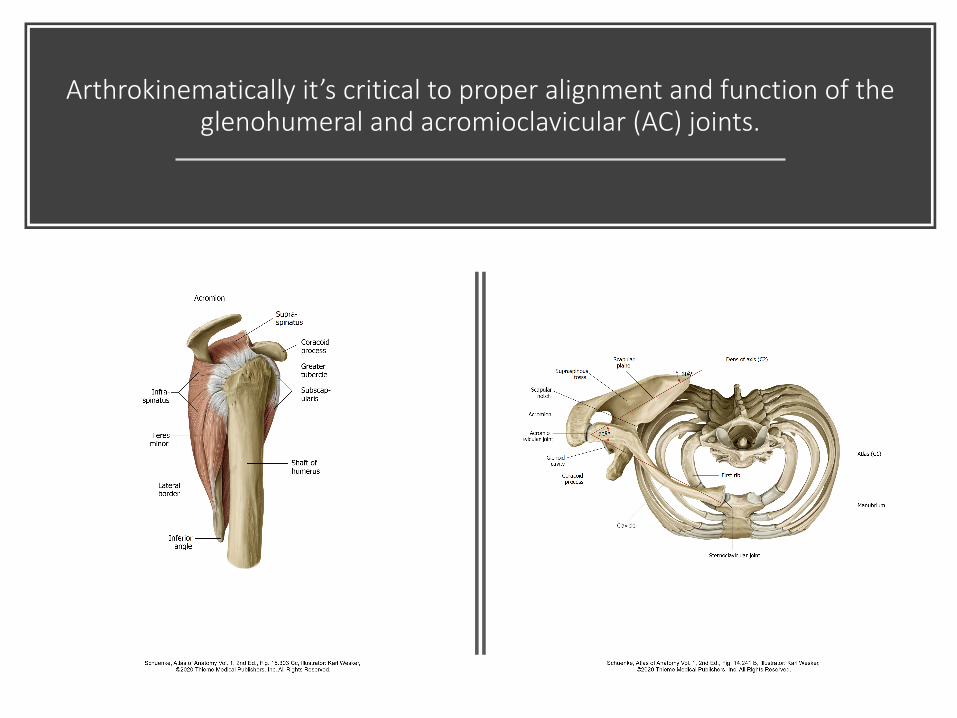
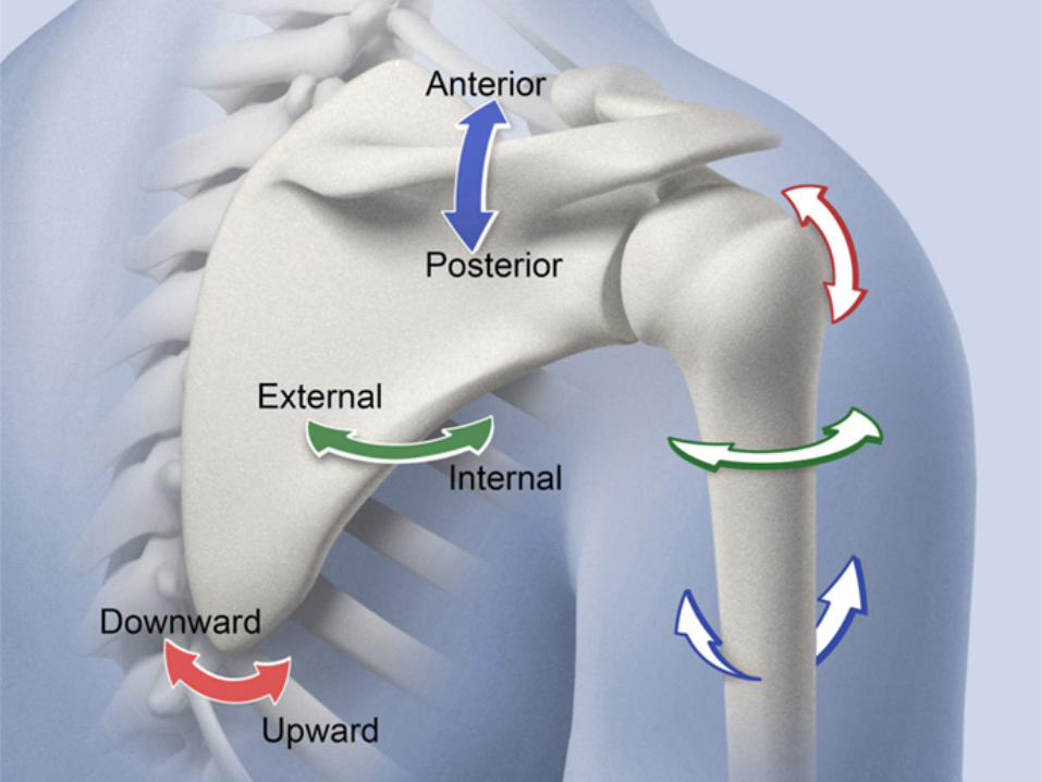
Arthrokinematically it’s critical to proper alignment and function of the glenohumeral and acromioclavicular (AC) joints.
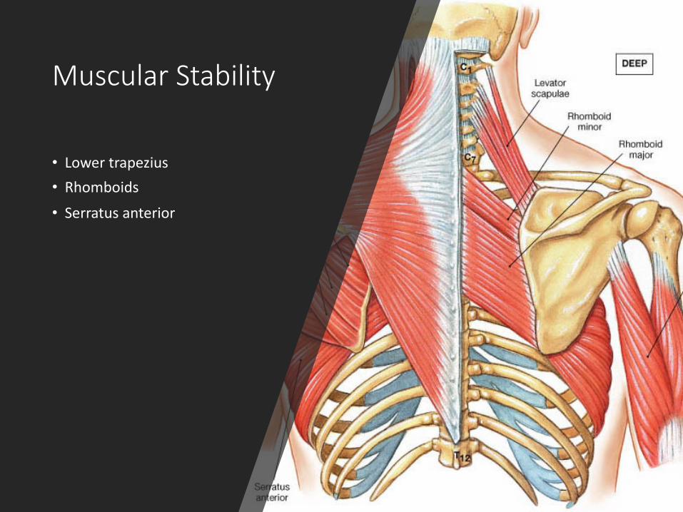
Muscular Stability
• Lower trapezius• Rhomboids
• Serratus anterior
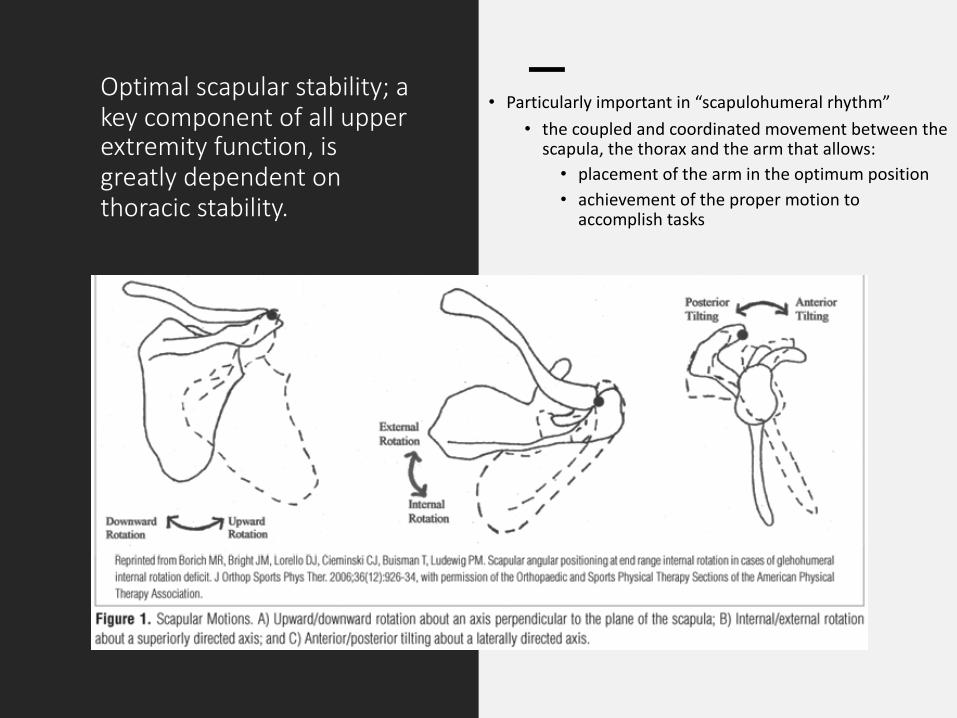
Optimal scapular stability; a key component of all upper extremity function, is greatly dependent on thoracic stability.
• Particularly important in “scapulohumeral rhythm”• the coupled and coordinated movement between the
scapula, the thorax and the arm that allows:• placement of the arm in the optimum position• achievement of the proper motion to
accomplish tasks
Optimal scapular stability is a key component of all upper extremity function.
• Biomechanically• the scapula provides a stable base for muscle activation and a moving
platform to maintain ball-and socket kinematics• It also serves as an efficient link between the core, which develops force,
and the arm, which delivers the force.
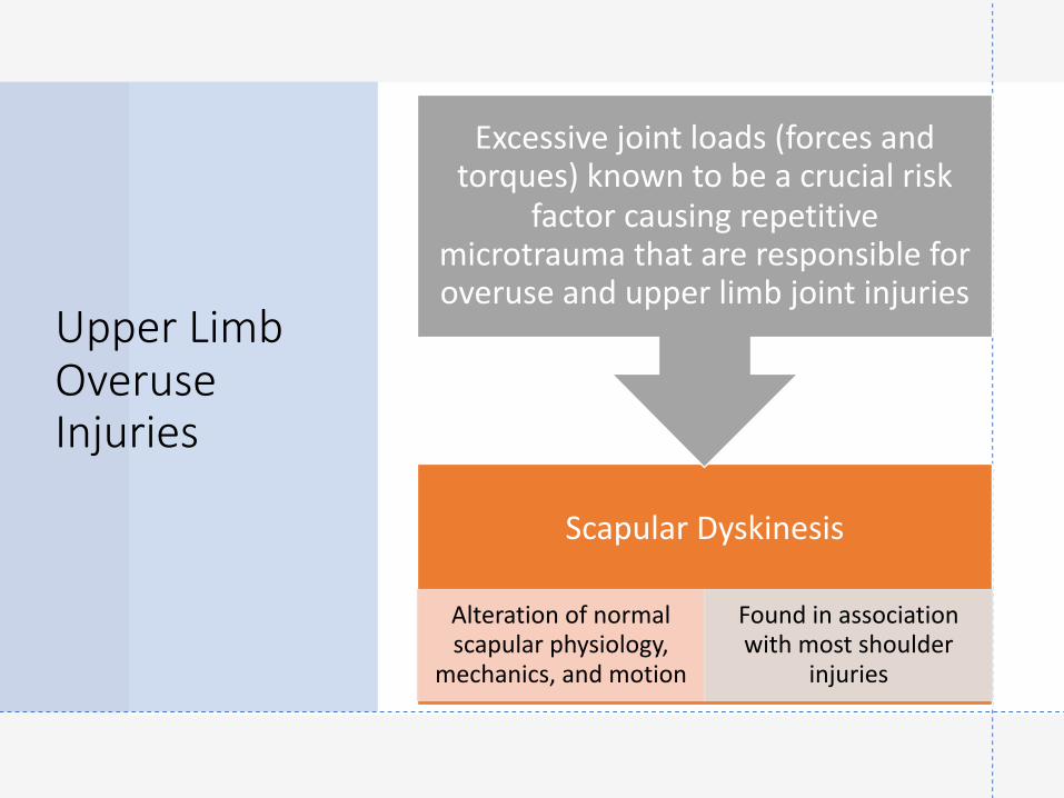
Upper Limb Overuse Injuries
Scapular Dyskinesis
Alteration of normal scapular physiology,
mechanics, and motion
Found in association with most shoulder
injuries
Excessive joint loads (forces and torques) known to be a crucial risk
factor causing repetitive microtrauma that are responsible for overuse and upper limb joint injuries
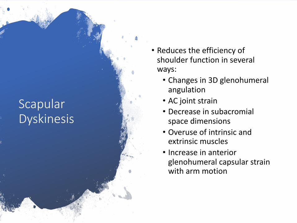
Scapular Dyskinesis
• Reduces the efficiency of shoulder function in several ways:• Changes in 3D glenohumeral
angulation• AC joint strain• Decrease in subacromial
space dimensions• Overuse of intrinsic and
extrinsic muscles• Increase in anterior
glenohumeral capsular strain with arm motion

Disintegration of pelvis, thoracic, cervical and scapular stability.
• Primary etiology of scapular dyskinesis and associated sequalae.
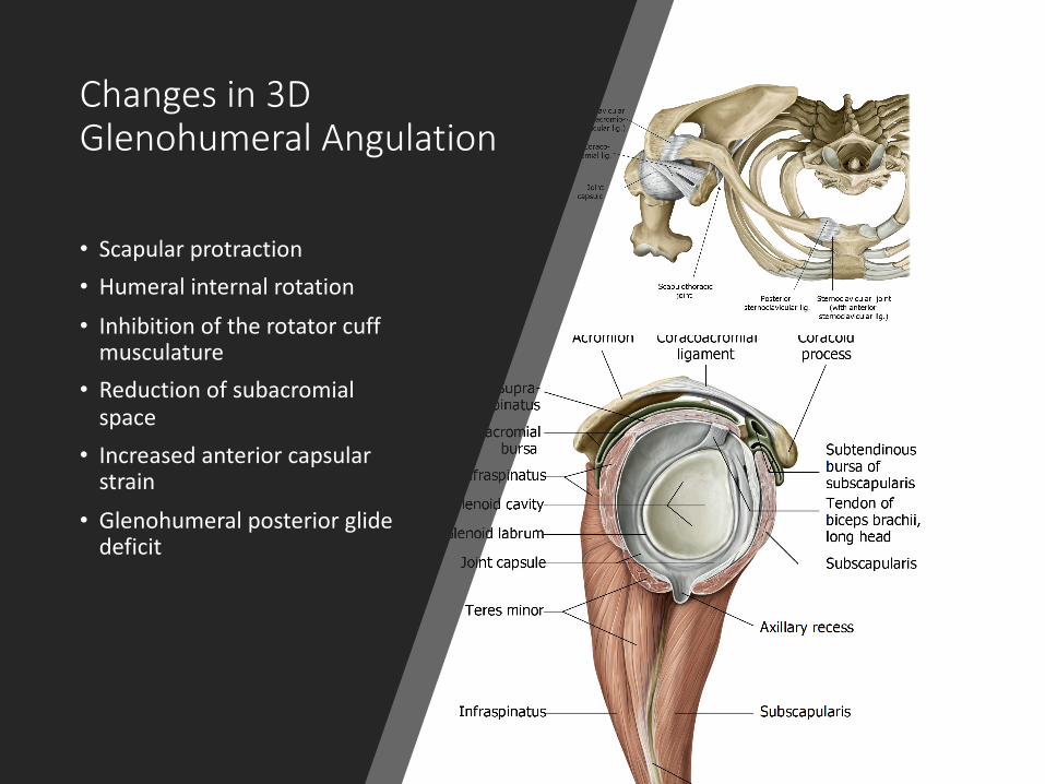
Changes in 3D Glenohumeral Angulation
• Scapular protraction• Humeral internal rotation
• Inhibition of the rotator cuff musculature
• Reduction of subacromial space
• Increased anterior capsular strain
• Glenohumeral posterior glide deficit
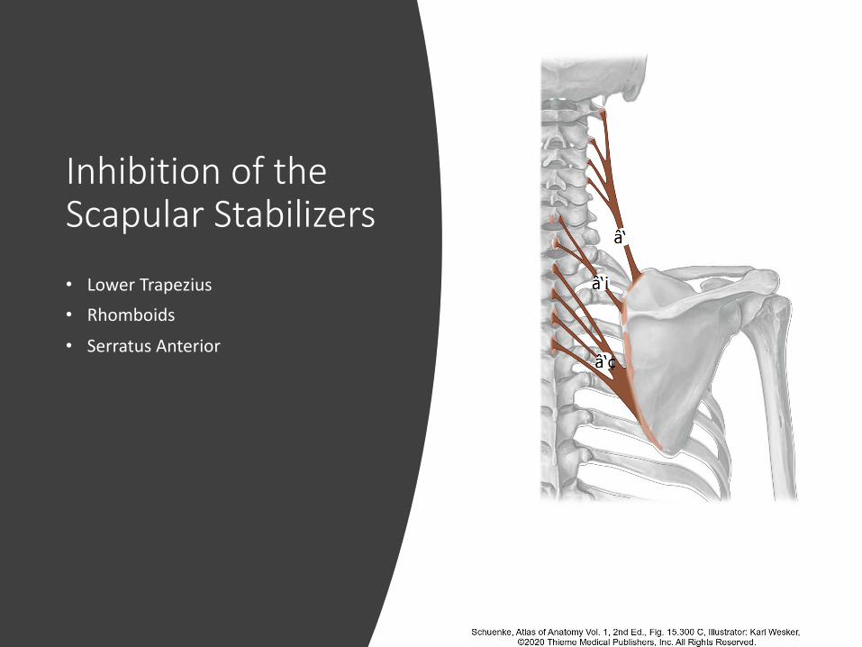
Inhibition of the Scapular Stabilizers
• Lower Trapezius• Rhomboids
• Serratus Anterior

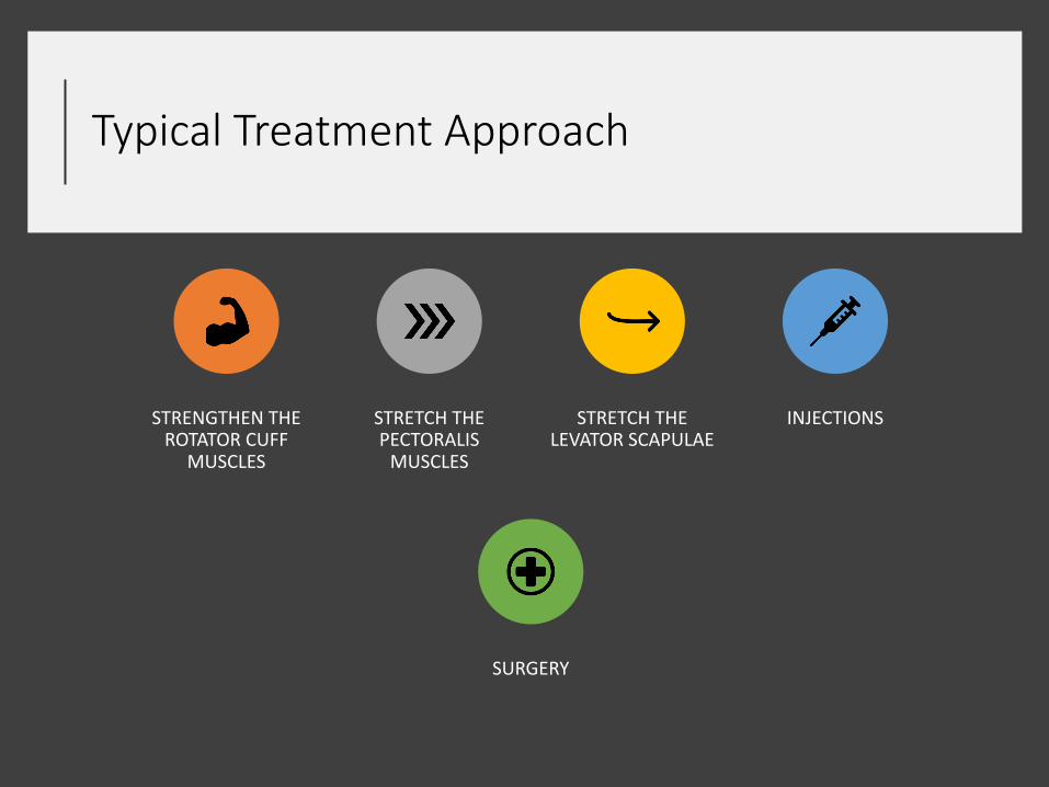
Typical Treatment Approach
STRENGTHEN THE ROTATOR CUFF
MUSCLES
STRETCH THE PECTORALIS
MUSCLES
STRETCH THE LEVATOR SCAPULAE
INJECTIONS
SURGERY
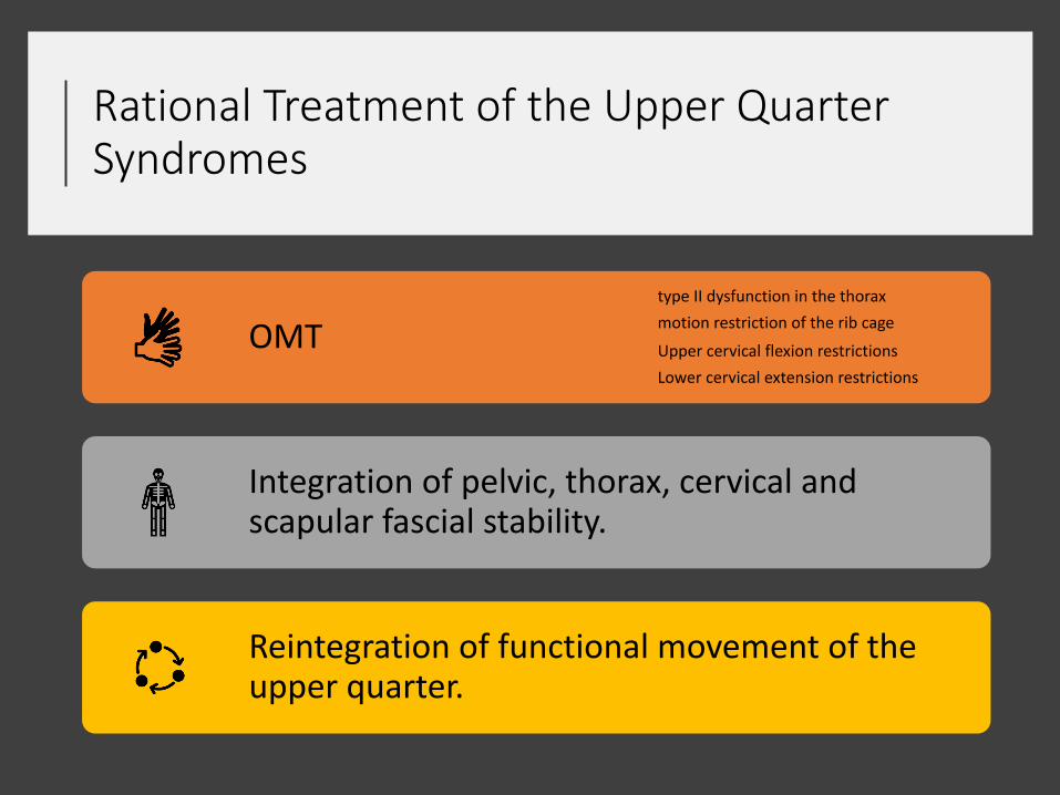
Rational Treatment of the Upper Quarter Syndromes
OMTtype II dysfunction in the thoraxmotion restriction of the rib cage
Upper cervical flexion restrictionsLower cervical extension restrictions
Integration of pelvic, thorax, cervical and scapular fascial stability.
Reintegration of functional movement of the upper quarter.

Thank You!