reindeer health aide manual - university of alaska fairbanks
TRANSCRIPT

Reindeer Health Aide Manual
Prepared byRobert A. Dieterich, D.V.M.
Jamie K. Morton, Ph.D.First Edition 1981
Second Edition 1990
Illustrated by Susan L. Kraft
AFES Misc. Pub 90-4CES 100H-00046
Agricultural and Forestry Experiment Station, Cooperative Extension Service,University of Alaska Fairbanks
and
U. S. Department of Agriculture cooperating

TABLE OF CONTENTSPage
1 ANATOMY OF REINDEERThe skeleton........................................................................................................................ 1Skull .................................................................................................................................... 1Vertebrae ............................................................................................................................. 1Ribs ..................................................................................................................................... 1Limbs .................................................................................................................................. 1The organs .......................................................................................................................... 3
2 PHYSIOLOGY OF REINDEERCirculation .......................................................................................................................... 5Respiration .......................................................................................................................... 5Digestion & Nutrition ......................................................................................................... 8Urogenital System ............................................................................................................ 10Antler Growth ................................................................................................................... 13Body Heat Control ............................................................................................................ 13Determination of Age by Tooth Wear ............................................................................... 14
3 DISEASES OF REINDEERBacteria and Viruses ......................................................................................................... 16Immunity to Disease ......................................................................................................... 17Brucellosis ........................................................................................................................ 21Rabies ............................................................................................................................... 26Respiratory Diseases......................................................................................................... 27Foot Rot ............................................................................................................................ 28Mandibular Lesions .......................................................................................................... 28Setaria ............................................................................................................................... 28Fibropapillomas ................................................................................................................29Keratitis (White-Eye) ....................................................................................................... 29Abortion ............................................................................................................................ 29Broken Antlers .................................................................................................................. 29
4 PARASITOLOGY OF REINDEERParasites ............................................................................................................................ 30Warbles ............................................................................................................................. 30Nasal Bots ......................................................................................................................... 33Internal Parasites............................................................................................................... 35Page
5 COMMON MEDICAL TREATMENTS FOR REINDEERFirst Aid ............................................................................................................................ 42Castration .......................................................................................................................... 42Aid in Fawning .................................................................................................................43
6 NECROPSY OF REINDEERNecropsy Procedures and techniques ............................................................................... 47Specimen Preparation ....................................................................................................... 47Tissue Collection ..............................................................................................................50Causative Agents .............................................................................................................. 51

APPENDICESAppendix I Tissue Collection ........................................................................................... 52Appendix II Serum Collection.......................................................................................... 53Appendix III Whole Blood Collection for Complete Blood Count .................................. 54Appendix IV Care and Use of Syringes ........................................................................... 55Appendix V Label and Use of Ivermectin ........................................................................ 59Appendix VI Specimen Submission for Rabies Assay ..................................................... 60Appendix VII Raising Orphan Reindeer Fawns ............................................................... 63Appendix VIII Nutrition ................................................................................................... 68Appendix IX Normal Blood Values for Reindeer ............................................................ 64Appendix X Glossary ....................................................................................................... 85
LIST OF FIGURESPage
Anatomy OF REINDEERFigure 1. Skeleton of the reindeer ........................................................................................... 2Figure 2. Internal organs of the reindeer ................................................................................. 4
PHYSIOLOGY OF REINDEERFigure 1. Structure of the heart ...............................................................................................6Figure 2. Structure of the lungs ...............................................................................................7Figure 3. Ruminant stomach of the reindeer ........................................................................... 9Figure 4. Female urogenital system in reindeer .................................................................... 11Figure 5. Male urogenital system in reindeer ....................................................................... 12Figure 6. Age determination of reindeer by tooth wear ........................................................ 15
DISEASE OF REINDEERFigure 1. Natural disease response ........................................................................................ 18Figure 2. Vaccination ............................................................................................................ 19Figure 3. Overwhelming infection ........................................................................................ 20
Parasites OF REINDEERFigure 1. Warble flies ............................................................................................................ 31Figure 2. Nasal bots .............................................................................................................. 34Figure 3. Roundworms .......................................................................................................... 35Figure 4. Lungworms ............................................................................................................36Figure 5. Tapeworms ............................................................................................................. 37Figure 6. Wild carnivore-wild ruminant tapeworms ............................................................. 38Figure 7. Echinococcus .........................................................................................................40Figure 8. Sarcocystis ............................................................................................................. 41
COMMON MEDICAL TREATMENTS FOR REINDEERFigure 1. Normal presentation of fawn at birth ..................................................................... 44Figure 2. Some abnormal presentations of the fawn ............................................................. 44
NECROPSY OF REINDEERFigure 1. Skinning the upper half of the body ...................................................................... 48Figure 2. Disarticulating the left hip joint ............................................................................. 49Figure 3. View of chest and abdomen fully exposed ............................................................ 50
APPENDICESFigure 1. Assembly of multiple dose syringe unit ................................................................ 56
INTRODUCTIONThe first edition of the Reindeer Health Aide Manual was printed in 1981. It represented a
written summary of information presented at a number of Reindeer Health Aide Workshops spon-sored by the University of Alaska and the Alaska Reindeer Herders’ Association. These workshopswere designed to provide herders with basic reindeer health information so they could recognizediseased animals, collect samples, and treat various ailments of reindeer in cooperation with andunder the supervision of agency personnel and veterinarians. Over the past several years there hasbeen an increasing interest in ownership of reindeer by individuals both within and outside the stateof Alaska. These individuals routinely request information on the care of their reindeer. New drugsand vaccines have been developed since this manual was last published. These products are nowdescribed and discussed in the text. Products no longer in use have been deleted. Additionally, basicblood test parameters are listed to aid owners working with veterinarians who have not had experi-ence in the treatment of reindeer diseases. It is not the purpose of this manual to describe eachsubject in great detail or in technical terms. Some items may seem overly simplified, but most peopleusing this manual are not biologists, and every effort has been made to address their needs.

The SkeletonThe skeleton of an animal is a hard, bony framework which provides internal support and form
to the body. It also supplies points of attachment for the muscles which enables an animal to makestrong physical movement. Generally, the types of bones making up the skeleton are the same in allanimals. Figure 1 identifies the major bones of the reindeer skeleton. These will be discussed in thefollowing paragraphs.
Skull: The skull is formed by the fusion of many smaller bones. It provides a protective case forthe brain and contains the bones of the nasal cavity, or turbinates. The upper jaw, or maxilla, isformed by the front lower surface of the skull. The lower jaw, or mandible, moves on a hinge to openand close the mouth. The reindeer’s teeth are embedded in the bone of the upper and lower jaw. Onthe crown of the skull are the two short permanent stems, or pedicles, from which the antlers growevery year.
Vertebrae: The vertebrae are bones which fit together in a column that makes up the neck andbackbone. Running through a canal in the vertebrae is the spinal cord which transmits messagesbetween the brain and body. The vertebrae, then, serves to protect the spinal cord from injury. Thecervical vertebrae are in the neck, the thoracic vertebrae in the chest area, and the lumbar vertebraein the lower back area. They fuse in the pelvic region and continue back to become the smaller tailbones.
Ribs: The ribs form a cage of bones that encase and protect the lungs and heart; they also giveform to the chest and play a role in breathing.
Limbs: The front limb attaches by a large muscle mass connecting the shoulder blade, or thescapula, to the body. The humerus is similar to a man’s upper arm, and the radius to man’s forearm.The reindeer’s “hand” bones are elongated and fused into a third long limb bone, the metacarpus. Inthe hind limb, the femur corresponds to a man’s thigh bone, and the tibia is similar to man’s calfbone. As with the ‘hand” bones, the “foot” bones have elongated and fused, creating the metatarsus.The reindeer’s hooves consist of two digits (fingers) plus two dewclaws representing a total of fourdigits.
The OrgansThe internal organs of a reindeer are illustrated in Figure 2. The esophagus is the tube which
carries food from the mouth to the stomach. The ruminant stomach has four chambers. The largest isthe rumen, a large sac in the abdomen, which lies mostly on the left side of the body. The other threechambers are the reticulum, omasum, and the abomasum. From the stomach, food passes into thecoiled intestine at the rear of the animal. The intestine leads to outside the body, and feces pass outby this route.
The trachea or windpipe is the tube below the esophagus and can be identified by the rings ofcartilage holding the tube open. Air passes through the trachea from the nostrils and mouth to thelungs. The lungs have several lobes and fill much of the chest area. The heart lies within the lungs atthe bottom of the chest. The diaphragm is the strong wall of muscle separating the chest from theabdomen.
The liver is the large mass of tissue lying behind the diaphragm. The smaller, reddish-greyorgan on top of the rumen is the spleen. The kidneys are paired and lie in the upper abdomen next tothe back towards the rear.
Anatomy of Reindeer

Figure 1.A - antlerCV - cervical vertebraeF - femurH - humerusHF - hoofLV - lumbar vertebraeMC - metacarpusMN - mandibleMT - metatarsusMX - maxillaP - pelvisR - ribsRD - radiusS - scapulaST - sternumT - tailTB - tibiaTV - thoracic vertebrae

Figure 2. Internal organs of the reindeer

Physiology of ReindeerPhysiology describes how cells, organs, or even whole systems such as the digestive tract
function to keep an animal alive. Physiology, then, deals with how an animal takes in food anddigests it, how it breathes in oxygen and gets rid of unwanted gases, transports nutrition and oxygento its body parts, and how it grows and reproduces. In discussing physiology, it is convenient to lookat digestion, respiration, circulation, and reproduction separately, but it is important to realize thateach of these systems in the deer’s body depends on and influences the others.
CirculationCirculation is the flow of blood through the body. Blood supplies nutrition and oxygen to body
parts and carries away carbon dioxide, a waste product exhaled by the lungs. Blood consists of redand white blood cells and clotting cells in a suspension of clear plasma. The red blood cells transportoxygen and carbon dioxide: white cells are important in fighting infection. Clotting cells gather at acut or wound to stop bleeding. The plasma is the portion that contains the antibodies. The heartserves as a pump that keeps the circulation flowing (see Figure 1). Blood from the body parts flow tothe heart through vessels called veins. This blood is low in oxygen. It enters the heart at the rightatrium and passes to the right ventricle, which pumps it through the pulmonary artery to the lungs.
In the lungs, oxygen is picked up from inhaled air, and carbon dioxide is exhaled. The bloodthen returns to the left atrium of the heart with its new supply of oxygen and is pumped from the leftventricle back to the body parts through the aorta.
The spleen aids in circulation by making and storing new red blood cells. It can contract andsend new red blood cells to the circulatory system when oxygen is in low supply; this may occur, forinstance, when an animal exercises or becomes very excited.
RespirationRespiration is the process of inhaling air into the lungs in order to extract oxygen for the body’s
use. Carbon dioxide, a waste product of many body functions, is given off with the exhaled breath.Figure 2 shows the structure of the lungs with their many air passages. Air is inhaled through thenostrils and mouth into the back of the throat and down the trachea, or windpipe. The windpiperemains open and stiff because rings of cartilage run the length of its walls. This differs from theesophagus which is soft and collapses after swallowing. In the chest the windpipe branches into aseries of air passages. The branching occurs again and again until the air passages are so thin thatthey become microscopic. At this level, carbon dioxide leaves the blood in which it has been trans-ported and oxygen flows in.
The diaphragm aids in respiration. It contracts and squeezes the chest forcing air out of thelungs. When the diaphragm relaxes, the chest expands and air flows in. During normal activity a deerbreathes through its nostrils. The air passage within the reindeer’s nostrils is a scroll-like pattern,formed by special bones called turbinates. Inhaled air must pass through this spiral route and indoing so, cold dry air is moistened and warmed by the animal’s nasal tissues before reaching thelungs. When this air is exhaled, it again passes through the turbinates and some of that heat andmoisture is returned to the tissues instead of leaving the body. In this way, heat and moisture areretained within the body.
When a reindeer runs hard, it breathes through its mouth to gain an increase in the flow ofoxygen and to cool itself. In this case, the turbinate bones are bypassed and cold dry air goes directlyto the lungs. The advantage of moisture retention is lost and if the animal is excessively run, stress incombination with drying of the mucous membranes of the respiratory system can lead to lung ill-nesses such as pneumonia.

Digestion and NutritionRemember that the reindeer’s stomach consists of four chambers, each with a special function
in digesting food. Figure 3 shows the path food takes when it reaches the stomach area. Food passesback and forth between the reticulum and rumen after being swallowed. The rumen is the largefermentation sac containing bacteria and other one celled organisms. The rumen is unique to grazinganimals whose diet consists of plants and grasses. Such plants are made of a tough material calledcellulose, which is indigestible to animals with a one-chambered stomach, like humans. The bacteriawithin the rumen have the ability to break down the cellulose in plants to a form usable by thereindeer’s body. Without the bacteria in its rumen, the reindeer could not survive on plants andlichen. The reticulum chamber has an inner lining which is folded into an intricate honeycombpattern, which strains finer food particles from coarse undigested particles.
Any food which is still coarse and indigestible leaves the rumen and reticulum and is broughtback to the mouth and rechewed. After it is swallowed again and further digested in the rumen andreticulum, it next goes to the omasum. The omasum has many folds of tissue inside that are pressedtogether like leaves. Here food is ground further, and water from the food is absorbed by the body.Food particles then pass to the abomasum, which is a true stomach like ours. Digestive juices act onthe food here and the particles pass on to the intestine.
The nutritive portion of the food particles is absorbed by the intestinal wall. The liver receivesthe nutritive particles and converts them into products that will be used as fuel by the rest of thebody. This fuel is used for movement, maintaining body heat, reproduction, and antler and bodygrowth. The liver also removes poisonous substances from the intestine, and stores and filters theblood. The leftover, unusable portion of the food particles is packaged into pellets, or fecal drop-pings, in the lower part of the intestine, and passed outside the body.
Seasonal migration of the reindeer is due, in part, to nutritional needs. In the summer, theanimals migrate to areas where lush, young, green plant shoots are emerging. This new growth is themost desirable to the deer because it provides energy and is very nutritious.
In the summer, the reindeer’s diet consists primarily of green vegetation such as shrubs, sedges,grasses, and herbs. As green vegetation ages and dies in late summer, the reindeer turns to lichen,which, by wintertime, forms a great part of its winter diet. Lichen is high in quick energy but lacksmany important nutrients and is also low in salts. The reindeer, however, seems to be well-adjustedto this type of winter diet.
Published lists exist which outline the plants that reindeer are observed to feed on, or that havebeen found in rumen sample contents. These lists can help in evaluating how nutritious a particularrangeland will be to the reindeer herd.
Water is essential to the life of all animals. A reindeer can do without feed for a longer periodthan it can go without water. Lack of water may become a serious problem to the herd when theanimals are driven hard, kept moving for a long time period, or contained in corrals with no watersource available. Generally, a good rule of thumb is that the animals should not go longer than 12hours without water.Urogenital System
This system is a combination of that part of the body which produces urine (uro-) and theportion dealing with reproduction (-genital). These two systems are often considered together be-cause they are so close anatomically.
Urine is a concentrated fluid that carries waste products from the body. It is formed in thekidneys which filter the wastes from circulating blood. From the kidneys, urine flows down a ductcalled the ureter. The bladder receives and stores this urine. From here it is voided to the outsidethrough the urethra. Both males and females have basically the same type of urinary system. Figures


4 and 5 show the makeup of the male and female urogenital tracts.The two ovaries produce eggs which travel to the uterus through the oviducts. When fertilized
by the male reindeer’s sperm, the egg will develop into a fawn within the female uterus. In additionto producing eggs, the ovaries make important hormones which regulate the female reproductivecycle and control the progress and maintenance of pregnancy.
The uterus in reindeer consists of two branches called horns. The two horns join into one unitnear the cervix. From the cervix the reproductive tract opens to the outside of the body through thevagina. The testicles of the male correspond to the female ovaries. Sperm is produced in the testicles.During breeding, sperm travels down the vas deferens duct, leaves through the penis, and unites withthe egg in the female which results in fertilization. Testicles also produce hormones that play a rolein the male breeding cycle.


Antler GrowthUnlike other members of the deer family, both sexes of reindeer grow antlers. Generally, the
bulls’ antler cycle is a few weeks ahead of the females of the same herd. Both males and femalesshed their antlers yearly. The following account describes a typical yearly antler cycle.
Bulls start the growth of a new set of antlers in early spring, just before and during the fawningperiod. The females’ antlers begin growing soon after giving birth. The outside of the growingantlers is covered with soft, furred skin, or velvet, which carries nerves and blood vessels to the newtissue. Blood is also supplied through the stem of the antler itself. By mid-June bulls’ antlers aregood-sized and are beginning to calcify. This is the usual time for cutting the male antlers. Females’antlers are cut two to three weeks later because of their slower development.
As antlers continue to grow through the summer, they harden to bone and the blood supply isgradually stopped. By the breeding season, bulls’ antlers are full-sized, bony, and hard. The velvethas dried and has been rubbed off. Some females may still have their velvet at this time.
When bulls lose their antlers in November-December, the females, who still have their racks,become more dominant. In February and March, those females who are barren (not pregnant) droptheir antlers. Pregnant cows drop their racks shortly after giving birth in late spring.
Fawns begin antler growth in their first summer. They retain this set through the winter anddrop them the following spring.Body Heat Control
It is of interest to look at the special features of reindeer which enable them to withstand thecold arctic winter.
Reindeer hair is unique because it is hollow, like a straw. Air is trapped inside the separate hairs,and this serves as good insulation. Air is also trapped close to the body by the long, thick winter haircoat, and in this way the body is further insulated. Hollow hair allows reindeer to float when swim-ming.
Why are the reindeer’s legs not well-protected with a thick hair coat in winter? It has beenfound that the reindeer has the ability to cool down its limbs; in other words, when the weather isvery cold (about – 300F) the deer doesn’t spend much heat and energy keeping its lower legs warm.Instead, the temperature in the lower legs is allowed to go down to about 330F just above freezing,while the chest and abdomen are still kept at the normal body temperature of 101.50F. Leg tempera-ture is lowered by the tightening, or constriction, of the blood vessels feeding the legs. In this way,very little warm blood can flow down into the legs. Most of the reindeer’s muscles are up high in thebody where they will stay warm and functional. The lower legs and hooves are primarily tendonsand ligaments. These can continue to function at low temperatures, and cool leg temperatures don’taffect the reindeer’s ability to move.
When the outside temperature warms above O0F, the blood vessels in the deer’s legs open, andwarm blood flows into the legs. This allows the legs to heat back to normal body temperature. Thisability to cool down or warm up the lower legs allows the reindeer to conserve heat within the body.In the long run, this means less fuel, and therefore, less food is needed by the body to keep warm.
How does a reindeer cool itself? People cool of through perspiring; sweat evaporates and coolsthe body. Reindeer have very few sweat glands, so perspiration is not important to them. Instead,they pant like a dog and heat is given off from the moist, hot air that they exhale.
Excess heat can also be lost by the legs and antlers where there is less hair cover. Blood vesselscarry more blood close to the surface of the skin to allow heat to escape. The nasal turbinates, dis-cussed in the respiration section of this manual, are also important body temperature regulators.Determination of Age by Tooth Wear
Reindeer have two sets of teeth. The fawn is born with milk teeth, which fall out and are re-

placed by a permanent set by the time the reindeer has reached two years of age.In the front of the mandible are the incisor teeth, which are used to nip and tear at plants while
feeding. In the rear of the mouth are the large molars. Molars are important in grinding and crushingfood to make it more digestible.
The age of a reindeer can be estimated by looking at the incisor teeth on the lower jaw. A fawnstill has the tiny milk teeth. A yearling has grown a permanent set which is new and unworn. As thedeer grows older, the front teeth will wear down to a flat surface. The middle two incisors are thefirst to show wear. With age the incisors on both sides progressively wear down. A very old reindeerm•v have only tiny nubs remaining, and feeding will be impaired. Figure 6 illustrates the wearingprocess on the teeth of a reindeer.

Bacteria and VirusesThink of the smallest thing you’ve ever seen: the tip of a needle, a snowflake, or a piece of
dust. Bacteria are 1.000 times smaller than anything you can see. Although they are so small, theyare alive. Bacteria can be found almost anywhere. They live in water, air, on surfaces and insideliving plants and animals. They can be helpful or harmful.
Helpful bacteria include those used to produce foods such as cheese and yogurt, sauerkraut,corned beef, and vinegar. Bacteria are important in decomposition of dead plants and animals,garbage, and sewage. Inside animals, bacteria can help with digestion. Bacteria are found in reindeerand cattle rumens and play an important part in digestive processes.
Some bacteria, however, are harmful. Bacteria can cause bad breath, food spoilage, foodpoisoning, and many diseases. Brucellosis, for example, is a disease found in reindeer and cattlewhich is caused by bacteria.
It is important to identify disease–causing bacteria. If the bacterial agent can be identified, thecorrect cure can be given to the diseased animal. Even if it is too late or impractical to cure diseasedanimals, if the bacteria is identified, other animals can be protected with vaccine, medications, orprecautions.
How are bacteria identified? They can be stained and seen under a microscope. Bacteria comein three basic shapes: round (cocci), rodshaped (bacilli), and spiral. Some bacteria turn differentcolors with a certain staining method. Bacteria can be divided into general categories by use of amicroscope, but there are hundreds of types of bacteria. They must be further identified by use ofwhat is called media. Media is simply food for bacteria. It contains different vitamins, proteins, andsugars. Some bacteria form acid or produce gas when grown on certain media and will cause it tochange colors. Some will only grow on specific types of media. Bacteria can be identified by theircharacteristics of growth on different media after they have been categorized by their shapes andstaining under the microscope.
Viruses can be 10,000 times smaller than bacteria. They cannot be seen with ordinary micro-scopes. Although they seem to be spread nearly everywhere, unlike bacteria they cannot live bythemselves. They must be inside another living cell to grow and reproduce. In doing so, they oftenkill the host cell. Viruses can be found in animal cells, plant cells, or even in bacteria. For thesereasons, viruses are much harder to grow and identify in the laboratory. It is much easier to detectantibodies to particular viruses by doing blood tests. If an animal has some antibodies to a virus, itmeans it has been exposed to that virus sometime earlier. If it has many antibodies to that virus, it isprobably infected with it at the time.
Virus diseases are much harder to treat than bacterial diseases because antibiotics are noteffective against viruses.
Immunity to DiseaseThe skin is the first line of defense in keeping harmful organisms such as bacteria and viruses
out of the body. However, these organisms can enter the body through a break in the skin, such as acut, by being swallowed, or by passing through the membranes of the eyes, nose, or mouth.
Once inside the body, organisms are met by the body’s army of antibodies. Antibodies are verysmall, microscopic molecules found in the clear plasma portion of blood. They are formed by thebody in response to a foreign invader and are specific for that one type of organism. Bacteria andviruses all have a slightly different shape, and antibodies are produced to combine with a particular–shaped organism causing the problem. The antibodies react with the bacteria in a lock and keyfashion:

When the antibody is stimulated by the organism, it begins dividing to make more antibodies. Ittakes about a week or 10 days to make enough antibodies to be effective; consequently the organismcan often cause disease symptoms before enough antibodies are produced to inactivate the organism.However, once these antibodies are produced, they remain in the body and are ready for immediateaction the next time the organism enters the body. This is why, for example, a person only getsmumps once. Enough antibodies are produced during the first exposure to prevent disease when theperson is exposed the second time (see Figure 1 on Natural Disease Response).
This principle is used to vaccinate animals against a disease. Disease organisms can often bechanged enough in the laboratory that they are able to stimulate antibody production but can’t causedisease. A vaccine contains the organisms that will stimulate the body to make antibodies. The bodyproduces antibodies to a vaccine just as it does in a natural disease response, and when the animal islater exposed to the real disease organisms, a full army of antibodies is ready to fight. Thus vaccina-tion prevents disease. Because it still takes the body at least a week or 10 days to make protectiveantibodies, a vaccine must be given well before natural disease exposure. A vaccination will do theanimal no good once it is already sick (see Figure 2 on Vaccination).
Antibiotics or certain drugs may be given to treat a sick animal. Sometimes the animal’s naturaldefense mechanisms are not effective against the disease process, and antibiotics help by killing thedisease organisms (see Figure 3 on Antibiotics and Treatment).
BrucellosisBrucellosis is a bacterial disease that also goes by the name of Bang’s disease in cattle, conta-
gious abortion, and undulant fever (in humans). Different species of the bacteria affect differentspecies of animals: Brucella abortus (cattle), B. suis (swine), B. melitensis (goats), B. ovis (sheep),B. canis (dogs), and B. neotomae (desert wood rats). Man can be infected by each type.
A variety of wild animals can become infected with brucellosis. These include elk, bison,caribou, reindeer, wolves, fox, and bears. In Alaska the disease is of primary concern in reindeer andcaribou which are infected by their own type of Brucella, Brucella suis type 4. It has been suggestedit should be better called B. rangifer.
HistoryThe disease organism was first isolated from humans dead of ”gastric fever” in 1887 by David
Bruce, whose name is the basis for the term brucellosis. In 1897 Frederick Bang isolated and identi-fied Brucella abortus from aborted bovine fetuses. As time passed, more was learned about theorganism, and it was found in a wide range of hosts. Important sources of infection for humans werefound to be milk, aborted fetuses, or slaughtered cattle carcasses. Symptoms in humans includemalaise, fever, weakness, aches, sweats, digestive and nervous upsets, liver and bone marrow in-flammations.
For a number of years, Brucella has been known to infect reindeer and caribou in the SovietUnion. It is not known whether the disease was introduced into Alaska with the importation ofreindeer from Siberia in the late 1800s or if it has been present since prehistoric times. The diseasewas identified in Alaska in humans with 49 cases being recorded between 1939 and 1953. Duringthat period it was believed that these cases were due to drinking raw milk or contact with cattle orswine. However, later studies suggested that caribou might have been the source of infection in somecases. Brucella was isolated in caribou in Alaska in 1963 which established the actual source of most


recent infections. The type of Brucella isolated in caribou was found to be the same type infectingseveral Native patients in rural Alaska. In 1964, about 20% of the residents of Fort Yukon and ArcticVillage were positive for brucellosis as determined by the rapid slide test. In a serologic study ofseven villages in 1962-1964, 11% of 763 individuals tested had evidence of past Brucella infection.It was also reported that eight cases occurred from 1962-1964 in Eskimos having frequent contactwith reindeer or caribou in northern Alaska. No cases were found at that time among people livingoutside that area. Seventeen cases of brucellosis in humans were reported in Alaska between 1966and 1975. Authors point out there is little doubt that many cases do not come to the attention ofmedical personnel. Physicians in Alaska bush communities are frequently short-term and many casesof brucellosis may go undiscovered because medical personnel are unaware of the many signs of thedisease.
Following the detection of brucellosis in Alaska reindeer and caribou, evidence of the diseasewas also found in Alaskan grizzly bears, wolves, red foxes, sled dogs, and Arctic ground squirrelsthat come into contact or feed upon tissues of reindeer and caribou. Serologic blood testing of cari-bou and reindeer for brucellosis was carried out during the early 1960s and 1970s. The highestpercentage of animals having a positive blood test at various times was 30% for the Arctic caribouherd, 6.5% for the Nelchina caribou herd and up to 15% for some of the reindeer herds on theSeward Peninsula. Recently, renewed testing of reindeer on the Seward Peninsula indicates that thedisease has spread into herds found on the northern part with incidence of serologically positiveanimals reaching 30%. Signs of brucellosis are commonly seen in these infected reindeer herds whenthe animals are closely observed.
Brucellosis is also recognized as a health problem in elk and bison herds in North America. Thedisease is now under study in these populations by several different research teams.
Transmission and PathogenesisBrucella bacteria grow well in the male and female reproductive organs, and the major impact
on herd health occurs because of abortion and sterility. Brucellosis causes abortion, retained placen-tas and impaired health in female reindeer and caribou. Infection in males is seen in the testicles andrelated reproductive tissue In both males and females there can be swelling of the joints with associ-ated lameness. It is believed that the primary spread of the disease is by contact with infectiveuterine discharges following abortion. Abortion in reindeer appears to occur one to two monthsbefore normal fawning time in early May. Fawns may also be born alive but weak and die within afew days. Other fawns born to infected females can survive but remain infected as carriers of thedisease. In domestic cattle. Females commonly abort when first infected. They may or may not abortthe next year, and after that they produce live calves. This same pattern appears to be true for rein-deer, but joint disease, abscesses, and other chronic signs of the disease appear as the infectionprogresses. The exact course of the disease and its impact on reindeer and caribou herds in Alaskahas yet to be determined. The role of male animals in passing on the disease through mating is notfully understood.
It should be stressed that the major impact of brucellosis on a reindeer herd is reduced repro-duction. Lame animals are commonly seen in a herd of infected reindeer. Infected individuals willmost often have enlarged knee or hoof joints and will only use the affected limb when being chased.These animals will be particularly lame when they first move after resting. Careful examination ofmales will reveal enlarged testicles, some as large as 12 to 20 cm in diameter. Others may only havea swollen epididymis (found attached to the testicle) which can only be detected by feeling theswelling or seeing it at necropsy. Such visible signs of the disease in reindeer or caribou herdsrepresent only a small part of the overall effect of the disease.

Abscesses containing an odorless, thick, light-green pus are found in Brucella–infected reindeerand caribou. These are most commonly located in the milk–producing tissue but can be found in thereproductive organs, liver, kidney, abdominal cavity, or as lumps under the skin.
DiagnosisDiagnosis of brucellosis in wildlife can be made by serologic testing or by actual isolation of
the bacteria (B. suis type 4 in Alaska) in the laboratory. In reindeer, it has been found that the com-monly used field tests (rapid card test and standard plate test) will not identify all chronically in-fected carrier animals. These tests do, however, accurately identify acutely infected animals.
The actual isolation of the bacteria in the laboratory proves that a Brucella infection is present,but this procedure depends on supplying the laboratory with suitable tissues for culture. Tissues(obvious abscesses or swellings, lymph nodes, organs) must be submitted within a few hours ofsterile collection or can be frozen immediately and submitted later. Isolation of the organism isdependent on the proper collection of infected tissues. Brucella–infected tissues may not showoutward signs of disease. Handling diseased animals should be done with extreme care using asepticprocedures to reduce the possibility of human infection.
Significance and ControlThe importance of brucellosis in reindeer appears to be substantial. It should be stressed that
brucellosis in reindeer is a disease that directly causes relatively few deaths in adults but does infectmany animals chronically. It does cause death of unborn young and greater loss through predationand weather–related mortality because of the associated lameness. Therefore, the occasional ob-server of a herd will not be struck with the effect of the disease because he will not see animalslaying about dead nor many obviously ill. The actual impact will be seen in herd reproduction whichis difficult to measure in wild populations. Other factors such as climate, nutrition, predators, etc.affect population numbers, but it is hard to determine how each factor affects groups of animals thatare only occasionally observed. In domestic animals that are herded closely or confined to pens, theowner will easily notice a retained placenta, dead calf, or a barren cow.
The predators of reindeer are known to be naturally infected with the Brucella organism. In thelaboratory, transmission of brucellosis from fox to reindeer has been demonstrated. Grizzly bearsharbor the disease for a prolonged period after being experimentally infected. The effects of brucel-losis on the predators themselves does not appear to be of major consequence.
Brucellosis has rarely been reported in moose even in areas which overlap with reindeer orcaribou range. Moose are highly susceptible to Brucella, and it has been postulated that infectedmoose may die in a short period of time and thus, are removed from any population being sampled.Further research is needed before any conclusion can be reached.
Brucellosis is a zoonotic disease, that is, a disease that can spread from animals to man. SomeAlaskans who handle reindeer or caribou tissues are infected each year. The number of infections isnot great. The degree of illness varies among individuals. Some humans with serologic evidence ofbrucellosis report few or no symptoms. Others are severely ill and require hospitalization. If brucel-losis is recognized early, it can be treated successfully with antibiotics. The chronic cases are moredifficult to cure. Brucella organisms rarely infect muscle tissue so most meat from infected animalsis safe to eat. Reproductive organs, internal organs, Iymph nodes, and bone marrow should behandled with care, preferably with protective gloves. Thorough cooking kills the organism butfreezing does not. Extreme care should be exercised in handling any fetal membranes or abortedtissues.
Two methods of brucellosis control are now being developed for reindeer. First, with the aid of

an accurate blood test, infected animals could be detected and removed from the herd. This removalwould have to be linked to the economics of herding because many animals would be involved.However, it is difficult, if not infeasible to round up all the reindeer at one time for testing, and thepossibility would exist of reinfection of “clean” herds by non-tested reindeer or by infected migra-tory caribou or predators. A vaccination program is the preferred method of controlling the disease.If most a herd is vaccinated, then the amount of infective organisms is greatly reduced. There is asignificant reduction in the incidence of the disease and the observation of clinical signs.
The current product used to control brucellosis in Alaska reindeer is call a killed homologousvaccine in adjuvant. This product provides protection for up to at least four years. Currently it isrecommended that all reindeer in a herd be vaccinated after reaching the age of six months. Inpractice this means that all non–vaccinated reindeer should be vaccinated when corralled during thenormal winter handlings for separation, marking, and warble treatment.
Vaccination stimulates the animal’s immune system to produce antibodies to the brucellosisorganism as discussed in the Immunity to Diseases section. These antibodies present a problem whenreindeer are blood tested after being vaccinated to determine if they are carriers of the disease.Current testing methods cannot determine the difference between a reindeer that has been vaccinatedand one that has contracted the disease naturally. Research is currently under way to develop amodified vaccine and new testing methods that would be able to distinguish between naturallyinfected and vaccinated reindeer. This differentiation becomes particularly important when reindeerare shipped from an infected herd to other locations. Non-vaccinated, blood test negative animals, orknown vaccinated animals are the safest to ship to other locations.
The current brucellosis vaccine is produced in Alaska and is approved for use in the state withthe permission of the state veterinarian. Administration of the vaccine is not difficult, but care mustbe taken to ensure that it is properly cared for (refrigerator temperature, not allowed to freeze, cleansyringes and needles, etc.), and that it is given subcutaneously (under the skin) in the side of theneck. It is not uncommon for a lump to form at the site of the injection. This lump can enlarge to thesize of a lemon and fill with a non-infective pus–like material. This material is the reindeer body’sreaction to the foreign matter and is not harmful. If it is found at slaughter time, it should be cutaway as a sack and discarded. These lumps do not affect the wholesomeness of the carcass and donot represent any threat to the reindeer or the people slaughtering the animals.
As more Alaskan reindeer are vaccinated, the prevalence of brucellosis will decrease resultingin a healthier herd. Increased profits will accrue resulting from increased herd productivity.
RabiesRabies is a very serious disease caused by a virus. The virus is carried in the saliva. Rabies is
spread through a bite by the infected animal. The virus can also be carried in saliva that gets into acut or wound in the skin. The disease affects all animals, including man, and if left untreated, resultsin death. Although rabies is not commonly found in reindeer, it is an important condition to be awareof. The disease is always present in a few red and arctic fox throughout Alaska, and the possibilityfor infection of reindeer or humans by local foxes or dogs does exist.
The disease virus does its damage by reproducing many times within the animal and migratingalong nerve fibers to the brain. Because it acts upon brain tissue, rabies usually causes an animal toact strangely and will often affect its ability to walk, swallow, or bark. A rabid wild animal may loseits fear of humans (it will act tame and unafraid), and will be seen in places that it would normallyavoid. The disease may cause an animal to become vicious and unpredictable, attacking anythingthat moves. In other cases, a rabid animal might avoid light and noise and simply seek out a quietdark corner to lie down.

A typical sign of a rabid animal is heavy drooling at the mouth; this occurs because it has lostthe ability to swallow.
In Alaska, rabies is most commonly found in foxes, and the number of affected animals in anarea seems to follow a cycle. When fox populations rise, the number of infected foxes also rises.Outbreaks of the disease occur most often in fall and winter. Rabies seems to occur less frequently inwolves, bear, caribou and moose.
In the past, rabies in reindeer has coincided with a high prevalence of the disease in foxes. Thenumber of foxes typically increases and peaks every few years. The great increase in foxes withrabies which attack and bite reindeer results in infection and death in herds. It has been estimatedthat about 100 reindeer have died on the Seward Peninsula during an outbreak. No deaths have beenreported in years of low rabies prevalence in foxes. The disease is self-limiting in reindeer as theyapparently do not spread the infection among themselves. Rabies vaccination of reindeer is normallynot recommended because of the low prevalence of the disease. No rabies vaccine currently avail-able is approved for use in reindeer. Vaccines are available for use in most domestic animals.
A reindeer with rabies typically has some degree of impairment of locomotion such as stagger-ing, posterior paralysis, or the appearance of being blind. They can be aggressive to humans or otherreindeer, attacking them with their head or front feet. Some will have saliva hanging from theirmouths. Infected reindeer should be killed. It is best not to shoot the animal in the head as the brainmay be needed by a diagnostic laboratory. Observations of reindeer with rabies symptoms should bereported to public health officials and appropriate agencies. Precautions, such as wearing rubbergloves should be taken if it is requested that the animal’s head be shipped to a laboratory for diagno-sis.
Although foxes are most commonly infected, they do transmit the disease to dogs. It is throughdogs that most human infections have occurred. If rabies is suspected in a certain area, it is wise towear gloves when skinning a fox and to avoid getting the fox’s saliva on the hands.
If a person has been bitten by a wild animal suspected of having rabies, the animal should bekilled immediately and the head sent to the nearest rabies unit (see Appendix VI). The laboratorywill test the brain matter to see if the animal truly had rabies. If so, the person must start rabiestreatments immediately. A doctor, nurse, or veterinarian should be notified immediately if someoneis bitten by an animal that could have rabies. In some cases, the treatments must be started immedi-ately without waiting for the laboratory results. This will depend on where on the body the bite islocated and how badly the person was bitten. It normally takes anywhere from two to eight weeksfor the signs of the disease to show. Once symptoms appear, it is too late to start treatments.
An important way to reduce the number of rabies infections in the state is to vaccinate dogsagainst the disease. By protecting dogs from rabies, you indirectly protect the people in your area.
Respiratory DiseasesMany factors can lead to diseases of the respiratory tract. If the reindeer is weakened by stress,
exhaustion, cold, wet weather, poor nutrition, lungworms, or other disease, the body’s immunesystem may also be weakened. Organisms normally present in the reindeer’s environment becomeharmful when the normal defense mechanisms are lowered.
Pneumonia is one of the most common results of a weakened condition. Lungs in a reindeerwith bacteria–caused pneumonia may be off-color with a white or green pus. Lungs may be “sticky,”i.e., stuck to the wall of the chest cavity. A dark color due to an abnormal accumulation of blood maybe seen. Badly diseased lungs may look like liver. A reindeer with respiratory disease may be ob-served to be slow, weak, and may travel with its head held low. It may have a thin, watery or a thick,mucous discharge from the nose. If the lungs are damaged, they simply can’t transfer enough oxygen

to the blood for survival.Recently, serologic (blood test) evidence has shown some reindeer have antibodies to a group
of bovine respiratory viruses. This indicates that the reindeer have been exposed to these viruses atsome time. This group of viruses includes BVD (bovine viral diarrhea), IBR (infectious bovinerhinotracheitis) and PI
3 (para-influenza-3). Although the signs and symptoms in reindeer are not yet
certain, these viruses cause many effects in domestic cattle including respiratory tract infections,diarrhea, reproductive tract infections, abortion, eye infections and brain infections. Signs in cattleare usually associated with stress. Antibiotics may be used to treat certain respiratory tract infections.Diseases of the respiratory tract can probably best be prevented by reducing or eliminating stress.Certain vaccines have been used in cattle, but their effectiveness in reindeer has not been deter-mined.
Foot RotTwo diseases cause most of the lameness seen in reindeer. One is brucellosis, discussed in a
separate section, and the other is foot rot.Foot rot probably starts when the hoof is damaged. Bacteria can then enter the foot through the
break in the hoof. It is believed the bacteria (Fusobacterium necrophorum) which causes foot rot incattle is the same one that causes foot rot in reindeer.
As the infection progresses, the foot and hoof may become large and deformed. An open,draining sore in the foot is usually seen in foot rot but not in brucellosis.
Foot rot can be treated with some success with penicillin injections or sulfa-containing pills.Certain chemical foot baths can also be used.
Certain other nonspecific events such as an inflammation or injury in the hoof or mineraldeficiencies can also cause large, abnormal hooves. Sometimes reindeer will quit using an injuredfoot or leg, and the lack of use will allow the hoof to grow longer than normal.
Mandibular LesionsAn abnormal swelling or deformity in the lower jaw (mandible) is called a mandibular lesion. It
is an infection of the bone. It begins from damage around the root of the tooth along the gum line.The same type of syndrome in people is called periodontal disease. Damaged teeth may fall out. Theincidence of mandibular lesions seems to increase with age; more mandibular lesions are seen inolder deer than younger. There is no specific treatment for this disease.
SetariaSetaria is a parasite but is discussed here because it affects the reindeer more like a disease than
a parasite. The adult worm lives free in the abdominal cavity outside the intestines. It is a whiteworm visible to the naked eye. Setaria causes a gray, cloudy surface on the liver.
Immature Setaria live as microscopic larvae in the bloodstream. Biting flies pick up thesemicroscopic larvae (called microfilariae) in the blood and carry them to another animal when theybite it.
Moose can also be infected with Setaria. Setaria appears to be more prevalent in reindeer ininterior Alaska probably because it is transmitted to reindeer from moose in the area.
FibropapillomasFibropapillomas are warts caused by a virus and may be seen on the side, head, or elsewhere on
reindeer. Although they can grow as large as a man’s fist, they are attached to the reindeer by a thinstalk. They are not harmful, but they can become quite annoying. These warts can be cut off at thestalk or base. An antiseptic or antibiotic powder should be applied afterwards to prevent infection.

Keratitis (white–eye)Keratitis is an infection of the eye. It is usually seen in the summer and is associated with dusty
conditions and flies. The affected eye may appear cloudy or white with redness in the white of theeye and around the border. Pus may be seen around the eyelids. It can lead to blindness.
Fortunately, keratitis can be treated with a combination of penicillin and cortisone.Reindeer with keratitis and blindness in both eyes probably die. Consequently, keratitis is
usually only seen in one eye.
AbortionAbortion, or premature birth of a dead fawn, can be caused by any one of several things. Bru-
cellosis is the most common cause in reindeer. Other diseases, malnutrition, and stress can also causeabortion.
Broken AntlersBroken antlers should be cut off if possible. Reindeer ‘go crazy’ with a broken antler flopping
around. They may starve to death from being so distracted. It is especially important in fawns as theskull is usually also broken. Continued movement of that antler moves the broken skull cap aroundand damages the sensitive brain tissue underneath. Fawns will probably die of brain damage ifuntreated.
Broken antlers on adults may be cut at the break or at the base. On fawns they should be cut offas close to the head as possible without breaking the skin.
An antiseptic or fly repellent should be sprayed to help prevent infection. Rubber bands may beapplied to the base to control bleeding if necessary on adults.

Parasitology of ReindeerParasites
A parasite lives in or on another animal and gets its nourishment and shelter from that animal,called a host. Parasites living inside the animal are called internal parasites and resemble worms.They are small, but most can be seen with the naked eye. Internal parasites may be found in theintestines, stomach, and lungs. Some, Setaria for example, may be found free within the abdominalcavity. Parasites living outside an animal are called external parasites and include lice, mites, andticks.
WarblesThe warble is a major pest infecting Alaskan reindeer. The larval stage of the warble is a para-
site that lives underneath the skin. It impairs the health of the reindeer. General management of theherds is affected by the widespread presence of the adult fly.
Figure 1 illustrates the life cycle of the warble fly. Adult flies can be found on the tundra fromlate May to late August. The flies do not feed during this time, and are not parasitic to reindeer asadults. Thee flies mate. After mating, females spend their time laying eggs. The eggs are laid ontothe base of the shaft of reindeer hair, near the warmth of the reindeer’s body. Eggs are most com-monly laid on the deer’s legs, rump, and back. Adults continue laying eggs through the summer.They die when temperatures drop in early fall. The eggs take about six days to hatch into tiny,wormlike larvae. The larvae are the true parasites of reindeer. They burrow into the skin and travelunderneath the skin to the deer’s back. When the larva comes to rest, it becomes walled off in a sacof tissue called a cyst. Here the larvae feed on the reindeer’s blood and body fluids. In late Septem-ber to October, the larvae chew a breathing hole in the skin where they are resting. Through thewinter they grow into thick grubs about an inch long. In late spring, these grubs emerge from theirholes in the reindeer’s skin and drop to the tundra. Once on the ground, they transform into adultflies, and the cycle begins again.
During summer, adult flies are a worrisome nuisance to the reindeer who frantically try to avoidthe buzzing flies that are trying to lay their eggs. The flies are most active on warm, sunny days. Atthese times, the reindeer may spend much less time feeding as they run or trot to keep away from theflies. For this reason, the warble flies can prevent the reindeer from fattening up during the summer.From the herder’s viewpoint, the deer are much harder to control and keep together because of theirnervous reaction to warble flies.
Infected reindeer have been found to have as many as 200 to 2000 larvae living under the skin.This can greatly weaken the deer and may especially harm young fawns who need all their nourish-ment to feed their growing bodies. Presence of the larvae can increase the risk of bacterial infectionor cause harmful bodily reactions in which the body tries to kill the large number of invading larvae.The generally weakened condition of infected reindeer may make them more vulnerable to badweather conditions, predators, or diseases.
The large number of larvae breathing holes and the scar tissue resulting from cysts greatlyreduces reindeer’s hide quality. This can have major economic importance on the value of the herdas the hides are worth less to the buyers. There are several reasons, then, to protect reindeer fromwarble parasites.
Treatment of reindeer infected with warbles involves the use of the drug Ivermectin which killsthe larvae but does not harm the reindeer. Ivermectin is used in most other domestic animals to

Effects: Control:1. Slow growth, decreased weight gain 1. Single yearly injection of drug2. Secondary infections along back. (Ivermectin) eliminated 98% or3. Death in severe cases. more of warbles.4. Increased susceptibility to disease, 2. Treatment must be done fromclimatic stress, and predators. September to the end of January5. Erratic behavior to avoid adult fly to reach larvae.(difficult herding).6. Lowered value of scarred hides.
Figure 1. Warble flies.

control both internal and external parasites. Tests were performed by the University of Alaska incooperation with the United States Department of Agriculture to determine Ivermectin’s safety,effectiveness, and body tissue residues. Results of these tests were submitted to the US Food andDrug Administration (FDA) and approval was granted to use Ivermectin in reindeer. These studiesdemonstrated that the drug could be given up to at least 10 times the normal dosage with no adverseeffects. The drug was shown to be more than 98% effective in killing warble fly larvae when admin-istered sometime between October and the first of February.
Ivermectin is not considered toxic to humans, but as with all drugs given to meat producinganimals, the FDA sets a limit on the number of days after treatment during which treated animals arenot to be slaughtered. This limit is called the withdrawal period. For reindeer it is 56 days. Thiswithdrawal period allows the reindeer to eliminate the drug from its tissues before the meat is eaten.If a herder wishes to slaughter some animals within 56 days after a marking and treatment handling,he should not inject the reindeer scheduled for slaughter. This will not affect the overall control ofwarbles in a herd as the larvae in the slaughtered animals will die.
Ivermectin is given by injection subcutaneously (under the skin) in the side of the neck. There isno reaction to the injection (no lumps, etc.). As mentioned earlier, reindeer should be treated betweenOctober and the first of February. This timing is important to kill the warble larvae while it is stillsmall and living in the body of the reindeer. None of the adult warble flies live past the end of thesummer (after a heavy frost), so it is possible to kill almost all the warbles in a herd if all the animalsare treated. Fawns and adults should be treated at a dosage rate of 1 ml per 110 pounds of bodyweight.
Another reason not to treat reindeer past the first of February is to avoid handling pregnantfemales. Handling stress can induce abortions in females in the last third of pregnancy.
Treatment with Ivermectin is an easy, economical way to control warbles and other parasites inreindeer. A good treatment program can do much to increase the general health of a herd, improvethe quality of hides, and make herding and management an easier process.
Nasal BotsNasal bots are similar to warbles in that it is not the adult that is the true parasite of the reindeer,
but its immature form or larvae (Figure 2). In the summer, the adult fly deposits larvae close to thenostril openings. This in itself causes strange, irregular, or erratic behavior in the reindeer as they tryto avoid the adult pest.
The first stage larvae travel to the nasal cavities where they live most of the winter. As theybegin to grow in the spring, they move to what are called the retropharyngeal pouches or sacs at theupper end of the nasal cavity. This causes considerable irritation, and reindeer can be heard snortingand sneezing in their attempt to get rid of the larvae.
By the time the nasal bot larvae make their way out of the nostrils or are sneezed out, they areabout the size of the warbles seen in the back. Once on the ground, the larvae develop into pupae andbecome adult flies continuing the cycle.
Both the adult fly and the larvae are severe pests for the reindeer. Large numbers of larvae inthe nasal pouches can cause suffocation. Sometimes larvae find their way to the lungs where theystart a pneumonia process.
Fortunately, the same drug that is used to kill the warble fly larvae also kills the nasal botlarvae. Control of this pest has progressed with the control of warbles (see Figure 1).

Effects :1. Erratic behavior to avoid adult fly.2. Nasal irritation, sneezing as mature larvae leave nostrils.3. Heavy infection may cause suffocation.4. Larvae sometimes migrate to lungs.
Control:Ivermectin (same drug for warble control) will aid in the control.
Figure 2. Nasal bots.

Internal ParasitesAdult roundworms living in the stomach and intestines lay eggs which pass out of the body
with the manure and fall to the ground. These microscopic eggs hatch and are eaten by other animalsas they graze. The immature worms develop into adults in the stomach or intestines, and the cyclebegins again (Figure 3).
Adult lungworms live in the lungs. After the eggs are laid, they are coughed up and swallowed.Eggs hatch in the intestines, and immature lungworms (larvae) are passed in the manure. When theselarvae are eaten by a grazing animal, they go first to the intestines, then migrate to their home in thelungs where they live as adults (Figure 4).
Tapeworms may live in the intestines of reindeer. They are white, flat, and quite long, resem-bling a miniature tape measure. They attach to the wall of the intestines with hooks or suckers intheir head. The adults are made of many sections called segments which contain the eggs. Periodi-cally, some segments break off and are passed with the manure. On the ground, these eggs must beeaten by a mite to develop. If the mite is eaten by a reindeer as it grazes, the immature tapeworm iscarried to the intestines where it will grow and live as an adult (Figure 5).


Effects:Dogs: Diarrhea in heavy infections.Reindeer: Liver damage; more serious in young animals.
Diagnosis:Dogs: Eggs in feces; adult in intestine at necropsy.Reindeer: Embryos (“bladder worm”) in abdominal cavity (or muscle) at necropsy.
Treatment:Dogs: Drugs to kill adults in intestine (domestic dogs).
Prevention:Dogs: Don’t feed reindeer viscera or meat with cysts to dogs.
Figure 6. Wild carnivore—wild ruminant tapeworms.
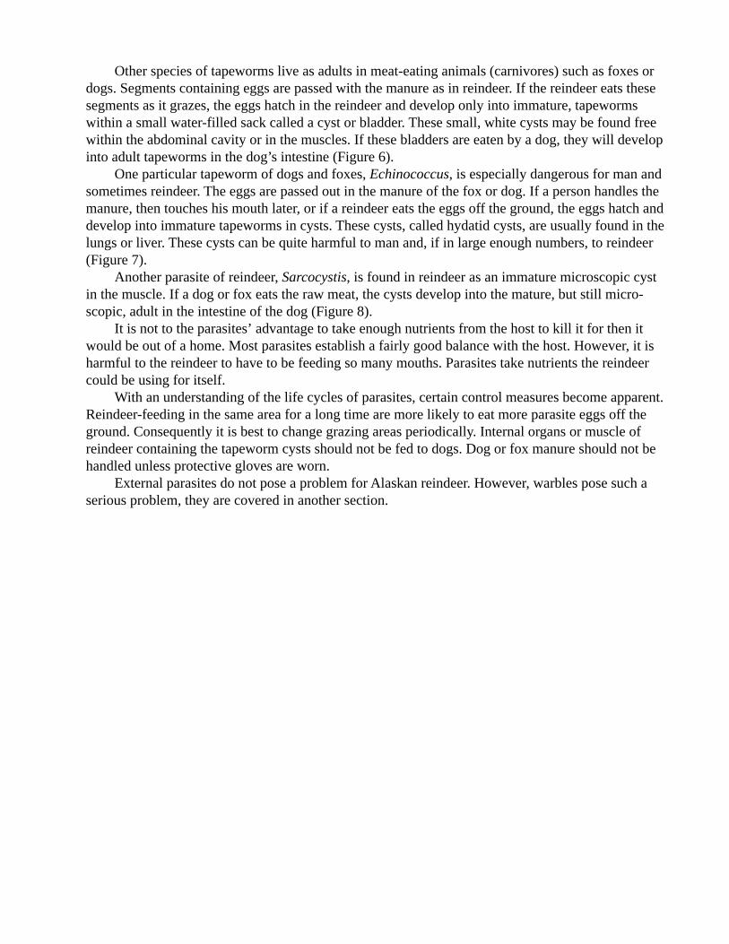
Other species of tapeworms live as adults in meat-eating animals (carnivores) such as foxes ordogs. Segments containing eggs are passed with the manure as in reindeer. If the reindeer eats thesesegments as it grazes, the eggs hatch in the reindeer and develop only into immature, tapewormswithin a small water-filled sack called a cyst or bladder. These small, white cysts may be found freewithin the abdominal cavity or in the muscles. If these bladders are eaten by a dog, they will developinto adult tapeworms in the dog’s intestine (Figure 6).
One particular tapeworm of dogs and foxes, Echinococcus, is especially dangerous for man andsometimes reindeer. The eggs are passed out in the manure of the fox or dog. If a person handles themanure, then touches his mouth later, or if a reindeer eats the eggs off the ground, the eggs hatch anddevelop into immature tapeworms in cysts. These cysts, called hydatid cysts, are usually found in thelungs or liver. These cysts can be quite harmful to man and, if in large enough numbers, to reindeer(Figure 7).
Another parasite of reindeer, Sarcocystis, is found in reindeer as an immature microscopic cystin the muscle. If a dog or fox eats the raw meat, the cysts develop into the mature, but still micro-scopic, adult in the intestine of the dog (Figure 8).
It is not to the parasites’ advantage to take enough nutrients from the host to kill it for then itwould be out of a home. Most parasites establish a fairly good balance with the host. However, it isharmful to the reindeer to have to be feeding so many mouths. Parasites take nutrients the reindeercould be using for itself.
With an understanding of the life cycles of parasites, certain control measures become apparent.Reindeer-feeding in the same area for a long time are more likely to eat more parasite eggs off theground. Consequently it is best to change grazing areas periodically. Internal organs or muscle ofreindeer containing the tapeworm cysts should not be fed to dogs. Dog or fox manure should not behandled unless protective gloves are worn.
External parasites do not pose a problem for Alaskan reindeer. However, warbles pose such aserious problem, they are covered in another section.

Effects:Dogs: Non-apparent effects.Reindeer, moose, etc.: Depends on number of cysts; may not be too serious.Man: Always serious.
Diagnosis:Dogs: Segments in feces or worms in intestine at necropsy.Reindeer, moose, etc.: Cysts in liver or lung at necropsy.
Treatment:Dogs: Give drugs to kill adults in intestine.Man: Surgery.
Prevention:Dogs: Don’t feed reindeer viscera to dogs.Man: Wear gloves when handling dog droppings.
Figure 7. Echinococcus

Effects:Dogs: Diarrhea?Reindeer: None apparent.Cattle: Anemia, weight loss.Mule deer: May be fatal
Diagnosis:Carnivore: Eggs in feces.Cattle, deer: Cysts in muscle tissue.Reindeer: Microscopic cysts in muscle tissue.
Treatment:Dogs: Drugs to kill mature coccidia in intestine.
Prevention:Dogs: Don’t feed raw reindeer meat.Man: Cook meat before eating (effects in man are not clear; probably usually not serious except
in pregnant women).
Figure 8. Sarcocystis

Treatment for ReindeerFirst Aid
1. Advice for particular problems can be obtained by calling the Applied Reindeer ResearchProgram in Fairbanks at (907) 474-7166.
2. For wounds, first clean the area as well as possible. Remove any dirt or hair in the wound. Toprevent infection, apply a topical antibiotic powder such as Furacin®. Gentian Violet® is another drugthat can be used on surface wounds.
In the summer a wound spray with fly repellent is useful in preventing infection by flies. Thisshould be used to spray around the scrotal area after castration and on the base of a cut-off brokenantler on a fawn (shield the fawn’s eyes with your hand as you spray). If there is bleeding, applydirect pressure on the wound as you would on yourself.
3. Hooves that are too long may be trimmed with cutters.4. Ivermectin is available for parasite control.5. Keratitis (White-eye) is treated by injecting a mixture of two parts of penicillin plus one part
vetalog into the white of the eye just under the first layer. One-half to one ml. of the drug combina-tion is carefully deposited under the conjunctiva (white part of eye) by just penetrating this thin layerand making a small blip of drug. Assistance will be needed to hold the reindeer’s head still to avoidinjuring the eye with the injection needle and to keep the eyelid open.
6. Pay attention to how your reindeer are acting. For instance, sick animals don’t shed their coatas fast as well ones. Many animals slow in shedding out in a herd may indicate a herd health prob-lem.
7. Many antibiotics are available and can be used on your own reindeer under the orders orsupervision of a veterinarian. Many drugs are available only through a veterinarian.
CastrationCastration takes away the bull’s desire to breed and reduces male traits such as aggressiveness
and dominant behavior. Besides making males easier to handle as work animals, it is used on bullsmeant for slaughter later in the year. Once castrated, the male will feed and put on weight throughthe fall instead of losing weight during the rut. Also, meat from a castrated male is thought by someto be of less gamey flavor and more tender than meat from a bull.
Castration methods fall under two categories—open and closed. Open castration is done bycutting the scrotal sac and removing the testes. It is a fast procedure, and if done properly will notharm the bull’s health. Once the testes are removed, the male’s sexual drive should be eliminated.The following steps outline the typical way for an open castration:
1. Take the loose, excess skin of the scrotal sac and slice out a semicircle of skin with a sharpknife.
2. Pop out the first testis, and slide your hand down along the spermatic cord pushing connec-tive tissue ahead and away from the testis. This exposes a 4 to 6 inch length of spermatic cordrunning from the testis to the body.
3. Fray and cut the spermatic cord close to the body opening in a back and forth motion tominimize bleeding. Repeat 2) and 3) with the other testis.
4. Spray the scrotal opening with a repellent/disinfectant in warm weather.Closed castration is done by crushing the spermatic cord without having to cut the scrotum
open. There are several types of plier–like tools available on the market to do this job. When thevessels, nerves, and vas deferens are crushed, passage of material to the testes is blocked. Eventually

the testes will wither and die. This method is effective when done accurately, but room for errorexists. If the tool is placed or used incorrectly, castration may be incomplete or totally ineffective.
Aid In Fawning
Although most reindeer will give birth normally without aid from people, it is possible toencounter a cow having difficulty while fawning. There are several conditions that create problemswhile fawning, the most common of which is called abnormal presentation. This refers to the fawn’sposition, or presentation, as it enters the birth canal to be born. An abnormal presentation is one inwhich the fawn is turned or twisted so that it gets caught in the relatively small opening of the birthcanal, making birth dangerously slow or impossible.
With knowledge of how the fawn should be positioned in order to emerge easily, a person canstraighten it so that a normal birth can take place. This is a process that can be done without specialtools or complex medical know–how. If carefully done, it is often successful and may prevent thedeath of the cow and also save the fawn,
In a normal birth the fawn is positioned, or presented, so that the front hooves and head areaimed down the birth canal. In this position, the fawn faces the rear of the cow (see Figure l). It isnot uncommon for the fawn to be facing the opposite way, that is, with the hind legs emerging firstand the head pointing in the same direction as the cow’s head (posterior presentation). This, too, canlead to a normal birth.
When beginning labor, the cow will experience hard muscular contractions and will appear tobe straining. Usually the fawn will emerge anywhere from a few minutes to a half hour after the cowbegins straining heavily. When the fawn is born, the cow licks off the membranes and mucus sur-rounding its body and tears the umbilical cord if it is still attached. The cow’s constant licking driesthe calf and stimulates its circulation. A healthy fawn will rise on its shaky legs within the next hourto begin suckling milk.
A cow having trouble giving birth will be in labor for an unusually long time. There are twosigns that will indicate that a cow is having difficulties:
1. If part of the fawn is visible at the entrance to the vagina for longer than a half hour withoutbeing born.
2. If the cow is straining hard with no results for an hour. If a cow is in this situation, steps canbe taken to straighten the fawn out into the normal position.
1. Approach the cow quietly and slowly. It is very important not to spook her because aftergiving birth she may abandon the fawn if she has been badly frightened. Although it is possible toraise the fawn by hand, the fawn’s natural mother is its best chance for survival.
2. If possible, wear gloves, or at least wash hands well before and after this process. This is toprotect not only the cow, but you. Remember that brucellosis can cause late abortion, and the cowyou are treating may be infected.
3. Gradually work your hand into the vagina to reach the fawn, avoiding injury to the birthpassage. Determine the position of the fawn and feel for the head. Figure 2 illustrates some typicalabnormal positions of the fawn. It may be necessary to twist the head back to a forward position,straighten a leg or unhook it from the pelvic girdle. If there are twins in the womb, the situation maybe more confusing. One fawn should be pushed back slightly to allow the other to emerge. Theproblem here is sorting out which legs belong to which fawn. If the fawn is facing backwards (poste-rior presentation), try to position it so that the rear legs are heading down the birth canal.
4. Once the fawn’s position has been straightened, leave the cow and watch from a distance. Ifshe still has the strength she will give a normal birth, and all will be well. If the fawn is still not born,


return and grasp the fawn by the forelegs (or hind legs). With a gentle , smooth motion, pull inunison with the cow’s contractions to help the fawn out. Pulling should be the last resort, because ifdone too hard or at the wrong time, it can do much damage to both the cow and fawn.
After the fawn is born, the cow should take over its care automatically. The cow will be morelikely to reject the fawn if she is frightened by your presence, or if the fawn has been handled toomuch by humans. The cow and fawn should be left alone after birth. The fawn should not be handledunless the cow is too weak to take over. In extreme cases it may be necessary to clean and dry thefawn, clear its nose and nostrils to breathe, and tie off the umbilical cord close to the fawn. Cut theumbilical cord between the knot and the mother. Make the cut two to three inches from the knot. Dipthe fawn end of the stump in iodine to prevent infection. If a fawn has been abandoned, followfeeding directions in the nursing care section of this manual.
Necropsy of ReindeerNecropsy Procedures and Techniques
Protection of humans against infection should always be the primary concern of everyoneinvolved in the sampling process. The essence of this is to “think clean”. Wear adequate protectiveclothing and follow clean techniques during the necropsy. Minimization of contamination results inbetter samples and reduced environmental contamination while decreasing the chance of infectionfor personnel.
Documentation (recording the facts) is an essential aspect of the sampling process. Complete,accurate, legible field notes specifying the circumstances of the animal’s death—species, age, sex,location, environmental characteristics, clinical signs, and other pertinent observations are necessaryfor generating reports that accompany tissue samples to the laboratory. Photographs of parasites orlesions are extremely useful for determining diagnoses and indicating disease causing potential(pathogenicity). A label showing the specimen number and some reference of scale (such as a coinor knife) should appear in the photo. Exposure numbers with brief descriptions should be recordedin the field notes if many exposures are taken.
Specimen PreparationThe procedure listed is for land (terrestrial) mammal (reindeer, bears, foxes, etc.) samples under
ideal conditions. Usually, conditions are somewhat less than ideal and one must salvage whateverpossible from a decomposing carcass under inclement weather conditions. Two people can conduct amore efficient necropsy than one. This allows one person (“dirty”) to remove tissues and one person(“clean”) to take photographs, record notes, label and hold sample bags. For large animals, a thirdperson is useful to help separate leg joints, move organs, and help prevent contamination of thepeople or parts of the carcass.
1. Collect a blood sample. This applies only to living animals or ones that can be sampledwithin minutes after death. The best sources are the jugular vein, femoral artery (in the leg), or theheart. Avoid taking blood from open wounds as it will contain unwanted material. Blood should notbe allowed to freeze or be exposed to excessive heat or agitation as this causes the red blood cells tobreak open. A suitable anticoagulant, such as EDTA, should be added to whole blood samples to beused for measuring cell counts, packed cell volume, glucose determinations (enzyme inhibitor alsoneeded) or other measurements. Blood put in a clean, dry tube or plastic bag will clot after standingabout 12 hours at room temperature. Blood serum, which separates after the blood clots, can becarefully removed with a syringe or eye dropper and put into a sterile vial for antibody detection.Serum may be stored frozen, although repeated freezing and thawing should be avoided.

If the animal is found dead, serum can often be collected from the heart clot. The whole clot, orthe yellow “chicken fat” part, if separation has occurred, is placed in a plastic bag, allowed to standfor 12 hours, and then free serum is collected.
2. Visually inspect the carcass for external parasites, hair loss, traumatic injuries, dischargesfrom the nose, mouth, or anus and other abnormalities. Photographs and external parasites should betaken at this time.
3. Position the animal right side down (to expose the spleen when opened) and remove its skinfrom the left (upper) side. The testes or mammary glands should not be exposed or damaged duringskinning. Take care when skinning around Iymph nodes under the skin if these are needed. Removethe nodes and the testes or mammary glands after the skinning is finished. Disinfect instrumentsbefore handling tissues to be sampled. Tissues should not contact the sides of sample containerswhen placed inside.
4. Raise the left foreleg and cut beneath the shoulder blade (scapula) until the leg and shouldercan be laid back (see Figure 1). Separate (disarticulate) the joint of the left rear leg at the pelvis bycutting the muscles through the joint (Figure 2). If time is critical, the carcass can be temporarily leftas cooling will proceed. Sampling should resume as soon as possible to prevent tissue deterioration,especially in high air temperatures.
5. Open the belly (abdomen) to the breast bone (sternum) taking care not to cut the intestines orrumen. Split the sternum with a knife or saw. Be careful not to puncture the chest (thoracic) organs.Free the diaphragm from its upper attachment to the ribs. Cut the muscles between every second or

third rib and break them back at their points of connection with the vertebrae. Split the front part ofthe pelvis (pubis) to expose the pelvic organs if desired. The specimen is now ready for removingtissue samples (Figure 3).
Tissue CollectionBefore disturbing the internal organs (viscera), record an estimate of the time of death, gross
internal abnormalities and any visible internal parasites (e.g. Setaria). Aseptically collect samples ofthe major visceral organs (heart, lung, liver, spleen, and kidney) approximately one inch in size andplace in containers labeled with specimen number and tissue type. Start with clean, disinfectedinstruments. If possible, disinfect instruments between each different tissue collected. Do not get hairor dirt on any sample. (Note: whirl packs make very good containers as they are unbreakable, rela-tively fluid tight, and require a minimum amount of space.) Collect internal Iymph nodes if wanted.Remove the reproductive organs and place in a separate container. These tissues should be frozen forlater culturing.
Samples of the major visceral organs, skeletal muscle, diaphragm and tongue can now becollected for microscopic (histologic) examination. These tissues can be placed in a single containerwith a suitable fixative (e.g. 10% buffered formalin). Tissues for histology should not be mutilatedsince the ultimate analysis is based on the structure and organization of cells.
Examine the carcass for endoparasites after collecting tissue samples. Warble (Hypodermatarandi) and bot fly (Cephenomyia trompe) larvae can readily be found from early spring through

midsummer beneath the skin and in pharyngeal pouches respectively. Other reindeer parasites, suchas lung worms (Dictyocaulus viuiparus), will occur less often and will require a more diligentsearch. Any tissue suspected of being parasitized should be collected.
Causative AgentsBacterial, mycotic (mold), and viral diseases may be indicated by:1. large epizootics which involve many individuals of a population;2. bloody mucous discharges from the mouth, nose, or anus;3. reddened and congested lungs, kidneys, and intestines;4. an enlarged, dark colored spleen;5. an enlarged, off-colored liver; and6. presence of pus.Preserve both frozen and formalin-fixed tissue samples and blood serum if bacterial, mycotic,
or viral causes are suspected.Ectoparasites (e.g. fleas, ticks, and lice) are usually visible with the naked eye. Host reactions to
endoparasites may result in cysts that obscure the parasitic organisms. Collect these cysts in theirentirety by removing the effected block of host tissue. Endoparasites are often specific as to hostspecies and anatomical region of attack. Therefore, it is important to note both aspects of the condi-tion as well as the number of parasites present.

Non-infectious diseases, i.e. toxicities and nutritional or mineral deficiencies, are often indis-tinct. Localized sick or dead animals may indicate a non-infectious etiology. Hair bundles approxi-mately 3/4 of an inch in diameter should be collected along with whole blood. Collect soil and watersamples, in addition to tissue samples, and place them in inert containers (i.e. glass or plastic vials).These samples must be analyzed as quickly as possible as many toxins rapidly break down in moistconditions. Containers holding water should not contain any air space after being covered. Soilsamples should extend a minimum of four inches deep. Plants for mineral assays or toxin analysesare collected in their entirety, quickly pressed, and dried. Stomach samples from ruminants can beplaced in porous cloth and excess water squeezed out before placing in 10% formalin. Stomachsamples from monogastric (one stomach) animals can be placed directly in 10% formalin, if smallenough, or portions of their contents removed and preserved.

APPENDIX IISerum Collection
Serum is the clear portion of blood that separates from the clot. Within the serum are the anti-bodies against specific diseases. Tests conducted on serum for antibodies are called serologic tests.Some common serologic tests for brucellosis that will be run in the laboratory are the standard platetest (SPT), rapid card test (BBA), rivanol (Riv) and complement fixation test (CFT).
FOR COLLECTION FOR STORAGEEquipmentneeded: Gloves Clean syringe & needle
Syringe (35 cc) & Needle Small plastic vialsWhirlpack bag (small size)Vacutainer® tube (red top)
Procedure:
1. Collectiona. If the animal is still alive, collect blood from the jugular vein. Inject blood carefully
into one or two Vacutainer® (red top) tubes.b. If the animal is dead, try to collect from the jugular vein or heart. Inject blood care
fully into one or two Vacutainer® tubes.c. If the blood has already clotted, open the heart, Remove the yellowish colored portion
of clot from the heart and place in a whirlpack bag.d. If none of the above are possible, collect blood from the body cavity and place in a
whirlpack bag.
APPENDIX ITissue Collection
Two sets of samples are to be obtained:1. The first set is for culture to check for bacteria (including Brucella).2. The second set is for formalin preservation.Wearing gloves and using clean instruments, expose the internal organs of the deer. Be careful
not to contaminate any structures.Use clean forceps and scalpel to collect one inch cubes of tissue from the major organs in this
order: heart, lung, spleen, kidney, liver, reproductive organs, muscle from tongue, and muscle fromrear leg. Put these samples in a large whirlpack bag and roll the bag closed. This bag may be frozen.
In a separate bag, put a second similar set of tissues measuring about 1/4 inch by 1 inch. Addenough formalin to this bag to cover the tissues. Roll the bag closed. Do not freeze this bag.
Contact:Applied Reindeer Research ProjectAgricultural and Forestry Experiment StationUniversity of Alaska FairbanksFairbanks, Alaska 99775–0100Tel: (907)–474–7166

APPENDIX IIIWhole Blood Collection For Complete Blood Count (CBC)
This is only to be done if:1. The blood can be taken immediately to a laboratory for testing.2. The animal is to be treated.
Equipment needed:GlovesSyringe (12 cc or 6 cc) and needleVacutainer® (purple top) (EDTA tube)
Procedure:1. Collect blood from the jugular vein.2. Inject blood very slowly and gently into the purple stopper Vacutainer® tube. Gently turn tube
upside down and right side up several times. Label the tube. Record time collected and importantinformation.
3. Keep the tube cool and take to a laboratory for testing immediately. (The blood will onlykeep for one day.) Do not freeze.
2. Record location, date, sex, approximate age (adult or fawn?), and other important information.Label the tubes.
3. Storagea. Let set at room temperature overnight. Keep blood sample upright. Don’t shake and
don’t freeze the blood.
4. Collection of seruma. After the clear serum has separated from the clot, use a syringe and needle or an eye
dropper to remove the serum from the clotted sample. Put the serum in a clean, smallplastic bottle (vial) or another Vacutainer® tube. The serum should be frozen. It willkeep for many months in the freezer, but will only keep a few days in the refrigerator.
b. As soon as possible, contact:
Applied Reindeer Research ProgramAgricultural and Forestry Experiment StationUniversity of Alaska FairbanksFairbanks, Alaska 99775-0100Tel: (907)-474-7166.

APPENDIX IVCare and Use of SyringesDisposable Syringe
This is useful when delivering a single dose of medicine. It is also valuable when it is crucialthat the syringe be sterile.
Keep the sterile syringe in its plastic container until needed. After using the syringe, throw theneedle away. The syringe itself can be cleaned by plunging warm water through the barrel four orfive times. Next, it should be disinfected with alcohol. The barrel can then be reused with a newsterile needle.
Choose a needle size appropriate for the task. For example, a larger needle (16 ga.) is neededwhen injecting thick medicine, or when drawing blood from a large animal. A smaller needle (18 ga.or 20 ga.) is used for more delicate processes, such as a smaller animal or thinner medicine.
Syringe barrels also are provided in several sizes. The long graduations on the barrel are in unitsof cubic centimeters (cc), which is the same as milliliters (ml). Fractions of the cubic centimeter aremeasured with the shorter graduated lines.
Automatic SyringeThis is useful when many doses of one type of medicine will be delivered to many animals.
Although normally not as sterile as disposable syringes, it can be kept adequately clean. It is practi-cal in large handling situations.
Before using, thoroughly wash all parts with warm water and dry. While using the syringe, keepa jar filled with alcohol to resterilize needles. Each time the barrel is refilled with medicine, put aclean sterile needle onto the syringe. If the syringe becomes contaminated with blood or dirt, themedicine should be evacuated in a safe place, and the syringe and needle thoroughly washed beforerefilling.
The syringe can be taken apart and reassembled according to Figure 1. The plunger or washersmay need replacing if they become dry or cracked. New glass barrels should be kept in stock in caseone is broken.
When using the multiple dose syringe, it is important to remain aware of the size of dose eachanimal needs. The graduated dial on the hand grip allows you to adjust the dosage. Each numberrepresents a cubic centimeter, as in the disposable syringe. Even when the same dose is administeredeach time, make it a habit to check the dial every two or three shots to make sure that it hasn’tslipped from the proper dosage.

Key0l Needle02 Metal barrel frame03 Forward end tablet04 Barrel washer05 Glass barrel06 Inner piston rod07 Rubber plunger08 Plunger upper disc09 Graduated ratchet sleeve10 Hand grip11 Plunger adjustment knob
Figure 1. Assembly of multiple dose syringe unit.

Care and Use of Automatic (Pistol Grip) Syringes
Cleaning1. Completely dissemble all parts. Wash in hot soapy water. Be sure to flush the inside portion
of the outer plunger shaft. Rinse well. Air dry.2. Re-assemble when dry. Tension on the barrel washer should be loose so it moves freely.
Tighten the large and small screws on the pistol-grip handle.3. Flush dirty needles with soapy water, rinse well. Check needles for sharpness. If barbed,
gently rub over sharpening stone. (If badly bent, discard). Rinse again, air dry.
Use1. Fill two needle cans with 70% ethanol. Use one for dirty needles and one for clean needles.
Place clean sharp needles in the clean needle alcohol can.2. Use clean 12 gauge needle from alcohol can on syringe for filling with brucellosis adjuvant
(or other thick) vaccine. A 16 gauge needle can be used to fill syringe with thinner solutionssuch as Ivermectin.
3. Adjust tension on barrel washer by turning the knob at the end of the plunger shaft so it isjust tight enough to draw vaccine from the bottle, but not so loose it allows vaccine to flowaround the washer. Maintain pressure to fill syringe.
4. When syringe is full (or bottle is empty), remove 12 gauge needle from syringe and place itin the dirty needle alcohol can.
5. Take a 16 gauge needle from the clean needle alcohol can and place on end of syringe. Thisneedle can be used for all the animals vaccinated with that syringe of vaccine providing theneedle stays clean and sharp. If there is any doubt, replace it with a clean needle from thealcohol can.
6. Set the dosage to be administered by aligning the number on the dial with the arrow. Eachsqueeze administers the number of cc’s indicated on the dial. If it is hard to squeeze thehandle, loosen the tension slightly on the barrel plunger washer. Be careful not to allowvaccine to run around the washer.
7. When the syringe is empty, remove the used needle and place it in the dirty needle alcoholcan. Remove a 12 gauge filling needle from the clean needle alcohol can and repeat thesyringe-filling process.
8. Regularly check that the two screws on the handle are tight.9. To clean needles in dirty needle alcohol can, flush alcohol up and down through needles can
using a 12 cc. syringe. When needles are clean, transfer them to the clean needle alcohol can.10. If any blood appears inside the glass barrel, stop using that syringe until it has been washed.11. Vaccine or drug left in the syringe should be discarded after 24 hours .

APPENDIX VLabel and Use of Ivermectin
Used for the treatment of parasites in reindeerIVOMECB (Ivermectin) 1% sterile solutionInject subcutaneously at a dosage rate of 200 mcg per kilogram of body weight(1 ml per 110 pounds body weight)WARNING: Do not treat reindeer within eight weeks (56 days) of slaughter
InjectionsIvermectin and brucellosis vaccine are two products commonly injected into reindeer. Both are
given subcutaneously (under the skin) in the loose skin on the sides of the neck. The site of theinjection is midway between the head and the shoulder about one half the way up the neck. This siteis termed mid-cervical. Using a short (1/2 inch) 16 gauge needle, it is easy to make a short jab at anangle into the skin in such a manner that the medication or vaccine is deposited into the proper area.Opposite sides of the animal should be used if an animal is being treated with two products at thesame time. Injection equipment should be exchanged and cleaned if the hair is dirty, or there is anyobvious contamination of the syringe or needle.
Other medication, such as antibiotics, may have to be given in the muscle or in a vein. Any ofthese products should be administered only after consultation with a veterinarian. Remember thatreindeer are used for human food and strict guidelines must be followed to ensure there are no drugresidues and that the meat is wholesome.

APPENDIX VISpecimen Submission For Rabies Assay
Alaska Department of Health and Social ServicesDivision of Public HealthSection of LaboratoriesVirology-Rabies Unit
Rabies laboratory services are provided through the Virology-Rabies Unit established in Fairbanks.To assure accurate and reliable reports, the directions listed below must be followed.
1. Specimens will be accepted from physicians, veterinarians, public health officials, health orsanitation aides, wildlife agents, authorized medical or physician assistants, or individuals desig-nated by the Division of Public Health.
2. Rabies laboratory examinations will be limited to warm–blooded animals of any type in whichthere is a clear history of human or possible human exposure, or animals deemed necessary byhealth or wildlife authorities.
3. Special Instructions:Virology-Rabies Unit must be notified prior to submission of rabies specimens. Call Fairbanks(907) 474–7017 or I907) 474–7018, or on weekends (907) 456–5974.
4. Minimum Information:The following information, or a completed Rabies Investigation Report (Form 06-1272) mustaccompany each specimen. This information is essential for the evaluation of each case andexpediting the transmission of results to the responsible physician.
a. Name and address of person(s) bitten and/or exposed.b. Date of exposure.c. Location of bite(s).d. Severity of bite(s).e. Date of first aid treatment and name of physician.f. In the case of a dog or cat, was the animal vaccinated?g. Provoked or unprovoked attack?h. Name, address and phone number of person sending the specimen.i. Name, address, and phone number of person to receive the laboratory report.
5. Conditions of Shipment:a. No Iiving animal(s) will be accepted for rabies diagnostic studies.b. When killing the animal, do not shoot in the head or mutilate the head in any way.c. Wear gloves when handling the animal, and send the head and part of the neck of large
animals, such as dogs, foxes, wolves, Iynx, etc. Sever the head at the neck and leave sufficienttissue attached to the region of the head to ensure inclusion of the salivary glands. No other partof the animal should be submitted.
d. Small animals such as mice, voles, and bats may be sent intact if recently expired, andONLY after consultation with the Chief of the Virology Unit or the State Medical Epidemiologist.
e. Specimens for rabies examination must be fresh. Decomposed specimens will be evalu-ated, and if unsuitable for assay, will be promptly incinerated, and no rabies assay will be at-tempted.
6. Packing:a. Wear gloves when handling the animal.b. Wrap the head in absorbent material, and place into two heavy, water-tight plastic bags.

Tie-off the bags to prevent leakage.c. Place the packaged head in a leak-proof container, and pack the bag next to a water-tight
can of frozen water or freeze packs. An alternative is to use a second set of heavy plastic bags andfill these with ice cubes and seal the top tightly. Remote areas may use river ice, but this type ofice has rough edges and will cut the bag. This can be avoided by wrapping the ice in newspaperand then packing with absorbent material to prevent movement of the ice in the carton.
d. Remove the gloves and either burn the gloves or enclose them with the head for disposal.Then wash hands thoroughly with soap.
e. Check the carton or container for security and include the Rabies Investigation Report.Please include contact telephone numbers.
7. Labeling:a. All rabies specimens must bear the special Rabies Biohazard Label or the following state-
ments:
b. Address all shipments to:Virology-Rabies UnitAlaska Division of Public HealthArctic Health Research Building, Room 233UAF, Fairbanks, Alaska 99706-0230
8. Shipping:a. Specimens may be brought directly to the Virology-Rabies Unit.b. Ship specimens by air freight or air special package service. If mail service must be used,
send via airmail-special delivery, or express mail, if available.c. All shipments must be prepaid unless prior arrangements have been worked out with the
Virology-Rabies Unit.d. Contact the SVL and advise the method of routing and the airway bill number, etc.
9. Reports:a. A preliminary fluorescent microscopic report will be called all on positive animal heads,
and human exposure cases within 24–48 hours after specimen receipt and confirmation. Copiesof the report will be mailed to the submitters.
b. An interim report will be forwarded at completion of the fluorescent microscopic (FA)examination. When the mouse inoculation test (MI) is performed, this final report will be for-warded subsequent to completion of a 28-day observation period.
10. For further information on rabies antiserum and vaccine for human use: Contact the AlaskaDivision of Public Health Regional Health Officer or Laboratory serving your area.
State Virology LaboratoryAlaska Division of Public Health232 Arctic Health Building901 KoyukukP.O. Box 60230Fairbanks, Alaska 99706-0230Phone:(907)474-7017,0r(907)474-7018
Medical EpidemiologistSection of Communicable Disease Control3601 C St., Suite 540P.O. 8ox 240249Anchorage. Alaska 99524-0249Phone: (907)-5614406
DIAGNOSTIC SPECIMENTHIS CARTON CONTAINS THE HEAD OF AN ANIMAL.PLEASE REFRIGERATE ON ARRIVAL AND CONTACTTHE VIROLOGY-RABIES UNIT AT 474-7017 or 474-7018, ON WEEKENDS 456-5974. IF NO Answer, CALL452-0467 AND LEAVE A MESSAGE.
MEDICAL MATERIALSPACKAGING CONFORMS WITHSTANDARDS IN 49 CFR173.386 (2)CFR72.25 (c), and NIH GUIDE OFFEBRUARY 10, 1975.

APPENDIX VIIRaising Orphan Reindeer Fawns
Orphan reindeer fawns are often observed at summer handlings. Sometimes an abandoned fawn is foundon the range. These fawns can be saved and raised for pets or for sale as live animals. There is an increasingdemand for live reindeer to be shipped to locations outside Alaska. Tame reindeer adapted to a commercialdiet can be a potential source of income for the herder as well as a rewarding project for the family. Thefollowing information is intended for reindeer herders in Alaska who do not have ready access to modernveterinary facilities or care. Raising a reindeer fawn on a bottle is one of the easiest ways to obtain a gentledeer. If started on a bottle within the first few days of life, a reindeer will quickly form a bond with humans.The older they are when started, the less likely they are to form a bond. However, there are other advantagesto feeding older fawns: they don’t have to be fed as often, and they can be fed solid foods. No matter what theage, feeding a reindeer fawn is very rewarding and potentially profitable. The following is intended to be aguideline for feeding a fawn. One thing to remember is that no two animals are exactly identical any morethan two human babies are exactly alike. Each must be treated as an individual.
For newly born fawns or fawns up to two months of age:1. In our experience and that of others raising reindeer fawns as well as other species of wild fawns
or calves, commercial formulas such as Land O’Lakes Lamb Replacer can be used as a milksubstitute for nursing fawns. Alaska feed stores might be able to special order a replacer formulafor you if they do not have it in stock.
2. A temporary substitute formula of: one quart of whole milk or reconstituted whole powdered milkfor example Milkman mixed with one can of evaporated milk can be used until the commercialreplacer is available. The substitute formula, however, should be used only as a temporary substi-tute. It is not satisfactory for prolonged use. Regular human baby vitamins can be added to theformula at the same dosage as specified for human babies. Warm the formula to body temperaturebefore feeding. Refrigerate any leftover formula or replacer between feedings.
3. Use either a regular baby bottle and nipple or a lamb nipple on a bottle to feed the fawn. The holein the nipple can be made a little larger by heating the sharp end of a large safety pin in a flame.Then stick the hot point through the hole in the nipple. Don’t cut big slits in the nipple to make ahuge hole. Just as human babies like to suck their thumbs, fawns also like to suck. If the hole inthe nipple is too big, fawns will suck too fast and finish too soon to satisfy their sucking reflex. Itis better that they take longer to finish and feel more satisfied. They can also choke if they sucktoo fast. This can lead to pneumonia.
4. Hold the nipple end of the bottle between the thumb and first two fingers with the nipple pointedtowards the palm of the hand. Cup the fawn’s chin in the palm of your hand. The bottle andfawn’s chin are now in the palm of your hand so your hand and the bottle can move with themovements of the fawn to help keep the fawn from swallowing air. The last two fingers can beused to help direct the nipple into the fawn’s mouth.
5. Start off feeding small amounts of formula or replacer.For example:Age (weeks) Ounces Times/Day
0-1 2-3 5-71-2 3-4 5-62-4 4-5 4-54-6 6-8 3-46-8 8-10 38-12 8 3

Another guideline is that a baby animal usually requires 1/10 of its body weight per day oftotal milk replacement if that is all it is eating. If a fawn weighs 10 pounds, which equals 160ounces (16 ounces per pound times 10), that fawn should have a total of 16 ounces (1/10 of 160ounces) of formula or replacer per day. That 16 ounces could be divided as four ounces four timesa day. A 15 pound fawn should have a total of 24 ounces per day, and a 20 pound fawn shouldhave a total of 32 ounces per day. By the time it is eight weeks old, the quantity of milk replace-ment fed should be decreasing as the quantity of grain and greens increases.
6. Make each increase gradually. Do not jump from one level to the next all at once.7. DO NOT OVERFEED. Fawns will always act like they want more. Overfeeding is the most
common cause of diarrhea. A fawn’s manure is normally soft but it should not be watery. Pro-longed diarrhea can cause death. Fawns can actually eat themselves to death.
If diarrhea does occur:a. Reduce on the amount of formula at each feeding until diarrhea is under control. You can
use less powdered milk with the usual volume of water until the manure is normal again. Thengradually increase the milk powder back to the proper amount according to the recipe.
b. If diarrhea continues, give a baby’s dose of human medicine for diarrhea such asKaopectate or Pepto-Bismol. Use the dosage directed by the instruction on the bottle.
ORc. Add a little baby rice cereal (Gerbers’ for example) to the formula.
NOTE: Diarrhea is usually watery and yellowish from eating too much milk or from a disease. It isusually green from eating too many greens.
9. After eating, at least two times a day until the fawn is well trained (generally one to two weeksold), gently rub its rear end with warm water on a washcloth to stimulate the fawn to pass manure.Female reindeer lick their fawn’s rear end to do the same thing.
1O. If your fawn is older than three weeks when you start, it may not be willing to drink from abottle. If it doesn’t, offer the milk replacement in a pan or bucket.
11. After the fawn is about five days old, offer it some grain immediately after it finishes the milkreplacement. To get the fawn started, dip your fingers in some milk replacement, then in the grainso some grain sticks to your finger. Put your finger into the fawn’s mouth. It will usually suckyour finger getting the grain at the same time. Do this until it gets used to the taste and texture ofthe grain and eats grain by itself. Don’t force it. Some fawns take to grain right away. Others justaren’t interested for a while.
During the first few weeks of life, use a commercially available feed prepared for youngcalves. Purina’s Nursing Chow and Calf Manna are two examples of the many brands on themarket that can be successfully used at this stage of the fawn’s life.
When a fawn is about six weeks old, mix your starter feed 1:1 with pelletted feed mixturesuitable for reindeer. The University of Alaska Fairbanks has had years of experience and successfeeding reindeer a mixture which contains:
Crude Protein 16% (minimum)Crude Fiber 8% (minimum)Crude Fat 3% (minimum)This feed is a mixture of rolled corn and/or barley; ground oats, barley and/or corn; cotton-
seed and/or soybean meal; wheat millrun; wheat middlings; beet pulp pellets; salt and molasses.Commercial feeds such as Wod-Lyn Feeds or a feed specifically formulated for reindeer fromAlaska Mill and Feed meet these requirements. Check with your local feed supplier for a rationthat meets these nutritional requirements.
Fawns will usually not eat more than about one cup of grain twice a day until they are older

than four weeks. From four to six weeks of age, they will eat about one and one half cups of graintwice a day. From six to eight weeks, they will eat about two cups of grain once a day.
12. When the fawn is about one week old, provide some willows for it to eat. Start with a few leavesat a time and gradually work up to a small branch. If you are feeding too many greens, the fawnwill develop a green colored diarrhea. Cut back on the quantity of greens until the manure isnormal again. If you are keeping the fawn in an outside pen, fawns usually have no trouble graz-ing on natural feed.
13. Keep a pan of clean drinking water available at all times.14. Provide some dirt for it to lick if you are keeping it inside.15. Keep the fawn in a clean, sanitary, dog-proof area. Provide shelter from the sun and the rain.
Summary
Age Quantity Frequency Greens Grainweeks milk times per
replmt day
0-1 2-3 5-71-2 3-4 4-5 Offer willow Offer
leaves commercialcalf feed
2-4 4-5 4-5 Willows 1/2-1 cupnatural feed twice a day
calf feed
4-6 6-8 4 Willows 1-1 1/2 cupsnatural feed twice a day
calf feed
6-8 8-10 3 Willows 2 cupsnatural feed twice a day
calf feed with equal parts of adult feed
8 8 3 willows 1 part calfnatural feed feed to 2
parts adult feed twice a day8-12 8 3 Wiilows 1 part calf
natural feed feed to 3parts adult feed twice a day

Appendix VIIINutrition
Free grazing reindeer consume a wide variety of plants that are selected on a seasonal basis.Many of these arctic plants are of an exotic nature and unusual chemical composition. A publicationby Dr. Jack Luick summarized information on the diet of freely grazing reindeer. This publication isreproduced as part of this appendix on reindeer nutrition. As Dr. Luick states, it seems hardly sur-prising that in some reindeer relocation attempts, more than 60 percent of the reindeer have diedwithin a few weeks of departure from tundra ranges. Although other factors such as handlingstresses, hypothermia, regurgitation and inhalation of rumen contents, injury, disease, and overdoseof immobilizing agents have compromised relocation attempts, diet is all-important in pen farmingreindeer. For more information on the relocation of reindeer, refer to Circular 70, Air Transport ofAlaskan Reindeer by Dr. Bob Dieterich. This circular can be obtained from the Agricultural andForestry Experiment Station, University of Alaska, Fairbanks, Alaska, 99775-0080.
Good husbandry practices are equally important when feeding pen farmed reindeer. The wholeprocess of adaptation to confinement and a new diet can prove very stressful to reindeer if it is notdone correctly. After arriving at a new location where they are to be pen farmed, reindeer should beunloaded as quietly as possible and taken to a dog-proofed fenced field and released. It is best if thisfield has some natural hiding areas such as trees, so the animals can stay out of sight if they wish.Adaptation to a new diet is one of the most important factors to ensure survival. Natural grasses, finestemmed grass hay, and commercial grain mixtures are used. The commercial feed Quality Texture(Purina Mills, St. Louis, Missouri) has been successfully used in many relocations of reindeer. Thisgrain mixture is first mixed with high quality fine stemmed hay and placed on the ground. Later,after the reindeer learn to accept the grain mixture, it is fed in”V” shaped feeding troughs placed onposts about two feet above the ground. These troughs prevent the animals from wasting feed bypawing and aid parasite control by keeping feces away from food sources. The grain mixture hasbeen used as the primary feed source for several generations of reindeer for at least 15 years. Onemanager reported good results with fine stemmed grass or hammer milled brome hay fed at 20percent of the diet mixed with Quality Texture. Many other successful feeding programs have beenused in zoos and private herds. It has been our experience that a diet containing a large amount ofalfalfa hay has led to the development of laminitis (foundering) in some reindeer.
Usually 90 to 95 percent of relocated reindeer will successfully adapt to captivity and a com-mercial diet. The remaining 5 to 10 percent will show signs of weight loss after two to three weeks.It appears that the time of greatest weakness in most newly transported reindeer occurs after theirnatural body reserves are depleted at three to four weeks post transport. Unless it is absolutelynecessary, newly transported reindeer should not be approached or handled during this criticaladjustment period.
After the initial conditioning period is completed, the reindeer should be treated for parasiteswith ivermectin at the standard rate of one ml per 110 pounds body weight and vaccinated with akilled bacterin-toxoid containing Clostridium chauvaei-septicum-novyiperfringens Type C and D.

DIETS FOR FREELY GRAZING Reindeer *J. R. LuickReindeer† are indigenous to arctic tundra and taiga range lands where they subsist (and indeed thrive)
under widely ranging nutritional circumstances and survive ostensibly unremitting environmental stresses.During the six- to eight-month winter grazing period, reindeer subsist chiefly on lichens, which are algal/fungal symbionts of poor nutritional quality. Additional stresses include predation by arctic carnivores, violentclimatic conditions, scarcity of food, and the necessity of locating and excavating food from beneath deep andfrequently encrusted snow.
Summer gazing conditions impose an entirely different set of nutritional and environmental stresses andwith the exception of the considerably improved nutritional quality of available food, appear to be no lesssevere than winter grazing conditions. Female reindeer calve during April and May when food is often scarce,and their body condition is extremely poor. The metabolic demands for producing an adequate supply ofhighly nutritious milk, as well as energy demands for almost continual movement of the herd in search offood impose additional stresses during late spring. Predators are always present, and harassment by mosqui-tos, black flies, and parasitic warble and nostril flies provide a significant deterrent to the attainment of thepeak body condition necessary for winter survival.
Thus, the annual movement patterns of reindeer and, hence, the plant foods they consume are dictatedseemingly by a number of environmental and climatic factors. Comprehensive discussions of these factorsand consideration of the adaptive significance of the observed migration patterns have been published re-cently by Kelsall1, Skogland2, and White et a1.3 In brief, it seems that motivation for herd movement isdictated mainly by the necessity of following the phenotypic progression and primary production of variousplant species. This seasonal usage of phytosociological plant communities in Norway and Siberia is welldescribed by Skogland2, and Glinka4, respectively (Table l).
Table 2 lists a number of vegetative types and plant species that are consumed selectively by reindeerduring migration. Interestingly, these plants are found in most of the reindeer grazing ranges of the circumpo-lar nations. 1,6-8 Perhaps the considerable differences found in the chemical composition of various food plants(Tables 3 through 5), the dry matter digestibility of individual plants and mixed forages (Table 6), and espe-cially the seasonal changes that occur in the nutritive value of these plants as they progress through thevarious stages of maturity are of greater interest (Table 7).5,9
*Data reported in this chapter that originated in the author’s laboratory resulted from research activities supportedby U.S. Atomic Energy Commission Contract AT(04-3)-310; Energy Research Development Administration ContractE(45-1)-2229; and the Office of Polar Programs and the International Biological Program of the National ScienceFoundation (NSF Grant Number GV-29342). By acceptance of this article, the publisher and/or recipient acknowledgesthe U.S. Government’s right to retain a nonexclusive, royalty-free Iicense in and to any copyright covering this paper.
†Unless otherwise noted, the term “reindeer” as used in this chapter refers to both semidomesticated (i.e., herded)and wild reindeer (in North America, “caribou”), classified taxonomically as Rangifer tarandus.
From: Diets for freely grazing reindeer, J. R. Luick, CRC Handbook Series in Nutrition and Food, Section G,Volume 1, (c) CRC Press, Inc. Used by permission of CRC Press, Inc.
TABLE 1Range Use by Reindeer in Southern Norway2
Plant community Annual Use Period of high intensity use(%)
Lichen heaths 60.0 December—April (95%)Grassy meadows 14.5 Early spring, late fall (50%)Salices and herb snow beds 22.5 Early, late summer (95%)Bogs 3.0 Midsummer (45%)

TABLE 2Food Plants Commonly Selected By Reindeer And Cariboua
Arboreal lichensAlectoria jubotaA. oreganaEvernia mesomorphaParmelia physodesP. saxalilisRamalina farinaceaUsnea hirtaU. plicata
Fruticose lichensCetraria islandicaC. nivalisCladonia alpestrisC. amaurocraeaC. arbusculaC. gracilisC. mitisC. multiformisC. rangiferinaC. uncialisPeltigera spp.Slereocaulon spp.Thamnolia vermicularis
Rock lichensUmbilicaria hyperborea
Herbs and forbsArlemisia arcticaA. richardsoniiEquisitum spp.Hedysanrm spp.Heraculum lanatumLuplnus spp.Oxytropis spp.Pedicularis spp.PerasitesfrigidusPolygonium bistortaSaxifraga oppositifolia
a Relative contribution to daily food intake varies seasonally with availability and state ofmaturity of the various plant species.
ShrubsBetula glandulosaB. nanaDryas integrifoliaD. octapetalaSalix arcticaS. glaucaS. lanataS. ovalifolilaS. pulchraS. rotundifoliaS. scirpoidia
GrassesArctophila fulvaCalamagrostic spp.Deschampsia spp.Dupontia fischeriFestuca spp.Hierochloe spp.Poa spp.
SedgesCarex aquatilisEriophorum angustifolium
Woody perennialsArctostaphylos alpinaA. rubraEmpetrum nigrumLedum decumbensL. groenlandicumRhododendron lapponicumVaccinium oxycoccusV. uliginosumV. vitis-idaea

TABLE 3Chemical Analyses
of Some Preferred Reindeer and Caribou Food Plants 1,5,9,31
Crude Crude Crude N-free gcalItem na protein fat fiber ash extract /100g Ref.
Fungi 2 34.76 4.76 20.80 8.12 31.55 1Musci spp. 2 4.81 3.39 28.26 6.88 56.64 1Arboreal lichenAlectoria jubata 1 4.81 2.55 39.70 31A. sarmmtosa 1 3.50 10.94 3.40 31
Fruticose lichenCefrarip cucullata 3 3.77 3.82 11.23 2.14 80.02 1
2 2.30 2.30 5.30 1.20 89.00 468 5C. islandica 5 4.60 3.38 7.85 1.51 82.66 1
3 2.60 1.10 6.20 1.20 89.30 446 5C. nivalis 4 2.47 3.95 5.11 2.15 88.34 1Cladonia alpestris 3 2.75 1.52 44.49 1.87 49.12 1
2 2.67 2.79 32.30 0.76 31C. crispata 1 3.50 1.10 40.80 1.30 53.30 431 5C. gracilis 2 2.60 0.80 30.70 1.10 64.90 451 5C. mitis 1 2.54 1.67 28.00 2.18 65.62 1C. rangifera 5 2.21 1.58 42.30 1.49 51.39 1
1 2.03 1.11 38.30 0.83 313 2.80 0.60 28.30 1.40 66.90 449 5
C. sylvatica 2 1.86 1.16 43.97 2.21 50.80 13 2.30 0.90 23.60 1.10 72.10 435 5
C. uncialis 1 2.30 1.60 19.80 0.90 75.40 437 5Peltigera sp. 1 19.77 1.29 25.33 9.14 44.47 1Stereocaulon spp. 4 8.02 1.78 24.57 2.55 63.07 1
Sedges and grassesArctagrostls 1atifolia 1 22.00 2.60 24.9 6.70 43.80 9Carex aquatilis 3 10.55 3.21 35.44 5.95 44.84 1
2 10.40 2.90 25.70 3.50 57.60 457 9C. bigelowii 3 11.30 1.50 28.50 4.30 54.50 475 5C. nesophila 1 25.10 2.80 22.30 5.10 44.70 9C.spp. 7 19.40 2.90 30.00 6.10 50.90 468 9Eriophorun angustifolium 2 14.10 170 21.IO 4.40 58.80 489 5E. chamissonis 1 15.10 2.50 15.60 4.00 62.80 472 9
Herbs and forbsArtemisia arctica 2 20.00 2.20 9.30 10.20 54.30 451 9Epilobium angustifolium 4 13.37 3.09 27.08 6.60 49.80 1Equisetum spp. 6 11.36 3.31 18.81 13.55 52.97 1Heraculum lanatum 2 38.70 2.90 8.70 16.00 338.00 461 9Ligusticum hultenii 3 25.00 2.80 9.90 7.90 57.00 478 9Potentilla palustris 1 18.80 3.10 17.50 5.10 55.5 463 5Ranunculus occidentalis 2 20.90 3.70 12.50 6.60 61.5 466 0
ShrubsBetula glandulosa& B. nana 15 15.59 7.16 16.21 3.32 57.70 1B. nana 3 15.80 2.70 19.60 2.80 58.00 546 5Dryas octapetala 2 13.20 5.20 19.30 3.30 54.50 9Salix sp. 1 21.90 1.60 4.30 9S. spp. 15 20.99 3.10 16.66 6.53 52.74 1S. pulchra 2 23.80 1.90 10.10 4.40 60.00 497 5
Woody perennialsArctostaphylos alpina 1 8.55 2.54 9.45 5.37 74.09 1mpetrum nigrum 4 4.60 9.98 23.11 3.33 58.97 1
1 5.4 10.2 23.8 1.8 58.8 573 5Ledum decumbens 3 6.52 6.52 27.30 2.17 57.49 1
1 7.4 6.2 25.9 1.8 58.7 555 5Vaccinium uliglnosum 2 6.75 3.14 31.39 3.07 55.64
3 14.2 2.4 21.1 2.6 59.6 511 5V. vitis-idaea 2 5.76 3. 13 20.20 2.66 68.24 1
1 6.2 3.6 26.9 2.3 61.0 518 5an =Number of analyses used to compute mean values.
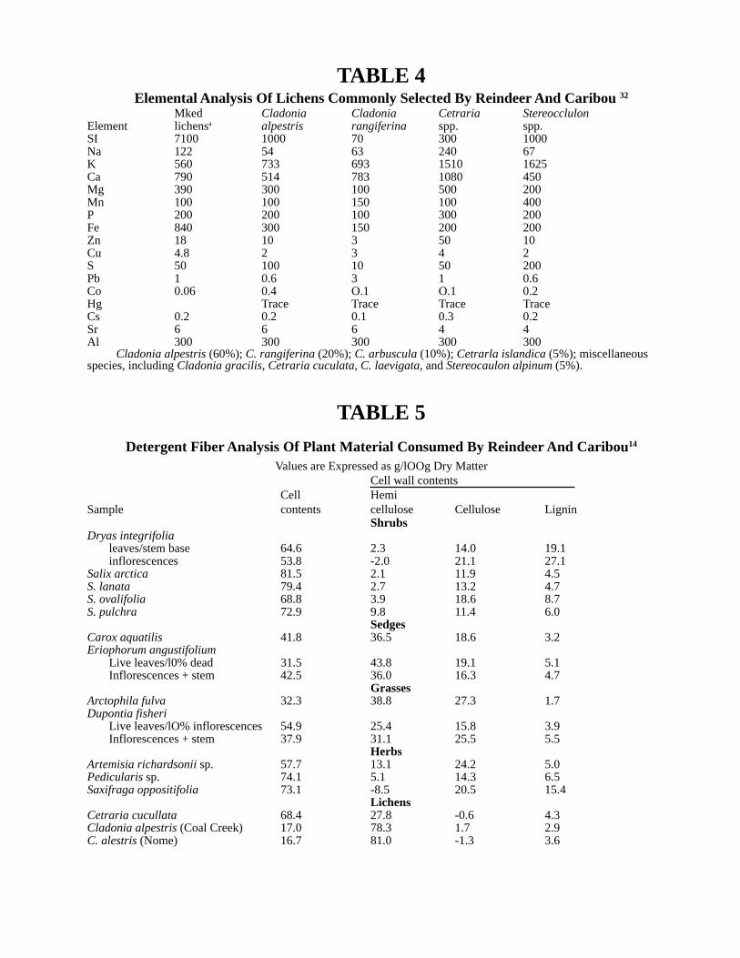
TABLE 4Elemental Analysis Of Lichens Commonly Selected By Reindeer And Caribou 32
Mked Cladonia Cladonia Cetraria StereocclulonElement lichensa alpestris rangiferina spp. spp.SI 7100 1000 70 300 1000Na 122 54 63 240 67K 560 733 693 1510 1625Ca 790 514 783 1080 450Mg 390 300 100 500 200Mn 100 100 150 100 400P 200 200 100 300 200Fe 840 300 150 200 200Zn 18 10 3 50 10Cu 4.8 2 3 4 2S 50 100 10 50 200Pb 1 0.6 3 1 0.6Co 0.06 0.4 O.1 O.1 0.2Hg Trace Trace Trace TraceCs 0.2 0.2 0.1 0.3 0.2Sr 6 6 6 4 4Al 300 300 300 300 300
Cladonia alpestris (60%); C. rangiferina (20%); C. arbuscula (10%); Cetrarla islandica (5%); miscellaneousspecies, including Cladonia gracilis, Cetraria cuculata, C. laevigata, and Stereocaulon alpinum (5%).
TABLE 5
Detergent Fiber Analysis Of Plant Material Consumed By Reindeer And Caribou14
Values are Expressed as g/lOOg Dry MatterCell wall contents
Cell HemiSample contents cellulose Cellulose Lignin
ShrubsDryas integrifolia
leaves/stem base 64.6 2.3 14.0 19.1inflorescences 53.8 -2.0 21.1 27.1
Salix arctica 81.5 2.1 11.9 4.5S. lanata 79.4 2.7 13.2 4.7S. ovalifolia 68.8 3.9 18.6 8.7S. pulchra 72.9 9.8 11.4 6.0
SedgesCarox aquatilis 41.8 36.5 18.6 3.2Eriophorum angustifolium
Live leaves/l0% dead 31.5 43.8 19.1 5.1Inflorescences + stem 42.5 36.0 16.3 4.7
GrassesArctophila fulva 32.3 38.8 27.3 1.7Dupontia fisheri
Live leaves/lO% inflorescences 54.9 25.4 15.8 3.9Inflorescences + stem 37.9 31.1 25.5 5.5
HerbsArtemisia richardsonii sp. 57.7 13.1 24.2 5.0Pedicularis sp. 74.1 5.1 14.3 6.5Saxifraga oppositifolia 73.1 -8.5 20.5 15.4
LichensCetraria cucullata 68.4 27.8 -0.6 4.3Cladonia alpestris (Coal Creek) 17.0 78.3 1.7 2.9C. alestris (Nome) 16.7 81.0 -1.3 3.6

TABLE 6Dry Matter Digestibility a of Individual And Mixed Specimens of Plants Selectively Grazed
By Reindeer And Carlbou 3, 14
In vitro digestibility techniqueSource of Inoculum
Plant Sample Caribou Reindeerb Mean Reindeerb
ShrubsDryas integrifolia Leaves/stem base 33 33 33
Heads 21D. octopetala Leaves and stems (a]pine) 12±2Salix arctica Leaves/buds 71 52±2 62S. lonata Live Ieaves 34±7S. ovalifolia Leaves/buds 35 46±6 41S. pulchra Leaves/buds 54 54 24±0.3S. reticulata Leaves/buds 38
Sedge and cotton grassCarex aquatilis Live leaves 56 68±4 62 39±2Eriopforum angustifolium Live leaves 48 59±3 55 32±3
Inflorescences + stem 56±3E. vaginatum Mature leaves 28±5GrassesArctophila fulva Live leaves 56 72 64Dupontia fishri Live Ieaves 79±2
Inflorescences 67 70 68Herbs
Artemisia richardsoiana Leaves 62 66±3 64Braya spp. Whole plant 68Oxytropis sp. Live leaves 70Parrva nudicalus Whole plant 67Pedicularis sp. Inflorescence + leaves 64±6Saxifraga oppositifolia Inflorescence + leaves 33±1
LichensAlectoria nigricans 45Cetraria cucullata 74±3 48±1 68 77±9C. islandica 48±9Cladonia alpestris 27±14 16±7 21 16±3C. arbuscula 10C. rangiferina 40C. uncialis 44Lobaria linita 45Peltigera aphthosa 44Stereocaulon alpinum 18Thamnolia vermiculoris 59
MossesHylocomium sp[endens Entire gametophyte 19±6Sphagnum magellanicum Entire gametophyte 6Other Entlre gametophyte 13
Esophageal egestaDryas heath Reindeer No. 12 49±3.3Dupontia brook bank Reindeer No. 31 50±0.6Eriophorum meadow Reindeer No. 10 62±O.2Eriophorum meadow Reindeer No. 12 49±1.4Combined reindeer 45±1.0
Caribou C2 37±2.2Caribou C3 43±1.6
aPercent ±SE.bRumen inoculum was obtained from tranquilized caribou and reindeer grazing at Prudhoe Bay, Alaska, or reindeer
given a mixed diet containing 67 percent (dry weight) lichens, 8 percent Carex aquatiIis and 25 percent brome hay. Thefood was ground in hammermill and thoroughly mixed.

TABLE 7Seasonal Changes In The Chemical Composition And Caloric Content Of Important Reindeer
Forage Plants Near Nome, Alaska
Protein Fat(%) Crude Ash Nitrogen–
Species Month (%; Nx 6.25) ether extract fiber (%) (%) free extract Kcal/lOOg
Betula nana March 5.3 2.6 29.3 1.3 61.5 560June 26.0 2.2 13.2 4.6 54.0 542August 16.0 3.5 16.3 2.8 61.4 536
Carex bigelowiia June 16.6 1.3 24.5 4.3 53.3 471August 12.9 1.8 3.6 4.0 52.8 479
Cetraria cucullata June 2.4 1.9 28.5 1.2 90.9 474August 2.2 2.6 7.0 1.2 87.0 463
C. islandica March 2.4 0.8 6.6 1.2 89.0 453June 2.6 0.3 3.7 1.1 92.3 426August 2.6 1.1 8.4 1.2 86.7 458
Cladonia rangiferina March 2.7 1.0 30.3 1.1 64.9 451June 2.9 0.4 27.7 2.3 66.7 466August 2.7 0.4 26.9 0.8 69.2 431
C. gracilis March 2.8 1.0 29.8 1.1 65.3 471August 2.4 0.6 31.6 1.0 64.4 431
C. sylvaticab March 2.5 1.4 21.9 1.1 73.1 426June 2.2 0.7 26.8 1.2 69.1 452August 2.0 0.6 22.2 0.9 74.3 426
Eriophorum angustifolium June 16.1 1.3 21.5 5.6 55.5 477August 12.1 2.0 20.7 3.1 62.1 501
Salix pulchra June 25.2 1.6 8.6 4.9 59.7 504August 22.4 2.1 11.5 3.8 60.2 490
Vaccinium uliginosum March 6.0 3.1 35.9 1.3 53.7 543June 23.4 2.2 11.0 4.0 59.4 523August 13.2 2.0 16.5 2.5 65.8 466
aIncludes a small portion of Carex aquatilisbIncludes Cladonia mitis

Spring DietsIn Alaska, spring grazing commences at the time of calving, or shortly thereafter, when snow is
disappearing rapidly and green shoots and other vegetative parts of grasses, shrubs, and sedgesbecome available. Reindeer feed extensively on dry heaths and wet grassy meadows, selectingDupontia fischeri, Dryas integrifolia, and especially Eriophoriun angustifolium3. Wet boggy areas,the habitat of Carex aquatilis, are not visited widely during the warm, wind-free, daylight hours,presumably because they harbor hordes of mosquitos and other stinging insects. Lactating reindeerattain peak milk production soon after calving; the milk contains high concentrations of fat andprotein but little lactose. The calves commence grazing two to three days following birth;1,lO,11 conse-quently, they gain body weight rapidly, whereas the adult females remain in poor body condition.l2
During early spring, reindeer spend about 50 percent of the day searching for and consuming plants.Eating rates, estimated at 70 to 90 percent of grazing rates, amount to about 10 hours/day during latewinter, but increase to nearly 19 hours/day during late spring, when plant foods become more abun-dant and are of increasing nutritional value. 1,2,5,10
Midsummer DietsDuring midsummer, reindeer gain body weight and improve in condition rapidly while feeding
on grasses such as Carex aquatilis and Dupontia fischeri; sedges, especially Eriophorumangustifolium; shrubs, including Salix spp.; and glandular birches, Betula glandulosa and B. nana.1, 2,
13 Lichens are also eaten selectively, when available in sufficient quantity. According to Kelsall,grasses and sedges make up 28.1 percent of the daily food intake of Canadian reindeer, lichens 31.3percent, and shrubs 31.2 percent; herbs and forbs are little grazed.l White et al. reported similarfindings for reindeer at Prudhoe Bay, Alaska, with green vegetation, grasses, and sedges beingconsumed in a somewhat higher proportion (40 percent) to compensate for the relatively low biom-ass of lichens.3 Personl4 and White et a1.3 found that the grasses appeared to be the most highlydigestible plants, followed in order by herbs and forbs, sedges, and shrubs. Digestibility of lichenswas highly variable, whereas the mosses were of little nutritional value. In fact, the mosses, whichoften comprise more than 50 percent of the vegetative biomass, were never found in significantamounts in rumen contents or egesta collected from esophageal fistula.14 Thus, in midsummer, whenfood plants attain peak nutritional content and quality and reindeer spend about 35 percent of the dayconsuming green vegetation, milk production begins to decline, insect harassment is high, and thereindeer attain the peak body condition necessary for winter survival.3,5,9,10,15,16
Late Summer And Fall DietsThe grazing behavior of reindeer during late summer and fall is characterized by a shift in plant
selection from grasses and sedges to green leafy parts of woody perennials such as Arctostaphylosalpina, A. rubra, Vaccinium uliginosum, Ledum decumbens, Empetrum nigrum and Rhododendronlapponicum. Dryas integrifolia, several salices, and various herbs and forbs (especially Pedicularissp.) are eaten in large quantities.1,3 Mushrooms are so highly prized that the intense search for themoften leads to straying and even stampeding of commercial herds of semidomesticated reindeer.33 Ofsimilar concern to reindeer herders are the effects of tramped dry and brittle lichen heaths during themidsummer to late fall grazing season. Pegau has determined that 35 percent of the edible lichenbiomass may be destroyed in this way during close herding of reindeer on the heaths.l7 Fortunately,lichens are so hygroscopic that a brief rain shower or period of high humidity is sufficient to convertthe brittle lichen heath to a soggy, sponge–like biomass. Finally, digestibility studies have shown thatreindeer make efficient use of the crude fiber in grass and sedge hays.18,19

Winter DietsThe singular nutritional characteristic of reindeer that distinguishes them from all other large
herbivores and that has enabled them to utilize holoarctic range lands is their preference for andability to survive on lichens during the long (six– to eight–months) winter grazing period.13,23 Rein-deer seem to have an uncanny ability to locate and excavate lichens, even where these food plantsare buried under snow cover of up to 60 cm (two feet) or more.20 This does not imply that reindeerthrive under these conditions. On the contrary, the very low protein ( ~2 to 3 percent DM), ash (~2percent DM), and fat (1.5 percent) content and the high crude fiber (45 percent) of lichens (Table 3)results in a considerable loss of body substance (but not necessarily body weight during prolongedwinter grazing.4,12,21 The data listed in Table 8 are intended to emphasize the great differences innutritive content that exist between lichens and the common livestock feed, alfalfa hay. The abovediscussion also should not be construed to imply that reindeer eat only lichens during winter; rein-deer also consume variable amounts of frozen grasses, sedges, the leaves of woody plants, andmushrooms which are found in craters that are dug in search of lichens. 4,22,23
TABLE 8Nutrient Content of the Preferred Lichen, Cladoinia Alpestris
Compared To Alfalfa Hay a,b
Ingredient Cladonia alpesaris Alfalfa hay
Crude protein (%) 2.75 18.2Crude fat (%) 1.52 2.5Crude fiber (%) 44.49 28.0Ash (%) 1.87 10.3N-free extract (%) 49.12 41.0Na (mg/kg) 54 190K (mg/kg) 733 2460Ca (mg/kg) 514 1350Mg(mg/kg) 300 340Mn (mg/kg) 100 46.5P (mg/kg) 200 300Fe(mg/kg) 300 50Zn (mg/kg) 10 35.1Cu(mg/kg) 2 18.7Co (mg/kg) 0.4 0.24
aValues for proximate analysis and mineral content are taken from Kelsall1 and Luick,32 respec-tively.
b Sun–cured, ground alfalfa hay (N.A.S. Ref. No. 1-00-111) as reported in United States–Canadian Table55 of Feed Composition, National Academy of Sciences Publication 1684, Washing-ton. D.C. 1969.
Although competition for grazing on winter lichen ranges is essentially nonexistent, thereindeer’s high dependence upon that food source is not without hazard. Massive die-offs have

occurred when early spring rains freeze and form an impenetrable crust of ice on top of the snow.23
Skogland states that reindeer spend about 62 percent of the day searching for and eating lichens, andvarious workers have estimated that lichen consumption rates are approximately 2 to 5 kg (four to 11pounds) per day per animal.1,2,24,25 The consumption of even these relatively small amounts of frozenfoodstuffs and solid water (snow) is of considerable energetic cost to reindeer, since the necessity ofraising the temperature of ingesta to deep body temperature has been estimated at approximately fivepercent of the resting metabolic rate.13,26
Lastly, rations have been compounded and tested in Norway and Siberia for use either assupplements to winter lichen diets or as complete rations during emergency feeding conditions.23,27,28
Formulas for these rations are listed in Table 9.
The Lichen ParadoxRecent findings at the Cantwell Reindeer Research Station, Cantwell, Alaska seem to establish
a paradoxical situation insofar as they apply to the winter grazing habits of reindeer. In preliminarystudies on the food and water preferences of penned reindeer fed widely differing basal rations,lichens were always selected (and consumed in toto) over nutritionally superior livestock ration(pelletized Purina Cattle Starter No. I ) and/or three species of long hay, Poa pretensis (KentuckyNugget Bluegrass), and Festuca rubra (mixed Arctared red fescue), and Calamagrostis canadensis(Bluejoint reedgass). Snow was preferred over cold, warm, or saline water as a source of waterintake.29
This strong preference for a nutritionally inadequate and energetically taxing diet, especiallywhen nutritional and climatic stresses are most severe, does not fit easily into the concept of naturalselection. However, the recent finding in our laboratory, that the daily water flux of freely grazingreindeer diminishes threefold to fivefold during winter suggests a reasonable explanation for thephenomenon. The survival value of ingesting small amounts of dietary protein and minerals such aslichen and distilled water (snow) may be attributed to the conservation of body water and, therefore,body heat by decreasing the amount of water required to eliminate metabolic nitrogen and bodyelectrolytes.12 This hypothesis is supported by Cameron’s finding that decreases in water flux ofpen–fed reindeer during severe cold are elicited chiefly by the level of dietary protein intake.26
Evidence contrary to this hypothesis stems from reports that reindeer crave urine and sea water,gnaw old bones and shed antlers, and kill and eat lemmings and mice, as well as bird’s eggs andnestlings.30 It is also known that reindeer maintain and even gain body weight during winter whenthere is an adequate supply of frozen green vegetation to supplement the lichen biomass.4,21 Similarresults have also been noted with reindeer that are fed supplemental and/or complete rations duringsevere winter weather (Table 9). 23,27,28

TABLE 9Diets for Supplementing or Replacing Normal FeedingRegimes of Reindeer During Periods of Severe Undernutrition
Supplemental rations a,23 Complete ration A23 Complete ration B27
Urea (50%) Cereal grains (41%) Ground barley (40%)Dicalcium phosphate (30%) Ground hay (25%) Ground oats (17%)Minerals (20%) Ground straw (15%) Wheat bran (15%)
Molasses beet chips (10%) Soybean oil (3%)Wheat bran (5%)Dicalcium phosphate (1.7%)Iodized Salt (0.3%)vitamins (2%)Mineral mixture (trace)
a This ration is mixed with lichens and fed ad libitum to reindeer as a supplement to typicalwinter diets.
In rebuttal to the above, it seems unlikely that significant quantities of urine, sea water, oldbones, lemmings, bird’s eggs, etc. would be available or accessible during deep winter; hence, theyshould not be considered as supplements to winter lichen diets. Additionally, Cameron and Luickl2
confirmed the earlier findings that reindeer can maintain body weight while grazing mixed vegeta-tion during winter, but discovered further that this was accomplished by replacing a substantial lossof body solids with an almost equivalent amount of body water. Thus, changes in body weight aredeceptive and must therefore be interpreted with great caution.

References1. Kelsall, J.P., The Caribou, Queen’s Printer, Ottawa, Ontario, Canada. 1969,1—340.2. Skogland, T., Range use and food selectivity by wild reindeer in southern Norway, in Proc. Ist Int.
Reindeer and Caribou Symp., Special Rep. No. 1, Luick, J.R, Lent. P.C., Klein, D.R., and White,R.C., Eds., Biological Papers, University of Alaska, Fairbanks, 1975, 342-354.
3. White, R.G., Thomson, B.R., Skogland, T., Person, S.J., Russell, D.E., Holleman, D.F., and Luick,J.R., Ecology of caribou at Prudhoe Bay, Alaska, in Ecological Investigations of the TundraBiome in the Prudhoe Bay Region, Alaska, Special Rep. No. 2, Brown, J., Ed., Biological Papers,University of Alaska, Fairbanks, 1975, 151—201.
4. Glinka, D.M., The seasons of reindeer pastures and significance of green forage in the winterfeeding of reindeer, Trans. Inst. Polar Agric., Animal Husbandry and Fishing and Hunting Indus-try Series, Reindeer Industry, 4, 45–46, 1939.
5. Pegau, R.E., Reindeer Range Appraisal in Alaska, M.Sci. thesis, University of Alaska, Fairbanks,1969, 1–130.
6. Andreev, A.N., Ed., Botanical Research in Yakutia, Yakutian Branch SO–AN Academy of Science.Yakuts, USSR, 1975, 1—153.
7. Skjenneberg, S., Ed., Reindeer and Reindeer Management. A.s. Fjeii–Nytt, Lesjaskog, Norway,1965, 1–326.
8. Kuvaev, V.B., Possibilities for rationalized use of the Verkhoyansk deer pastures, Probl. Severa, 8,280–289, 1964.
9. Klein, D.R., Nutritive Quality of Alaskan Range Forage and Associated Growth and Welfare ofReindeer and Caribou, Completion Rep., Contract No. 14-11-001-3748, Bureau or Land Manage-ment, U.S Department of the Interior, 1970, 1—30.
10. Luick, J.R., Ed., Nutrition and Metabolism of Reindeer and Caribou in Alaska, Progress Rep.1973/1974, Contract No. AT(45 -1) 2229, U.S. Atomic Energy Commission, 1974, 1-109.
11. Luick, J.R., White, R.C., Cau, A.M., and Jenness, R., Compositional changes in the milk secretedby grazing reindeer. I. Cross composition and ash, J. Dairy Sci. 57, 1325-1333, 1974.
12. Cameron, R.D. and Luick, J.R., Seasonal changes in total body water, extracellular fluid, andblood volume in grazing reindeer, Can. J. Zool., 50, 107—116, 1972.
13. White, R.C., Some aspects of nutritional adaptations of arctic herbivorous mammals, in Physi-ological Adaptation to the Environment, Vernberg, F.J., Ed., Intext Educational Publications, NewYork, 1975, 234-268.
14. Person, S.J., Digestibility of Indigenous Plants Utilized by Rangifer tarandus, Ph.D. dissertation,Institute of Arctic Biology, University of Alaska, Fairbanks, 1975, 1-97.
15. White, R.C., Factors regulating the food intake of caribou and reindeer on tundra ranges, in Proc.Circumpolar Conf. on Northern Ecology, Ottawa, Ontario, Canada, 1975, 1-5.
16. McEvvan, E.H., Growth and development of the barren-ground caribou. II. Postnatal growthrates, Can. J. Zool., 46, 1023-1029, 1968.
17. Pegau, R.E., Effect of reindeer trampling and grazing on lichens, J. Range Manage., 23, 95—97,1970.
18. Nordfelt, S., Cagell, W., and Nordkvist, M., Digestibility experiments with reindeer, SartryckOch ForhandsmeddeIande, 151, 1—14, 1961.
19. Eriksson, S. and Schmekel, J., A comparison between the ability of reindeer and sheep to digesttheir food, Kungi Lantbrukshogskolans Annaler,. 28, 175—180, 1962.
20. Pruitt, W.O., Jr., Behavior of Barren-ground Caribou, Biological Papers, Special Rep. No. 3,

University of Alaska, Fairbanks, 1960, 1-43.21. Terentiev, N.D., Experimental stall-feeding of reindeer on reindeer moss and hay. Soviet Rein-
deer Industry II STVO, 8, 86, 1936.22. Auramchik, M.N., The winter feeding of reindeer on the Yamal North, Trans. Inst. Polar Agric.,
Animal Husbandry and Fishing and Hunting Industry Series, Reindeer Industry, 4, 65-66, 1939.23. Steen, E., Some aspects of the nutrition of semidomestic reindeer, in Comparatiue Nutrition of
Wild Animals, Crawrord, M.A., Ed., Academic Press, New York, 1968, 117-128.24. Holleman, D.F. and Luick, J.R., Radiocesium cycling in reindeer/carlbou, ln Proc. Ist Int. Rein-
deer and Caribou Symp., Special Rep. No. 1., Luick, J.R., Lent, P.C., Klein, D.R., and White, R.C., Eds., Biol. Papers, University of Alaska, Fairbanks, 1975, 64—70.
25. Hanson, W.C., Whicker, F.W., and Lipscomb, J.F., Lichen forage ingestion rates of free-roamingcaribou estimated with fallout cesium-137, in Proc. Ist Int. Reindeer and Caribou Symp., SpecialRep. No. 1, Luick, J.R., Lent, P.C., Klein, D.R. and White, R.G., Eds., Biological Paper, Univer-sity of Alaska, Special Rep. No. 1, 1975, 71-79.
26. Cameron, R.D., Water metabolism by reindeer (Rangifer tarandus), Ph.D. dissertation, Instituteof Arctic Biology, University of Alaska, Fairbanks, 1972, 1—98.
27. Jacobsen, E. and Skjenneberg. S., Some results from feeding experiments with reindeer, in Proc.Ist Int. Reindeer and Caribou Symp., Special Rep. No. 1, Luick, J.R., Lent, P.C., Klein, D.R. andWhite, R.G., Eds., Biological Papers, University of Alaska, Fairbanks, 1975, 95-107.
28. Luick, J.R., Reindeer research in northern Siberia, Reindeer Herders NewsL, 1, I-12, 1976.29. Luick, J.R, Ed., Industrial Development in Alaska and its Effects on the Nutritional and Physi-
ological Status of Arctic Animals, Progress Rep. 1975/1976, Contract No. E(45-l) 2229, U.S.Energy Research Development Administration, 1976, 1-60.
30. Llano, G.A., Utilization of lichens in the arctic and subarctic, Econ Bot. 10, 367–392. 1956.31. Pulliainen, E., Nutritive values of some lichens used as food by reindeer in northeastern Lapland,
Ann Zool Fenn, 8, 285-289, 1971.32. Luick, J.R., Ed., Nutrition and Metabolism of Reindeer and Caribou in Alaska, Progress Rep.
1970/71, Contract No. AT(04 -3)-310, U.S. Atomic Energy Commission 1971,1-100.33. Borodzin, E., personal communication.

APPENDIX IXNormal Blood Values for Reindeer
Hemoglobin (8/dl) 15.1±4Red Blood Cells (x 106) 9.5±1.5Packed Cell Volume (%) 45.0±5White Blood Cells (x 103) 7.8±2Neutrophils (%) 45.0±10Lymphocytes (%) 40.0±10Monocytes (%) 3.0±2Eosinophils (%) 5.0±5Basophils (%) 3.0±2Plasma Protein (g/dl) 7 5±1Fibrinogen (mg/dl) 300-500Glucose (mg/dl) 101±22Blood Urea Nitrogen (mg/dl) 35.0±6Uric Acid (mg/dl) 0.36±0.07Total Bilirubin (mg/dl) 0.31±0.09Cholesterol (mg/dl) 68.0 ±12Calicum (mg/dl) 9.8±0.8Inorganic Phosphate (mg/dl) 6.8±0.9Sodium (mEq/l) 145±8Potassium (mEq/l) 5.0±0.8Magnesium (mg/dl) 2.1±0.3
APPENDIX XGlossary Of Health Related Terms
Abdomen - Area or space in the body between the chest and pelvis; the part of the body behind thediaphragm
Abdominal - Cavity Area or space in the abdomen containing the digestive organs, reproductiveorgans, kidneys, liver, and spleen
Abomasum - Fourth stomach in a reindeer
Abortion - Premature birth of a dead fetus
Abscess - Pocket of pus
Acute Infection - Infection that lasts a short time

Anatomically - Referring to the structure of the animal
Anatomy - Science of the structure of the animal body
Anemia - Thin blood, a decrease in the red blood cells or hemoglobin in the blood
Antibiotic - Drug given to an animal to fight infection
Antibodies - Produced by the body to fight a specific disease: found in the clear serum portion of theblood
Anticoagulant - A chemical added to blood being collected that keeps it from clotting
Anus - The rear end opening of the digestive tract
Artery - Carries blood away from the heart
Aseptic - Sterile, without contamination
Aspirate - To pull back on the plunger of the syringe (after the needle has been inserted into theanimal for an injection) to see if blood can be sucked into the syringe
Atrium - One of the two upper chambers of the heart; blood is received in an atrium and passed to aventricle
Bacteria - Very small germs; one cell in size; many kinds found in the air, soil, in and on humans andanimals; some cause disease, others are necessary for life
Barren - Not pregnant
Bovine - Referring to domestic cattle
Brucellosis - Disease caused by a bacteria named Brucella; causes female reindeer to abort (lose thebaby before it is ready to be born); causes males to have swollen testicles; lameness is caused in bothsexes in long-term infections; in humans the disease causes fevers, aches and pain
Carbon Dioxide - Gas formed by the tissues and eliminated by the lungs as a waste product
Carnivore - Meat-eating animal
Cartilage - Makes up most of the skeleton before an animal is born and when it is very young; inadults it is found in such places as the ends of the ribs, the joint surfaces of bones and rings of thetrachea
Castrate - To remove the testicles of a male

Cervical - Referring to the neck
Chronic - Infection Infection that lasts a long time
Colony - A group of bacteria that can be seen on a culture plate
Congestion - An abnormal accumulation of fluid, usually blood, in the body
Conjunctivitis - Reddening of the white of the eye
Contagious - Can be spread from one animal to another
Contaminant - An unwanted object, usually a bacteria
Cyst - Fluid-filled sac
Debilitate - To make weak, not strong
Diagnosis - Identification of the nature of the disease
Diaphragm - Muscle separating the abdominal cavity from the chest cavity
Disarticulate - Separation of a joint
Discharge - Fluid oozing from an opening
Ectoparasite - Parasite on the outside of an animal such as fleas, ticks, lice, and mites
Endoparasite - Parasite living inside an animal such as lungworms, tapeworms, roundworms, etc.
Epididymis - Structure attached to the testicle in which the sperm are stored
Epizootic - A disease affecting many animals in an area at the same time
Esophagus - Tube carrying food from the mouth to the stomach
Etiology - Study of the events causing a disease
Feces - Manure
Fecal Sample - Sample of manure
Femur - Thigh bone; extends from the pelvis to the tibia
Fetus - Unborn baby inside the mother
Foot Rot - Foot infection caused by a bacteria

Formalin - Chemical used to preserve tissues
Heart - Pumps blood to the lungs to pick up oxygen, then throughout the rest of the body to deliveroxygen and other nutrients
Hemolyze - To break the red blood cells which release hemoglobin
Histologic - Microscopic study of the structure of tissues and their cells
Hormones - Chemicals produced by the body to regulate specific body functions
Humerus - Upper front leg bone; extends from the scapula to the radius
Hydatid Cyst - Fluid-filled sac containing immature Echinococcus; usually found in liver or lung
Infection - Invasion and multiplication of germs in the body
Incisor - One of the front four teeth; adapted for cutting
Inflammation - Protective response of the body to injury; characterized by heat, swelling, redness,and pain
Injection - The act of forcing a liquid (usually a drug or medicine) into the body with a syringe andneedle
Intestines - Coiled, hose-like portion in the digestive tract between the stomach and anus wherewater and nutrients from the food are absorbed into the body
Isolation - Removal and growth of a disease-causing agent for identification
Intramuscular - Within a muscle
Kidneys - One on each side of the backbone in the small of the back; filter blood to keep body fluidsbalanced; selects out unwanted materials and eliminates them from the body in the urine
Larvae - Immature worm or fly
Lesion - Abnormality caused by bacteria, viruses, parasites or chemical or physical injury
Liver - Large red organ located in the top portion of the abdomen; stores and filters blood; receivesmost of the absorbed foodstuffs from the intestines and changes them into products that can be usedin other parts of the body; receives and degrades toxic (poisonous) substances from the intestines;the liver is essential for life
Lumbar - Pertaining to the back between the chest and the pelvis

Lungs - In the chest cavity on each side; puts oxygen in the blood to be used by the rest of the body;in breathing the lungs exchange oxygen which is breathed in for carbon dioxide which is breathedout
Lymph - Node Small round glands that act as filters to clean body fluids
Mammary Gland - Breast; milk-producing gland
Mandible - Lower jaw
Maxilla - Upper jaw
Metacarpus - Lower front leg bone
Metatarsus - Lower rear leg bone
Media - “Food” used to grow bacteria in the laboratory
Micro organism - Living microscopic cell
Microscopic - Something that can only be seen with a microscope
Mycotic - Referring to a mold or fungus
Necropsy - Examination of a dead animal
Necrosis - Cell death
Omasum - Third stomach in a reindeer
Organ - A group of cells organized together in the body to perform a particular function
Ovary - Makes eggs for reproduction: two in normal females
Oviduct - Tube that carries eggs from the ovary to the uterus
Oxygen - A gas that is essential for animal life; it is carried by the blood from the lungs to the rest ofthe body
Parasite - Worms or insects that live inside the animal (for example, in the lungs or intestines) oroutside the animal and take their nourishment from the animal
Pathogenic - Causes disease
Pathogenicity - Ability to cause disease
Path Report - Pathology report; report of the history and examinations performed to determine thecause of death of an animal

Pedicle - Base or stem of the antler
Pelvic Girdle - Union of the two hipbones
Peritonitis - Inflammation (reddening) of the lining around the abdominal cavity and intestines
Petri Dish - Used to grow bacteria in the laboratory
Pink-eye - Infection of the eye
Placenta - Membrane that attaches the fetus to the uterus and through which it receives its nourish-ment
Pneumonia - Infection of the lungs
Pubis - The part of the hipbone forming the front part of the pelvis
Pus - Accumulation of dead white blood cells and products of tissuedeath: usually caused by a bacterial infection
Rabies - Viral disease usually carried by dogs, wolves, foxes, etc. in their saliva; transmitted tohumans by a bite or by getting saliva on the body by handling an infected animal; can be preventedin animals by a vaccine; always causes death if not treated
Radius - Upper front leg bone between the humerus and metacarpus
Rectum - Lower part of the large intestine
Reticulum - Second stomach in a reindeer
Retropharyngeal - Pouch Located in the back of the throat, above the windpipe
Rumen - First stomach in a reindeer
Scapula - Shoulder blade
Scrotum - Bag on the outside of the male body that holds the testicles
Serology - Study of the disease reactions of blood serum
Serum - Clear part remaining after blood clots are removed; contains antibodies; the part of theblood that is collected and sent to the laboratory for testing; can be frozen after it is separated fromthe clot
Spinal Cord - Part of the nervous system carrying messages from the brain to the body
Spleen - Found in the abdominal cavity of the left side of the rumen; flat dark red in color: destroysold red blood cells; makes new red blood cells in young animals: stores blood

Steer - Castrated male
Sternum - Breastbone
Symptom - A non-specific indication of a disease, such as a headache
Testicle (testes-pl.) - Two in normal males: makes sperm which fertilize eggs in the female forreproduction (‘balls”)
Thoracic - Referring to the chest
Tibia - A bone of the upper rear leg between the femur and metatarsus
Tissue - A group of cells that together make a particular structure
Topical - Upon the skin
Trachea - Windpipe: carries air (with oxygen) into the lungs and out ofthe lungs (with carbon dioxide)
Trauma (traumatic) - Injury caused by physical or chemical means
Treatment - A drug or medicine given to an animal to kill disease-causing germs
Turbinate - Bones in the reindeer nasal cavity
Ulcer - A break or hole
Undulant Fever - Brucellosis in man; symptoms include fever, aches, and pains; can be treated withantibiotics
Ureter - Tube carrying urine•from the kidney to the bladder
Uterus - Organ that receives and holds the fertilized egg to develop into the fetus
Vaccine - A suspension of germs that have been killed or changed so they cannot cause disease butdo cause the body to make antibodies that can fight the real disease later; a vaccine prevents disease
Vas Deferens - Tube that carries sperm from the testes to the penis
Veins - Carry blood from the parts of the body to the heart
Ventricle - One of the lower. thick-walled chambers of the heart that pumps blood out to the body
Vertebra - One of the sections of bone of the backbone
Virus - Very small germ, smaller than bacteria; cannot be seen with ordinary microscopes; must live
within another living cell
Viscera - Internal organs of the body
Zoonosis - Disease that can be transmitted from animals to man
Agricultural and Forestry Experiment StationSchool of Agriculture and Land Resources Management
University of Alaska FairbanksJames V. Drew, Dean and Director
andCooperative Extension Service
Irvin W. Skelton, Acting DirectorUniversity of Alaska Fairbanks
andU.S. Department of Agriculture
It is the policy of the University of Alaska to provide equal education and employment opportu-nities and to provide services and benefits to all students and employees without regard to race,color, religion, national origin, sex, age, disability, status as a Vietnam era or disabled veteran,marital status, changes in marital status, pregnancy or parenthood, pursuant to laws enforced by theDepartment of Education and the Department of Labor, including Presidential Executive Order11246, as amended, Title VI and Title VII of the 1964 Civil Rights Act, Title IX of the EducationAmendments of 1972, the Public Health Service Act of 1971, the Veteran’s Readjustment AssistanceAct of 1974, the Vocational Rehabilitation Act of 1973, the Age Discrimination in Employment Act of1967, the Equal Pay Act of 1963, the 14th Amendment, EEOC’s Sex Discrimination Guidelines, andAlaska Statues 18.80.010 et seq., and 14.18.010 et seq., and 36.30.010 et seg. Inquiries regardingapplication of these and other regulations should be directed either to the University of AlaskaFairbanks, Director of Employee Relations; the Office of Civil Rights, Department of Education,Washington, DC; or to the Office of Federal Contract Compliance Programs, Department of Labor,Washington, DC.
Issued in furtherance of Cooperative Extension work, acts of May 8 and June 30, 1914, incooperation with the U.S. Department of Agriculture, Irvin W. Skelton. Acting Director, CooperativeExtension Service, University of Alaska Fairbanks.
In order to simplify terminology, trade names of products or equipment may have been used inthis publication. No endorsement of products or firms mentioned is intended, nor is criticism impliedof those not mentioned. Material appearing herein may be reprinted provided no endorsement of acommercial product is stated or implied. Please credit the researchers involved and the Agriculturaland Forestry Experiment Station, Cooperative Extension Service and the University of AlaskaFairbanks.