regulation of osteoblast metabolism by wnt signaling · wnt signaling pathway that is critical for...
TRANSCRIPT

318 www.e-enm.org
Endocrinol Metab 2018;33:318-330https://doi.org/10.3803/EnM.2018.33.3.318pISSN 2093-596X · eISSN 2093-5978
ReviewArticle
Regulation of Osteoblast Metabolism by Wnt Signaling Megan C. Moorer1,2, Ryan C. Riddle1,2
1Department of Orthopaedic Surgery, Johns Hopkins University School of Medicine; 2Baltimore Veterans Administration Medical Center, Baltimore, MD, USA
Wnt/β-catenin signaling plays a critical role in the achievement of peak bone mass, affecting the commitment of mesenchymal pro-genitors to the osteoblast lineage and the anabolic capacity of osteoblasts depositing bone matrix. Recent studies suggest that this evolutionarily-conserved, developmental pathway exerts its anabolic effects in part by coordinating osteoblast activity with interme-diary metabolism. These findings are compatible with the cloning of the gene encoding the low-density lipoprotein related receptor-5 (LRP5) Wnt co-receptor from a diabetes-susceptibility locus and the now well-established linkage between Wnt signaling and me-tabolism. In this article, we provide an overview of the role of Wnt signaling in whole-body metabolism and review the literature re-garding the impact of Wnt signaling on the osteoblast’s utilization of three different energy sources: fatty acids, glucose, and gluta-mine. Special attention is devoted to the net effect of nutrient utilization and the mode of regulation by Wnt signaling. Mechanistic studies indicate that the utilization of each substrate is governed by a unique mechanism of control with β-catenin-dependent signal-ing regulating fatty acid β-oxidation, while glucose and glutamine utilization are β-catenin-independent and downstream of mamma-lian target of rapamycin complex 2 (mTORC2) and mammalian target of rapamycin complex 1 (mTORC1) activation, respectively. The emergence of these data has provided a new context for the mechanisms by which Wnt signaling influences bone development.
Keywords: Wnt signaling; Beta catenin; Osteoblasts; Intermediary metabolism
INTRODUCTION
The dynamic nature of bone tissue requires a homeostatic bal-ance between new bone formation and the resorption of old or damaged matrix to maintain skeletal architecture and strength. The skeleton must balance the need to provide a rigid structure that can protect vital organs and facilitate locomotion against its function as a mineral reserve for the entire body. Osteoclastic cells, which degrade bone matrix and liberate the calcium and phosphate stored as hydroxyapatite, are derived from the hema-topoietic lineage, while bone-forming osteoblasts responsible for the deposition and mineralization of new bone matrix are of
mesenchymal origin. Understanding the local, hormonal, and genetic effectors that influence the activity of these two cell types is critical to our understanding of human disease and the development of new therapeutics that increase bone mass and strength [1,2].
In our aging population, the close association and often co-existing conditions of osteopenia, obesity, diabetes, and cancer have peaked an interest in the effects of intermediary metabo-lism on bone cell function. The initial analyses of fuel selection by bone cells were performed more than 50 years ago, were fo-cused on the osteoblast and the role that metabolites might play in the liberation of mineral ions, but were forgotten by the field.
Received: 23 June 2018, Revised: 1 July 2018, Accepted: 8 July 2018Corresponding author: Ryan C. RiddleDepartment of Orthopaedic Surgery, Johns Hopkins University School of Medicine, 1721 E. Madison St, Baltimore, MD 21205, USA Tel: +1-410-502-6412, Fax: +1-443-287-4428, E-mail: [email protected]
Copyright © 2018 Korean Endocrine SocietyThis is an Open Access article distributed under the terms of the Creative Com-mons Attribution Non-Commercial License (http://creativecommons.org/licenses/by-nc/4.0/) which permits unrestricted non-commercial use, distribu-tion, and reproduction in any medium, provided the original work is properly cited.
Wnt and Osteoblast Metabolism
Copyright © 2018 Korean Endocrine Society www.e-enm.org 319
Endocrinol Metab 2018;33:318-330https://doi.org/10.3803/EnM.2018.33.3.318pISSN 2093-596X · eISSN 2093-5978
Glucose was proposed as the primary energy source for the os-teoblast and a carbon source for amino acid and collagen syn-thesis, while the metabolites citrate and lactate were expected to provide an acidic environment sufficient for the release of calci-um from the bone matrix [3-7]. The oxidation of palmitate, the most abundant fatty acid in animals, by osteoblasts was sug-gested to contribute between 40% and 80% of the energy pro-duced by glucose oxidation [8]. More recently, observations made in genetic mouse models or in cell culture using more so-phisticated bioenergetic analyses have provided confirmation of these classic studies and elaborated on the changes in metabolic flux that accompany each stage of osteoblast differentiation [9,10]. Glucose uptake via glucose transporter 1 (Glut1) is now recognized as a key regulator of the molecular events that initi-ate early osteoblast commitment by regulating the stability of Runt-related transcription factor 2 (Runx2) [11]. Likewise, stud-ies using radiolabeled lipoproteins and fatty acids indicate that the skeleton plays a role in lipid homeostasis [12,13].
Emerging evidence suggests that the utilization of specific fuel substrates by the osteoblast is governed by key develop-mental and hormonal signals [14-18]. Key among these is the Wnt signaling pathway that is critical for normal bone mass ac-crual and exerts control of over nearly all facets of osteoblast maturation and function. In the sections below, we provide overviews of the Wnt signaling pathway and its role in whole body metabolism before describing the effect of Wnt signaling on fatty acid, glucose and glutamine catabolism by the osteo-blast.
Wnt SIGNALING
Wnt signaling plays a central role in the coordination of a num-ber of cellular and organismal processes including proliferation, tissue development and repair, and metabolism [19-21]. The most thoroughly studied pathway, referred to as Wnt/β-catenin signaling or the “canonical” pathway, regulates the proteasomal degradation of the transcription factor, β-catenin [20]. In the ab-sence of Wnt ligands, glycogen synthase kinase-3β (Gsk3β) and casein kinase-1 (Ck-1), in collaboration with a destruction com-plex that contains the adenomatous polyposis coli (Apc) protein [22], the Wilms tumor suppressor protein (WTX) [23], and Axin [24,25], sequentially phosphorylate cytosolic β-catenin [26]. These modifications facilitate the recognition of β-catenin by β-transducing repeat-containing protein (β-TrCP), a component of an E3 ubiquitin ligase complex, and its targeting for degrada-tion [27,28]. Wnt ligands inhibit the proteolysis of newly syn-
thesized β-catenin [29] by stimulating the formation of a multi-protein receptor complex composed of a seven transmembrane Frizzled receptor [30,31] and a low-density lipoprotein related receptor-5 (Lrp5) or Lrp6 co-receptor [32,33]. Ligand engage-ment leads to the phosphorylation of the intracellular domain of Lrp5 and Lrp6 [34], the recruitment of disheveled (Dvl) [35-37] and Axin [38], and ultimately the cytoplasmic accumulation and then nuclear translocation of β-catenin. Within the nucleus, β-catenin regulates target gene expression by interacting with DNA-bound T-cell factor (TCF) [39,40] and disrupting its asso-ciation with the transcriptional repressor, Groucho [41,42]. Transcriptional activity is further enhanced by the phosphoryla-tion of β-catenin and TCF [43,44] and the recruitment of co-ac-tivators and histone modifying enzymes that interact with the C- and N-terminal tails of β-catenin [45-47].
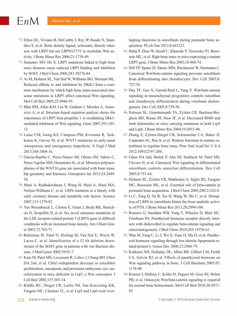
The binding of Wnt ligands to Frizzled and Lrp5/6 can also initiate signaling downstream of the mammalian target of ra-pamycin complex 1 (mTORC1) and mammalian target of ra-pamycin complex 2 (mTORC2) complexes. Gsk3β, the kinase that phosphorylates β-catenin and targets it for degradation, phosphorylates the tuberous sclerosis 2 (Tsc2) protein at two serine residues to enhance its inhibition of mTORC1. Therefore, by inhibiting Gsk3β activation, Wnt ligands increase protein synthesis and cell growth [48,49]. Activation of the mTORC2 complex by Wnts has not been as well studied, though Wnt3a, Wnt7b, and Wnt10b are able to stimulate its activity in osteo-blastic cells [50] and the complex is required for the osteoana-bolic effect of sclerostin neutralization [51]. In vitro gene knockdown studies indicated that the signaling mechanism in-volves the small GTPase, Rac family small GTPase 1 (Rac1) [50].
Other “non-canonical” pathways that do not activate β-catenin or require a Lrp5/Lrp6 co-receptor are also activated by the in-teraction of Wnt ligands with Frizzled receptors. These path-ways predominately affect processes like cellular migration and polarity [52-54] and their activation may antagonize the activa-tion of Wnt/β-catenin signaling [55-58]. In the Wnt-Ca2+ path-way, Wnt stimulation induces calcium transients [58-60] that activate calcium/calmodulin-dependent kinase II, calcineurin, and protein kinase C [61,62]. In another pathway, known as Wnt-Frizzled planar cell polarity, Frizzled and the four trans-membrane protein, Vangl, together with four other core proteins interact across cell membranes to regulate cellular directionality [54,63]. The role of Wnt ligands in this pathway is less clear, but both Wnt-5a and Wnt-11 [64-66] have been implicated in the process.
Moorer MC, et al.
320 www.e-enm.org Copyright © 2018 Korean Endocrine Society
CONTRIBUTIONS OF Wnt SIGNALING TO SKELETAL HOMEOSTASIS
The concept that Wnt signaling regulates skeletal development and homeostasis was first evident in mouse mutants deficient or hypomorphic for Wnt3a [67,68]. In these models, global disrup-tion results in an axial truncation caudal to the forelimbs with a lack of somites and extensive death of mesodermal cells, while hypomorphic alleles lead to deficiencies in ossification with malformations and fusions of caudal vertebrae. However, the notion that Wnts are required for normal bone acquisition gained significant momentum when three publications linked mutations in the human LRP5 gene that encodes the Wnt co-re-ceptor to conditions with high and low bone mass in humans. In 2001, Gong and colleagues [69] from the Osteoporosis Pseudo-glioma (OPPG) Syndrome Collaborative Group reported that loss-of-function mutations in LRP5 were causal for the develop-ment of OPPG, a condition characterized by severe, early-onset osteoporosis as well as disruptions in ocular structure or the per-sistence of vitreal vascularization. Less than a year later, Little et al. [70] and Boyden et al. [71] independently identified muta-tions leading to a glycine-to-valine amino acid change (G171V) in LRP5 in kindreds with a high bone mass (HBM) phenotype. This missense mutation was revealed to inhibit the binding of dickkopf and sclerostin, two secreted Wnt signaling antagonists, to LRP5 thereby enhancing signaling capacity [71-75]. Subse-quent studies have identified additional mutations in LRP5 as well as LRP6 and other Wnt signaling components that influ-ence bone mass and strength [76-80].
Numerous transgenic mouse models have also now been cre-ated to examine the cellular and molecular basis by which Wnt signaling governs skeletal modeling/remodeling. Most of these models and especially mice globally deficient for Lrp5 and those expressing HBM alleles recapitulate the OPPG and HBM phenotypes, respectively [81-83]. Wnt/β-catenin signaling is re-quired for the initial fate specification of cells committing to the osteoblast lineage [84,85], regulates the performance of matur-ing osteoblasts [82,86,87], controls osteoclastogenesis [88,89], and also influences responsivity of osteoblasts to anabolic hor-mones [90-93]. Dramatic examples of the central role of Wnt/β-catenin signaling in skeletal homeostasis are evident in the work of Holmen et al. [89] who generated mice in which the gene encoding β-catenin or the Apc protein were ablated specif-ically in the osteoblast. The β-catenin deficient mice developed severe osteopenia due to a reduction in osteoblast numbers and a dramatic increase in the prevalence of osteoclasts, while Apc
mutants exhibited increased β-catenin activation and bone over-growth. Strikingly, neither model was compatible with pro-longed postnatal life. Osteocyte-specific β-catenin knockout mice (via expression of the dentin matrix protein 1 [DMP1]-Cre transgene) also have a severe skeletal phenotype that resembles the osteoblast-specific mutant models, with an expanded mar-row cavity and thin cortical bone due to increased resorption by osteoclasts and changes in the osteoprotegerin (OPG)/receptor activator of nuclear factor kappa-Β ligand (RANKL) ratio, but without a change in numbers or activity of osteoblasts [94]. Thus, it is possible that β-catenin actions in the osteocyte regu-late the activity of osteoclasts, while osteoblastic β-catenin reg-ulates cell maturation.
IMPACT OF Wnt SIGNALING ON WHOLE BODY METABOLISM
The notion that Wnt signaling contributes to the regulation of whole body metabolism was evident from the initial cloning of the gene encoding the LRP5 co-receptor in humans. Located on the q-arm of chromosome 11, LRP5 was identified as one of four genes in a 400 kb region surrounding the insulin dependent diabetes mellitus 4 (IDDM4) locus that exhibited strong genetic linkage with the development of type 1 diabetes [95,96]. Subse-quent studies would reveal that LRP5 was not the causative gene at this locus [97], but polymorphisms in LRP5 (A1330V, N740N, Q89R) have been linked to increased total and low-density lipoprotein cholesterol levels, hypertension, increased body mass index, and obesity [98-102]. Indeed, recent work from Loh et al. [103] reported that HBM mutations in LRP5 in humans are associated with a metabolically favorable body fat distribution and increased insulin sensitivity, while low bone mass, loss of function mutations are associated with increased abdominal fat accumulation.
The extracellular domain of LRP5 contains 3 low-density li-poprotein receptor (Ldlr) domains that retain the capacity to bind apolipoprotein E (ApoE) [104] and mouse studies per-formed by Fujino and colleagues [105] suggest a direct role for the protein in glucose and lipoprotein metabolism. On a stan-dard chow diet, Lrp5–/– mice exhibit age-related impairments in glucose-stimulated insulin secretion which is likely due to alter-ations in glucose-stimulated ATP production and Ca2-transients. With high fat diet feeding, the mutants develop hypercholester-olemia secondary to a reduction in hepatic chylomicron clear-ance [105]. Crossing the Lrp5–/– mice onto an ApoE–/– back-ground, to generate double mutants, results in hypercholesterol-

Wnt and Osteoblast Metabolism
Copyright © 2018 Korean Endocrine Society www.e-enm.org 321
emia even on a chow diet and to a greater extent than ApoE-de-ficiency alone, as well as impaired fat tolerance, and advanced atherosclerosis [106]. Lrp5 appears, therefore, to contribute to lipoprotein metabolism in a pathway that works in parallel with the Ldlr.
Genome wide association studies have implicated other Wnt signaling components in the development of metabolic disease, with polymorphisms in WNT5B [107] and WNT10B [108] linked to the development of type 2 diabetes and obesity, re-spectively. More strikingly, Grant and colleagues [109] identi-fied a strong association between variants in the gene encoding the Wnt effector transcription factor 7-like 2 (TCF7L2, also re-ferred to as TCF4) with susceptibility for type 2 diabetes in an Icelandic cohort and then replicated the finding in Danish and American cohorts. Additional studies have now replicated this association in a number of other ethnic populations [110-112]. The expression of Tcf7l2 appears to be regulated by alterations in the metabolism of a number of tissues, including the pancre-as, adipose tissue, and the liver [113-115]. Direct examination of the transcriptional regulator’s mechanism of action were ini-tially hampered by the perinatal death of Tcf7l2–/– mice [116], but heterozygous mice were shown to be protected from the de-velopment of a diabetic phenotype, while those that overex-pressed the human gene exhibited an increased susceptibility when fed a high fat diet [117]. TCF7L2 was expected to exert its effect on metabolism via the β-cell and two groups demon-strated that loss of Tcf7l2 function in the pancreas of transgenic mouse models resulted in impaired glucose tolerance [118,119]. However, tissue-specific knockouts generated by Boj and col-leagues [120], in which Tcf7l2 expression was ablated in either the pancreatic β-cell or the hepatocyte, suggested a different mechanism of action. Their β-cell-specific Tcf7L2 knockouts exhibited normal islet development and function, but hepato-cyte-specific ablation reduced glucose production and improved glucose homeostasis. These results remain controversial and the mechanisms of TCF7L2 actions in metabolism continue to be an area of intense interest [121]. It is likely that the actions of TCF7L2 in metabolic control represent combinatorial effects across a number of tissues.
Wnt-STIMULATED β-OXIDATION IN THE OSTEOBLAST
Our group’s interest in the metabolic actions of Wnt signaling in the skeleton stems from a serendipitous observation made dur-ing an evaluation of the unique and overlapping functions of the
Wnt co-receptors, Lrp5 and Lrp6, in the osteoblast. Consistent with the contributions of Lrp5 and Lrp6 to the activation of β-catenin-dependent signaling and the roles of Wnt signaling in regulating osteoblast function noted above, genetic ablation of either Wnt co-receptor in the mature osteoblast (Lrp5flox; Oc-Cre or Lrp6flox; Oc-Cre) impaired skeletal homeostasis and re-sulted in the development of an osteopenic phenotype [82]. Sur-prisingly, Frey et al. [122] demonstrated that the Lrp5 mutants also developed alterations in body composition that were not evident in the Lrp6 mutants. Loss of Lrp5 function increased the size of white adipose tissue depots, reduced whole body en-ergy expenditure indexed by indirect calorimetry, and resulted in the development of dyslipidemia marked by increased levels of serum triglycerides and free fatty acids. Subsequent gene ex-pression profiling of Lrp5-deficient osteoblasts cultured in vitro suggested that the phenotype was a result of altered fatty acid catabolism, as the expression of a number of genes involved in mitochondrial long-chain fatty acid oxidation were down-regu-lated in the mutant osteoblasts (Fig. 1). Indeed, Lrp5-deficient osteoblasts exhibited an impaired ability to fully oxidize oleate to CO2.
In a follow-up study, Frey et al. [123] investigated whether the regulation of long-chain fatty acid oxidation by Lrp5 re-quired the activation of β-catenin. Initially, cultures of primary osteoblasts were treated with Wnt ligands expressed in the bone microenvironment [124-126] and their ability to influence β-oxidation was assessed. Only ligands like Wnt3a, Wnt10b and Wnt16 that are able to induce β-catenin activation en-hanced oleate oxidation, which suggested that Wnt-induced al-terations in metabolism proceeded via the canonical mecha-nism. As a more direct test of this hypothesis, Frey et al. [123] generated mice in which the genetic ablation of the catenin beta 1 (Ctnnb1) gene in the osteoblast could be controlled by the administration of tamoxifen (Ctnnb1flox; Oc-CreERT2) to avoid the early lethality associated with constitutive disruption of β-catenin expression in this cell population [89]. In vitro, the loss of β-catenin function in cultures of maturing osteoblasts re-sulted in the expected inhibition of osteoblast maturation as well as a nearly 50% reduction in the capacity for oleate oxidation and a significant reduction in cellular ATP content, despite an increase in glucose uptake and glycolytic metabolism. In vivo, manipulation of β-catenin expression mirrored the effect of Lrp5 loss of function as the mutants developed an increase in adipose tissue mass and an increase in serum fatty acids. Sur-prisingly, the β-catenin mutants also developed impairments in glucose tolerance and insulin sensitivity that were not evident in
Moorer MC, et al.
322 www.e-enm.org Copyright © 2018 Korean Endocrine Society
Lrp5 mutants and are likely secondary to ectopic lipid accumu-lation. The more severe metabolic phenotype that develops in the β-catenin mutants is likely a result of an impairment in all effects of canonical Wnt signaling with the deletion of the path-way’s target transcription factor, while the presence of Lrp6 may be able to partially compensate and maintain a level of Wnt signaling in Lrp5 mutants.
To explore the absolute requirement for fatty acid catabolism during bone development and to determine if bone contributes to whole body lipid homeostasis, Kim et al. [13] examined the skeletal and metabolic phenotypes of a mouse model in which the expression of carnitine palmitoyltransferase 2 (Cpt2), an ob-
ligate enzyme for mitochondrial fatty-acid β-oxidation, was dis-rupted in osteoblasts and osteocytes. Consistent with the obser-vation that bone takes up a significant quantity of circulating fatty acids, skeletal homeostasis was impaired by Cpt2 deficien-cy, but the severity was dependent on the sex of the mutant. Male Cpt2 mutants exhibited only a transient decrease in tra-becular bone volume that was most evident at 6 weeks of age, but female mutants failed to reach peak trabecular bone volume and exhibited a decrease in trabecular bone volume in both the distal femur and L5 vertebrae at all timepoints examined after 1 month of age. Cortical tissue area in the femur of female mice was increased although cortical thickness did not change, im-
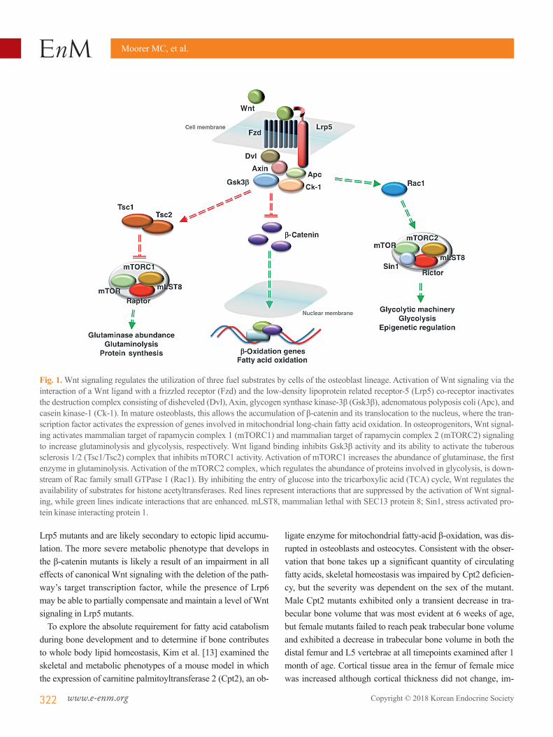
Fig. 1. Wnt signaling regulates the utilization of three fuel substrates by cells of the osteoblast lineage. Activation of Wnt signaling via the interaction of a Wnt ligand with a frizzled receptor (Fzd) and the low-density lipoprotein related receptor-5 (Lrp5) co-receptor inactivates the destruction complex consisting of disheveled (Dvl), Axin, glycogen synthase kinase-3β (Gsk3β), adenomatous polyposis coli (Apc), and casein kinase-1 (Ck-1). In mature osteoblasts, this allows the accumulation of β-catenin and its translocation to the nucleus, where the tran-scription factor activates the expression of genes involved in mitochondrial long-chain fatty acid oxidation. In osteoprogenitors, Wnt signal-ing activates mammalian target of rapamycin complex 1 (mTORC1) and mammalian target of rapamycin complex 2 (mTORC2) signaling to increase glutaminolysis and glycolysis, respectively. Wnt ligand binding inhibits Gsk3β activity and its ability to activate the tuberous sclerosis 1/2 (Tsc1/Tsc2) complex that inhibits mTORC1 activity. Activation of mTORC1 increases the abundance of glutaminase, the first enzyme in glutaminolysis. Activation of the mTORC2 complex, which regulates the abundance of proteins involved in glycolysis, is down-stream of Rac family small GTPase 1 (Rac1). By inhibiting the entry of glucose into the tricarboxylic acid (TCA) cycle, Wnt regulates the availability of substrates for histone acetyltransferases. Red lines represent interactions that are suppressed by the activation of Wnt signal-ing, while green lines indicate interactions that are enhanced. mLST8, mammalian lethal with SEC13 protein 8; Sin1, stress activated pro-tein kinase interacting protein 1.
Wnt and Osteoblast Metabolism
Copyright © 2018 Korean Endocrine Society www.e-enm.org 323
plying that the skeleton adapted to reduced bone quality by changing geometrical properties of the femur to maintain bone strength. Histomorphometric analyses revealed that the skeletal phenotype in the mutant mice was secondary to a mineralization defect, as the female mutants accumulated unmineralized ma-trix and exhibited a reduction in the mineral apposition rate and increase in the mineralization lag time. A combination of in vivo and in vitro studies suggested that the sexually dimorphic phe-notype is related to the ability of estrogen to influence adjust-ments in fuel selection. Glucose uptake was increased in Cpt2 deficient osteoblasts cultured in vitro and the skeletal tissue of male Cpt2 deficient mice, but not in the bone of female mutants or in mutant osteoblast cultures treated with exogenous estro-gen. Estrogen treatment of primary cultures of mutant osteo-blasts also exacerbated the downregulation of genes associated with osteoblast differentiation and resulted in a more severe im-pairment in matrix mineralization. A similar influence of estro-gen on cellular metabolism has been noted in a number of other tissues [127-129].
Like genetic ablation of Lrp5 or β-catenin, inhibition of long-chain fatty acid metabolism in the osteoblast resulted in an in-crease in serum fatty acids, but on a normal chow diet the male Cpt2 mutants exhibited a reduction in body fat fraction and in the weight of the gonadal fat pad. This body composition phe-notype is likely due to a shift in glucose utilization and storage because glucose uptake by adipose was repressed while skeletal glucose uptake was increased. Intriguingly, when the male Cpt2 mutants were fed a high fat diet that increased the levels of es-trogen, bone loss ensued, the weights of all major fat pads in-creased, and the mutant mice performed poorly in glucose toler-ance and insulin tolerance tests. Overall, these data demonstrat-ed that fatty acid catabolism is required for normal osteoblast function and bone mass acquisition and is strongly influenced by both sex and diet.
Wnt-STIMULATED GLYCOLYSIS IN THE OSTEOBLAST
As indicated above, glucose is required for normal osteoblast function and likely represents an important energy source. Since components of the Wnt signaling cascade have been linked to the regulation of whole-body glucose metabolism, Esen and colleagues [50] explored the effect of Wnt signaling on glucose utilization by osteoblasts. Relying primarily on the ST2 cell line, which models mouse bone marrow stromal cells, Wnt3a and Wnt10b were demonstrated to increase glucose acquisition
by stimulating the expression of Glut1, hexokinase-2, lactate dehydrogenase, and pyruvate dehydrogenase kinase 1 and were more effective than high dose insulin stimulation. Intriguingly, the increase in glucose uptake was not accompanied by an in-crease in the oxygen consumption rate, which suggests that it was not processed via oxidative phosphorylation. Rather, Wnt3a stimulated lactate production and increased the extracellular acidification rate, suggesting that Wnt signaling activated aero-bic glycolysis, a metabolic process most closely associate with cancer cell metabolism [130].
Using pharmacological antagonists and gene knockdown studies to elaborate on the mechanism by which Wnts stimulate glycolysis, Esen et al. [50] demonstrated that the response re-quired the Wnt co-receptor Lrp5. Indeed, Lrp5–/– mice as well as those in which Lrp5 expression was abolished via osterix-Cre expression exhibited a reduction in glycolytic enzyme expres-sion in bone. However, it did not proceed via alterations in the activity of either Gsk3β or β-catenin activity, as inhibiting the activity of these effectors did not impact glucose consumption. Instead, Wnt3a activated mTORC2 via Rac1, which in turn co-ordinated the increase in glucose consumption and the increase in glycolytic gene expression.
Why Wnt signaling, which has profound anabolic effects on osteoblast differentiation and function, should lead early osteo-blasts to rely on a less efficient mode of ATP generation from glucose remains an open question. One possibility is that like cancer cells, the products of aerobic glycolysis are used by im-mature cells as the starting material for biosynthetic pathways that produce amino acids and nucleotides [130]. A second pos-sibility suggested by the Long Laboratory is that this metabolic paradigm contributes to the epigenetic regulation of osteoblast differentiation [131]. Using RNA sequencing, Karner et al. [131] demonstrated that the number of genes exhibiting an in-crease in expression after Wnt3a stimulation of ST2 cells was surpassed by the number of genes exhibiting a decrease in ex-pression and that many of the downregulated genes were associ-ated with differentiation toward the chondrocyte or adipocyte lineages (including peroxisome proliferator-activated receptor gamma [Pparg] and CCAAT enhancer binding protein alpha [Cebpa]). Since gene activation is expected to be β-catenin’s major mode of action, the group postulated that Wnts contribut-ed to gene suppression via the regulation of histone modifica-tion. Consistent with this idea, bulk histone acetylation was re-duced in ST2 cells after Wnt3a treatment, but the activity levels of histone deacetylases and histone acetyltransferases were not affected. Instead, Wnt signaling reduced the availability of the

Moorer MC, et al.
324 www.e-enm.org Copyright © 2018 Korean Endocrine Society
histone acetyltransferase substrate, acetyl coenzyme A (acetyl-CoA), as well as its precursor, citrate. Therefore, by inhibiting the entry of glucose into the tricarboxylic acid (TCA) cycle and thereby reducing citrate and acetyl-CoA production, Wnt signal-ing regulates the genomic landscape and fate-specification of osteoprogenitors.
Wnt-STIMULATED GLUTAMINOLYSIS IN THE OSTEOBLAST
The amino acid, glutamine, represents a third energy source whose catabolism is under the control of Wnt signaling in the osteoblast. Glutamine is abundant in the circulation (approxi-mately 20% of the free amino acid pool) and in the neoplastic cells and normal cells (i.e., lymphocytes, fibroblasts, etc.) that utilize Warburg-type metabolism it can be utilized in a process referred to as anaplerosis. This process maintains TCA function when intermediates like citrate are removed from the cycle for anabolic reactions. In this reaction, glutaminase, a mitochondri-al enzyme, deaminates glutamine to form glutamate which is then converted to α-ketoglutarate, a TCA intermediate, by gluta-mate dehydrogenase or via transamination with pyruvate by ala-nine aminotransferase. Biltz and colleagues [132] demonstrated more than 30 years ago that rat calvarial cells oxidize glutamine while more recent in vitro studies have suggested a requirement for glutamine supplementation for mineralization of bone ma-trix by osteoblasts [133].
Karner and colleagues [134] examined glutamine metabolism in the context of Wnt-stimulated osteoblast differentiation and the coordinate increases in protein synthesis necessary to pre-pare bone matrix. Wnt stimulation of the ST2 cell line increased glutamine uptake, but it was quickly metabolized, as the cells exhibited a nearly 40% decrease in cellular glutamine levels 24 hours after treatment due to an increase in glutaminase activity and the entry of glutamine-derived carbons into the TCA cycle via anaplerosis. Using pharmacological inhibition of glutamin-ase activity, the group demonstrated that glutamine catabolism was required for Wnt-stimulated osteoblast differentiation in vi-tro as well as the increase in bone mass resulting from the ex-pression of a HBM variant of the Lrp5 co-receptor in vivo. In parallel, glutaminolysis initiated the activation of the integrated stress response and the associated increase in the expression of genes necessary for amino acid uptake and protein folding, which were also required for Wnt-stimulated bone acquisition. Consistent with this close association with protein synthesis, mechanistic studies indicated that the regulation of glutamine
utilization is dependent on the activation of mTORC1. Thus, glutamine acquisition and utilization in response to Wnt signal-ing appear to represent a molecular rheostat that acts to maintain cellular energetics, endoplasmic reticulum status [134] and re-dox balance [17].
CONCLUSIONS
Taken together the studies reviewed above highlight an exciting and newly appreciated effect of Wnt signaling on the biology of the osteoblast. This evolutionarily-conserved pathway that is critical for the attainment of normal bone mass and structure co-ordinates one of the most essential cellular functions: the gener-ation of energy necessary to fuel other cell processes. The exist-ing data suggest that the governance of osteoblast fuel selection by Wnt is highly dependent on the state of osteoblast differenti-ation, with immature osteoblasts exhibiting an increase in glu-cose and glutamine utilization and mature cells switching to and increasing the utilization of fatty acids. These findings accord with well-established concepts in developmental biology and the changing metabolic demands of osteoblast differentiation [9]. Differentiated osteoblasts that prepare and mineralize the bone matrix maintain abundant mitochondria likely as a result of the tremendous energetic demands of protein synthesis [135,136]. It follows that this stage of cellular differentiation is associated with fatty acid β-oxidation, which has the capacity to produce approximately 131 ATP per molecule of palmitate, in response to anabolic Wnt stimulation. Immature bone cells in-stead seek to maintain the redox balance, checks on endoplas-mic reticulum status, and substrates for epigenetic regulation and increasing cellular biomass offered by glycolysis and gluta-minolysis as Wnt signaling stimulates their commitment to the osteoblast lineage.
Additional basic and translational studies are necessary to fur-ther probe the contributions of Wnt signaling to osteoblast me-tabolism and how this contributes to global energy balance. The sheer size of the skeleton and its cellular biomass suggests that anabolic signals like Wnt should lead to the reallocation of en-ergy sources. Indeed, models of Lrp5 deficiency in bone exhib-ited changes in serum lactate and lipids. It is also likely that Wnt contributes to the regulation of mitochondrial biogenesis. This interaction has been examined in other tissues [137,138], but not yet in the osteoblast. Finally, while compelling evidence for the role of Wnt signaling in whole body metabolism and the regulation of skeletal dynamics exists in humans, data on the ef-fects of Wnt on the intermediary metabolism of human osteo-
Wnt and Osteoblast Metabolism
Copyright © 2018 Korean Endocrine Society www.e-enm.org 325
blasts is still lacking. Such data should be an essential part of our understanding of Wnts actions in the skeleton as therapeu-tics that target the Wnt pathway enter the clinic.
CONFLICTS OF INTEREST
No potential conflict of interest relevant to this article was re-ported.
ACKNOWLEDGMENTS
The authors are grateful to the many other investigators whose work has not been cited here due to space limitations. This work was supported by National Institutes of Health grant DK099134 and Biomedical Laboratory Research and Development Service of the Veterans Affairs Office of Research and Development grant BX003724.
ORCID
Megan C. Moorer https://orcid.org/0000-0001-6581-3502Ryan C. Riddle https://orcid.org/0000-0001-7265-6939
REFERENCES
1. Long F, Ornitz DM. Development of the endochondral skeleton. Cold Spring Harb Perspect Biol 2013;5:a008334.
2. Long F. Building strong bones: molecular regulation of the osteoblast lineage. Nat Rev Mol Cell Biol 2011;13:27-38.
3. Neuman MW, Neuman WF. Emerging concepts of the structure and metabolic functions of bone. Am J Med 1957; 22:123-31.
4. Neuman WF, Neuman MW, Brommage R. Aerobic glycol-ysis in bone: lactate production and gradients in calvaria. Am J Physiol 1978;234:C41-50.
5. Borle AB, Nichols N, Nichols G Jr. Metabolic studies of bone in vitro. II. The metabolic patterns of accretion and re-sorption. J Biol Chem 1960;235:1211-4.
6. Borle AB, Nichols N, Nichols G Jr. Metabolic studies of bone in vitro. I. Normal bone. J Biol Chem 1960;235:1206-10.
7. Flanagan B, Nichols G Jr. Metabolic studies of bone in vi-tro. V. Glucose metabolism and collagen biosynthesis. J Biol Chem 1964;239:1261-5.
8. Adamek G, Felix R, Guenther HL, Fleisch H. Fatty acid oxidation in bone tissue and bone cells in culture. Charac-
terization and hormonal influences. Biochem J 1987;248: 129-37.
9. Guntur AR, Le PT, Farber CR, Rosen CJ. Bioenergetics during calvarial osteoblast differentiation reflect strain dif-ferences in bone mass. Endocrinology 2014;155:1589-95.
10. Komarova SV, Ataullakhanov FI, Globus RK. Bioenerget-ics and mitochondrial transmembrane potential during dif-ferentiation of cultured osteoblasts. Am J Physiol Cell Physiol 2000;279:C1220-9.
11. Wei J, Shimazu J, Makinistoglu MP, Maurizi A, Kajimura D, Zong H, et al. Glucose uptake and runx2 synergize to orchestrate osteoblast differentiation and bone formation. Cell 2015;161:1576-91.
12. Niemeier A, Niedzielska D, Secer R, Schilling A, Merkel M, Enrich C, et al. Uptake of postprandial lipoproteins into bone in vivo: impact on osteoblast function. Bone 2008;43: 230-7.
13. Kim SP, Li Z, Zoch ML, Frey JL, Bowman CE, Kushwaha P, et al. Fatty acid oxidation by the osteoblast is required for normal bone acquisition in a sex- and diet-dependent man-ner. JCI Insight 2017;2:e92704.
14. Fulzele K, Riddle RC, DiGirolamo DJ, Cao X, Wan C, Chen D, et al. Insulin receptor signaling in osteoblasts regu-lates postnatal bone acquisition and body composition. Cell 2010;142:309-19.
15. Li Z, Frey JL, Wong GW, Faugere MC, Wolfgang MJ, Kim JK, et al. Glucose transporter-4 facilitates insulin-stimulat-ed glucose uptake in osteoblasts. Endocrinology 2016;157: 4094-103.
16. Dirckx N, Tower RJ, Mercken EM, Vangoitsenhoven R, Moreau-Triby C, Breugelmans T, et al. Vhl deletion in os-teoblasts boosts cellular glycolysis and improves global glucose metabolism. J Clin Invest 2018;128:1087-105.
17. Stegen S, van Gastel N, Eelen G, Ghesquiere B, D’Anna F, Thienpont B, et al. HIF-1α promotes glutamine-mediated redox homeostasis and glycogen-dependent bioenergetics to support postimplantation bone cell survival. Cell Metab 2016;23:265-79.
18. Regan JN, Lim J, Shi Y, Joeng KS, Arbeit JM, Shohet RV, et al. Up-regulation of glycolytic metabolism is required for HIF1α-driven bone formation. Proc Natl Acad Sci U S A 2014;111:8673-8.
19. Clevers H, Nusse R. Wnt/β-catenin signaling and disease. Cell 2012;149:1192-205.
20. Angers S, Moon RT. Proximal events in Wnt signal trans-duction. Nat Rev Mol Cell Biol 2009;10:468-77.
Moorer MC, et al.
326 www.e-enm.org Copyright © 2018 Korean Endocrine Society
21. Jin T. The WNT signalling pathway and diabetes mellitus. Diabetologia 2008;51:1771-80.
22. Rubinfeld B, Souza B, Albert I, Muller O, Chamberlain SH, Masiarz FR, et al. Association of the APC gene product with beta-catenin. Science 1993;262:1731-4.
23. Major MB, Camp ND, Berndt JD, Yi X, Goldenberg SJ, Hubbert C, et al. Wilms tumor suppressor WTX negatively regulates WNT/beta-catenin signaling. Science 2007;316: 1043-6.
24. Ikeda S, Kishida S, Yamamoto H, Murai H, Koyama S, Ki-kuchi A. Axin, a negative regulator of the Wnt signaling pathway, forms a complex with GSK-3beta and beta-catenin and promotes GSK-3beta-dependent phosphoryla-tion of beta-catenin. EMBO J 1998;17:1371-84.
25. Kishida S, Yamamoto H, Ikeda S, Kishida M, Sakamoto I, Koyama S, et al. Axin, a negative regulator of the Wnt sig-naling pathway, directly interacts with adenomatous polyp-osis coli and regulates the stabilization of beta-catenin. J Biol Chem 1998;273:10823-6.
26. Liu C, Li Y, Semenov M, Han C, Baeg GH, Tan Y, et al. Control of beta-catenin phosphorylation/degradation by a dual-kinase mechanism. Cell 2002;108:837-47.
27. Aberle H, Bauer A, Stappert J, Kispert A, Kemler R. Beta-catenin is a target for the ubiquitin-proteasome pathway. EMBO J 1997;16:3797-804.
28. Kitagawa M, Hatakeyama S, Shirane M, Matsumoto M, Ishida N, Hattori K, et al. An F-box protein, FWD1, medi-ates ubiquitin-dependent proteolysis of beta-catenin. EMBO J 1999;18:2401-10.
29. Li VS, Ng SS, Boersema PJ, Low TY, Karthaus WR, Ger-lach JP, et al. Wnt signaling through inhibition of β-catenin degradation in an intact Axin1 complex. Cell 2012;149: 1245-56.
30. He X, Saint-Jeannet JP, Wang Y, Nathans J, Dawid I, Var-mus H. A member of the Frizzled protein family mediating axis induction by Wnt-5A. Science 1997;275:1652-4.
31. Bhanot P, Brink M, Samos CH, Hsieh JC, Wang Y, Macke JP, et al. A new member of the frizzled family from Dro-sophila functions as a Wingless receptor. Nature 1996;382: 225-30.
32. Wehrli M, Dougan ST, Caldwell K, O’Keefe L, Schwartz S, Vaizel-Ohayon D, et al. Arrow encodes an LDL-receptor-related protein essential for Wingless signalling. Nature 2000;407:527-30.
33. Tamai K, Semenov M, Kato Y, Spokony R, Liu C, Kat-suyama Y, et al. LDL-receptor-related proteins in Wnt sig-
nal transduction. Nature 2000;407:530-5.34. Zeng X, Tamai K, Doble B, Li S, Huang H, Habas R, et al.
A dual-kinase mechanism for Wnt co-receptor phosphory-lation and activation. Nature 2005;438:873-7.
35. Smalley MJ, Sara E, Paterson H, Naylor S, Cook D, Jayati-lake H, et al. Interaction of axin and Dvl-2 proteins regu-lates Dvl-2-stimulated TCF-dependent transcription. EMBO J 1999;18:2823-35.
36. Kishida S, Yamamoto H, Hino S, Ikeda S, Kishida M, Ki-kuchi A. DIX domains of Dvl and axin are necessary for protein interactions and their ability to regulate beta-catenin stability. Mol Cell Biol 1999;19:4414-22.
37. Zeng X, Huang H, Tamai K, Zhang X, Harada Y, Yokota C, et al. Initiation of Wnt signaling: control of Wnt coreceptor Lrp6 phosphorylation/activation via frizzled, dishevelled and axin functions. Development 2008;135:367-75.
38. Mao J, Wang J, Liu B, Pan W, Farr GH 3rd, Flynn C, et al. Low-density lipoprotein receptor-related protein-5 binds to Axin and regulates the canonical Wnt signaling pathway. Mol Cell 2001;7:801-9.
39. Molenaar M, van de Wetering M, Oosterwegel M, Peter-son-Maduro J, Godsave S, Korinek V, et al. XTcf-3 tran-scription factor mediates beta-catenin-induced axis forma-tion in Xenopus embryos. Cell 1996;86:391-9.
40. Behrens J, von Kries JP, Kuhl M, Bruhn L, Wedlich D, Grosschedl R, et al. Functional interaction of beta-catenin with the transcription factor LEF-1. Nature 1996;382:638-42.
41. Cavallo RA, Cox RT, Moline MM, Roose J, Polevoy GA, Clevers H, et al. Drosophila Tcf and Groucho interact to re-press Wingless signaling activity. Nature 1998;395:604-8.
42. Roose J, Molenaar M, Peterson J, Hurenkamp J, Brantjes H, Moerer P, et al. The Xenopus Wnt effector XTcf-3 interacts with Groucho-related transcriptional repressors. Nature 1998;395:608-12.
43. Lee W, Swarup S, Chen J, Ishitani T, Verheyen EM. Home-odomain-interacting protein kinases (Hipks) promote Wnt/Wg signaling through stabilization of beta-catenin/Arm and stimulation of target gene expression. Development 2009; 136:241-51.
44. Hikasa H, Ezan J, Itoh K, Li X, Klymkowsky MW, Sokol SY. Regulation of TCF3 by Wnt-dependent phosphoryla-tion during vertebrate axis specification. Dev Cell 2010;19: 521-32.
45. Hecht A, Vleminckx K, Stemmler MP, van Roy F, Kemler R. The p300/CBP acetyltransferases function as transcrip-
Wnt and Osteoblast Metabolism
Copyright © 2018 Korean Endocrine Society www.e-enm.org 327
tional coactivators of beta-catenin in vertebrates. EMBO J 2000;19:1839-50.
46. Takemaru KI, Moon RT. The transcriptional coactivator CBP interacts with beta-catenin to activate gene expression. J Cell Biol 2000;149:249-54.
47. Barker N, Hurlstone A, Musisi H, Miles A, Bienz M, Clev-ers H. The chromatin remodelling factor Brg-1 interacts with beta-catenin to promote target gene activation. EMBO J 2001;20:4935-43.
48. Inoki K, Zhu T, Guan KL. TSC2 mediates cellular energy response to control cell growth and survival. Cell 2003;115: 577-90.
49. Valvezan AJ, Huang J, Lengner CJ, Pack M, Klein PS. On-cogenic mutations in adenomatous polyposis coli (Apc) acti-vate mechanistic target of rapamycin complex 1 (mTORC1) in mice and zebrafish. Dis Model Mech 2014;7:63-71.
50. Esen E, Chen J, Karner CM, Okunade AL, Patterson BW, Long F. WNT-LRP5 signaling induces Warburg effect through mTORC2 activation during osteoblast differentia-tion. Cell Metab 2013;17:745-55.
51. Sun W, Shi Y, Lee WC, Lee SY, Long F. Rictor is required for optimal bone accrual in response to anti-sclerostin ther-apy in the mouse. Bone 2016;85:1-8.
52. van Amerongen R. Alternative Wnt pathways and recep-tors. Cold Spring Harb Perspect Biol 2012;4:a007914.
53. Kohn AD, Moon RT. Wnt and calcium signaling: beta-catenin-independent pathways. Cell Calcium 2005;38:439-46.
54. Yang Y, Mlodzik M. Wnt-Frizzled/planar cell polarity sig-naling: cellular orientation by facing the wind (Wnt). Annu Rev Cell Dev Biol 2015;31:623-46.
55. Torres MA, Yang-Snyder JA, Purcell SM, DeMarais AA, McGrew LL, Moon RT. Activities of the Wnt-1 class of se-creted signaling factors are antagonized by the Wnt-5A class and by a dominant negative cadherin in early Xenopus development. J Cell Biol 1996;133:1123-37.
56. Stoick-Cooper CL, Weidinger G, Riehle KJ, Hubbert C, Major MB, Fausto N, et al. Distinct Wnt signaling path-ways have opposing roles in appendage regeneration. De-velopment 2007;134:479-89.
57. Topol L, Jiang X, Choi H, Garrett-Beal L, Carolan PJ, Yang Y. Wnt-5a inhibits the canonical Wnt pathway by promot-ing GSK-3-independent beta-catenin degradation. J Cell Biol 2003;162:899-908.
58. Westfall TA, Brimeyer R, Twedt J, Gladon J, Olberding A, Furutani-Seiki M, et al. Wnt-5/pipetail functions in verte-
brate axis formation as a negative regulator of Wnt/beta-catenin activity. J Cell Biol 2003;162:889-98.
59. Slusarski DC, Corces VG, Moon RT. Interaction of Wnt and a Frizzled homologue triggers G-protein-linked phos-phatidylinositol signalling. Nature 1997;390:410-3.
60. Slusarski DC, Yang-Snyder J, Busa WB, Moon RT. Modu-lation of embryonic intracellular Ca2+ signaling by Wnt-5A. Dev Biol 1997;182:114-20.
61. Kuhl M, Sheldahl LC, Malbon CC, Moon RT. Ca(2+)/calmodulin-dependent protein kinase II is stimulated by Wnt and Frizzled homologs and promotes ventral cell fates in Xenopus. J Biol Chem 2000;275:12701-11.
62. Sheldahl LC, Park M, Malbon CC, Moon RT. Protein ki-nase C is differentially stimulated by Wnt and Frizzled ho-mologs in a G-protein-dependent manner. Curr Biol 1999; 9:695-8.
63. Gao B, Yang Y. Planar cell polarity in vertebrate limb mor-phogenesis. Curr Opin Genet Dev 2013;23:438-44.
64. Andre P, Song H, Kim W, Kispert A, Yang Y. Wnt5a and Wnt11 regulate mammalian anterior-posterior axis elonga-tion. Development 2015;142:1516-27.
65. Gros J, Serralbo O, Marcelle C. WNT11 acts as a direction-al cue to organize the elongation of early muscle fibres. Na-ture 2009;457:589-93.
66. Tada M, Smith JC. Xwnt11 is a target of Xenopus Brachy-ury: regulation of gastrulation movements via Dishevelled, but not through the canonical Wnt pathway. Development 2000;127:2227-38.
67. Takada S, Stark KL, Shea MJ, Vassileva G, McMahon JA, McMahon AP. Wnt-3a regulates somite and tailbud forma-tion in the mouse embryo. Genes Dev 1994;8:174-89.
68. Greco TL, Takada S, Newhouse MM, McMahon JA, Mc-Mahon AP, Camper SA. Analysis of the vestigial tail muta-tion demonstrates that Wnt-3a gene dosage regulates mouse axial development. Genes Dev 1996;10:313-24.
69. Gong Y, Slee RB, Fukai N, Rawadi G, Roman-Roman S, Reginato AM, et al. LDL receptor-related protein 5 (LRP5) affects bone accrual and eye development. Cell 2001;107: 513-23.
70. Little RD, Carulli JP, Del Mastro RG, Dupuis J, Osborne M, Folz C, et al. A mutation in the LDL receptor-related pro-tein 5 gene results in the autosomal dominant high-bone-mass trait. Am J Hum Genet 2002;70:11-9.
71. Boyden LM, Mao J, Belsky J, Mitzner L, Farhi A, Mitnick MA, et al. High bone density due to a mutation in LDL-re-ceptor-related protein 5. N Engl J Med 2002;346:1513-21.
Moorer MC, et al.
328 www.e-enm.org Copyright © 2018 Korean Endocrine Society
72. Ellies DL, Viviano B, McCarthy J, Rey JP, Itasaki N, Saun-ders S, et al. Bone density ligand, sclerostin, directly inter-acts with LRP5 but not LRP5G171V to modulate Wnt ac-tivity. J Bone Miner Res 2006;21:1738-49.
73. Semenov MV, He X. LRP5 mutations linked to high bone mass diseases cause reduced LRP5 binding and inhibition by SOST. J Biol Chem 2006;281:38276-84.
74. Ai M, Holmen SL, Van Hul W, Williams BO, Warman ML. Reduced affinity to and inhibition by DKK1 form a com-mon mechanism by which high bone mass-associated mis-sense mutations in LRP5 affect canonical Wnt signaling. Mol Cell Biol 2005;25:4946-55.
75. Bhat BM, Allen KM, Liu W, Graham J, Morales A, Aniso-wicz A, et al. Structure-based mutation analysis shows the importance of LRP5 beta-propeller 1 in modulating Dkk1-mediated inhibition of Wnt signaling. Gene 2007;391:103-12.
76. Laine CM, Joeng KS, Campeau PM, Kiviranta R, Tark-konen K, Grover M, et al. WNT1 mutations in early-onset osteoporosis and osteogenesis imperfecta. N Engl J Med 2013;368:1809-16.
77. Garcia-Ibarbia C, Perez-Nunez MI, Olmos JM, Valero C, Perez-Aguilar MD, Hernandez JL, et al. Missense polymor-phisms of the WNT16 gene are associated with bone mass, hip geometry and fractures. Osteoporos Int 2013;24:2449-54.
78. Mani A, Radhakrishnan J, Wang H, Mani A, Mani MA, Nelson-Williams C, et al. LRP6 mutation in a family with early coronary disease and metabolic risk factors. Science 2007;315:1278-82.
79. Van Wesenbeeck L, Cleiren E, Gram J, Beals RK, Benich-ou O, Scopelliti D, et al. Six novel missense mutations in the LDL receptor-related protein 5 (LRP5) gene in different conditions with an increased bone density. Am J Hum Gen-et 2003;72:763-71.
80. Balemans W, Patel N, Ebeling M, Van Hul E, Wuyts W, Lacza C, et al. Identification of a 52 kb deletion down-stream of the SOST gene in patients with van Buchem dis-ease. J Med Genet 2002;39:91-7.
81. Kato M, Patel MS, Levasseur R, Lobov I, Chang BH, Glass DA 2nd, et al. Cbfa1-independent decrease in osteoblast proliferation, osteopenia, and persistent embryonic eye vas-cularization in mice deficient in Lrp5, a Wnt coreceptor. J Cell Biol 2002;157:303-14.
82. Riddle RC, Diegel CR, Leslie JM, Van Koevering KK, Faugere MC, Clemens TL, et al. Lrp5 and Lrp6 exert over-
lapping functions in osteoblasts during postnatal bone ac-quisition. PLoS One 2013;8:e63323.
83. Babij P, Zhao W, Small C, Kharode Y, Yaworsky PJ, Boux-sein ML, et al. High bone mass in mice expressing a mutant LRP5 gene. J Bone Miner Res 2003;18:960-74.
84. Hill TP, Spater D, Taketo MM, Birchmeier W, Hartmann C. Canonical Wnt/beta-catenin signaling prevents osteoblasts from differentiating into chondrocytes. Dev Cell 2005;8: 727-38.
85. Day TF, Guo X, Garrett-Beal L, Yang Y. Wnt/beta-catenin signaling in mesenchymal progenitors controls osteoblast and chondrocyte differentiation during vertebrate skeleto-genesis. Dev Cell 2005;8:739-50.
86. Holmen SL, Giambernardi TA, Zylstra CR, Buckner-Ber-ghuis BD, Resau JH, Hess JF, et al. Decreased BMD and limb deformities in mice carrying mutations in both Lrp5 and Lrp6. J Bone Miner Res 2004;19:2033-40.
87. Zhong Z, Zylstra-Diegel CR, Schumacher CA, Baker JJ, Carpenter AC, Rao S, et al. Wntless functions in mature os-teoblasts to regulate bone mass. Proc Natl Acad Sci U S A 2012;109:E2197-204.
88. Glass DA 2nd, Bialek P, Ahn JD, Starbuck M, Patel MS, Clevers H, et al. Canonical Wnt signaling in differentiated osteoblasts controls osteoclast differentiation. Dev Cell 2005;8:751-64.
89. Holmen SL, Zylstra CR, Mukherjee A, Sigler RE, Faugere MC, Bouxsein ML, et al. Essential role of beta-catenin in postnatal bone acquisition. J Biol Chem 2005;280:21162-8.
90. Li C, Xing Q, Yu B, Xie H, Wang W, Shi C, et al. Disrup-tion of LRP6 in osteoblasts blunts the bone anabolic activi-ty of PTH. J Bone Miner Res 2013;28:2094-108.
91. Romero G, Sneddon WB, Yang Y, Wheeler D, Blair HC, Friedman PA. Parathyroid hormone receptor directly inter-acts with dishevelled to regulate beta-catenin signaling and osteoclastogenesis. J Biol Chem 2010;285:14756-63.
92. Wan M, Yang C, Li J, Wu X, Yuan H, Ma H, et al. Parathy-roid hormone signaling through low-density lipoprotein-re-lated protein 6. Genes Dev 2008;22:2968-79.
93. Kulkarni NH, Halladay DL, Miles RR, Gilbert LM, Frolik CA, Galvin RJ, et al. Effects of parathyroid hormone on Wnt signaling pathway in bone. J Cell Biochem 2005;95: 1178-90.
94. Kramer I, Halleux C, Keller H, Pegurri M, Gooi JH, Weber PB, et al. Osteocyte Wnt/beta-catenin signaling is required for normal bone homeostasis. Mol Cell Biol 2010;30:3071-85.

Wnt and Osteoblast Metabolism
Copyright © 2018 Korean Endocrine Society www.e-enm.org 329
95. Twells RC, Metzker ML, Brown SD, Cox R, Garey C, Hammond H, et al. The sequence and gene characterization of a 400-kb candidate region for IDDM4 on chromosome 11q13. Genomics 2001;72:231-42.
96. Hey PJ, Twells RC, Phillips MS, Nakagawa Y, Brown SD, Kawaguchi Y, et al. Cloning of a novel member of the low-density lipoprotein receptor family. Gene 1998;216:103-11.
97. Twells RC, Mein CA, Payne F, Veijola R, Gilbey M, Bright M, et al. Linkage and association mapping of the LRP5 lo-cus on chromosome 11q13 in type 1 diabetes. Hum Genet 2003;113:99-105.
98. Suwazono Y, Kobayashi E, Uetani M, Miura K, Morikawa Y, Ishizaki M, et al. G-protein beta 3 subunit polymorphism C1429T and low-density lipoprotein receptor-related pro-tein 5 polymorphism A1330V are risk factors for hypercho-lesterolemia in Japanese males: a prospective study over 5 years. Metabolism 2006;55:751-7.
99. Suwazono Y, Kobayashi E, Uetani M, Miura K, Morikawa Y, Ishizaki M, et al. Low-density lipoprotein receptor-relat-ed protein 5 variant Q89R is associated with hypertension in Japanese females. Blood Press 2006;15:80-7.
100. Suwazono Y, Kobayashi E, Dochi M, Miura K, Morikawa Y, Ishizaki M, et al. Combination of the C1429T polymor-phism in the G-protein beta-3 subunit gene and the A1330V polymorphism in the low-density lipoprotein receptor-relat-ed protein 5 gene is a risk factor for hypercholesterolaemia. Clin Exp Med 2007;7:108-14.
101. Guo YF, Xiong DH, Shen H, Zhao LJ, Xiao P, Guo Y, et al. Polymorphisms of the low-density lipoprotein receptor-re-lated protein 5 (LRP5) gene are associated with obesity phenotypes in a large family-based association study. J Med Genet 2006;43:798-803.
102. Lappalainen S, Saarinen A, Utriainen P, Voutilainen R, Jaaskelainen J, Makitie O. LRP5 in premature adrenarche and in metabolic characteristics of prepubertal children. Clin Endocrinol (Oxf) 2009;70:725-31.
103. Loh NY, Neville MJ, Marinou K, Hardcastle SA, Fielding BA, Duncan EL, et al. LRP5 regulates human body fat dis-tribution by modulating adipose progenitor biology in a dose- and depot-specific fashion. Cell Metab 2015;21:262-73.
104. Kim DH, Inagaki Y, Suzuki T, Ioka RX, Yoshioka SZ, Ma-goori K, et al. A new low density lipoprotein receptor relat-ed protein, LRP5, is expressed in hepatocytes and adrenal cortex, and recognizes apolipoprotein E. J Biochem 1998; 124:1072-6.
105. Fujino T, Asaba H, Kang MJ, Ikeda Y, Sone H, Takada S, et al. Low-density lipoprotein receptor-related protein 5 (LRP5) is essential for normal cholesterol metabolism and glucose-induced insulin secretion. Proc Natl Acad Sci U S A 2003;100:229-34.
106. Magoori K, Kang MJ, Ito MR, Kakuuchi H, Ioka RX, Ka-mataki A, et al. Severe hypercholesterolemia, impaired fat tolerance, and advanced atherosclerosis in mice lacking both low density lipoprotein receptor-related protein 5 and apolipoprotein E. J Biol Chem 2003;278:11331-6.
107. Kanazawa A, Tsukada S, Sekine A, Tsunoda T, Takahashi A, Kashiwagi A, et al. Association of the gene encoding wing-less-type mammary tumor virus integration-site family member 5B (WNT5B) with type 2 diabetes. Am J Hum Genet 2004;75:832-43.
108. Christodoulides C, Scarda A, Granzotto M, Milan G, Dalla Nora E, Keogh J, et al. WNT10B mutations in human obe-sity. Diabetologia 2006;49:678-84.
109. Grant SF, Thorleifsson G, Reynisdottir I, Benediktsson R, Manolescu A, Sainz J, et al. Variant of transcription factor 7-like 2 (TCF7L2) gene confers risk of type 2 diabetes. Nat Genet 2006;38:320-3.
110. Florez JC, Jablonski KA, Bayley N, Pollin TI, de Bakker PI, Shuldiner AR, et al. TCF7L2 polymorphisms and pro-gression to diabetes in the Diabetes Prevention Program. N Engl J Med 2006;355:241-50.
111. Saxena R, Gianniny L, Burtt NP, Lyssenko V, Giuducci C, Sjogren M, et al. Common single nucleotide polymor-phisms in TCF7L2 are reproducibly associated with type 2 diabetes and reduce the insulin response to glucose in non-diabetic individuals. Diabetes 2006;55:2890-5.
112. Cauchi S, El Achhab Y, Choquet H, Dina C, Krempler F, Weitgasser R, et al. TCF7L2 is reproducibly associated with type 2 diabetes in various ethnic groups: a global me-ta-analysis. J Mol Med (Berl) 2007;85:777-82.
113. Columbus J, Chiang Y, Shao W, Zhang N, Wang D, Gaisa-no HY, et al. Insulin treatment and high-fat diet feeding re-duces the expression of three Tcf genes in rodent pancreas. J Endocrinol 2010;207:77-86.
114. Ip W, Shao W, Chiang YT, Jin T. The Wnt signaling path-way effector TCF7L2 is upregulated by insulin and repress-es hepatic gluconeogenesis. Am J Physiol Endocrinol Metab 2012;303:E1166-76.
115. Kaminska D, Kuulasmaa T, Venesmaa S, Kakela P, Vaittin-en M, Pulkkinen L, et al. Adipose tissue TCF7L2 splicing is regulated by weight loss and associates with glucose and
Moorer MC, et al.
330 www.e-enm.org Copyright © 2018 Korean Endocrine Society
fatty acid metabolism. Diabetes 2012;61:2807-13. 116. Korinek V, Barker N, Moerer P, van Donselaar E, Huls G,
Peters PJ, et al. Depletion of epithelial stem-cell compart-ments in the small intestine of mice lacking Tcf-4. Nat Genet 1998;19:379-83.
117. Savic D, Ye H, Aneas I, Park SY, Bell GI, Nobrega MA. Alterations in TCF7L2 expression define its role as a key regulator of glucose metabolism. Genome Res 2011;21: 1417-25.
118. da Silva Xavier G, Mondragon A, Sun G, Chen L, McGinty JA, French PM, et al. Abnormal glucose tolerance and insu-lin secretion in pancreas-specific Tcf7l2-null mice. Diabe-tologia 2012;55:2667-76.
119. Mitchell RK, Mondragon A, Chen L, Mcginty JA, French PM, Ferrer J, et al. Selective disruption of Tcf7l2 in the pancreatic β cell impairs secretory function and lowers β cell mass. Hum Mol Genet 2015;24:1390-9.
120. Boj SF, van Es JH, Huch M, Li VS, Jose A, Hatzis P, et al. Diabetes risk gene and Wnt effector Tcf7l2/TCF4 controls hepatic response to perinatal and adult metabolic demand. Cell 2012;151:1595-607.
121. Jin T. Current understanding on role of the Wnt signaling pathway effector TCF7L2 in glucose homeostasis. Endocr Rev 2016;37:254-77.
122. Frey JL, Li Z, Ellis JM, Zhang Q, Farber CR, Aja S, et al. Wnt-Lrp5 signaling regulates fatty acid metabolism in the osteoblast. Mol Cell Biol 2015;35:1979-91.
123. Frey JL, Kim SP, Li Z, Wolfgang MJ, Riddle RC. β-Catenin directs long-chain fatty acid catabolism in the osteoblasts of male mice. Endocrinology 2018;159:272-84.
124. Andrade AC, Nilsson O, Barnes KM, Baron J. Wnt gene expression in the post-natal growth plate: regulation with chondrocyte differentiation. Bone 2007;40:1361-9.
125. Ayturk UM, Jacobsen CM, Christodoulou DC, Gorham J, Seidman JG, Seidman CE, et al. An RNA-seq protocol to identify mRNA expression changes in mouse diaphyseal bone: applications in mice with bone property altering Lrp5 mutations. J Bone Miner Res 2013;28:2081-93.
126. Tan SH, Senarath-Yapa K, Chung MT, Longaker MT, Wu JY, Nusse R. Wnts produced by Osterix-expressing osteo-lineage cells regulate their proliferation and differentiation. Proc Natl Acad Sci U S A 2014;111:E5262-71.
127. Campbell SE, Febbraio MA. Effect of ovarian hormones on
mitochondrial enzyme activity in the fat oxidation pathway of skeletal muscle. Am J Physiol Endocrinol Metab 2001; 281:E803-8.
128. Herrero P, Soto PF, Dence CS, Kisrieva-Ware Z, Delano DA, Peterson LR, et al. Impact of hormone replacement on myocardial fatty acid metabolism: potential role of estro-gen. J Nucl Cardiol 2005;12:574-81.
129. Hatta H, Atomi Y, Shinohara S, Yamamoto Y, Yamada S. The effects of ovarian hormones on glucose and fatty acid oxidation during exercise in female ovariectomized rats. Horm Metab Res 1988;20:609-11.
130. Vander Heiden MG, Cantley LC, Thompson CB. Under-standing the Warburg effect: the metabolic requirements of cell proliferation. Science 2009;324:1029-33.
131. Karner CM, Esen E, Chen J, Hsu FF, Turk J, Long F. Wnt protein signaling reduces nuclear acetyl-coa levels to sup-press gene expression during osteoblast differentiation. J Biol Chem 2016;291:13028-39.
132. Biltz RM, Letteri JM, Pellegrino ED, Palekar A, Pinkus LM. Glutamine metabolism in bone. Miner Electrolyte Metab 1983;9:125-31.
133. Brown PM, Hutchison JD, Crockett JC. Absence of gluta-mine supplementation prevents differentiation of murine calvarial osteoblasts to a mineralizing phenotype. Calcif Tissue Int 2011;89:472-82.
134. Karner CM, Esen E, Okunade AL, Patterson BW, Long F. Increased glutamine catabolism mediates bone anabolism in response to WNT signaling. J Clin Invest 2015;125:551-62.
135. Rolfe DF, Brown GC. Cellular energy utilization and mo-lecular origin of standard metabolic rate in mammals. Physiol Rev 1997;77:731-58.
136. Buttgereit F, Brand MD. A hierarchy of ATP-consuming processes in mammalian cells. Biochem J 1995;312(Pt 1): 163-7.
137. Ning X, He J, Shi X, Yu T, Yang G. Wnt3a regulates mito-chondrial biogenesis through p38/CREB pathway. Biochem Biophys Res Commun 2016 May 2 [Epub]. https://doi.org/10.1016/j.bbrc.2016.05.004.
138. Yoon JC, Ng A, Kim BH, Bianco A, Xavier RJ, Elledge SJ. Wnt signaling regulates mitochondrial physiology and in-sulin sensitivity. Genes Dev 2010;24:1507-18.