regulation of dopamine release in the forebrain by … · alpha2 adrenoceptors and nmda glutamate...
TRANSCRIPT

DEPARMENT OF NEUROLOGY SERIES OF REPORTS NO 78, 2005
JOUNI IHALAINEN
Regulation of Dopamine Release in the Forebrain by Alpha2 Adrenoceptors and NMDA
Glutamate Receptors
A Microdialysis Study
Doctoral dissertion
To be presented with assent of the Medical Faculty of the University of Kuopio for public examination in Auditorium L3, Canthia building, University of Kuopio,
on Friday 25th November 2005, at 12 noon
Department of Neurology, University of Kuopio Department of Neurology, Kuopio University Hospital

Distributor: Department of Neurology University of Kuopio P.O. Box 1627 FIN-70211 Kuopio FINLAND Tel. +358 17 162 682 Fax +358 17 162 048 Author's address: Department of Neuroscience and Neurology University of Kuopio P.O. Box 1627 FIN-70211 Kuopio FINLAND E-mail: [email protected] Supervisors: Professor Heikki Tanila, M.D., Ph.D. Department of Neurobiology A. I. Virtanen Institute
University of Kuopio Docent Jouni Sirviö, Ph.D. Orion Pharma Turku Docent Jukka Jolkkonen, Ph.D. Department of Neuroscience and Neurology University of Kuopio Reviewers: Professor emerita Liisa Ahtee, M.D., Ph.D. Division of Pharmacology and Toxicology Faculty of Pharmacy University of Helsinki Docent Tarja Stenberg, M.D., Ph.D. Institute of Biomedicine/Physiology University of Helsinki Opponent: Docent Juha-Matti Savola, M.D., Ph.D. Juvantia Pharma Ltd. Turku ISBN 951-781-370-8 ISBN 951-27-0208-8 (PDF) ISSN 0357-6043 Kopijyvä Kuopio 2005 Finland

Ihalainen, Jouni. Regulation of dopamine release in the forebrain by alpha2-adrenoceptors and NMDA glutamate receptors - a microdialysis study. Series of Reports, No. 78, Department of Neurology, University of Kuopio 2005. 97 p. ISBN 951-781-370-8 ISBN 951-27-0208-8 (PDF) ISSN 0357-6043 ABSTRACT
The dopaminergic system is involved in many behavioural and biological functions in the brain. The treatments for medical conditions such as schizophrenia, Parkinson’s disease, attention deficit hyperactivity disorder, restless legs syndrome and addiction are, at least, partly based on the drugs affecting the dopaminergic system. However, many neurochemical mechanisms that modulate the dopaminergic system are still unclear. The purpose of this study was to investigate the function of the brain dopaminergic system and its interaction with adrenergic and glutamatergic systems. In vivo brain microdialysis was used to study the extracellular concentrations of dopamine (DA) and noradrenaline (NA). First, the effects of stressful stimuli, such as mild handling, novel environment and needle injection, on the modulation of DA and NA release were compared in neocortex, hippocampus, nucleus accumbens (NAc) and striatum in mice and rats. Second, the role of alpha2-adrenoceptor (α2-AR) subtypes in the regulation of DA and NA release in the medial prefrontal cortex (mPFC) and NAc was investigated by using α2A-AR knockout (KO) and wild type (WT) mice and α2-AR specific pharmacological tools. Third, the effects of a specific α2-AR agonist and antagonist were studied on the locomotor activity in α2-AR KO and WT mice. Fourth, the effect of the non-competitive NMDA-antagonist, ketamine, was investigated on DA release in the retrosplenial cortex in rats. Our results indicate that in vivo extracellular concentrations of DA in mouse brain reflect neuronal release and are sensitive to activation by unconditioned stimuli such as handling, novel environment and injection stress. The dopaminergic system showed regional differences in the response to the stressful stimuli in that mPFC, hippocampus and retrosplenial cortex were sensitive to mildly stressful stimuli, whereas striatum and NAc were unresponsive. However, a robust increase in the extracellular levels of NA was seen also in the striatum and NAc after exposure to stressful stimuli. Furthermore, the α2A-AR subtype appears to be the main regulator of both DA and NA release in the mPFC in response to stressful stimulation. However, both α2A- and α2C-ARs regulate DA release in the mPFC during rest. In contrast, α2A-ARs regulate NA release, but not DA release, at the terminal level in NAc, although they influence DA release indirectly via ventral tegmental area DA neurons. Additionally, modulation of locomotor activity by the α2-AR agonist or the antagonist seems to be mediated via α2A-ARs. Finally, the NMDA-antagonist, ketamine, markedly increased the extracellular DA concentration in the retrosplenial cortex in rats. In conclusion, adrenergic α2A-ARs and NMDA glutamate-receptors appear to be important regulators of DA neurotransmission in the mouse and rat brain National Library of Medicine Classification: QU 60, QY 60.R6, WK 725, WL 102.8, WM 172 Medical Subject Headings: brain/drug effects; brain/metabolism; dopamine; mice, knockout; microdialysis; noradrenaline; receptors, adrenergic, alpha-2; receptors, N-Methyl-D-Aspartate/antagonists and inhibitors; stress, psychological

ACKNOWLEDGEMENTS
This study was carried out in the Department of Neuroscience and Neurology, University of Kuopio during the years 1998-2005. I warmly thank my supervisors, Professor Heikki Tanila, Docent Jouni Sirviö, Docent Jukka Jolkkonen and Docent Paavo Riekkinen Jr. for their teaching and supervision. I wish to thank Doctor Matthijs Feenstra for his excellent guidance and teaching during my visit in Amsterdam. I thank Professor Hilkka Soininen for the possibility to carry out this work. I would like to thank Professor emerita Liisa Ahtee and Docent Tarja Stenberg, the official pre-examiners of this thesis, for their constructive criticisms and suggestions for improving the manuscript. I am grateful for Päivi Räsänen, Pasi Miettinen, Henna-Riikka Iivonen and Anna-Liisa Gidlund for technical assistance. I thank Esa Koivisto, Sari Palviainen, Nilla Nykänen, Tuija Parsons and Mari Tikkanen for their indispensable assistance. I also thank the personnel of National Laboratory Animal Center of the University of Kuopio. I thank Ewen MacDonald for revising the language of the manuscript. I wish to thank Juhana Aura, Markus Björklund, Kaj Djupsund, Irina Gureviciene, Kestutis Gurevicius, Taneli Heikkinen, Sanna-Kaisa Herukka, Mikko Hiltunen, Jari Huuskonen, Anne Hämäläinen, Maaria Ikonen, Sami Ikonen, Giedrius Kalesnykas, Petri Kerokoski, Miia Kivipelto, Petri Kolehmainen, Pauliina Korhonen, Minna Korolainen, Erkki Kuusisto, Li Liu, Mia Mikkonen, Rimante Minkeviciene, Tapio Nuutinen, Mari Oksman, Laura Parkkinen, Mia Pirskanen, Jukka Puoliväli, Anna Rissanen, Tero Tapiola, Jun Wang and Iain Wilson for their friendship and collaboration. I want to thank all the personnel of the Department of Neuroscience and Neurology for creating a unique and pleasant working atmosphere. I wish to thank all my relatives and friends for their support during these years. Finally I owe my deepest gratitude to my family, my parents Juhani and Sinikka for their love and support during these years, and my brothers Jari and Petri.

This study was financially supported by the University of Kuopio and the Kuopio University Foundation, the Finnish Cultural Foundation, the Finnish Cultural Foundation of Northern Savo, the Research and Science Foundation of Farmos, Academy of Finland and the Maud Kuistila foundation. Kuopio, November 2005 Jouni Ihalainen

ABBREVIATIONS
AMPA alpha-amino-3-hydroxy-5-methyl-4-isoxazole propionic acid
AR adrenoceptor
ATZ atipamezole
cAMP cyclic adenosine monophosphate
CNS central nervous system
COMT catechol-O-methyltransferase
DA dopamine
DAT dopamine transporter
DMT dexmedetomidine
DOPAC 3,4-dihydroxyphenylacetic acid
5-HT 5-hydroxytryptamine, serotonin
GABA gamma-aminobutyric acid
GPCR G-protein coupled receptor
HVA homovanillic acid
KO knockout
LC locus coeruleus
L-DOPA dihydroxyphenylalanine
MANOVA multivariate analysis of variance
MAO monoamine oxidase
mPFC medial prefrontal cortex
NA noradrenaline
NAc nucleus accumbens
NET noradrenaline transporter
NMDA N-methyl D-aspartate
OE overexpressing
PET positron emission tomography
PFC prefrontal cortex
SPECT single-photon emission computed tomography
VTA ventral tegmental area
WT wild type


LIST OF ORIGINAL PUBLICATIONS
This thesis is based on the following original publications that are referred to in the text
by the Roman numerals I-IV.
I. Ihalainen J.A., Riekkinen P. Jr., Feenstra M.G.: Comparison of dopamine and
noradrenaline release in mouse prefrontal cortex, striatum and hippocampus using
microdialysis. Neurosci. Lett. 277(2), 71-74 (1999).
II. Ihalainen J.A., Tanila H.: In vivo regulation of dopamine and noradrenaline release
by alpha2A-adrenoceptors in the mouse prefrontal cortex. Eur. J. Neurosci. 15(11),
1789-1794 (2002).
III. Ihalainen J.A., Tanila H.: In vivo regulation of dopamine and noradrenaline release
by alpha2A-adrenoceptors in the mouse nucleus accumbens. J. Neurochem. 91(1), 49-
56 (2004).
IV. Aalto S., Ihalainen J.A., Hirvonen J., Kajander J., Scheinin H., Tanila H., Någren K.,
Vilkman H., Gustafsson L.L., Syvälahti E., Hietala J.: Ketamine-induced psychotic
symptoms in man – role of cortical glutamate-dopamine interaction.
Psychopharmacology (Berl) 7, 1-9 (2005).

TABLE OF CONTENTS
1. INTRODUCTION 15
2. REVIEW OF THE LITERATURE 17
2.1. THE BRAIN DOPAMINERGIC SYSTEM 17
2.1.1. Dopamine as a neurotransmitter 17
2.1.2. Dopaminergic innervation of forebrain structures 17
2.1.3. Metabolism of dopamine 19
2.1.3.1. Synthesis 9 1222
222
33
2.1.3.2. Storage and release 0 2.1.3.3. Uptake 1 2.1.3.4. Degradation 2
2.1.4. Dopamine receptors 23
2.1.5. Presynaptic regulation of dopaminergic neurotransmission 26
2.1.5.1. Dopamine transporter 6 2.1.5.2. D2-autoreceptors 7 2.1.5.3. Degradation 8
2.1.6. Dopamine and other neurotransmitter systems 28
2.1.7. Adrenergic regulation of the dopaminergic system 30
2.1.7.1. Interaction sites and mechanisms 0 2.1.7.2. Alpha2-adrenoceptors 2
2.1.8. NMDA-receptor regulation of dopaminergic system 34
2.1.9. Dopamine and schizophrenia 35
2.2. IN VIVO MICRODIALYSIS 38
2.2.1. History 38
2.2.2. Principle 39
2.2.3. Specific features 40
3. AIMS OF THE STUDY 43
4. MATERIALS AND METHODS 44
4.1. ANIMALS 44
4.2. DRUGS 45
4.2.1. Dexmedetomidine hydrochloride (II, III) 45
4.2.2. Atipamezole hydrochloride (III) 45
4.2.3. Ketamine hydrochloride (IV) 46
4.3. EXPERIMENTS 46
4.3.1. In vivo microdialysis (I, II, III, IV) 46
4.3.2. High performance liquid chromatography (I, II, III, IV) 48
4.3.3. Stress (I, II, III, IV) 48
4.3.4. Locomotor activity (III) 49
4.4. HISTOLOGY 49 4.5. STATISTICAL ANALYSIS 50
5. RESULTS 51
5.1. GENERAL 51
5.1.1. Basal levels of dopamine and noradrenaline (I, II, III) 51
5.1.2. Stressful stimuli (I, II, III, IV) 51
5.1.3. Repeated stressful stimulus (II) 53
5.1.4. Effect of calcium concentration on dopamine release 54
5.1.5. Diffusion of dexmedetomidine into the brain 55
5.2. ALPHA2-ADRENERGIC DRUGS 56
5.2.1. Locally infused alpha2-adrenoceptor agonist, dexmedetomidine, (II, III) and
antagonist, atipamezole (III) 56
5.2.2. Systemically administrated alpha2-adrenoceptor agonist, dexmedetomidine,
(III) and antagonist, atipamezole (III) 57
5.2.3. Locomotor activity (III) 58
5.3. NMDA ANTAGONIST KETAMINE (IV) 59
6. DISCUSSION 61
6.1. METHODOLOGICAL ASPECTS 61
6.1.1. Mice 61
6.1.2. In vivo microdialysis method 62
6.1.3. Stressful stimuli 63
6.2. ALPHA2-ADRENOCEPTOR SUBTYPES AND DOPAMINE
NEUROTRANSMISSION 64
6.2.1. Alpha2-adrenoceptor subtypes and the regulation of dopamine release in the
medial prefrontal cortex (II) 64
6.2.2. Alpha2-adrenoceptor subtypes and the regulation of dopamine release in the
nucleus accumbens (III) 67
6.2.3. Differences in the regulation of dopamine release in the medial prefrontal
cortex and the nucleus accumbens by alpha2-adrenoceptors 69
6.2.4. Alpha2-adrenoceptors and the modulation of locomotor activity (III) 69
6.3. NMDA-RECEPTOR ANTAGONIST MEDIATED REGULATION OF DOPAMINE NEUROTRANSMISSION IN THE RETROSPLENIAL CORTEX (IV) 70
7. CONCLUSIONS 73
REFERENCES 74
APPENDIX: ORIGINAL PUBLICATIONS (I-IV)


15
1. INTRODUCTION
The dopaminergic system is one of the most widely investigated neurotransmitter
systems in the central nervous system (CNS) in both humans and experimental animals.
The main interest in studies of dopamine (DA) neurotransmission has focused on the
basal ganglia and prefrontal cortex (PFC), brain areas in which DA has a crucial role in
both physiology and pathology. Several lines of evidence indicate that the dopaminergic
system interacts with other neurotransmitter systems in the CNS, such as the adrenergic
and glutamatergic systems. Indeed, the interaction sites of the dopaminergic and
adrenergic systems comprise the overlap of their neuronal projections in the PFC and
NAc (Taghzouti et al. 1988, Tassin 1992), heterosynaptic regulation of neurotransmitter
release (Gobert et al. 1998, Trendelenburg et al. 1994), heterologous re-uptake sites via
the same transporter (Carboni et al. 1990, Pozzi et al. 1994, Tanda et al. 1997) and even
the release of two different neurotransmitters (e.g. DA and noradrenaline) from the
same synapse (Devoto et al. 2001, Devoto et al. 2003, Devoto et al. 2004).
In vivo microdialysis has been routinely performed in rodents since mid the 1980's. The
majority of microdialysis studies have been done in rats. However, the availability of
genetically modified mouse strains has greatly increased the number of in vivo
microdialysis studies in mice. For example, in many cases the lack of subtype selective
agonists and antagonists has restricted the possibility to the study the function of
neurotransmitter receptors in the CNS. Thus, the mouse models with targeted
inactivation or overexpression of certain receptor protein have provided valuable
information about how receptors function. In addition, the microdialysis technique has
offered the possibility to administer drugs through the microdialysis cannula, so called
reverse dialysis, to a discrete brain area, and to study the function of neurotransmitter
systems locally in the brain in awake animals.
The present series of experiments combined the in vivo microdialysis method with
pharmacological interventions in mice deficient for the alpha2A-adrenoceptor (α2A-AR)
subtype to investigate the dopaminergic and noradrenergic neurotransmission in the
medial prefrontal cortex (mPFC) and NAc in awake mice. The lack of subtype selective
16
α2-agonists or -antagonists has made it difficult to study the role of different α2-AR
subtypes in the CNS. Thus the mouse model with targeted inactivation of the α2-AR
gene and the corresponding lack of functional α2-AR protein could provide valuable
information on the role of different α2-AR subtypes in the regulation of DA and
noradrenaline (NA) release in the CNS.
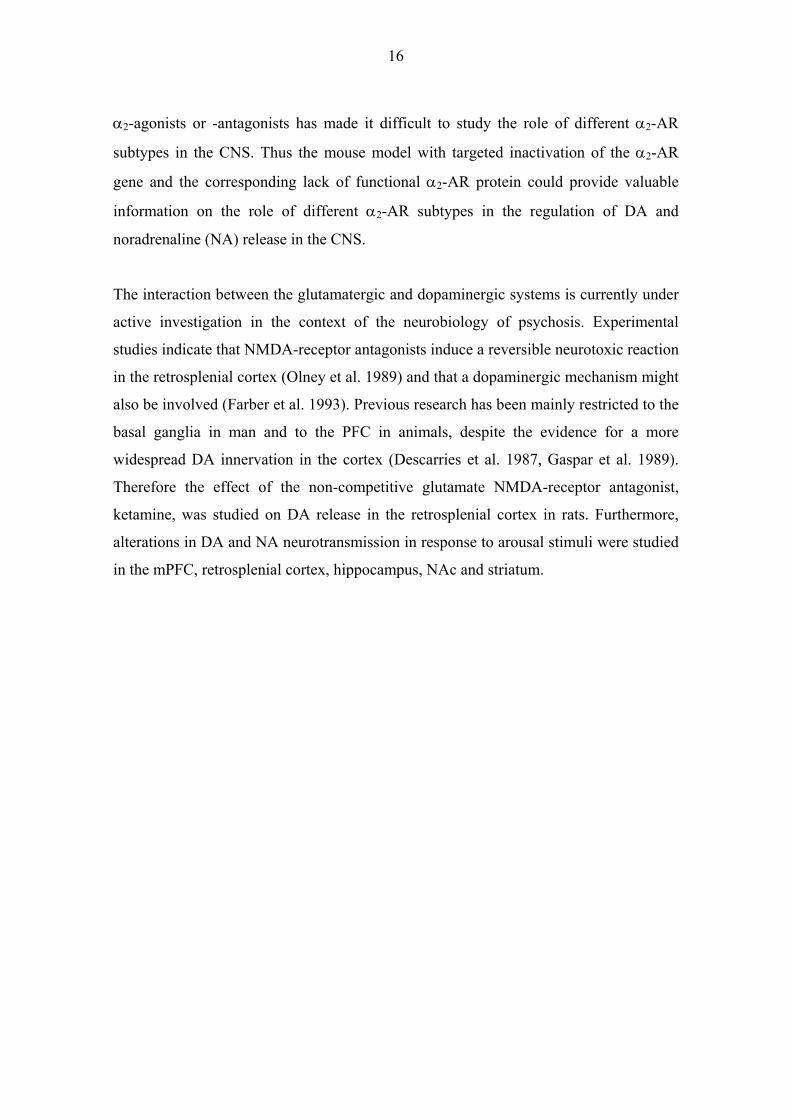
The interaction between the glutamatergic and dopaminergic systems is currently under
active investigation in the context of the neurobiology of psychosis. Experimental
studies indicate that NMDA-receptor antagonists induce a reversible neurotoxic reaction
in the retrosplenial cortex (Olney et al. 1989) and that a dopaminergic mechanism might
also be involved (Farber et al. 1993). Previous research has been mainly restricted to the
basal ganglia in man and to the PFC in animals, despite the evidence for a more
widespread DA innervation in the cortex (Descarries et al. 1987, Gaspar et al. 1989).
Therefore the effect of the non-competitive glutamate NMDA-receptor antagonist,
ketamine, was studied on DA release in the retrosplenial cortex in rats. Furthermore,
alterations in DA and NA neurotransmission in response to arousal stimuli were studied
in the mPFC, retrosplenial cortex, hippocampus, NAc and striatum.

17
2. REVIEW OF THE LITERATURE
2.1. THE BRAIN DOPAMINERGIC SYSTEM
2.1.1. Dopamine as a neurotransmitter
Dopamine (3,4-dihydroxyphenylethylamine) was found to be a neurotransmitter in 1958
(Benes 2001, Carlsson and Waldeck 1958, Carlsson 2001). Before this finding, DA was
assumed to be simply a precursor of noradrenaline. During the following decades,
knowledge of the role of DA in neurotransmission increased enormously and it was
linked to many biological functions and neurological disorders in the central nervous
system. DA in the brain has an important role in many behavioural and biological
functions, such as motivation and reward (Bassareo et al. 2002, Berridge and Robinson
1998, Olds and Milner 1954, Robbins and Everitt 1996, Salamone et al. 2005, Wise and
Rompre 1989); learning (Ljungberg et al. 1992, Schultz et al. 1993); memory (Arnsten
1997, Setlow and McGaugh 1998); feeding (Bassareo and Di Chiara 1999a, Hernandez
and Hoebel 1988a, Hernandez and Hoebel 1988b); vision (Djamgoz and Wagner 1992,
Ehinger 1983, Nguyen-Legros 1988); lactation (Ben-Jonathan and Hnasko 2001,
Thorner 1977); nausea and vomiting (Yoshida et al. 1995, Yoshikawa et al. 1996);
stress (Abercrombie et al. 1989, Imperato et al. 1993); sexual behaviour (Giuliano and
Allard 2001, Melis and Argiolas 1995, Pfaus and Phillips 1991); and control of
locomotor activity (Damsma et al. 1992, Fink and Smith 1980). The crucial role of DA
in different biological functions has made it an interesting target for drug development.
Indeed, there are treatments for medical conditions that are, at least, partly caused by a
failure in dopaminergic system such as schizophrenia, Parkinson’s disease, attention
deficit hyperactivity disorder, restless legs syndrome and addiction (Bloom and
Lazerson 1988, Nieoullon 2002, Nutt 1996, Self and Nestler 1995, Trenkwalder et al.
2005).
2.1.2. Dopaminergic innervation of forebrain structures
The cell bodies of the neurons forming the major ascending dopaminergic pathways to
forebrain arise from the ventral tegmental area (VTA, A10 region) and substantia nigra
pars compacta (A9 region) (Albanese and Minciacchi 1983, Björklund and Lindvall

18
1984, Fuxe et al. 1985). The nigrostriatal (or mesostriatal) DA system originates from
substantia nigra pars compacta and innervates mainly the caudate and putamen. A minor
proportion of substantia nigra pars compacta DA neurons innervate the NAc. The
dopaminergic fibers from the VTA to NAc, olfactory tubercle and other limbic regions
such as the amygdala, hippocampus and septum comprise the mesolimbic DA system
and VTA fibers to cortical regions, such as the prefrontal cortex (densest dopaminergic
innervation in infralimbic and prelimbic regions), entorhinal cortex and cingulate cortex
comprise the mesocortical DA system (Thierry et al 1973; Berger et al 1974). The third
major dopaminergic pathway is the tuberoinfundibular pathway that projects from the
median eminence (A12 region) to the pituitary gland and is involved in the control of
the secretion of prolactin levels. The dopaminergic cell bodies are also found in the
retina and olfactory bulb.
In primates, motor, premotor and supplementary motor areas are densely innervated
with DA fibers, whereas parietal, temporal and posterior cingulate cortices have a less
extensive dopaminergic input. The prefrontal, anterior cingulate, insular, piriform,
perirhinal and entorhinal cortices are densely innervated, while visual areas are only
sparsely innervated in both rodents and primates (Berger et al. 1985a, Berger et al.
1988, Lewis et al. 1987, Parnavelas and Papadopoulos 1989). In rodents, dopaminergic
neurons projecting to the cerebral cortex are differentiated into two main classes. The
first group of dopaminergic fibers originates from medial VTA and is distributed mainly
to the deep cortical layers, V-VI or VI (Berger et al. 1991, Emson and Koob 1978,
Lindvall et al. 1984). The second class of dopaminergic neurons originates from lateral
VTA and medial substantia nigra and distributes to the superficial cortical layers, I-III,
especially to the cingulate cortex (Berger et al. 1985a, Berger et al. 1985b, Descarries et
al. 1987). Both classes of dopaminergic neurons exhibit a clear rostro-caudal gradient
with a higher density in the PFC and a lower density in the posterior cortex. This
contrasts with cortical noradrenergic innervation, which is evenly distributed throughout
the cortex (fig. 1) (Lindvall et al. 1978, Seguela et al. 1990). On the other hand, the
laminar distribution of cortical dopaminergic projections is more evenly distributed in
primates compared to rodents, with the densest distribution being in layers I, III and V
(Goldman-Rakic et al. 1990, Goldman-Rakic et al. 1992).
19
A.
B.
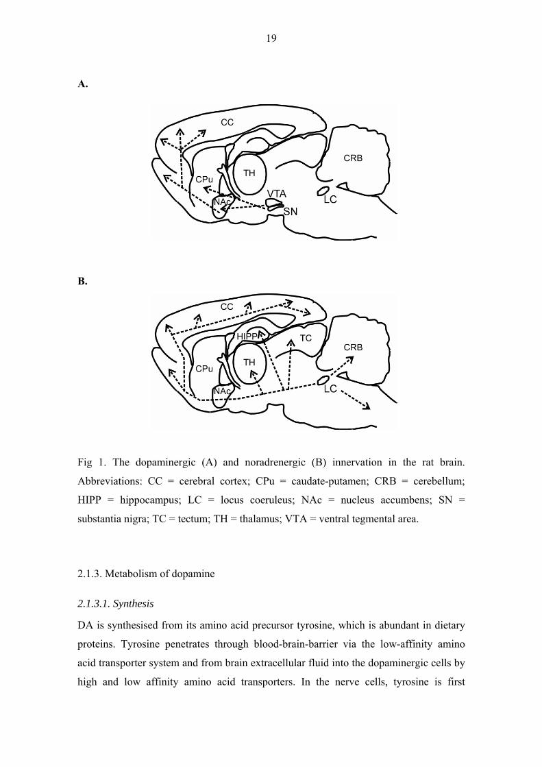
Fig 1. The dopaminergic (A) and noradrenergic (B) innervation in the rat brain.
Abbreviations: CC = cerebral cortex; CPu = caudate-putamen; CRB = cerebellum;
HIPP = hippocampus; LC = locus coeruleus; NAc = nucleus accumbens; SN =
substantia nigra; TC = tectum; TH = thalamus; VTA = ventral tegmental area.
2.1.3. Metabolism of dopamine
2.1.3.1. Synthesis
DA is synthesised from its amino acid precursor tyrosine, which is abundant in dietary
proteins. Tyrosine penetrates through blood-brain-barrier via the low-affinity amino
acid transporter system and from brain extracellular fluid into the dopaminergic cells by
high and low affinity amino acid transporters. In the nerve cells, tyrosine is first
20
hydroxylated by tyrosine hydroxylase to dihydroxyphenylalanine (L-DOPA). This
enzymatic reaction is normally the rate-limiting step in DA synthesis (Feldman et al.
1997). Tyrosine hydroxylase requires tetrahydrobiopterin as a cofactor for the
biosynthesis of L-DOPA. Aromatic amino acid decarboxylase (AADC or DOPA
decarboxylase) is the enzyme that converts L-DOPA to DA in the cytosol. In the
noradrenergic cells, DA is further converted by dopamine β-hydroxylase to NA inside
the synaptic vesicles.
2.1.3.2. Storage and release
e end product, DA, is transported into the storage vesicles In the dopaminergic cells, th
and is concentrated approximately 10-1000 times compared to the DA levels in the
cytosol (Johnson 1988, Kanner and Schuldiner 1987, Njus et al. 1986). Accumulation of
DA in the storage vesicles depends on the proton electrochemical gradient generated by
the vesicular hydrogen-ATPase and involves the vesicular monoamine transporter
mediated exchange of two luminal protons with one cytoplasmic amine. In addition to
storage in axon terminals, DA can be released also from dendrites (Björklund and
Lindvall 1975, Kalivas et al. 1989, Nieoullon et al. 1977b, Santiago and Westerink
1991). There DA is stored both in vesicles but also in the smooth endoplasmic reticulum
(Hattori et al. 1979, Mercer et al. 1979). The arrival of the axon potential to the nerve
terminal evokes the passage of calcium ions into the cell and this is the key element for
the fusion of storage vesicles with the cell membrane. The synaptic vesicles release their
soluble content into the synaptic cleft by exocytosis (Hanson et al. 1997, Matsuda et al.
1994). DA release is dependent on the nerve stimulus rate and pattern. Indeed, an
increase in DA cell activity is typically accompanied by a shift from an irregular single-
spiking pattern to one of burst firing (Tong et al. 1996; Carr et al. 1999). Stimulation
studies have shown that activation of the DA neuron axon in patterns resembling burst
discharge will release two to three times more DA than is released by an equivalent
number of evenly spaced stimuli (Bonci et al. 1997).
21
2.1.3.3. Uptake
The presynaptic dopaminergic terminals contain a transporter (DAT) that is responsible
for the homeostasis and which terminates the action of the neurotransmitter. These high-
affinity membrane carriers work in both directions depending on the concentration
gradient. Under normal conditions, the concentration of DA is lower in the cytosol than
in the synaptic cleft, and DA is recycled back to the storage vesicles. Some drugs such
as tricyclic antidepressants and cocaine can inhibit the action of DAT and in that way
increase the extracellular levels of DA (Giros and Caron 1993, Kuhar et al. 1991,
Randrup and Braestrup 1977, Ritz et al. 1987). On the other hand, amphetamine
reverses the function of DAT, transporting DA to the synaptic cleft from the cytosol
(Fischer and Cho 1979, Heikkilä et al. 1975, Jones et al. 1998, Raiteri et al. 1979,
Schmitz et al. 2001, Sulzer et al. 1995). It has also been assumed that other neurons and
glial cells can participate in the removal of DA from the extracellular fluid. Indeed, in
the PFC NA transporter (NET) has a prominent role in the uptake of DA from the
extracellular space (Gresch et al. 1995, Mazei et al. 2002, Valentini et al. 2004,
Yamamoto and Novotney 1998), whereas in the striatum DAT is mainly responsible for
the clearance of DA from the extracellular space (Gresch et al. 1995, Mazei et al. 2002).
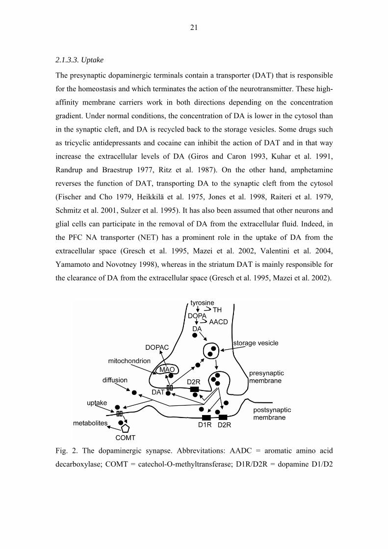
Fig. 2. The dopaminergic synapse. Abbrevitations: AADC = aromatic amino acid
decarboxylase; COMT = catechol-O-methyltransferase; D1R/D2R = dopamine D1/D2

22
receptor; DAT = dopamine transporter; DOPAC = dihydroxyphenylacetic acid; MAO =
monoamine oxidase; DA = dopamine; TH = tyrosine hydroxylase.
2.1.3.4. Degradation
The two main enzymes that take care of DA clearance are monoamine oxidase (MAO)
and catechol-O-methyltransferase (COMT). MAO is located in the nerve cells and also
in the glial cells, whereas COMT is found mainly extraneuronally (Kopin 1994). MAO
is located on the outer membrane of the mitochondrion. It metabolises DA by oxidative
deamination to aldehyde 3,4-dihydroxyphenylacetic acid that is further metabolised to
alcohols and acids. There are two isoenzymes of MAO: MAO A and MAO B. MAO A
preferentially metabolizes serotonin and NA while MAO B has a higher affinity for
phenylethylamine (Fowler and Tipton 1982, Fowler and Benedetti 1983). Both isoforms
can metabolize DA. In the mouse, DA is largely metabolized by MAO A under normal
physiological conditions, though at higher concentrations the contribution of MAO B
also becomes significant (Fornai et al. 1999). In contrast, in humans, DA is mainly
oxidized by MAO B (Glover et al. 1977). Both lesion (Kaakkola et al. 1987, Rivett et al.
1983) and immunohistochemical studies (Karhunen et al. 1995, Lundstrom et al. 1995)
have demonstrated that there is no significant COMT activity in presynaptic
dopaminergic neurons, but some activity is present in postsynaptic neurons and
substantial activity is located in glial cells. COMT inactivates DA by methylation of
hydroxyls on the catechol ring. COMT is able to methylate DA itself or metabolites that
have been first produced by MAO. The two main metabolites of DA are homovanillic
acid (HVA) and 3,4-dihydroxyphenylacetic acid (DOPAC). HVA is a methylated
compound that is produced by both MAO and COMT. DOPAC is an un-methylated
metabolite and it is produced only by MAO (fig. 3).

23
Fig. 3. Metabolism of dopamine. Abbreviations: DOPA = 3,4-dihydroxyphenylalanine;
DA = dopamine; DOPAC = 3,4-dihydroxyphenylacetic acid; HVA = homovanillic acid;
3-MT = 3-methoxytyramine; MAO = monoamine oxidase; COMT = catechol-O-
methyltransferase; AD = aldehydedehydrogenase.
2.1.4. Dopamine receptors
All DA receptors belong to the G-protein coupled receptor (GPCR) superfamily. The
dopaminergic receptors can be divided into D1- and D2-like receptors. D1-like
receptors consist of D1 and D5 receptors, and D2-like receptors of D2, D3 and D4
receptors (Jackson and Westlind-Danielsson 1994). This classification is based on the
mechanisms that link these GPCRs to the second messenger system. Thus D1-like
receptors stimulate the adenylate cyclase activity via Gs subunit leading to an increased
cAMP (cyclic adenosine monophosphate) concentration (Kebabian and Calne 1979,
Missale et al. 1998). On the other hand, D2-like receptors are negatively coupled via the
Gi subunit to the adenylate cyclase, which leads to a decline in the cAMP concentration
(Vallar et al. 1988). Structurally all GPCRs consist of seven transmembrane domains
that are connected with three extra- and three intracellular loops (Bockaert et al. 2002).
The D1 receptor is the most widespread DA receptor and is expressed at a higher level
than the other DA receptor subtypes (Dearry et al. 1990, Fremeau et al. 1991, Lidow et

24
al. 1990, Weiner et al. 1991). The D5 receptor is expressed at a much lower level than
the D1 receptor, with a distribution restricted mainly to the hippocampus and the
parafascicular nucleus of the thalamus (Meador-Woodruff et al. 1992, Tiberi et al.
1991). The D2 receptor is also widely distributed in the brain, with the highest densities
in the striatum, NAc and olfactory tubercle. The D2 receptor gene encodes two
molecularly distinct isoforms, named long (D2L) and short (D2S) (Picetti et al. 1997).
The D2L mainly acts at postsynaptic sites and the D2S serves presynaptic autoreceptor
functions (Usiello et al. 2000). The D3 receptor is specifically distributed in limbic
areas such as the shell of the NAc, olfactory tubercle and islands of Calleja, but has a
low expression in the striatum (Bouthenet et al. 1991, Sokoloff et al. 1990). The D4
receptor is highly expressed in the frontal cortex, amygdala, hippocampus,
hypothalamus and mesencephalon (O'Malley et al. 1992, Van Tol et al. 1991). The
relative abundance of the DA receptors in the rat central nervous system has been
claimed to be D1>D2>D3>D5>D4 (Jaber et al. 1996).
Table 1. Distribution of DA receptors in the brain (Jaber et al. 1996, Missale et al.
1998). Abbreviations: OT = olfactory tubercle; MN = mamillary nucleus; CC = cerebral
cortex; SR = septal region; SN = substantia nigra; VTA = ventral tegmental area; NAc =
nucleus accumbens; FC = frontal cortex.
DA receptor subtype
Function Brain region (high expression)
D1 stimulate adenylate cyclase via Gs
striatum, NAc, OT, hypothalamus, thalamus, limbic system
D5 stimulate adenylate cyclase via Gs
hippocampus, thalamus, MN, CC, striatum, hippocampus, SN
D2 negative coupling via Gi to adenylyl cyclase
striatum, NAc, OT, CC, SR, amygdala, hippocampus hypothalamus, SN, VTA
D3 negative coupling via Gi to adenylyl cyclase
limbic region (NAc, OT), SN, VTA, hippocampus
D4 negative coupling via Gi to adenylyl cyclase
FC, amygdala, hippocampus hypothalamus, mesencephalon, SN, thalamus

25
The unavailability of subtype selective DA receptor ligands has hampered the study of
DA receptor functions in the CNS. There are both agonists and antagonists that can
discriminate between D1-like and D2-like receptors but relatively few agents are
selective for DA receptor subtypes within the subfamilies. The D1 ligands have no more
than 10-fold greater selectivity for D1- vs. D5 receptors (Bourne 2001, Neumeyer et al.
2003, Shiosaki et al. 1996, Tice et al. 1994). However, there are D2 ligands that can
differentiate more selectively between D2-like receptor subtypes. Indeed, there exist
antagonists for the D4 receptor subtype having up to 1000-fold greater sensitivity for
D4 vs. D2 or D3 receptors (Kula et al. 1997, Kulagowski et al. 1996, Patel et al. 1996).
Moreover, there are still no D2 receptor selective agonists and antagonists available. In
recent years, mouse lines have been generated that have a targeted inactivation of the
receptor protein for all five DA receptor subtypes. These animals have helped to unravel
the function of the individual DA receptors in the CNS.
Studies in D1 receptor KO (knockout) mice have shown that the D1 receptor is essential
for locomotor activating effects of psychostimulants (Drago et al. 1998, Xu et al. 1994).
However, the D1 receptors do not affect the rewarding and reinforcing effects of
cocaine (Miner et al. 1995). Also, the D1 receptor was found to play a role in the
motivation to work for food reward but not in reward perception. (El-Ghundi et al.
2003). Cortical D1 receptors have also been implicated in the control of working
memory (Sawaguchi and Goldman-Rakic 1991) and have an important role in
modulating the extinction of fear memory (El-Ghundi et al. 2001).
Rouge-Pont et al. (2002) found that the lack of the D2 receptor produced higher
extracellular DA levels in response to morphine and cocaine administration, indicating a
key role of D2 receptor in the modulation of DA release in drug abuse. In the same
study, the neurochemical effects of morphine and cocaine were unchanged in mice with
selective deletion of the long isoform of D2L receptor, in support of role of the D2S
isoform in mediating the presynaptic autoreceptor function. Interestingly, the D2L
receptor seems to mediate parkinsonian-like syndromes induced by typical
antipsychotic drugs in D2L KO mice (Xu et al. 2002). On the other hand, the
amphetamine-induced disruption of sensorimotor gating phenomenon, known as

26
prepulse inhibition of startle reflex induced by loud sound, is mediated via the D2
receptors (Ralph et al. 1999). In addition, Xu et al. (2002) reported that amphetamine
and the antipsychotic drug clozapine mediate their effect on prepulse inhibition via the
D2S isoform. The D3 receptor has been found to play an inhibitory role in the control of
locomotor activity and rearing behaviour (Accili et al. 1996). Like the D1 receptor, the
D4 receptor has been implicated in drug abuse, since in mice lacking the D4 receptor
exhibited supersensitivity to the locomotor stimulating effects of methamphetamine and
cocaine (Rubinstein et al. 1997). Studies in D5 receptor KO mice have revealed a
modulatory role for the D5 receptor in acetylcholine release in the hippocampus
(Laplante et al. 2004) and the regulation of sexual behaviour both in males and females
(Kudwa et al. 2005).
2.1.5. Presynaptic regulation of dopaminergic neurotransmission
There are several mechanisms by which DA transmission is regulated presynaptically in
the CNS. These include DA reuptake by DAT, inhibition of DA synthesis and release
by presynaptic D2-like autoreceptors, degradation of released DA by metabolizing
enzymes and heterosynaptic regulation of DA release by other neurotransmitter
systems.
2.1.5.1. Dopamine transporter
Dopaminergic neurons exhibit both single spike and burst firing modes of activity, the
latter yielding much higher extracellular DA levels. The elevated DA levels in the
extracellular space after burst stimulation are more likely to be a result of saturated DA
reuptake sites rather than facilitated DA release from the presynaptic terminal (Chergui
et al. 1994). Namely, during tonic activity, DA released by single spikes is cleared from
the synaptic cleft before the next pulse, but during burst stimulation DA accumulates in
the synaptic cleft. This accumulation is more pronounced in brain areas other than
dorsal striatum, which is dense with DAT (Chergui et al. 1994, Suaud-Chagny et al.
1995). Extracellular accumulation of DA by burst activity is also needed to activate D2
autoreceptors (Benoit-Marand et al. 2001) and postsynaptic D1 receptors (Chergui et al.
1996, Chergui et al. 1997, Gonon 1997). Thus, tonic and bursting activities result in

27
different extracellular DA levels due to DA reuptake mechanism. On the other hand,
studies in DAT KO mice point to a key role of DAT in the refilling of intracellular DA
stores after prolonged DA release (Gainetdinov et al. 1998).
2.1.5.2. D2-autoreceptors
Several studies in D2 KO mice have indicated that the D2 receptor is the only functional
autoreceptor (Benoit-Marand et al. 2001, Mercuri et al. 1997, Schmitz et al. 2002).
However, some studies suggest that also D3 receptors might have an autoreceptor
function (Kuzhikandathil and Oxford 1999, Kuzhikandathil and Oxford 2000, O'Hara et
al. 1996, Tang et al. 1994). The D2 autoreceptor activation inhibits axon terminal
(Cragg and Greenfield 1997, Dwoskin and Zahniser 1986, Mayer et al. 1988, Palij et al.
1990, Starke et al. 1978) and somatodendritic (Cragg and Greenfield 1997) DA release.
There is also evidence that D2 receptors modulate DA synthesis by decreasing tyrosine
hydroxylase activity (Kehr et al. 1972, Roth et al. 1975, Strait and Kuczenski 1986,
Wolf et al. 1986). This action is probably mediated via inhibition of adenylyl cyclase
and a cAMP-dependent change in phosphorylation of tyrosine hydroxylase (Lindgren et
al. 2001, Onali and Olianas 1989). Studies in pheochromocytoma 12 cell cultures
indicate that D2 autoreceptors regulate DA release on two different time scales (Pothos
et al. 1998), through a fast mechanism that lasts for a few seconds and modulates ion
channels and through a slow one that lasts for minutes to hours and involves regulation
of DA synthesis. On the other hand, the D2 receptors on the soma and dendrites of the
neuron inhibit impulse flow by activating G protein coupled inwardly rectifying
potassium channels. This effect hyperpolarises the cell membrane (Lacey 1993, White
1996). D2 autoreceptors may also participate in the regulation of intracellular vesicular
transporter and modulate DA reuptake from the extracellular space (Meiergerd et al.
1993, Parsons et al. 1993, Schmitz et al. 2002, Wu et al. 2002). Indeed, D2 receptor
agonists can increase vesicular DA uptake and D2 receptor antagonists block increase of
vesicular DA uptake induced by cocaine (Brown et al. 2001). These studies indicate that
DAT activity is increased by the D2 receptor agonist quinpirole and decreased by the
D2 receptor antagonists pimozide, sulpiride and raclopride. Interestingly, the lack of the
D2 receptor in the mutant mouse line altered striatal DAT activity but did not affect DA
release (Brown et al. 2001). On the other hand, chronic treatment with D2 receptor

28
agonists is reported to increase DAT expression in the NAc and decrease DAT
expression in the striatum (Kimmel et al. 2001). Also, altered DAT levels have been
observed in schizophrenic patients and this may be attributable to the antipsychotic drug
treatment (Laakso et al. 2001). However, the exact mechanisms and conditions how D2
autoreceptors regulate the functions of DAT are still unclear.
2.1.5.3. Degradation
In the striatum, DA clearance from the synaptic cleft is thought to be largely dependent
on the function of DAT. However, the DAT expression is much lower in the cortex
compared to the striatum and the rate of DA uptake by DAT is slow (Garris et al. 1993,
Lewis et al. 2001, Sesack et al. 1998, Wayment et al. 2001). Thus, some other
mechanism, such as enzymatic degradation by COMT and MAO, and DA uptake by
NET, may participate in the DA clearance from the synaptic cleft. Indeed, studies in
COMT KO mice have revealed that DA levels are increased in the PFC but not in the
striatum, pointing to a role of COMT in DA clearance in the cortical areas but not in the
striatum (Gogos et al. 1998). Also, Matsumato et al. (2003) have noted that the COMT
mRNA expression is higher in the PFC than in the striatum in both humans and rats. On
the other hand, it has been assumed that MAO inhibitors attenuate the velocity of DA
clearance by 30-50 % in the mPFC (Wayment et al. 2001). Taken together, these results
indicate that enzymatic degradation of DA might regulate synaptic DA concentration in
the brain areas where DAT activity is low. The heterosynaptic regulation of DA release
in the CNS is discussed more detailed in the next chapters.
2.1.6. Dopamine and other neurotransmitter systems
Several neurotransmitter systems modulate the release of DA in the CNS. The most
well-studied neurotransmitter systems mediate their effect via NA, 5-hydroxytryptamine
(5-HT), glutamate and GABA. The receptors of these neurotransmitter systems and
their effect on DA cell firing in the VTA are listed in table 2. In this study, the main
interest was the α2-AR- and to a lesser extent - NMDA receptor mediated modulation of
DA release in the CNS. Therefore, the next chapters will focus on these two
neurotransmitter systems.

29
Table 2. Neurotransmitter systems modulating DA cell firing in the VTA.
* Only after blockade of somatodendritic D2 autoreceptors ** NC = no change
Effect on DA cell Transmitter Receptor firing in VTA References Noradrenaline: α1 DA cell firing ↑ * (Grenhoff et al. 1995) α2 DA cell firing ↓ (Gobbi et al. 2001, Millan et al. 2000b) β not studied Glutamate: Ionotropic: - NMDA DA cell firing↑ (Suaud-Chagny et al. 1992, Wang et al. 1993) - AMPA/Kainate DA cell firing ↑ (Suaud-Chagny et al. 1992, Wang et al. 1993) Metabotropic: - Group 1 DA cell firing ↑ (Zheng et al. 2002) - Group 2 not studied - Group 3 not studied 5-HT: 5HT1A DA cell firing NC**/↑ (Arborelius et al. 1993, Prisco et al. 1994) 5HT2A DA cell firing ↑ (Pessia et al. 1994) 5HT2C DA cell firing ↓ (Di Giovanni et al. 2000, Prisco et al. 1994) 5HT3 DA cell firing ↑ (Campbell et al. 1996) 5HT5 not studied 5HT6 not studied 5HT7 not studied GABA: GABAA DA cell firing ↓ (Lacey 1993, White 1996) GABAB DA cell firing ↓ (Lacey 1993)

30
2.1.7. Adrenergic regulation of the dopaminergic system
The main source of NA in the CNS is the LC, a bilateral adrenergic nucleus in the
dorsolateral tegmentum near the fourth ventricle. The axons of LC branch extensively
throughout the neuraxis innervating most regions in the CNS (Foote et al. 1983). The
ascending projections of the LC comprise the dorsal noradrenergic bundle and the
dorsal periventricular pathways. Other sources of NA innervation consist of the lateral
tegmental (A1, A5, A7) and dorsal medullary (A2) noradrenergic cell groups that give
rise to the ventral noradrenergic bundle, which innervates mainly the thalamus,
hypothalamus, preoptic area, propriobulbar networks in the brainstem and send
descending fibers to the spinal cord (Björklund and Lindvall 1986) (Fig. 1).
2.1.7.1. Interaction sites and mechanisms
Dopaminergic and noradrenergic systems interact at many levels in the CNS. First,
adrenergic neurons from the LC project into the VTA (Jones et al. 1977, Phillipson
1979, Simon et al. 1979) and VTA efferent axons descend into the LC (Beckstead et al.
1979, Ornstein et al. 1987), which enables direct crosstalk between these systems.
Several studies have indicated that the adrenergic regulation of dopaminergic VTA
neurons is mediated by α1-ARs postsynaptic to fibres originating from LC (Grenhoff et
al. 1993, Grenhoff et al. 1995). On the other hand, α2-ARs can also participate in the
regulation of DA neurons by acting as autoreceptors on noradrenergic afferent terminals
and thus indirectly inhibit DA release in the VTA (Grenhoff et al. 1993). In addition,
cells expressing D1 and D2 receptor mRNA are present in the LC (Meador-Woodruff et
al. 1991).
Second, dopaminergic and noradrenergic projections overlap in their terminal regions
such as mPFC, NAc and striatum, and there is a body of growing evidence for an
interaction between these two neurotransmitter systems at the terminal level (Carboni et
al. 1990, Devoto et al. 2001, Devoto et al. 2002, Di Chiara et al. 1992, Feenstra 2000,
Feenstra et al. 2000, Gresch et al. 1995, Kawahara et al. 2001, Moron et al. 2002, Tassin
1992, Tassin et al. 1992). Interestingly, DA may have even higher affinity for NET than
NA (Horn 1973, Raiteri et al. 1977), and NET mainly contributes to the removal of DA

31
from the extracellular space in the mPFC, and to a lesser extent in the NAc (Carboni et
al. 1990, Cass and Gerhardt 1995, Pozzi et al. 1994, Tanda et al. 1997, Yamamoto and
Novotney 1998). On the other hand, a recent study by Valentini et al. (2004) suggests
that in the parietal and occipital cortex, NET is not involved in the clearance of DA
from the extracellular fluid in rat. In contrast, in the DA rich striatum DAT is believed
to be solely responsible for the DA removal from the extracellular fluid (Carboni et al.
1990, Di Chiara et al. 1992, Gresch et al. 1995, Moron et al. 2002, Pozzi et al. 1994).
Third, the adrenergic system can indirectly regulate DA release in the VTA via other
neurotransmitter systems, such as glutamate- and GABAergic systems. The VTA
receives an intense glutamatergic projection from the frontal cortex (Rossetti et al.
1998) that enhances DA release via NMDA-receptor mediated mechanism (Kretschmer
1999). These glutamatergic neurons are under adrenergic regulation, so that α1-AR
stimulation enhances their firing (Marek and Aghajanian 1999) whereas α2-AR
stimulation mainly inhibits their firing (Kovacs and Hernadi 2003). In addition,
pyramidal neurons in the VTA can be indirectly inhibited via α-ARs on GABAergic
interneurons (Kawaguchi and Shindou 1998).
Fourth, Devoto et al. have proposed a co-release theory for NA and DA, which claims
that DA is released from NA terminals in posterior cortical areas (Devoto et al. 2001,
Devoto et al. 2002, Devoto et al. 2003, Devoto et al. 2004). This hypothesis is based on
findings that in some studies the extracellular DA levels in the parietal and occipital
cortices are only modestly lower than in the mPFC where DA innervation is known to
be much denser and that drug treatments that modify mainly noradrenergic activity
modulate also extracellular DA levels in these cortical areas. However, several other
mechanisms, such as adrenergic heteroceptors and competition of DA and NA for the
same transporter, might underlie these findings, leaving the origin of released DA in
posterior cortical regions an open question.

32
2.1.7.2. Alpha2-adrenoceptors
Adrenoceptors are divided into three main families, α1-, α2- and β-ARs, which all are
further divided into three subfamilies: α1A-, α1B- and α1C-ARs; α2A-, α2B- and α2C-ARs;
and β1-, β2- and β3-ARs, respectively (Bylund 1988). All adrenoceptors belong to the
GPCR family. The α2-ARs are negatively coupled to GPCR via the Gi/o signaling
system, which inhibits the adenylyl cyclase activity and the opening of the voltage-
gated calcium and potassium channels (Limbird 1988, Surprenant et al. 1992). α2A-ARs
are located mainly in the cortex, LC, hippocampus and brainstem; α2C-ARs are found in
the cortex, hippocampus, LC and striatum; in contrast α2B-ARs in the brain are located
almost exclusively in the thalamic nuclei (Aoki et al. 1994, Holmberg et al. 2003, Lee et
al. 1998, MacDonald and Scheinin 1995, Nicholas et al. 1993, Scheinin et al. 1994). α2-
ARs are present in the CNS both as prejunctional autoreceptors, inhibiting further NA
release (Docherty 1998, Starke 1977, Starke 1987), and as postjunctional receptors
either on the bodies and dendrites of target cells (Docherty 1998, MacMillan et al.
1996) or as heteroceptors, inhibiting the release of other modulatory neurotransmitters,
such as DA (Gobert et al. 1998, Trendelenburg et al. 1994).
The α2A-AR has been considered the predominant α2-AR subtype in the brain and the
main regulator of presynaptic autoinhibition of NA release in the CNS (Altman et al.
1999, Hein et al. 1999, Trendelenburg et al. 1999, Trendelenburg et al. 2001a,
Trendelenburg et al. 2001b). In vitro superfusion studies in α2A-AR wild type (WT) and
KO mice have revealed that the α2-AR agonist UK 14304 inhibited NA release
maximally by 96 % in the occipito-parietal cortex in α2A-AR WT and also in a similar
manner in α2B- and α2C-AR KO mouse preparations but UK 14304 only evoked a 24 %
reduction in NA release in α2A-AR KO mouse preparations (Bucheler et al. 2002). I.e.
reduced by nonetheless, some inhibition by UK 14304 remained in α2A-AR KO mouse
brain tissue. Studies in the double mutant α2AC-AR KO mouse line indicated that the
remaining autoinhibtion was mediated by α2C-ARs (Bucheler et al. 2002, Hein et al.
1999, Trendelenburg et al. 2001a). Taken together, these results suggest that α2A-
autoreceptors predominate in the CNS while the role of α2C-autoreceptors becomes
more pronounced when the α2A-AR is absent. A recent report by Trendelenburg et al.
33
(2003) has proposed that also the third α2-AR subtype, α2B-AR, may serve as an
autoreceptor in the postganglionic sympathetic neurons.
α2A-ARs are likely to mediate most of the heteroceptor function in the CNS (Gobert et
al. 1998, Scheibner et al. 2001, Trendelenburg et al. 1994). Scheibner et al. (2001),
using in vitro superfusion technique for 5-HT analysis, found that α2-heteroreceptors in
the hippocampus were a mixture of predominantly α2A-ARs and to a lesser extent α2C-
ARs, based on the finding that 5-HT release-inhibiting effect of the α2-agonist
medetomidine was reduced in α2A-AR KO and α2C-AR KO mice in hippocampal tissue
and disappeared completely in α2AC-AR KO mice. On the other hand, an in vivo
microdialysis study in the rat frontal cortex found markedly decreased extracellular DA
levels after a local infusion of α2-agonists, DMT and guanabenz, and increased levels
after local infusion of α2-antagonists, RX 821002 and BRL 44408 (Gobert et al. 1998).
In the same study, the α1/α2-antagonist prazosin, which is preferentially an antagonist
for α2C- and α2B-ARs but not for α2A-AR, did not affect DA release, indicating a
predominant role of α2A-AR subtype also in the regulation of DA release in the CNS.
However, so far the lack of subtype-specific α2-AR drugs has precluded a direct
comparison between the α2-AR subtypes on the regulation of neurotransmitter release.
Therefore, mutant mouse lines that either lack or overexpress a particular α2-AR
subtype offer the best tool to investigate the role of α2-AR subtypes in the regulation of
transmitter release. However, most of the in vivo studies that have investigated the
modulatory role of different α2-AR subtypes in the regulation DA metabolism have
been done only in post mortem brain material (Lähdesmäki et al. 2003, Sallinen et al.
1997).
Sallinen et al. (1997) using mice that either overexpress or lack α2C-ARs, found that the
α2-agonist, DMT, inhibited NA and DA turnover in whole brain homogenates in a
similar manner in OE (overexpressing) and KO mice and their wild-type controls.
Interestingly, drug-naive KO mice had lower HVA concentrations in the striatum and
OE mice higher HVA concentrations in the frontal cortex indicating the involvement of
α2C-ARs in the dopaminergic regulation, not only in the striatum, but also in the cortex.

34
On the other hand, Lähdesmäki et al. (2003) found that DMT inhibited DA turnover
(HVA/DA ratio) in the striatum and thalamus-hypothalamus of α2A-AR WT mice,
whereas in α2A-AR KO mice DMT was without any significant effect. However, DMT
and α2-antagonist, ATZ, failed to induce any major changes in DA turnover in mice
lacking the α2A-AR subtype (Lähdesmäki et al. 2003).
2.1.8 NMDA-receptor regulation of dopaminergic system
Glutamate is the major excitatory neurotransmitter in the mammalian CNS. It acts
through ligand- gated ion channels (ionotropic receptors) and G-protein coupled
(metabotropic) receptors. The ionotropic glutamate receptors have four to five subunits,
and are further subdivided into three groups, AMPA, NMDA and kainate receptors.
This classification is based on both receptor pharmacology and structural similarities.
Activation of glutamate receptors is responsible for basal excitatory synaptic
transmission and many forms of synaptic plasticity such as long-term potentiation and
depression, which is thought to underlie learning and memory (Abbott and Nelson
2000, Sourdet and Debanne 1999).
The glutamatergic innervation from the mPFC is the major excitatory input to VTA
(Hurley et al. 1991, Sesack et al. 1989). The mPFC glutamatergic neurons innervate
dopaminergic and also non-dopaminergic cells in the VTA (Sesack and Pickel 1992).
The mPFC glutamatergic input has been shown to synapse on dopaminergic cells that
project back to the mPFC and onto the GABAergic cells that project to the NAc (Carr
and Sesack 2000). Thus glutamate can control dopaminergic activity in the VTA
through glutamate receptors located in the VTA and via other neurotransmitter receptors
located in the pyramidal neurons of the mPFC, which determine the output activity of
glutamatergic projections, thereby modulating the release of glutamate in the VTA.
Systemic administration of non-competitive NMDA receptor antagonists appears to
facilitate burst firing of mesolimbic VTA neurons (Freeman and Bunney 1984, French
and Ceci 1990, French et al. 1993, Murase et al. 1993b) and thereby increase DA
release in the NAc (Gonon and Buda 1985). In contrast, competitive NMDA receptor

35
antagonists have no effect on these parameters when given systemically (French and
Ceci 1990, French et al. 1993). Biochemical studies have also demonstrated increased
DA turnover or release in ventral striatum after intra-VTA application of glutamate or
the NMDA receptor agonist (Kalivas et al. 1989, Suaud-Chagny et al. 1992, Wang et al.
1994). On the other hand, stimulation of the PFC increases levels of extracellular DA
within the NAc (Karreman and Moghaddam 1996, Murase et al. 1993a, Taber et al.
1995a, Taber and Fibiger 1995b), an effect that is blocked by infusion of glutamate
antagonists into the VTA but not into the NAc (Karreman and Moghaddam 1996, Taber
et al. 1995a, Taber and Fibiger 1995b). Inactivation of the PFC produces the opposite
response (Murase et al. 1993a), pointing to a role of the PFC in the regulation of tonic
levels of DA in the NAc.
2.1.9. Dopamine and schizophrenia
Schizophrenia is a complex psychiatric disorder with heterogenous symptoms. It is
characterized by the presence of positive and negative symptoms. Positive symptoms
include behaviour such as delusions, hallucinations, extreme emotions, excited motor
activity and incoherent thoughts and speech. In contrast, negative symptoms are
described as behavioural deficits such as blunting of emotions, language deficits, and
lack of energy. Even though no single organic cause for schizophrenia has been found,
there is evidence for anatomical changes in brain of schizophrenic patients, such as an
increase in brain ventricular volume and decreased volume of temporal lobe (Johnstone
et al. 1976, Weinberger et al. 1979a, Weinberger et al. 1979b); the presence of a genetic
component (Kety 1975, Kety 1987, Kety et al. 1994); imbalance in DA receptor density
in the striatum and PFC (Hess et al. 1987, Seeman 1985) and involvement of several
neurotransmitter systems, such as DA, glutamate, serotonin and NA in this disease.
In the 1950s, the first antipsychotic drug, chlorpromazine, was discovered accidentally
as the original idea was to develop an effective antihistamine drug. Carlsson and
Lindqvist (1963) found that typical antipsychotic drugs such as haloperidol and
chlorpromazine increased the turnover of monoamines as reflected by increased levels
of their metabolites, leading to the concept that antipsychotic drugs may block
monoamine receptors. This hypothesis received from findings that amphetamine
36
induced stereotypic behaviour in animals that resembles the positive symptoms of
schizophrenia. High doses of amphetamine evokes excessive gnawing, licking,
chewing, sniffing and scanning in rats and chronic amphetamine administration in non-
human primates elicits behaviours such as hypervigilance, abnormal tracking, grasping
and manipulation of thin air (Ellinwood et al. 1973, Ellison et al. 1981, Ellison and
Eison 1983, Ridley et al. 1982). On the other hand, the observation that antipsychotic
drugs potently blocked the psychostimulant actions of amphetamine in animals and
humans (Angrist et al. 1980, Kelly and Miller 1975, Randrup and Munkvad 1965,
Snyder 1973) suggested that dopaminergic signal transduction did play a role in the
development of schizophrenia. These findings were further linked to the DA hypothesis
of schizophrenia (Carlsson 1977, Matthysse 1973) as antipsychotic drugs were found to
block D2-receptors (Creese et al. 1976). However, this D2-receptor blocking is able to
alleviate only positive symptoms of schizophrenia, leaving negative symptoms
unchanged. A more recent modification of the DA hypothesis of schizophrenia is that a
mesolimbic dopaminergic hyperfunction coexists with hypofunction of dopaminergic
terminals in the PFC, and it is the latter that accounts for the negative symptoms (Davis
et al. 1991, Svensson et al. 1995, Svensson 2000, Weinberger 1988).
The non-competitive NMDA-receptor antagonists, such as phencyclidine and ketamine,
induce both negative and positive symptoms of schizophrenia in normal individuals
(Javitt and Zukin 1991, Snyder 1980) and also profoundly exacerbate both negative and
positive symptoms in schizophrenic patients (Itil et al. 1967, Lahti et al. 2001). These
findings provided evidence for the role of glutamate in schizophrenia, suggesting that
the disease is accompanied by a hypoglutamatergic state in the brain (Olney and Farber
1995). Another finding speaking indirectly in favour of the glutamate hypothesis of
schizophrenia was the development of atypical neuroleptics, such as clozapine. These
drugs relieved also the negative symptoms of schizophrenia and caused fewer side
effects compared to typical antipsychotic drugs (Meltzer 1995, Remington et al. 1996,
Stephens 1990). Notably, clozapine has lower affinity for the D2 receptor than the
typical neuroleptics (Farde et al. 1997, Nordström et al. 1995) and binds even more
effectively to D4 than D2 receptors (Tarazi et al. 1997, Van Tol et al. 1991). Clozapine
has also high affinity to many other receptors such as adrenergic α1- (Cohen and
37
Lipinsky 1986, Peroutka and Snyder 1980); serotonergic 5-HT1C- and 5-HT2- (Canton
et al. 1990, Hoenicke et al. 1992, Schmidt et al. 1995); glutamatergic NMDA- (Banerjee
et al. 1995, Lidsky et al. 1997) and GABAA- (Michel and Trudeau 2000, Squires and
Saederup 2000) receptors.
The DA and glutamate hypotheses of schizophrenia are not necessarily mutually
exclusive. Namely, as a general rule, DA receptors inhibit glutamate release and
therefore, mesolimbic DA overactivity can result in the continued and excessive
suppression of glutamate release. This in turn could cause NMDA receptor
hypofunction, which could disrupt DA firing pattern in the VTA. Interestingly, Olney et
al. (1989) showed that phencyclidine and several other non-competitive NMDA
receptor antagonists, such as MK-801, ketamine and tiletamine, induced acute
neurodegenerative changes in the adult rat brain. These neurodegenerative changes
consisted of vacuolar changes involving endoplasmic reticulum and mitochondria and
were confined especially to the posterior cingulate and retrosplenial cortices. However,
certain typical antipsychotic agents such as haloperidol and thioridazine, and more
potently atypical antipsychotics such as clozapine, could prevent NMDA antagonist
induced neurotoxicity in the posterior cingulate and retrosplenial cortices (Farber et al.
1993, Farber et al. 1996).
The brain imaging techniques, positron emission tomography (PET) and single-photon
emission computed tomography (SPECT), have made it possible to study psychosis and
schizophrenia in humans. The first brain imaging studies were performed in the 1980s
and focused on measurements of striatal D2 receptors to obtain further support for DA
hypothesis of schizophrenia (Farde et al. 1986, Wong et al. 1986). Studies in drug-free
schizophrenic subjects suggest that psychotic symptoms might be related to augmented
release of DA in brain, especially an abnormal hyperresponsiveness of the mesolimbic
DA projection (Breier et al. 1997, Laruelle et al. 1996). So far, the majority of the brain
imaging studies has focused on the neostriatum where D2 receptor density is higher
than in cortical and limbic regions of the brain. Also in vivo microdialysis studies in rats
have demonstrated that non-competitive NMDA-receptor antagonists, such as MK-801,
evoke a long-lasting increase in DA output within the terminal regions of the

38
mesocorticolimbic and the nigrostriatal DA systems (Mathe et al. 1999, Wedzony et al.
1993). However, the development of the more specific tracers for D2 receptors has
allowed brain imaging studies also in the brain areas with high interest in schizophrenia
but low density of DA receptors. For instance, there is evidence for a more widespread
DA innervation in other cortical regions, including the posterior cingulate/retrosplenial
cortex (Descarries et al. 1987, Gaspar et al. 1989, Hall et al. 1996, Lewis et al. 2001)
where the most severe symptoms of glutamate neurotoxicity have been found (Olney et
al. 1989). However, only a few animal or human studies on these areas have been
conducted so far.
2.2. IN VIVO MICRODIALYSIS
2.2.1. History
The last decades have witnessed the introduction of many methods to study the
extracellular compartment of intact brain. The early approaches to investigate the brain
extracellular environment were ventricular perfusion, cortical cup perfusion and push-
pull cannulae (Gaddum 1961, Nieoullon et al. 1977a). In 1973 the in vivo voltammetry
method was developed where carbon paste electrodes were used for the detection of
oxidizable molecules, such as DA, in the extracellular fluid (Kissinger et al. 1973).
The first steps towards the in vivo microdialysis technique were taken by Bito et al.
(1966) who implanted a dialysis membrane, containing saline solution, into the
parenchyma of the cerebral hemispheres of dogs. These saline containing membrane
sacs were removed ten weeks later from the tissue and the amino acids were analysed.
The next improvement to the microdialysis method came by Delgado et al. (1972) who
developed a dialytrode, which resembles the microdialysis cannulae used nowadays.
The modern microdialysis method was discovered by Swedish workers in 1980's who
had the idea that the microdialysis cannula would mimic the function of capillary blood
vessels. The use of small diameter hollow dialysis fibres together with very sensitive
analytical techniques strongly stimulated the development of the modern microdialysis
method (Jacobson et al. 1985, Ungerstedt 1984).

39
2.2.2. Principle
In vivo microdialysis is a sampling method that measures the chemical composition of
the interstitial tissue fluid that surrounds cells and other organs in the body. In vivo
microdialysis can be performed in almost every organ of the body such as blood,
muscles, adipose tissue and brain tissue. In the brain, the microdialysis technique is
based on the assumption that the extracellular neurotransmitter levels equilibrate with
the solution flowing through the dialysis cannula implanted in a discrete brain area. The
microdialysis cannula consists of small diameter hollow microdialysis inlet and outlet
tubings that are covered with a porous dialysis membrane. The dialysis membrane
allows the entry of small molecules such as neurotransmitters and their metabolites
inside the microdialysis cannula but prevents the removal of large molecules and
proteins from the extracellular fluid (fig. 4). The dialysis fluid that resembles the
extracellular fluid in the tissue by its chemical composition is perfused at a constant
flow (normally 0.5-3 μl/min) through the microdialysis cannula. The exchange of
substances through the membrane takes place by diffusion in both directions while the
small volume dialysis samples (usually 10-50 μl) are collected. Since the in vivo
microdialysis method is only a sampling method, it needs to be accompanied by a
sensitive analysing system for the detection of neurotransmitters. The most frequently
used analysing method is high performance liquid chromatography (HPLC) coupled
either with electrical or fluorescence detections.
Fig. 4. The principle of in vivo microdialysis technique.

40
2.2.3. Specific features
The crucial question with the in vivo microdialysis method is whether the collected
samples represent the synaptic release or is it mixed with release from non-synaptic
sources, such as glial cells. Many monoamine neurotransmitters, such as DA, NA and
serotonin do fulfil the criteria for synaptic release, whereas glutamate and GABA are
more complex in this respect (Timmerman and Westerink 1997). Usually the neuronal
release is indicated in microdialysis studies by infusing with the dialysis fluid a
selective sodium channel blocker, e.g. tetrodotoxin, or omitting calcium ions from the
dialysis fluid. Another important aspect in the in vivo microdialysis is whether the
dialysis samples represent the "true" extracellular concentration in the studied brain
area. The microdialysis cannula is in the extracellular space but not in the immediate
vicinity of the nerve endings. In the brain tissue, the clearance of neurotransmitters from
the synaptic cleft is a rapid process - what is being measured is the neurotransmitter
content of the transmitter that has left the synaptic cleft and reached the microdialysis
membrane. For this reason, it may not be reasonable to concentrate on the measure of
absolute concentration of a neurotransmitter in the sample but rather the relative change
in the neurotransmitter concentration from its baseline. However, there are some
dialysis methods that can give a relatively good estimation of measured
neurotransmitter concentration in the tissue. The most commonly used method is in
vitro recovery calibration of the microdialysis cannulla. In vitro recovery refers to the
ratio of the concentration of a substance in the dialysate and the concentration of the
same substance in the medium in which the cannula is positioned. However, due to
difference in diffusion coefficients between water and tissue extracellular fluid, in vitro
recovery calibration does not give a reliable estimate of the substance concentration in
the tissue. The more reliable method for the estimation of extracellular neurotransmitter
concentration in the tissue is the no-net-flux method, where the tissue is perfused with
varying concentrations of the studied substance, and then the equilibrium constant for
the substance is calculated (Hooks et al. 1992, Justice 1993, Lonnroth et al. 1987).
Alternatively the perfusion flow is varied during the experiment and the change of
substance emerging from the cannula is measured and extrapolated to zero flow
(Jacobson et al. 1985). Both of these methods require that the extracellular levels of
neurotransmitter remain constant during the experiment.

41
The in vivo microdialysis method has some limitations that need to be taken into
account when planning the experiments. First, most of the microdialysis studies are
nowadays done in rodents. Due to small brain volume of rodents, the microdialysis
cannula (diameter normally 250-350 μm) causes a relatively large lesion in the brain
and the collection of microdialysis samples is mainly localised to the scared tissue area
around the cannula. Furthermore, after the insertion of the cannula into the tissue,
several disturbing processes, such as bleeding and reduced oxygen levels, might affect
the condition of the cells and surrounding tissue (Benveniste et al. 1987, Bungay et al.
2003, Georgieva et al. 1993). Second, clogging of the cannula membrane by
extracellular substances or the growth of glial cells around the membrane limits the time
scale of a single dialysis experiment usually to 3-4 days (Georgieva et al. 1993,
Imperato and Di Chiara 1985, Jacobson and Hamberger 1984, Pei et al. 1989, Sandberg
and Lindstrom 1983, Westerink and Tuinte 1986). Third, the microdialysis method does
not allow for the measurement of neurotransmitter release from a single neuron or even
a small population neurons but more likely from tens of thousands of neurons. Thus, in
vivo microdialysis is applicable to relatively large areas and nuclei in the brain whereas
smaller structures are more difficult to reach. Fourth, the continuous removal of
neurotransmitters from the brain may have disturbing effect on the biological balance of
the studied brain structure. Fifth, the sample collection interval in the in vivo
microdialysis method is normally 5-30 minutes, which is a relatively long period for the
detection of rapid biological processes in the brain. This feature limits the use of in vivo
microdialysis in behavioural studies, where rapid processes are of interest. However, the
development of more sensitive analysing methods has made it possible to decrease the
time needed to collect dialysis samples (Feenstra and Botterblom 1996, Sauvinet et al.
2003, Shou et al. 2004).
Despite the above mentioned limiting factors, the in vivo microdialysis method has
several advantages for studies of neurochemistry in the CNS. First, in vivo microdialysis
can be performed in conscious animals, which allows experiments in their natural
environment and without the disturbing effects of anaesthetics. Also the possibility to
combine in vivo microdialysis and behavioural tasks broaden the use of microdialysis to
studies on the relationship between neurochemical effects and behaviour, such as

42
classical conditioning (Cheng et al. 2003, Feenstra et al. 2001, Mingote et al. 2004),
circadian rhythm (Kametani and Kawamura 1991, Paulson and Robinson 1994), feeding
(Bassareo and Di Chiara 1999a, Bassareo and Di Chiara 1999b), stress (Abercrombie et
al. 1989, Cenci et al. 1992, Enrico et al. 1998, Kawahara et al. 1999), sexual behaviour
(Becker et al. 2001, Fiorino and Phillips 1999), reward (Di Chiara et al. 2004,
Hernandez and Hoebel 1988b, Ventura et al. 2003). Second, the dialysis membrane is a
barrier between the cannula and surrounding tissue that prevents the removal of large
molecules and proteins from the tissue, minimizing the perturbation to the neural
environment. Third, as in vivo microdialysis is a sample collection method, the collected
samples represent all substances that pass through the dialysis membrane. This makes
them accessible to the very sensitive analytical techniques, which include the majority
of known neurotransmitters and their metabolites. Fourth, a very important aspect in the
in vivo microdialysis method is the possibility to infuse drugs locally through the
cannula to target tissue, so called reverse microdialysis. The local application of drugs
into the specific part of the brain helps to study local effects of treatments without the
drug affecting the entire brain.

43
3. AIMS OF THE STUDY
The purpose of this study was to investigate the brain dopaminergic system and its
interaction with adrenergic α2-receptors and NMDA glutamate-receptors. In vivo brain
microdialysis was used in the present series of experiments to study the extracellular
concentrations of DA and NA in mouse and rat brain. The specific aim of this study was
to address the following questions:
• How different stressors such as handling, novel environment and needle injection
modulate DA and NA release in different brain areas in mice and rats (I, II, III, IV)?
• What is the specific role of α2A-AR and α2C-AR subtypes in the regulation of DA
and NA release in the mPFC and NAc (II, III)?
• Do α2A-AR and α2C-AR subtypes regulate DA and NA release differently during
rest or under stressful stimulation in the mPFC (II)?
• Do α2-ARs regulate DA and NA release at the terminal level in the NAc or
indirectly e.g. from the VTA (III)?
• How the α2-AR agonist or antagonist mediated pharmacological effect on DA
release correlates with the locomotor activity (III)?
• Does the non-competitive NMDA-antagonist, ketamine, increase DA release also in
the posterior region of the rat brain as it does in medial frontal cortex or striatum
(IV)?
44
4. MATERIALS AND METHODS
4.1. ANIMALS
The species, strain, gender and age of the animals in these experiments were the
following:
Study I: male C57BL/6J OLA-Hsd mice (n=30, mean weight 25 g, Harlan/CPB, The
Netherlands), age 4-5 months.
Study II: female alpha2A-adrenoceptor knockout (n=6, mean weight 27 g, Turku,
Finland), female wild type C57BL/6J (n=6, mean weight 25 g, Kuopio, Finland) and
male C57BL/6J (n=8, mean weight 29 g, Kuopio, Finland) mice, age 10-13 months.
Study III: female alpha2A-adrenoceptor knockout (n=13, mean weight 27, Turku,
Finland) and wild type C57BL/6J (n=18, mean weight 31, Kuopio, Finland) mice, age
5-13 months.
Study IV: male Wistar rats (n=8, mean weight 500-700 g, Kuopio, Finland) age 12-13
months.
The mutant mouse line, α2A-AR KO, was generated in the laboratory of Dr. Brian
Kobilka at the Stanford University by Dr. John Altman (Altman et al. 1999). The
behavioural phenotype of α2A-AR KO mouse line has been described by Lähdesmäki et
al. (2002) and Schramm et al. (2001). Heterozygous α2A-AR KO mice were back-
crossed for five generations to C57BL/6J mice to create a congenic line. The α2A-AR
KO mice were bred in the Central Animal laboratory of the University of Turku and
transported to the National Laboratory Animal Centre at the University of Kuopio at the
age of 4 months. The environmental conditions were controlled and constant (21±1 °C,
humidity at 50±10 %, lights on 0700 – 1900 hours) with water and food freely
available. All animal experiments were conducted according to the guidelines of
Council of Europe (Directive 86/609), and were approved by the State Provincial Office
of Eastern Finland (II, III, IV) and the Animal Experimentation Committee of the Royal
Netherlands Academy of Arts and Sciences (I).

45
4.2. DRUGS
4.2.1. Dexmedetomidine hydrochloride (II, III)
Dexmedetomidine (Orion Corporation, Orion Pharma, Turku, Finland) is a specific but
subtype non-selective α2-AR agonist (Millan et al. 2000a, Scheinin et al. 1989, Virtanen
et al. 1988, Virtanen 1989). DMT has a very low affinity for 5-HT (Newman-Tancredi
et al. 1998), imidazoline (Millan et al. 2000a) and α1-adrenergic receptors (Millan et al.
2000a). In Study II, the effect of dexmedetomidine (DMT) was investigated on DA and
NA release in the mPFC with local infusion (right hemisphere: 10-9 – 10-8 M). In Study
III, DMT was administered both locally (right hemisphere: 10-9 – 10-7 M) in the NAc
and with systemic injections (10 or 25 μg/kg). DMT was dissolved in deionized water
and kept frozen (-40 ˚C) in small volumes. These stock solutions were diluted with fresh
Ringer or physiological saline solution on the day of the experiment. Drug or vehicle
was injected subcutaneously in a volume of 5 ml/kg. The drug concentrations were
selected on the basis of literature and pilot studies.
4.2.2. Atipamezole hydrochloride (III)
Atipamezole (Orion Corporation, Orion Pharma, Turku, Finland) is a specific but
subtype non-selective α2-AR antagonist (Millan et al. 2000a, Newman-Tancredi et al.
1998, Virtanen et al. 1989). Like DMT, it has a very low affinity for 5-HT (Newman-
Tancredi et al. 1998), imidazoline (Millan et al. 2000a) and α1-adrenergic receptors
(Millan et al. 2000a). In Study III, the effect of atipamezole (ATZ) was investigated on
DA and NA release in the NAc both with local infusions (right hemisphere: 10-8 – 10-6
M) and systemic injections (300 μg/kg). ATZ was dissolved in deionized water and kept
frozen (-40 ˚C) in small volumes. These stock solutions were diluted with fresh Ringer
or saline solution on the day of the experiment. Drug or vehicle was injected
subcutaneously in a volume of 5 ml/kg. The drug concentrations were selected on the
basis of literature and pilot studies.

46
4.2.3. Ketamine hydrochloride (IV)
Ketamine (Ketalar, Parke-Davis Scandinavia, Sweden) is non-competitive glutamate N-
methyl-D-aspartate antagonist (Brockmeyer and Kendig 1995, Harrison and Simmons
1985). In Study IV, the effect of subanaesthetic doses of ketamine (10 or 30 mg/kg, i.p.)
was investigated on the DA release in the retrosplenial cortex. Ketamine was ready for
use solution (50 mg/ml). The drug doses were selected on the basis of earlier studies
(Moghaddam et al. 1997).
4.3. EXPERIMENTS
4.3.1. In vivo microdialysis (I, II, III, IV)
On the day of surgery the animals were removed to individual perspex cages (25 x 25 x
32 cm, free access to food and water), where also the microdialysis procedure was
performed. Animals were anaesthetised with a mixture of pentobarbital and
chloralhydrate (each 10 mg/ml, 3.5 ml/kg i.p.), except in Study I where chloralhydrate
was used as an anaesthetic (3.5 % solution, 350 mg/kg). In addition, the local
anaesthetic, lidocaine (10 mg/ml; Medipolar, Orion Corporation, Oulu, Finland), was
applied on the skull. The mice were mounted in a Kopf stereotactic frame equipped with
a DKI 921 mouse adapter (David Kopf Instr., Tujunga, CA, USA) and rats with a DKI
920 rat adapter (David Kopf Instr., Tujunga, CA, USA). Flat horizontal positioning of
the skull was assumed when ventral coordinates of bregma and lambda, and two
positions ± 1.0 mm lateral to bregma were within 0.1 mm limits of bregma.
The microdialysis cannulae were placed in the targeted brain regions according to the
mouse (Franklin and Paxinos 1997) and rat (Paxinos and Watson 1986) brain atlas and
secured with dental cement (Selectaplus, Densply Limited, Surrey, England) and two
cranial screws (stainless steel screws 0.5 mm diameter). The detailed implantation
coordinates can be found in fig. 5. The microdialysis cannula was mainly implanted into
the dorsal striatum in Study I but the tip of the cannula was found to be in the shell of
the NAc (which is part of the ventral striatum). For this reason this area is simply called
striatum in the text as was done in the original publication. In Study III, the
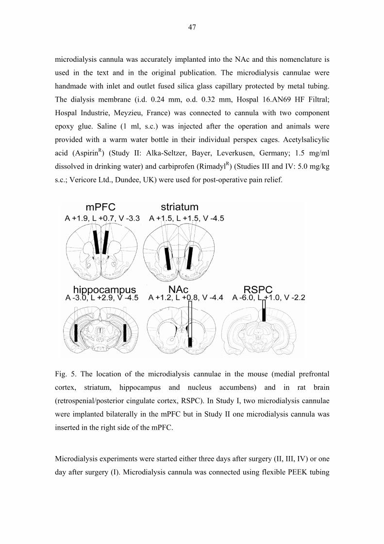
47
microdialysis cannula was accurately implanted into the NAc and this nomenclature is
used in the text and in the original publication. The microdialysis cannulae were
handmade with inlet and outlet fused silica glass capillary protected by metal tubing.
The dialysis membrane (i.d. 0.24 mm, o.d. 0.32 mm, Hospal 16.AN69 HF Filtral;
Hospal Industrie, Meyzieu, France) was connected to cannula with two component
epoxy glue. Saline (1 ml, s.c.) was injected after the operation and animals were
provided with a warm water bottle in their individual perspex cages. Acetylsalicylic
acid (AspirinR) (Study II: Alka-Seltzer, Bayer, Leverkusen, Germany; 1.5 mg/ml
dissolved in drinking water) and carbiprofen (RimadylR) (Studies III and IV: 5.0 mg/kg
s.c.; Vericore Ltd., Dundee, UK) were used for post-operative pain relief.
Fig. 5. The location of the microdialysis cannulae in the mouse (medial prefrontal
cortex, striatum, hippocampus and nucleus accumbens) and in rat brain
(retrospenial/posterior cingulate cortex, RSPC). In Study I, two microdialysis cannulae
were implanted bilaterally in the mPFC but in Study II one microdialysis cannula was
inserted in the right side of the mPFC.
Microdialysis experiments were started either three days after surgery (II, III, IV) or one
day after surgery (I). Microdialysis cannula was connected using flexible PEEK tubing
48
(o.d. 0.51 mm; i.d. 0.13 mm; Upchurch, Oak Harbour, WA, USA) to rotating head-
piece (375/D/22QM, Instech Laboratories Inc, PA, USA) without further tethering.
Ringer solution (Ringer: 145 mM NaCl, 2.7 mM KCl, 2.4 mM CaCl2, 1.0 mM MgCl2;
except 1.2 mM CaCl2 in Study I) was perfused at a rate of 2.8 μl/min (CMA/100
Microinjection Pump, Solna, Sweden). The dialysate was on-line introduced into the
HPLC injection loop and automatically injected every 15.5 min.
4.3.2. High performance liquid chromatography (I, II, III, IV)
Extracellular concentrations of DA and NA were measured using the high performance
liquid chromatography system consisted of a Shimadzu LC-9AD solvent delivery
system (Shimadzu Corporation, Kyoto, Japan) and an ANTEC Decade electrochemical
detector (Antec Leyden, Leyden, The Netherlands). The column oven of the Decade (33
°C) contained a high efficiency pulse dampener, an electrically actuated injector (Valco
EHN6W; Antec Leyden, The Netherlands) with 50 μl loop, Supelcosil LC-18-DB
column (5 μm particles; 250 x 4.6 mm) with Supelguard LC-18-DB guard column (20 x
4.6 mm) (Supelco, Bellefonte, PA, USA). A Coulochem 5011 detector cell (electrode 1
operated on + 250 mV, electrode 2 on - 300 mV) (ESA Inc., Chelmsford, MA, USA)
was controlled by the Decade using the TWIN option. The mobile phase consisted of
mQ water with 10.4 mM citric acid (2 g/l), 6.1 mM sodium acetate (5 g/l), 1.85 mM
heptanesulphonic acid (375 mg/l), 0.3 mM EDTA and 12.5% methanol. Data
acquisition and analysis was carried out with the Shimadzu Class-VP software
(Shimadzu, Duisburg, Germany). The detection limit for DA and NA was 0.1 pg.
4.3.3. Stress (I, II, III, IV)
The first way to induce mild stress was to take the mouse out of its home cage and place
it on a towel on the experimenter's forearm. This is referred to as handling. During
handling, the mouse usually sat quietly for the first few minutes and thereafter explored
its surroundings on the towel. The mouse was restrained only if it tried to jump off the
towel. The novel environment was a clean cage with sawdust, but no food or water.
After sitting quietly for the first few minutes, the mouse usually chewed on and
tunnelled into the sawdust and explored the cage. After handling or exposure to a novel
49
environment, the mouse was returned to its home cage. The duration of handling and
exposure to a novel environment was 15 min, which was the time needed to collect one
dialysate sample. The dialysate sample collection was continued during and after
handling or exposure to the novel environment.
The injection stress consisted of a saline injection (needle size 25 G) in a volume of 5
ml/kg either subcutaneously (Study III) or intraperitoneally (Study IV). The animal was
taken out of its home cage and immediately injected with saline. After injection, the
animal was returned to the home cage. The dialysate sample collection was continued
during and after the saline injection.
4.3.4. Locomotor activity (III)
Locomotor activity was tested in intact animals before in vivo microdialysis
experiments in an automated activity monitor based on infrared detection (TruScan®,
Coulbourn Instruments, Allentown, PA, USA). The system had four 26×26×39 cm
perspex cages with two photobeam sensor rings that were connected to a computer for
recording and data analysis. The mice were gently placed at the centre of the arenas and
the recording was started individually in all cages. After 30 minutes, the recording was
paused and the mice were injected either with saline or drug and the recording was
continued for 1.5 h. Movement parameters were recorded in 15- min epochs and the
total measurement time was set to 2 hours.
4.4. HISTOLOGY
After completion of the microdialysis studies, the animals were decapitated and the
brains were quickly removed and immersed in 4 % formalin. Coronal sections (50 μm)
were stained with cresyl violet to verify the accurate placement of the microdialysis
cannulae.

50
4.5. STATISTICAL ANALYSIS
The statistical analyses were made by using SPSS 10.0 for Windows computer program.
Multivariate analyses (MANOVA), Scheffe’s t-test, contrast analysis (simple) and t-test
were used to analyse line differences and the group and treatment interactions on
different variables. Values of P < 0.05 were considered statistically significant. Methods
for statistical analyses are described in detail in publications I-IV.

51
5. RESULTS
5.1. GENERAL
5.1.1. Basal levels of dopamine and noradrenaline (I, II, III)
The basal levels of DA and NA are presented in table 3. The basal levels are not directly
comparable with each other due to some differences in the measurements. Namely, two
microdialysis cannulae were used bilaterally in Study I, but Studies II and III used only
one microdialysis cannula. Also, in Studies II and III, the Ca2+-concentration (2.4 mM)
was higher than in Study I (1.2 mM).
Table 3. The basal levels of DA and NA in the mouse brain. The values are presented as
concentrations of DA or NA per 50 μl (±SEM) per 1 mm active membrane length.
Abbreviations: KOf = α2A-AR knockout female mouse; WTf = wild-type C57Bl/6J
female mouse; WT = wild type C57Bl/6J male mouse.
Brain Mouse pgDA/50μl pgNA/50μl area line per mm per mm Study I: mPFC (n=16) WT 0.34 ± 0.05 0.52 ± 0.06 striatum (n=7) WT 4.47 ± 0.73 0.32 ± 0.04 hippocampus (n=7) WT 0.14 ± 0.03 0.42 ± 0.04 Study II: mPFC (n=6) KOf 0.28 ± 0.04 0.97 ± 0.11 mPFC (n=6) WTf 0.33 ± 0.03 1.10 ± 0.10 Study III: NAc (n=15) KOf 7.87 ± 1.27 2.13 ± 0.31 NAc (n=20) WTf 5.31 ± 0.79 1.74 ± 0.24
5.1.2. Stressful stimuli (I, II, III, IV)
Handling, exposure to a novel environment and saline injection induced a marked
increase in DA and NA levels in the mPFC, retrosplenial cortex and hippocampus in
mice and rats, whereas in the striatum/accumbens an increase was seen only in the NA
levels.
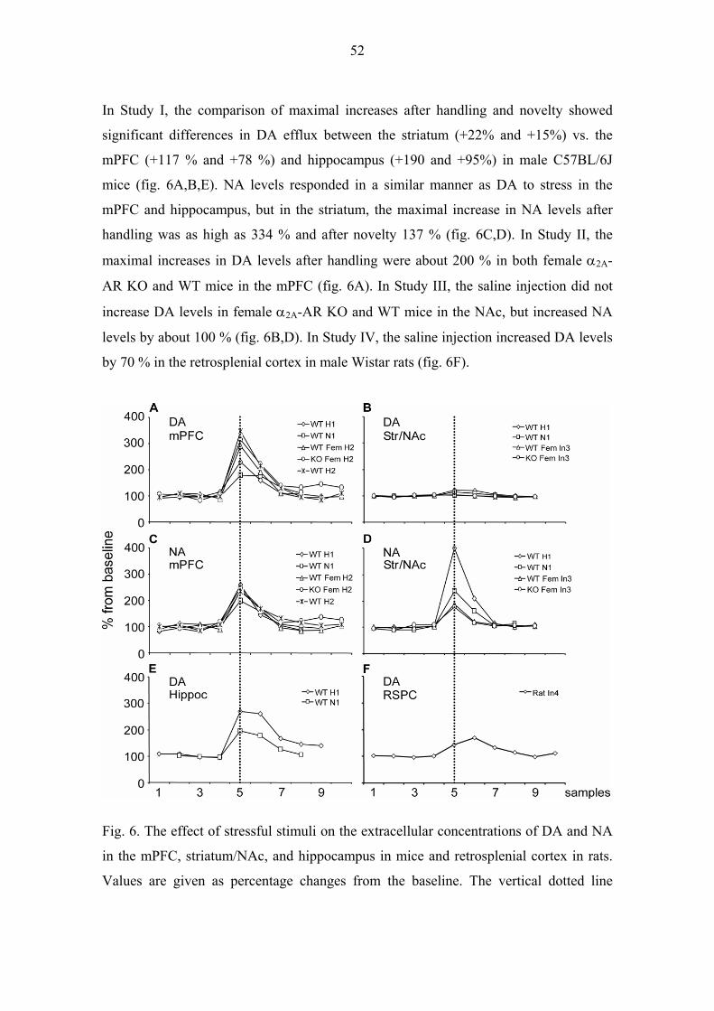
52
In Study I, the comparison of maximal increases after handling and novelty showed
significant differences in DA efflux between the striatum (+22% and +15%) vs. the
mPFC (+117 % and +78 %) and hippocampus (+190 and +95%) in male C57BL/6J
mice (fig. 6A,B,E). NA levels responded in a similar manner as DA to stress in the
mPFC and hippocampus, but in the striatum, the maximal increase in NA levels after
handling was as high as 334 % and after novelty 137 % (fig. 6C,D). In Study II, the
maximal increases in DA levels after handling were about 200 % in both female α2A-
AR KO and WT mice in the mPFC (fig. 6A). In Study III, the saline injection did not
increase DA levels in female α2A-AR KO and WT mice in the NAc, but increased NA
levels by about 100 % (fig. 6B,D). In Study IV, the saline injection increased DA levels
by 70 % in the retrosplenial cortex in male Wistar rats (fig. 6F).
Fig. 6. The effect of stressful stimuli on the extracellular concentrations of DA and NA
in the mPFC, striatum/NAc, and hippocampus in mice and retrosplenial cortex in rats.
Values are given as percentage changes from the baseline. The vertical dotted line

53
indicates the dialysate that was obtained during the handling, novelty or injection stress.
Abbreviations: WT H1(2) = wild-type male mouse handling Study I(II); WT N1 = wild-
type male mouse novelty Study I; WT Fem H2 = wild-type female mouse handling
Study II; KO Fem H2 = α2A-AR knockout female mouse handling Study II; WT Fem
In3 = wild-type female mouse injection Study III; KO Fem In3 = α2A-AR knockout
female mouse injection Study III; Rat In4 = rat injection Study IV; Hippoc =
hippocampus; Str = striatum; RSPC = retrosplenial cortex.
5.1.3. Repeated stressful stimulus (II)
The handling-induced stress was repeated twice in the same day in male C57Bl/6 mice
to assess possible habituation to the DA and the NA increase after the first stimulus.
Our results show that there was no habituation to the handling-induced stress as DA and
NA increases were equally high between two stressful stimuli in male C57Bl/6J mice
(fig. 7).
Fig. 7. The effect of repeated handling stimulus on the extracellular concentrations DA
and NA in the mPFC in male C57Bl/6 mice. The arrow indicates the dialysate that was
obtained during the handling. Values represent percentage changes from the baseline
(±SEM).

54
5.1.4. Effect of calcium concentration on dopamine release
In Studies II, III and IV, the Ca2+-concentration was twice as high as the "physiological"
Ca2+-concentration (1.2 mM) in Study I. The higher Ca2+-concentration was used
because the α2-agonist, DMT, induced a decrease in DA and NA levels compared to
baseline and the detection of DMT-induced decreases on DA and NA levels would have
been impossible since they would have been below the HPLC-system detection limits
for DA in mPFC and NA in mPFC and NAc. However, our unpublished observations in
rats indicate that the amphetamine-induced increase in DA levels were similar in the
mPFC and retrosplenial cortices whit either high Ca2+-concentration (2.4 mM) or
physiological Ca2+-concentration (1.2 mM) (fig. 8). The baseline DA concentrations
were lower in rats that received 1.2 mM Ca2+-concentration in the dialysis fluid but the
relative increase in DA levels after amphetamine treatment was equally high as that
obtained in the rats that received 2.4 mM Ca2+-concentration in the dialysis fluid.
Fig. 8. The effect of dialysate calcium concentration on the extracellular concentrations
of DA and NA in the mPFC and retrosplenial cortex in rats. Amphetamine was infused
locally (10-6-10-4 M) into the mPFC and retrosplenial cortex. Values represent
percentage changes from the baseline. The vertical dotted lines indicate the changes in
the dialysate amphetamine concentration. Abbreviations: aCSF = artificial cerebrospinal
fluid; A 10-6 (A10-5 and A10-4) = amphetamine 10-6 M (amphetamine 10-5 M and
amphetamine 10-4 M); mPFChigh = 2.4 Mm calcium concentration in the dialysate in
the medial prefrontal cortex; mPFC = 1.2 mM calcium concentration in the dialysate in

55
the medial prefrontal cortex; RSPChigh = 2.4 Mm calcium concentration in the
dialysate in the retrosplenial cortex; RSPC = 1.2 Mm calcium concentration in the
dialysate in the retrosplenial cortex.
5.1.5. Diffusion of dexmedetomidine into the brain
The diffusion of DMT into the brain was studied before local infusion of DMT in the
mPFC in Study II, to verify that the DMT effect was indeed restricted to the studied
brain area. Our unpublished results revealed that simultaneous, local infusion of DMT
(10-10 – 10-8 M) through microdialysis cannula in the ipsilateral mPFC did not affect DA
and NA levels in the contralateral side of mPFC, which supports the belief that DMT
exerted only local effects when administered via the microdialysis cannula (fig. 9).
Fig 9. The effect of ipsilaterally infused α2-agonist, DMT, on the extracellular
concentrations of DA and NA release in the contralateral side of the mPFC in male
C57Bl/6 mice. DMT was locally infused into the mPFC (each concentration 60 min).
Values represent percentage changes from the baseline (±SEM). Abbreviations: DMT-
10 (DMT-9 and DMT-8) = dexmedetomidine 10-10 M (dexmedetomidine 10-9 M and
dexmedetomidine 10-8 M).
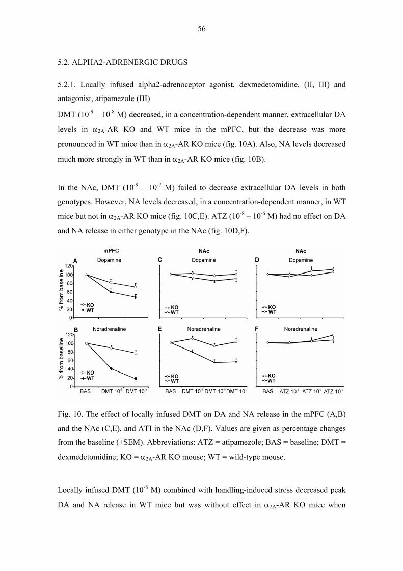
56
5.2. ALPHA2-ADRENERGIC DRUGS
5.2.1. Locally infused alpha2-adrenoceptor agonist, dexmedetomidine, (II, III) and
antagonist, atipamezole (III)
DMT (10-9 – 10-8 M) decreased, in a concentration-dependent manner, extracellular DA
levels in α2A-AR KO and WT mice in the mPFC, but the decrease was more
pronounced in WT mice than in α2A-AR KO mice (fig. 10A). Also, NA levels decreased
much more strongly in WT than in α2A-AR KO mice (fig. 10B).
In the NAc, DMT (10-9 – 10-7 M) failed to decrease extracellular DA levels in both
genotypes. However, NA levels decreased, in a concentration-dependent manner, in WT
mice but not in α2A-AR KO mice (fig. 10C,E). ATZ (10-8 – 10-6 M) had no effect on DA
and NA release in either genotype in the NAc (fig. 10D,F).
Fig. 10. The effect of locally infused DMT on DA and NA release in the mPFC (A,B)
and the NAc (C,E), and ATI in the NAc (D,F). Values are given as percentage changes
from the baseline (±SEM). Abbreviations: ATZ = atipamezole; BAS = baseline; DMT =
dexmedetomidine; KO = α2A-AR KO mouse; WT = wild-type mouse.
Locally infused DMT (10-8 M) combined with handling-induced stress decreased peak
DA and NA release in WT mice but was without effect in α2A-AR KO mice when
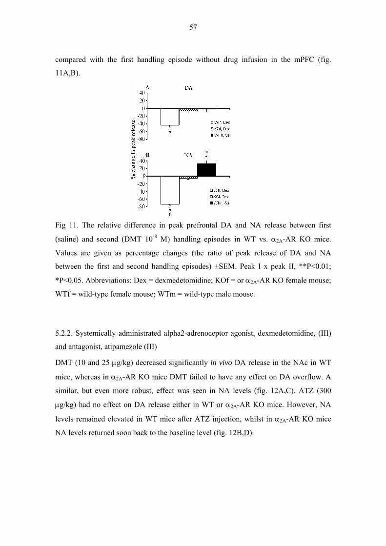
57
compared with the first handling episode without drug infusion in the mPFC (fig.
11A,B).
Fig 11. The relative difference in peak prefrontal DA and NA release between first
(saline) and second (DMT 10-8 M) handling episodes in WT vs. α2A-AR KO mice.
Values are given as percentage changes (the ratio of peak release of DA and NA
between the first and second handling episodes) ±SEM. Peak I x peak II, **P<0.01;
*P<0.05. Abbreviations: Dex = dexmedetomidine; KOf = or α2A-AR KO female mouse;
WTf = wild-type female mouse; WTm = wild-type male mouse.
5.2.2. Systemically administrated alpha2-adrenoceptor agonist, dexmedetomidine, (III)
and antagonist, atipamezole (III)
DMT (10 and 25 μg/kg) decreased significantly in vivo DA release in the NAc in WT
mice, whereas in α2A-AR KO mice DMT failed to have any effect on DA overflow. A
similar, but even more robust, effect was seen in NA levels (fig. 12A,C). ATZ (300
μg/kg) had no effect on DA release either in WT or α2A-AR KO mice. However, NA
levels remained elevated in WT mice after ATZ injection, whilst in α2A-AR KO mice
NA levels returned soon back to the baseline level (fig. 12B,D).

58
Fig 12. Effect of systemically injected saline (sample 5), DMT (10 or 25 μg/kg; s.c.)
(A,C) and ATZ (300 μg/kg, s.c.) (B,D) (sample 10) on DA and NA release in NAc in
α2A-AR KO and WT mice. The number of animals used were as follows: DMT25, KO
n=7, DMT10, WT n=5, DMT25, WT n= 8 and ATZ300, KO n=4, ATZ300, WT n=5.
Values are given as percentage changes from the baseline (±SEM). Arrows indicate the
first affected sample after saline and drug injections and filled symbols significant
differences from the baseline (p<0.05).
5.2.3. Locomotor activity (III)
DMT (10 and 25 μg/kg) significantly decreased locomotor activity in WT mice,
whereas in α2A-AR KO mice locomotor activity did not differ between saline and DMT
treated animals (fig. 13A,C). ATZ (300 μg/kg) treated WT mice maintained a higher
locomotor activity for a longer time period than saline treated WT mice. On the other
hand, ATZ and saline treated α2A-AR KO mice did not differ from each other (fig.
13B,D).

59
Fig. 13. Spontaneous locomotor activity expressed as the total number of movement
episodes recorded in α2A-AR KO and wild WT mice. Saline (Sal) and DMT (10 or 25
μg/kg, s.c.) (A, B) or ATZ 300 μg/kg) (C, D) were injected 30 min after the onset of
recording. Total duration of the recording was set to 2 hours. The number of animals
used were as follows: DMT, KO n=7, WT, n=9 and ATZ, KO n=6, WT, n=10. Filled
symbols indicate significant differences from the saline group and arrows the injection
time.
5.3. NMDA ANTAGONIST KETAMINE (IV)
Ketamine (10 and 30 mg/kg i.p.) injection increased in vivo release of extracellular DA
in the retrosplenial cortex up to 200 % in male Wistar rats. The handling stress related
to the saline injection also increased DA release in the retrosplenial cortex up to 70 %
from the baseline but the ketamine-induced increase of DA levels was significantly
higher (fig. 14).

60
Fig 14. Effect of saline injection (sample 5, i.p.) and ketamine injection (sample 11, i.p.)
on DA release in the retrosplenial cortex in male Wistar rats. The circles denote the
ketamine dose of 10 mg/kg and the triangles represent the ketamine dose of 30 mg/kg.
Values are given as percentage changes from the baseline (±SEM). Significant increases
from the baseline are shown as filled symbols.

61
6. DISCUSSION
6.1. METHODOLOGICAL ASPECTS
6.1.1. Mice
The present series of experiments combined the in vivo microdialysis method with
pharmacological interventions in mice deficient for a α2A-AR subtype to investigate the
dopaminergic and noradrenergic neurotransmission in conscious mice (except for rats
used in Study IV). These results give important new information about the role α2-AR
subtypes in the regulation of DA and NA release in the mPFC and NAc. In Study IV,
the effect of the non-competitive NMDA-antagonist, ketamine, was studied on DA
release in the retrosplenial cortex. Furthermore, modulation of DA and NA
neurotransmission in response to stressful stimuli was studied in the mPFC,
hippocampus and striatum.
One practical limitation of the study was that the collaborator was able to offer only
female α2A-AR KO mice and no littermates to the α2A-AR KO mouse line. Thus female
α2A-AR KO and WT mice were used in the experiments of Studies II and III. In these
studies, the period of the oestrous cycle was not controlled. The oestrous cycle of
female rodents has been reported to modulate dopaminergic neurotransmission in
striatum and NAc (Becker 1999, Castner et al. 1993, Morissette and Di Paolo 1993,
Xiao and Becker 1994). Oestradiol has also been shown to affect the number of α2-ARs
in the PFC (Karkanias et al. 1997). These findings raise the question of whether the
different phase of oestrous cycle in female mice would have had a disturbing effect on
the results. However, it should be noted that generalization of the results is limited to 50
% of the population if either female or male mice are used. Even though in this study
the phase of the oestrous cycle was not monitored, the low level of variation in the basal
and stimulated DA release suggests that the phase of oestrous cycle was unlikely to
have any effect on the results. Indeed, the individual variation in monoamine release
after stressful stimulus was no greater than in our previous studies in males (Ihalainen
and Tanila 2002).

62
It should be noted that the WT mice did not represent the F1 hybrid population of the
α2A-AR KO mouse line as is recommended (Silva et al. 1997). The origin of C57BL/6J
mouse line that was used for backcrossing of α2A-AR KO mice was derived from
Jackson Laboratories (Altman et al. 1999), whereas the α2A-AR WT mouse line was the
locally bred C57BL/6J of the National Laboratory Animal Centre in Kuopio. Thus, the
possibility cannot be ruled out that some differences in the background genome in
addition to α2A-AR gene modification might have accounted for the observed
differences between the α2A-AR KO and WT mice. However, the risk of such non-
specific background gene effects would be much higher in a direct comparison of KO
and WT lines than in our approach of comparing the pharmacological effects on specific
α2-AR drugs.
The DA and NA levels were measured in two time periods in Study III. Thus the age
variability was quite high (5-13 months) between the animals. However, the α2A-AR
KO and WT mice were divided equally in both groups and there were no differences in
the results between the mice that were measured in the first vs. second phase of the
study.
6.1.2. In vivo microdialysis method
The in vivo microdialysis method has been used since the 1980's to determine the
neurotransmitter contents in rats. However, the microdialysis has been routinely
performed in mice only for 5-7 years when the availability of genetically modified
mouse strains started to increase. Study I was one of the first in vivo microdialysis
studies where the DA neurotransmission was investigated outside the striatum in
conscious mice. Technically, the in vivo microdialysis method applied for mice differs
little from the method used in rats, as specific microdialysis equipment such as small
diameter and size microdialysis cannulae, low torque liquid swivels etc. have been
developed, which are suitable for the mice studies. However, the relatively small brain
volume in mice restricts the studies to larger brain structures as in rats. Also, shorter
active dialysis membrane lengths are needed in mice due to the smaller brain size. This
leads to a lower concentrations of collected neurotransmitters in the samples and highly

63
sensitive analysing methods are needed, especially in brain areas where the
concentrations of studied substances are low.
The chemical composition of dialysis perfusates varies between research groups.
Different modifications of basic Ringer's solutions are common. The concentrations of
basic ions such as sodium, potassium and chloride are in most cases in accordance with
estimated physiological concentrations in the extracellular space of the brain (Jones and
Keep 1987, Jones and Keep 1988). However, the composition of other ions in the
perfusion fluid varies extensively. Indeed, magnesium is added to the perfusate in most
cases, and some more complete artificial cerebrospinal fluids consist also of amino
acids, glucose and sodium bicarbonate. There are also variations in dialysate calcium
concentrations. It has been estimated that the physiological calcium concentration in the
extracellular fluid is 1.2 mM (Jones and Keep 1988). Previous studies suggest that the
high calcium concentration (2.4 and 3.4 mM) in the dialysis fluid changes the stimulus-
induced DA release in the nigrostriatal dopaminergic system compared to the
physiological calcium concentration (Moghaddam and Bunney 1989, Tepper et al.
1991). Nowadays most microdialysis studies are performed with a calcium
concentration near 1.2 mM but in some cases higher calcium concentrations are needed.
Indeed, in studies where the neurotransmitter concentrations are near the detection limit
of the analysing system, calcium concentrations are increased in the dialysate. In this
study, the 2.4 mM calcium concentration was used in the dialysate to aid in detecting
the decrease of DA and NA levels induced by the α2-agonist. Importantly, the responses
of DA and NA levels to handling-induced stressful stimulus in the mPFC and
amphetamine-induced increase in the mPFC and retrosplenial cortex indicate that the
results are basically similar with physiological and double (2.4 mM) calcium
concentrations (fig. 6A,C and fig. 8).
6.1.3. Stressful stimuli
The dopaminergic system has been shown to be sensitive to stressful stimuli. Several
microdialysis studies have reported most pronounced stress-related increase in DA
release in the PFC, whereas in the striatum/accumbens the increase has been smaller
(Abercrombie et al. 1989, Cenci et al. 1992, Enrico et al. 1998, Feenstra et al. 1998,
64
Imperato et al. 1991, Kawahara et al. 1999). These results support the idea that the
dopaminergic system is globally activated by stress in the CNS but regional differences
exist in the regulation of DA release in the brain. Indeed, our results reveal that different
kinds of stressors such as mild handling, exposure to a novel environment and needle
injection cause a significant increase in DA release in the cortex (mPFC and
retrosplenial cortex) and hippocampus but failed to increase DA levels in the
striatum/NAc (Fig. 4A,B,E,F). Several studies have shown a small increase in the DA
levels after stressful stimuli in the NAc (Cenci et al. 1992, Feenstra et al. 1998, Inglis
and Moghaddam 1999). In this study, only mild stressors were used, which might partly
explain the lack of increase of DA levels in the NAc and also the fact that mice were
used in this study whilst the former studies used rats.
However, accumbal NA release was markedly increased after saline injection in WT
mice. It is possible that NA projections to the NAc are more readily activated by the
mild and short-lived stressors than the DA projections. In addition, the mildly stressful
stimuli such as handling and novelty elicited an even higher response of NA release in
the striatum/NAc than in the mPFC and hippocampus in mice in Study I. One possible
explanation for the high sensitivity of accumbal NA to mildly stressful stimuli is the
fact that NAc shell receives its noradrenergic projections mainly from the nucleus
tractus solitarius (A2 group), a region that is intimately involved in pain and autonomic
control (Delfs et al. 1998). Other possibly explanations for the different responsiveness
of DA and NA systems to the stressful stimuli, such as DA reuptake by NET, are
discussed in the next chapters.
6.2. ALPHA2-ADRENOCEPTOR SUBTYPES AND DOPAMINE
NEUROTRANSMISSION
6.2.1. Alpha2-adrenoceptor subtypes and the regulation of dopamine release in the
medial prefrontal cortex (II)
The PFC possesses both α2A- and α2C-ARs (Holmberg et al. 2003, Lee et al. 1998,
Nicholas et al. 1993, Scheinin et al. 1994). Previous in vitro slice preparation studies
indicate that α2A-ARs function as the predominant subtype in the brain and are the main

65
regulator of presynaptic autoinhibition of NA release in the CNS (Trendelenburg et al.
1999, Trendelenburg et al. 2001a, Trendelenburg et al. 2001b). On the other hand, α2A-
ARs mediate also most of the heteroceptor function in the CNS (Gobert et al. 1998,
Scheibner et al. 2001, Trendelenburg et al. 1994). However, the importance of α2C-ARs
has also been highlighted in the regulation of NA and DA release in the PFC (Hein et al.
1999, Scheibner et al. 2001). So far, the in vivo studies have been restricted to animals
with an unaltered genetic background and drugs that lack real subtype selectivity for α2-
ARs. In this study we investigated the role of the α2A-AR subtype on the regulation of
DA and NA release in the mPFC in a mouse line lacking a functional α2A-AR subtype.
Interestingly, the basal extracellular levels of DA and NA did not differ between α2A-
AR KO and WT mice in the mPFC. The failure to express the α2A-AR subtype, the
main presynaptic regulator of NA release, would have been expected to increase NA
and also DA levels in the mPFC. On the other had, the loss of α2A-AR subtype resulted
in increased levels of MHPG, the main metabolite of NA, in the cortex measured from
brain homogenates (Lähdesmäki et al. 2002). Also, in the same study, the NA turnover
was increased in the cortex, whereas no significant differences were found in the DA
turnover. Even though our results do not support the theory that lack of α2A-AR has an
effect on baseline release of NA or DA in the mPFC in the resting condition, the
findings should be interpreted with care. The use of in vivo microdialysis technique with
the special zero flow or no-net-flux method would be needed to give a more reliable
estimate for the baseline levels of NA and DA in the mPFC in α2A-AR KO and WT
mice.
The highly specific α2-AR agonist, DMT, concentration-dependently decreased both
NA and DA release in the mPFC in the WT mice, which is consistent with the previous
studies where other α2-AR agonists were used in rats (Dalley and Stanford 1995, Gobert
et al. 1997, Gobert et al. 1998, Gresch et al. 1995, van Veldhuizen et al. 1993). The
concentration-response curve of DMT indicates that NA release is almost entirely
regulated by α2A-ARs in the mPFC, whereas the role of α2C-ARs is minor in this regard.
On the other hand, α2C-ARs seem to have a more prominent role in the regulation of DA

66
release in the mPFC. Indeed, our results reveal that both α2A-ARs and α2C-ARs regulate
DA release at the terminal level in the mPFC. The maximum reduction of DA release
was 50 % of the baseline level, and 40 % of this DMT-induced (10-8 M) effect was
accounted for by α2A-ARs and 60 % by α2C-ARs. On the other hand, the maximum
reduction of NA release was 80 % of baseline level and 75 % of this effect was
mediated by α2A-ARs and only 25 % by α2C-ARs.
These results are in line with the electrophysiological findings that both α2A- and α2C-
ARs mediate auto- and heteroceptor function in the cortex (Gobert et al. 1998,
Scheibner et al. 2001, Trendelenburg et al. 1994, Trendelenburg et al. 1999,
Trendelenburg et al. 2001a). However, this interpretation is complicated by the findings
that DA in cortex may be regulated by NA uptake sites and even be directly released
from NA terminals. Indeed, specific NET blockers elevate extracellular DA levels
effectively in the PFC but not in the caudate nucleus, which provides evidence that the
NET is involved in clearing DA in the PFC in rat (Carboni et al. 1990, Gresch et al.
1995, Mazei et al. 2002, Moron et al. 2002, Pozzi et al. 1994, Yamamoto and Novotney
1998). On the other hand, it has been hypothesized that DA and NA are co-released
from NA neurons since the extracellular DA levels in the parietal and occipital cortices
are only modestly lower than in the mPFC, where DA innervation is known to be much
denser, and that drug treatments that modify mainly noradrenergic activity modulate
also extracellular DA levels in these cortical areas. (Devoto et al. 2001, Devoto et al.
2002, Devoto et al. 2003, Devoto et al. 2004). This hypothesis is mainly based on the
evidence that α2-agonists reduce both DA and NA levels and α2-antagonists increase
both DA and NA levels in cortical areas. However, it is possible that DA has a greater
possibility to be taken up by NET when the α2-agonist reduces the release of NA.
Likewise, the α2-antagonist-induced increase of NA levels might cause a parallel
increase in the DA concentration due to competition of the same transporter (Carboni
and Silvagni 2004). Nevertheless, our finding that the maximum effect of DMT (10-8
M) was a 80 % reduction of NA release and only a 50 % reduction of DA release
suggests that the regulation of DA can be independent of NA release in the mPFC.

67
Interestingly, the peak DA release was almost entirely controlled by α2A-ARs when the
local infusion of α2-agonist, DMT, was repeated during handling-induced stress. In fact,
the peak release of DA during the second handling episode under the influence of DMT
was markedly lower than during the first, predrug, handling episode in the WT mice,
whilst no such reduction was observed in the α2A-AR KO mice. One possible
explanation for the more pronounced role of α2A-AR in the stressful situation compared
to the resting condition could be the difference between the abilities of α2A- and α2C-
ARs to regulate DA release under conditions where extracellular levels of DA are either
low or high. Indeed, electrophysiological stimulation of occipito-parietal cortex and
heart slices has demonstrated that α2C-AR mediates autoinhibition by low frequency
stimulation, whilst α2A-AR operates to inhibit NA release after high frequency
stimulation (Hein et al. 1999, Scheibner et al. 2001).
6.2.2. Alpha2-adrenoceptor subtypes and the regulation of dopamine release in the
nucleus accumbens (III)
Several studies indicate that NA projections can regulate dopaminergic activity in the
NAc via α2-ARs (de Villiers et al. 1995, Murai et al. 1998, Russell et al. 1993,
Whittington et al. 2001, Yavich et al. 1997). The in vitro slice preparation studies
suggest that α2-ARs are able to regulate DA release at the terminal level of the NAc as
evidenced by the decrease of DA release by locally applied α2-agonists (de Villiers et
al. 1995, Russell et al. 1993). However, in our study where in vivo microdialysis were
used in conscious mice, no effect on the DA release could be seen after a local infusion
of the α2-agonist, DMT, whereas there was a clear decrease in NA levels in the NAc.
These discrepant results may derive from the use of rats in the earlier in vitro studies
and mice in this study. However, studies using systemic administration of α2-agonists
have yielded consistent results in rats and mice (Murai et al. 1998, Whittington et al.
2001, Yavich et al. 1997). Therefore, it is more likely that in vitro and in vivo models
measure different aspects of monoamine release. Indeed, one notable difference is that
in vitro slice studies have measured stimulated release of DA whereas our experiment
with reverse microdialysis measured baseline release. Also, the released

68
neurotransmitter is diluted into the perfusate in in vitro models whereas in in vivo
methods the neurotransmitter is recycled back to the cell from the extracellular space.
Interestingly, all effects of α2-agonist, DMT, on accumbal DA and NA release, whether
local or systemic, were absent in α2A-AR KO mice. Indeed, the local administration of
the α2-agonist, DMT, markedly inhibited the release of DA in the mPFC but was
without effect in the NAc. Furthermore, an almost 50 % decline in the DA levels
occured in the α2A-AR KO mice, evidence in favour of a α2C-ARs in the mPFC. In
comparison to these findings, the absence of α2C-ARs mediated regulation of
monoamine release in the NAc is surprising, especially as NAc is among the brain areas
with the densest distribution of α2C-AR subtype in the mouse CNS (Dossin et al. 2000,
Holmberg et al. 2003). It is worth noting, however, that lack of α2C-ARs has been
reported to augment and an overexpression of these receptors to decrease the locomotor
response to amphetamine (Sallinen et al. 1998). Therefore, it is possible that α2C-ARs
play a role in the control of excessive accumbal DA release under strong stimulation
conditions. There are several mechanisms by which systemic but not local
administration of α2-agonist may inhibit DA release in the NAc. These possible
interaction sites have already been discussed in the review of the literature. On the other
hand, the locally infused DMT concentration-dependently decreased NA levels in the
NAc in WT mice but was without effect in α2A-AR KO mice emphasizing the almost
exclusive role of α2A-AR subtype in the regulation of NA release in the terminal level of
NAc.
The locally administered α2-antagonist, ATZ, had no effect on DA and NA release in
the NAc. This observation is consistent with an earlier in vivo microdialysis study
(Hertel et al. 1999) that reported enhanced DA release in the PFC in rats after locally
administered α2-antagonist, idazoxan, but found no effect on the DA release in the NAc.
Also a recent in vivo voltammetry study in rats (Yavich et al. 2003) reported that ATZ
treatment has no effect on its own on DA release in the NAc in response to stimulation
of the medial forebrain bundle, but it enhanced the effect of L-DOPA. Interestingly, NA
levels remained elevated for 1 hour in WT mice after systemic injection of ATZ while

69
in α2A-AR KO mice NA levels returned soon back to baseline. Collectively, these
findings support the notion that α2-AR autoreceptor mediated control of NA release
plays a minor role during baseline release of the neurotransmitter. It is possible that only
when the capacity of reuptake and metabolizing enzymes is exceeded, is there enough
neurotransmitter in the synapse to activate the α2-AR autoreceptors. Under such
conditions of stimulated release, blockade of α2-AR autoreceptors could prolong the
action of NA.
6.2.3. Differences in the regulation of dopamine release in the medial prefrontal cortex
and the nucleus accumbens by alpha2-adrenoceptors
Taken together, our results indicate that DA release is differently regulated in the mPFC
and NAc by α2-ARs. In the mPFC, both α2A-ARs and α2C-ARs regulate DA release in
the condition when the animal is at rest. However, during stimulated DA release such as
occurred during handling of the animal, α2A-ARs seems to the main regulator of DA
release in the mPFC. On the other hand, our results suggest that α2-ARs do not regulate
DA release locally at the terminals in NAc. However, α2A-ARs regulate DA release in
the NAc indirectly by their effect on DA neurons in VTA via some yet unknown
mechanism.
6.2.4. Alpha2-adrenoceptors and the modulation of locomotor activity (III)
The effects of systemic injections of DMT on monoamine release in the NAc and on
exploratory activity were parallel to each other as the time courses of the drug actions
were markedly similar. There is a well-documented connection between the extent of
DA release in the NAc and spontaneous or psychostimulant-induced locomotion (Fink
and Smith 1980, Sharp et al. 1987, Steinpreis and Salamone 1993). On the other hand,
forced locomotion in a running wheel does not increase accumbal DA when measured
using in vitro microdialysis, although it leads to an increase in DOPAC levels (Damsma
et al. 1992). Therefore, it is unlikely that reduced locomotion by some mechanisms
independent of the NAc resulted in dose-dependent decrease of accumbal DA release
after DMT administration, especially as during the microdialysis experiment, mice were
70
immobile for most of the time. Therefore, it is more likely that DMT inhibited the
release of accumbal DA, which leads to decreased spontaneous locomotion. Thus, DMT
may act on the α2-AR autoreceptors on noradrenergic terminals impinging upon the
VTA and/or PFC. An interaction at the level of the PFC is supported by a recent finding
that locomotor hyperactivity by systemic administration of amphetamine is prevented
by blockade of α1-ARs (probably α1B-ARs) in the PFC by local microinfusion of
prazosin (Auclair et al. 2002, Darracq et al. 1998). In this study, the effects of DMT on
both accumbal DA release and spontaneous locomotor activity were totally absent in
α2A-AR KO mice, highlighting the predominant role of α2A-AR auto- and heteroceptors
in this regulation. Thus it is possible that α2A- and α1B-AR at least partially regulate the
same synaptic contacts, such that α2A-ARs are presynaptic and α1B-ARs postsynaptic.
The systemic administration of ATZ also had parallel effects on accumbal monoamine
release and spontaneous locomotion. Locomotor activity gradually decreased during the
locomotor activity task as the environment became familiar to the mice. However, mice
treated with ATZ maintained a high level of locomotion for a longer time than vehicle
treated mice. Interestingly, the effect of ATZ was seen only on NA release but not on
DA release in the NAc. One likely explanation for this apparent discrepancy is the fact
that the situation in the microdialysis experiment was different than that in the
exploratory task. The injection stress selectively increased accumbal NA release, which
was prolonged by the treatment of ATZ. On the other hand, exposure to a new
environment was likely to result in increased activity of both monoamine systems, and
prolonged NA release by the α2A-AR autoreceptor blockade by ATZ resulted in
sustained accumbal DA release and concomitant locomotor activity.
6.3. NMDA-RECEPTOR ANTAGONIST MEDIATED REGULATION OF
DOPAMINE NEUROTRANSMISSION IN THE RETROSPLENIAL CORTEX (IV)
Previous research on the glutamate-dopamine interaction has focused on the prefrontal
cortex and basal ganglia, although the DA system is more widespread in the brain
(Descarries et al. 1987, Gaspar et al. 1989). Notwithstanding some controversial
findings, the NMDA receptor antagonists, phencyclidine and ketamine, do not induce

71
notable DA release in the striatum in animals (Adams et al. 2002, Verma and
Moghaddam 1996) or in humans (Aalto et al. 2002, Kegeles et al. 2000, Kegeles et al.
2002). The glutamate-dopamine interaction is likely to be different in the cortical
regions, as NMDA antagonists have been reported convincingly to induce DA release in
the rat frontal cortex (Adams and Moghaddam 1998, Lindefors et al. 1997, Lorrain et al.
2003, Verma and Moghaddam 1996). The results of this study indicate that ketamine
does increase extracellular DA concentrations also in the retrosplenial cortex in rats.
The functions of the posterior cingulate/retrosplenial cortex are poorly known at
present, but animal studies indicate that it is an important location for spatial learning
(Cooper et al. 2001, Vann and Aggleton 2002) and also the most sensitive brain region
for the NMDA receptor antagonist-induced neurotoxicity in rats (Olney and Farber
1995). In humans, lesions in the right cingulate/retrosplenial cortex have been related to
topographical disorientation (Katayama et al. 1999), and imaging studies have reported
bilateral activation of the retrosplenial cortex to be associated with navigation (Maguire
2001) and spatial attention (Mesulam et al. 2001).
The microdialysis findings in rats indicate that ketamine increases extracellular DA
levels in the retrosplenial cortex. Although there are known species differences in
cortical DA innervation patterns, this microdialysis observation support the role of an
increased synaptic DA concentration underlying the PET findings. At subanesthetic
doses (below 0.5 mg/kg), ketamine is known to be relatively selective for the NMDA
receptor (Javitt and Zukin 1991). Ketamine has some affinity for the DAT in vitro but
only at micromolar concentrations (Nishimura and Sato 1999). Moreover, a direct effect
on D2 receptors (Kapur and Seeman 2001) is unlikely, as such an effect should have
been evident in the three recent PET/SPECT experiments using ketamine intervention
(Aalto et al. 2002, Kegeles et al. 2000, Kegeles et al. 2002). Thus, the ketamine-induced
increase in DA concentration is the most probable explanation for the decreased D2/D3
receptor ligand [11C]FLB 457 binding in the limbic posterior cingulate cortex, although
other indirect mechanisms, such as agonist-mediated D2 receptor internalization cannot
fully be excluded (Laruelle 2000). Cortical glutamatergic afferents project to the VTA
and synapse directly onto VTA dopaminergic neurons that reciprocally connect with

72
glutamatergic pyramidal neurons, at least in the PFC (Sesack et al. 2003). Thus,
disruption of the glutamatergic corticofugal control over DA release is the most
plausible neuroanatomical hypothesis for these findings (Carlsson et al. 1999, Moore et
al. 1999, Sesack et al. 2003).

73
7. CONCLUSIONS
These data demonstrate that in vivo extracellular concentrations of DA in the mouse
brain reflect neuronal release and are sensitive to activation by unconditioned stimuli
such as handling, novel environment and injection stress. The dopaminergic system
exhibited regional differences in the response to the stressful stimuli, as mPFC,
hippocampus and retrosplenial cortex were sensitive to mildly stressful stimuli, whereas
striatum and NAc were unresponsive. However, a robust increase in the extracellular
levels of NA was seen also in the striatum and NAc after stressful stimuli.
In general, α2A-AR seems to be the main regulator of both DA and NA release in the
mPFC and NAc, especially during stress. Nevertheless, α2C-ARs have an important role
in the regulation of DA release in the mPFC in rest. In NAc, α2A-ARs regulate NA but
not DA release at the terminal level, but do regulate DA release indirectly through their
effect on DA neurons in the VTA.
The α2-AR agonist, DMT, and the antagonist, ATZ, mediate their effect on locomotor
activity via α2A-ARs.
The non-competitive NMDA-antagonist, ketamine, markedly increased DA release in
the retrosplenial cortex in rats. This finding indicates that a functional dopaminergic
system also exists in the posterior region of the rat brain, and supports the idea that the
altered DA binding in posterior cortical areas in response to ketamine in human
volunteers in the parallel PET study indeed reflects increased dopamine release.

74
REFERENCES
Aalto S, Hirvonen J, Kajander J, Scheinin H, Nagren K and Vilkman H et al. Ketamine does not decrease striatal dopamine D2 receptor binding in man. Psychopharmacology (Berl) 2002;164:401-406.
Abbott LF and Nelson SB. Synaptic plasticity: taming the beast. Nat Neurosci 2000;3 Suppl:1178-1183.
Abercrombie ED, Keefe KA, DiFrischia DS and Zigmond MJ. Differential effect of stress on in vivo dopamine release in striatum, nucleus accumbens, and medial frontal cortex. J Neurochem 1989;52:1655-1658.
Accili D, Fishburn CS, Drago J, Steiner H, Lachowicz JE and Park BH et al. A targeted mutation of the D3 dopamine receptor gene is associated with hyperactivity in mice. Proc Natl Acad Sci U S A 1996;93:1945-1949.
Adams B and Moghaddam B. Corticolimbic dopamine neurotransmission is temporally dissociated from the cognitive and locomotor effects of phencyclidine. J Neurosci 1998;18:5545-5554.
Adams BW, Bradberry CW and Moghaddam B. NMDA antagonist effects on striatal dopamine release: microdialysis studies in awake monkeys. Synapse 2002;43:12-18.
Albanese A and Minciacchi D. Organization of the ascending projections from the ventral tegmental area: a multiple fluorescent retrograde tracer study in the rat. J Comp Neurol 1983;216:406-420.
Altman JD, Trendelenburg AU, MacMillan L, Bernstein D, Limbird L and Starke K et al. Abnormal regulation of the sympathetic nervous system in alpha2A-adrenergic receptor knockout mice. Mol Pharmacol 1999;56:154-161.
Angrist B, Rotrosen J and Gershon S. Differential effects of amphetamine and neuroleptics on negative vs. positive symptoms in schizophrenia. Psychopharmacology (Berl) 1980;72:17-19.
Aoki C, Go CG, Venkatesan C and Kurose H. Perikaryal and synaptic localization of alpha 2A-adrenergic receptor-like immunoreactivity. Brain Res 1994;650:181-204.
Arborelius L, Chergui K, Murase S, Nomikos GG, Hook BB and Chouvet G et al. The 5-HT1A receptor selective ligands, (R)-8-OH-DPAT and (S)-UH-301, differentially affect the activity of midbrain dopamine neurons. Naunyn Schmiedebergs Arch Pharmacol 1993;347:353-362.
Arnsten AF. Catecholamine regulation of the prefrontal cortex. J Psychopharmacol 1997;11:151-162.
Auclair A, Cotecchia S, Glowinski J and Tassin JP. D-amphetamine fails to increase extracellular dopamine levels in mice lacking alpha 1b-adrenergic receptors: relationship between functional and nonfunctional dopamine release. J Neurosci 2002;22:9150-9154.
Banerjee SP, Zuck LG, Yablonsky-Alter E and Lidsky TI. Glutamate agonist activity: implications for antipsychotic drug action and schizophrenia. Neuroreport 1995;6:2500-2504.
Bassareo V, De Luca MA and Di Chiara G. Differential Expression of Motivational Stimulus Properties by Dopamine in Nucleus Accumbens Shell versus Core and Prefrontal Cortex. J Neurosci 2002;22:4709-4719.
Bassareo V and Di Chiara G. Differential responsiveness of dopamine transmission to food-stimuli in nucleus accumbens shell/core compartments. Neuroscience 1999a;89:637-641.
75
Bassareo V and Di Chiara G. Modulation of feeding-induced activation of mesolimbic dopamine transmission by appetitive stimuli and its relation to motivational state. Eur J Neurosci 1999b;11:4389-4397.
Becker JB. Gender differences in dopaminergic function in striatum and nucleus accumbens. Pharmacol Biochem Behav 1999;64:803-812.
Becker JB, Rudick CN and Jenkins WJ. The role of dopamine in the nucleus accumbens and striatum during sexual behavior in the female rat. J Neurosci 2001;21:3236-3241.
Beckstead RM, Domesick VB and Nauta WJ. Efferent connections of the substantia nigra and ventral tegmental area in the rat. Brain Res 1979;175:191-217.
Benes FM. Carlsson and the discovery of dopamine. Trends Pharmacol Sci 2001;22:46-7.
Ben-Jonathan N and Hnasko R. Dopamine as a prolactin (PRL) inhibitor. Endocr Rev 2001;22:724-763.
Benoit-Marand M, Borrelli E and Gonon F. Inhibition of dopamine release via presynaptic D2 receptors: time course and functional characteristics in vivo. J Neurosci 2001;21:9134-9141.
Benveniste H, Drejer J, Schousboe A and Diemer NH. Regional cerebral glucose phosphorylation and blood flow after insertion of a microdialysis fiber through the dorsal hippocampus in the rat. J Neurochem 1987;49:729-734.
Berger B, Gaspar P and Verney C. Dopaminergic innervation of the cerebral cortex: unexpected differences between rodents and primates. Trends Neurosci 1991;14:21-27.
Berger B, Trottier S, Verney C, Gaspar P and Alvarez C. Regional and laminar distribution of the dopamine and serotonin innervation in the macaque cerebral cortex: a radioautographic study. J Comp Neurol 1988;273:99-119.
Berger B, Verney C, Alvarez C, Vigny A and Helle KB. New dopaminergic terminal fields in the motor, visual (area 18b) and retrosplenial cortex in the young and adult rat. Immunocytochemical and catecholamine histochemical analyses. Neuroscience 1985a;15:983-998.
Berger B, Verney C, Febvret A, Vigny A and Helle KB. Postnatal ontogenesis of the dopaminergic innervation in the rat anterior cingulate cortex (area 24). Immunocytochemical and catecholamine fluorescence histochemical analysis. Brain Res 1985b;353:31-47.
Berridge KC and Robinson TE. What is the role of dopamine in reward: hedonic impact, reward learning, or incentive salience? Brain Res Brain Res Rev 1998;28:309-369.
Bito L, Davson H, Levin E, Murray M and Snider N. The concentrations of free amino acids and other electrolytes in cerebrospinal fluid, in vivo dialysate of brain, and blood plasma of the dog. J Neurochem 1966;13:1057-1067.
Björklund A and Lindvall O. Dopamine-containing system in the CNS. In: Björklund A and Hökfelt O, eds. Classical transmitters in the CNS, Part I (Handbook of chemical neuroanatomy). Amsterdam: Elsevier, 1984, pp. 55-122.
Björklund A and Lindvall O. Dopamine in dendrites of substantia nigra neurons: suggestions for a role in dendritic terminals. Brain Res 1975;83:531-537.
Björklund A and Lindvall O. Catecholaminergic brainstem regulatory systems. In: Mountcastle VB, Bloom FE and Geiger SR, eds. Handbook of Physiology: The Nervous System. Maryland: American Physiological Society, 1986, pp. 155-235.
76
Bloom FE and Lazerson A. Brain, Mind and Behavior. W.H. Freeman & Company, New York, 1988.
Bockaert J, Claeysen S, Becamel C, Pinloche S and Dumuis A. G protein-coupled receptors: dominant players in cell-cell communication. Int Rev Cytol 2002;212:63-132.
Bonci A, Grillner P, Siniscalchi A, Mercuri NB, Bernardi G. Glutamate metabotropic receptor agonists depress excitatory and inhibitory transmission on rat mesencephalic principal neurons. Eur J Neurosci 1997;9:2359-2369.
Bourne JA. SCH 23390: the first selective dopamine D1-like receptor antagonist. CNS Drug Rev 2001;7:399-414.
Bouthenet ML, Souil E, Martres MP, Sokoloff P, Giros B and Schwartz JC. Localization of dopamine D3 receptor mRNA in the rat brain using in situ hybridization histochemistry: comparison with dopamine D2 receptor mRNA. Brain Res 1991;564:203-219.
Breier A, Su TP, Saunders R, Carson RE, Kolachana BS and de Bartolomeis A et al. Schizophrenia is associated with elevated amphetamine-induced synaptic dopamine concentrations: evidence from a novel positron emission tomography method. Proc Natl Acad Sci U S A 1997;94:2569-2574.
Brockmeyer DM and Kendig JJ. Selective effects of ketamine on amino acid-mediated pathways in neonatal rat spinal cord. Br J Anaesth 1995;74:79-84.
Brown JM, Hanson GR and Fleckenstein AE. Cocaine-induced increases in vesicular dopamine uptake: role of dopamine receptors. J Pharmacol Exp Ther 2001;298:1150-1153.
Bucheler MM, Hadamek K and Hein L. Two alpha(2)-adrenergic receptor subtypes, alpha(2A) and alpha(2C), inhibit transmitter release in the brain of gene-targeted mice. Neuroscience 2002;109:819-826.
Bungay PM, Newton-Vinson P, Isele W, Garris PA and Justice JB. Microdialysis of dopamine interpreted with quantitative model incorporating probe implantation trauma. J Neurochem 2003;86:932-946.
Bylund DB. Subtypes of alpha 2-adrenoceptors: pharmacological and molecular biological evidence converge. Trends Pharmacol Sci 1988;9:356-361.
Campbell AD, Kohl RR and McBride WJ. Serotonin-3 receptor and ethanol-stimulated somatodendritic dopamine release. Alcohol 1996;13:569-574.
Canton H, Verriele L and Colpaert FC. Binding of typical and atypical antipsychotics to 5-HT1C and 5-HT2 sites: clozapine potently interacts with 5-HT1C sites. Eur J Pharmacol 1990;191:93-96.
Carboni E and Silvagni A. Dopamine reuptake by norepinephrine neurons: exception or rule? Crit Rev Neurobiol 2004;16:121-128.
Carboni E, Tanda GL, Frau R and Di Chiara G. Blockade of the noradrenaline carrier increases extracellular dopamine concentrations in the prefrontal cortex: evidence that dopamine is taken up in vivo by noradrenergic terminals. J Neurochem 1990;55:1067-1070.
Carlsson A. A paradigm shift in brain research. Science 2001;294:1021-1024.
Carlsson A. Does dopamine play a role in schizophrenia? Psychol Med 1977;7:583-597.
Carlsson A and Lindqvist M. Effect of chlorpromazine or haloperidol on formation of 3methoxytyramine and normetanephrine in mouse brain. Acta Pharmacol Toxicol (Copenh) 1963;20:140-144.
Carlsson A and Waldeck B. A fluorimetric method for the determination of dopamine (3-hydroxytyramine). Acta Physiol Scand 1958;44:293-28.
77
Carlsson A, Waters N and Carlsson ML. Neurotransmitter interactions in schizophrenia--therapeutic implications. Biol Psychiatry 1999;46:1388-1395.
Carr DB, O'Donnell P, Card JP, Sesack SR. Dopamine terminals in the rat prefrontal cortex synapse on pyramidal cells that project to the nucleus accumbens. J Neurosci 1999;19:11049-60.
Carr DB and Sesack SR. Projections from the rat prefrontal cortex to the ventral tegmental area: target specificity in the synaptic associations with mesoaccumbens and mesocortical neurons. J Neurosci 2000;20:3864-3873.
Cass WA and Gerhardt GA. In vivo assessment of dopamine uptake in rat medial prefrontal cortex: comparison with dorsal striatum and nucleus accumbens. J Neurochem 1995;65:201-207.
Castner SA, Xiao L and Becker JB. Sex differences in striatal dopamine: in vivo microdialysis and behavioral studies. Brain Res 1993;610:127-134.
Cenci MA, Kalen P, Mandel RJ and Björklund A. Regional differences in the regulation of dopamine and noradrenaline release in medial frontal cortex, nucleus accumbens and caudate-putamen: a microdialysis study in the rat. Brain Res 1992;581:217-228.
Cheng JJ, de Bruin JP and Feenstra MG. Dopamine efflux in nucleus accumbens shell and core in response to appetitive classical conditioning. Eur J Neurosci 2003;18:1306-1314.
Chergui K, Nomikos GG, Mathe JM, Gonon F and Svensson TH. Burst stimulation of the medial forebrain bundle selectively increase Fos-like immunoreactivity in the limbic forebrain of the rat. Neuroscience 1996;72:141-156.
Chergui K, Suaud-Chagny MF and Gonon F. Nonlinear relationship between impulse flow, dopamine release and dopamine elimination in the rat brain in vivo. Neuroscience 1994;62:641-645.
Chergui K, Svenningsson P, Nomikos GG, Gonon F, Fredholm BB and Svennson TH. Increased expression of NGFI-A mRNA in the rat striatum following burst stimulation of the medial forebrain bundle. Eur J Neurosci 1997;9:2370-2382.
Cohen BM and Lipinski JF. In vivo potencies of antipsychotic drugs in blocking alpha 1 noradrenergic and dopamine D2 receptors: implications for drug mechanisms of action. Life Sci 1986;39:2571-2580.
Cooper BG, Manka TF and Mizumori SJ. Finding your way in the dark: the retrosplenial cortex contributes to spatial memory and navigation without visual cues. Behav Neurosci 2001;115:1012-1028.
Cragg SJ and Greenfield SA. Differential autoreceptor control of somatodendritic and axon terminal dopamine release in substantia nigra, ventral tegmental area, and striatum. J Neurosci 1997;17:5738-5746.
Creese I, Burt DR and Snyder SH. Dopamine receptor binding predicts clinical and pharmacological potencies of antischizophrenic drugs. Science 1976;192:481-483.
Dalley JW and Stanford SC. Contrasting effects of the imidazol(in)e alpha 2-adrenoceptor agonists, medetomidine, clonidine and UK 14,304 on extraneuronal levels of noradrenaline in the rat frontal cortex: evaluation using in vivo microdialysis and synaptosomal uptake studies. Br J Pharmacol 1995;114:1717-1723.
Damsma G, Pfaus JG, Wenkstern D, Phillips AG and Fibiger HC. Sexual behavior increases dopamine transmission in the nucleus accumbens and striatum of male rats: comparison with novelty and locomotion. Behav Neurosci 1992;106:181-191.

78
Darracq L, Blanc G, Glowinski J and Tassin JP. Importance of the noradrenaline-dopamine coupling in the locomotor activating effects of D-amphetamine. J Neurosci 1998;18:2729-2739.
Davis KL, Kahn RS, Ko G and Davidson M. Dopamine in schizophrenia: a review and reconceptualization. Am J Psychiatry 1991;148:1474-1486.
de Villiers AS, Russell VA, Sagvolden T, Searson A, Jaffer A and Taljaard JJ. Alpha 2-adrenoceptor mediated inhibition of [3H]dopamine release from nucleus accumbens slices and monoamine levels in a rat model for attention-deficit hyperactivity disorder. Neurochem Res 1995;20:427-433.
Dearry A, Gingrich JA, Falardeau P, Fremeau RT,Jr, Bates MD and Caron MG. Molecular cloning and expression of the gene for a human D1 dopamine receptor. Nature 1990;347:72-76.
Delfs JM, Zhu Y, Druhan JP and Aston-Jones GS. Origin of noradrenergic afferents to the shell subregion of the nucleus accumbens: anterograde and retrograde tract-tracing studies in the rat. Brain Res 1998;806:127-140.
Delgado JM, DeFeudis FV, Roth RH, Ryugo DK and Mitruka BM. Dialytrode for long term intracerebral perfusion in awake monkeys. Arch Int Pharmacodyn Ther 1972;198:9-21.
Descarries L, Lemay B, Doucet G and Berger B. Regional and laminar density of the dopamine innervation in adult rat cerebral cortex. Neuroscience 1987;21:807-824.
Devoto P, Flore G, Longu G, Pira L and Gessa GL. Origin of extracellular dopamine from dopamine and noradrenaline neurons in the medial prefrontal and occipital cortex. Synapse 2003;50:200-205.
Devoto P, Flore G, Pani L and Gessa GL. Evidence for co-release of noradrenaline and dopamine from noradrenergic neurons in the cerebral cortex. Mol Psychiatry 2001;6:657-664.
Devoto P, Flore G, Pira L, Diana M and Gessa GL. Co-release of noradrenaline and dopamine in the prefrontal cortex after acute morphine and during morphine withdrawal. Psychopharmacology (Berl) 2002;160:220-224.
Devoto P, Flore G, Pira L, Longu G and Gessa GL. Alpha2-adrenoceptor mediated co-release of dopamine and noradrenaline from noradrenergic neurons in the cerebral cortex. J Neurochem 2004;88:1003-1009.
Di Chiara G, Bassareo V, Fenu S, De Luca MA, Spina L and Cadoni C et al. Dopamine and drug addiction: the nucleus accumbens shell connection. Neuropharmacology 2004;47 Suppl 1:227-241.
Di Chiara G, Tanda GL, Frau R and Carboni E. Heterologous monoamine reuptake: lack of transmitter specificity of neuron-specific carriers. Neurochem Int 1992;20 Suppl:231S-235S.
Di Giovanni G, Di Matteo V, Di Mascio M and Esposito E. Preferential modulation of mesolimbic vs. nigrostriatal dopaminergic function by serotonin(2C/2B) receptor agonists: a combined in vivo electrophysiological and microdialysis study. Synapse 2000;35:53-61.
Djamgoz MB and Wagner HJ. Localization and function of dopamine in the adult vertebrate retina. Neurochem Int 1992;20:139-191.
Docherty JR. Subtypes of functional alpha1- and alpha2-adrenoceptors. Eur J Pharmacol 1998;361:1-15.
Dossin O, Mouledous L, Baudry X, Tafani JA, Mazarguil H and Zajac JM. Characterization of a new radioiodinated probe for the alpha2C adrenoceptor in the mouse brain. Neurochem Int 2000;36:7-18.
Drago J, Padungchaichot P, Accili D and Fuchs S. Dopamine receptors and dopamine transporter in brain function and addictive behaviors: insights from targeted mouse mutants. Dev Neurosci 1998;20:188-203.

79
Dwoskin LP and Zahniser NR. Robust modulation of [3H]dopamine release from rat striatal slices by D-2 dopamine receptors. J Pharmacol Exp Ther 1986;239:442-453.
Ehinger B. Connexions between retinal neurons with identified neurotransmitters. Vision Res 1983;23:1281-1291.
El-Ghundi M, O'Dowd BF, Erclik M and George SR. Attenuation of sucrose reinforcement in dopamine D1 receptor deficient mice. Eur J Neurosci 2003;17:851-862.
El-Ghundi M, O'Dowd BF and George SR. Prolonged fear responses in mice lacking dopamine D1 receptor. Brain Res 2001;892:86-93.
Ellinwood EH,Jr, Sudilovsky A and Nelson LM. Evolving behavior in the clinical and experimental amphetamine (model) psychosis. Am J Psychiatry 1973;130:1088-1093.
Ellison G, Nielsen EB and Lyon M. Animal model of psychosis: hallucinatory behaviors in monkeys during the late stage of continuous amphetamine intoxication. J Psychiatr Res 1981;16:13-22.
Ellison GD and Eison MS. Continuous amphetamine intoxication: an animal model of the acute psychotic episode. Psychol Med 1983;13:751-761.
Emson PC and Koob GF. The origin and distribution of dopamine-containing afferents to the rat frontal cortex. Brain Res 1978;142:249-267.
Enrico P, Bouma M, de Vries JB and Westerink BH. The role of afferents to the ventral tegmental area in the handling stress-induced increase in the release of dopamine in the medial prefrontal cortex: a dual-probe microdialysis study in the rat brain. Brain Res 1998;779:205-213.
Farber NB, Foster J, Duhan NL and Olney JW. Olanzapine and fluperlapine mimic clozapine in preventing MK-801 neurotoxicity. Schizophr Res 1996;21:33-37.
Farber NB, Price MT, Labruyere J, Nemnich J, St Peter H and Wozniak DF et al. Antipsychotic drugs block phencyclidine receptor-mediated neurotoxicity. Biol Psychiatry 1993;34:119-121.
Farde L, Hall H, Ehrin E and Sedvall G. Quantitative analysis of D2 dopamine receptor binding in the living human brain by PET. Science 1986;231:258-261.
Farde L, Mack RJ, Nyberg S and Halldin C. D2 occupancy, extrapyramidal side effects and antipsychotic drug treatment: a pilot study with sertindole in healthy subjects. Int Clin Psychopharmacol 1997;12 Suppl 1:S3-7.
Feenstra MG. Dopamine and noradrenaline release in the prefrontal cortex in relation to unconditioned and conditioned stress and reward. Prog Brain Res 2000;126:133-163.
Feenstra MG and Botterblom MH. Rapid sampling of extracellular dopamine in the rat prefrontal cortex during food consumption, handling and exposure to novelty. Brain Res 1996;742:17-24.
Feenstra MG, Botterblom MH and Mastenbroek S. Dopamine and noradrenaline efflux in the prefrontal cortex in the light and dark period: effects of novelty and handling and comparison to the nucleus accumbens. Neuroscience 2000;100:741-748.
Feenstra MG, Botterblom MH and van Uum JF. Local activation of metabotropic glutamate receptors inhibits the handling-induced increased release of dopamine in the nucleus accumbens but not that of dopamine or noradrenaline in the prefrontal cortex: comparison with inhibition of ionotropic receptors. J Neurochem 1998;70:1104-1113.

80
Feenstra MG, Vogel M, Botterblom MH, Joosten RN and de Bruin JP. Dopamine and noradrenaline efflux in the rat prefrontal cortex after classical aversive conditioning to an auditory cue. Eur J Neurosci 2001;13:1051-1054.
Feldman RS, Meyer.J S and Quenzer LF. Catecholamines. In: Feldman RS, Meyer.J S and Quenzer LF, eds. Principles of Neuropsychopharmacology. Sunderland: Sinauer Associates, Inc, 1997, pp. 277-344.
Fink JS and Smith GP. Mesolimbicocortical dopamine terminal fields are necessary for normal locomotor and investigatory exploration in rats. Brain Res 1980;199:359-384.
Fiorino DF and Phillips AG. Facilitation of sexual behavior and enhanced dopamine efflux in the nucleus accumbens of male rats after D-amphetamine-induced behavioral sensitization. J Neurosci 1999;19:456-463.
Fischer JF and Cho AK. Chemical release of dopamine from striatal homogenates: evidence for an exchange diffusion model. J Pharmacol Exp Ther 1979;208:203-209.
Foote SL, Bloom FE and Aston-Jones G. Nucleus locus ceruleus: new evidence of anatomical and physiological specificity. Physiol Rev 1983;63:844-914.
Fornai F, Chen K, Giorgi FS, Gesi M, Alessandri MG and Shih JC. Striatal dopamine metabolism in monoamine oxidase B-deficient mice: a brain dialysis study. J Neurochem 1999;73:2434-2440.
Fowler CJ and Benedetti MS. The metabolism of dopamine by both forms of monoamine oxidase in the rat brain and its inhibition by cimoxatone. J Neurochem 1983;40:1534-1541.
Fowler CJ and Tipton KF. Deamination of 5-hydroxytryptamine by both forms of monoamine oxidase in the rat brain. J Neurochem 1982;38:733-736.
Franklin KBJ and Paxinos G. The mouse brain in stereotactic coordinates. Academic Press, San Diego, 1997.
Freeman AS and Bunney BS. The effects of phencyclidine and N-allylnormetazocine on midbrain dopamine neuronal activity. Eur J Pharmacol 1984;104:287-293.
Fremeau RT,Jr, Duncan GE, Fornaretto MG, Dearry A, Gingrich JA and Breese GR et al. Localization of D1 dopamine receptor mRNA in brain supports a role in cognitive, affective, and neuroendocrine aspects of dopaminergic neurotransmission. Proc Natl Acad Sci U S A 1991;88:3772-3776.
French ED and Ceci A. Non-competitive N-methyl-D-aspartate antagonists are potent activators of ventral tegmental A10 dopamine neurons. Neurosci Lett 1990;119:159-162.
French ED, Mura A and Wang T. MK-801, phencyclidine (PCP), and PCP-like drugs increase burst firing in rat A10 dopamine neurons: comparison to competitive NMDA antagonists. Synapse 1993;13:108-116.
Fuxe K, Agnati LF, Kalia M, Goldstein M, Andersson K and Härfstrand A. Dopaminergic system in the brain and pituitary. In: Fluckiger E, Muller EE, Thorner MO, eds. The dopaminergic system (Basic and clinical aspects of neuroscience). Berlin: Springer-Verlag, 1985, pp. 11-25.
Gaddum JH. Push-pull cannulae. J Physiol 1961;159:1-2.
Gainetdinov RR, Jones SR, Fumagalli F, Wightman RM and Caron MG. Re-evaluation of the role of the dopamine transporter in dopamine system homeostasis. Brain Res Brain Res Rev 1998;26:148-153.
Garris PA, Collins LB, Jones SR and Wightman RM. Evoked extracellular dopamine in vivo in the medial prefrontal cortex. J Neurochem 1993;61:637-647.

81
Gaspar P, Berger B, Febvret A, Vigny A and Henry JP. Catecholamine innervation of the human cerebral cortex as revealed by comparative immunohistochemistry of tyrosine hydroxylase and dopamine-beta-hydroxylase. J Comp Neurol 1989;279:249-271.
Georgieva J, Luthman J, Mohringe B and Magnusson O. Tissue and microdialysate changes after repeated and permanent probe implantation in the striatum of freely moving rats. Brain Res Bull 1993;31:463-470.
Giros B and Caron MG. Molecular characterization of the dopamine transporter. Trends Pharmacol Sci 1993;14:43-49.
Giuliano F and Allard J. Dopamine and sexual function. Int J Impot Res 2001;13 Suppl 3:S18-28.
Glover V, Sandler M, Owen F and Riley GJ. Dopamine is a monoamine oxidase B substrate in man. Nature 1977;265:80-81.
Gobbi G, Muntoni AL, Gessa GL and Diana M. Clonidine fails to modify dopaminergic neuronal activity during morphine withdrawal. Psychopharmacology (Berl) 2001;158:1-6.
Gobert A, Rivet JM, Audinot V, Newman-Tancredi A, Cistarelli L and Millan MJ. Simultaneous quantification of serotonin, dopamine and noradrenaline levels in single frontal cortex dialysates of freely-moving rats reveals a complex pattern of reciprocal auto- and heteroreceptor-mediated control of release. Neuroscience 1998;84:413-429.
Gobert A, Rivet JM, Cistarelli L, Melon C and Millan MJ. Alpha2-adrenergic receptor blockade markedly potentiates duloxetine- and fluoxetine-induced increases in noradrenaline, dopamine, and serotonin levels in the frontal cortex of freely moving rats. J Neurochem 1997;69:2616-2619.
Gogos JA, Morgan M, Luine V, Santha M, Ogawa S and Pfaff D et al. Catechol-O-methyltransferase-deficient mice exhibit sexually dimorphic changes in catecholamine levels and behavior. Proc Natl Acad Sci U S A 1998;95:9991-9996.
Goldman-Rakic PS, Lidow MS and Gallager DW. Overlap of dopaminergic, adrenergic, and serotoninergic receptors and complementarity of their subtypes in primate prefrontal cortex. J Neurosci 1990;10:2125-2138.
Goldman-Rakic PS, Lidow MS, Smiley JF and Williams MS. The anatomy of dopamine in monkey and human prefrontal cortex. J Neural Transm Suppl 1992;36:163-177.
Gonon F. Prolonged and extrasynaptic excitatory action of dopamine mediated by D1 receptors in the rat striatum in vivo. J Neurosci 1997;17:5972-5978.
Gonon FG and Buda MJ. Regulation of dopamine release by impulse flow and by autoreceptors as studied by in vivo voltammetry in the rat striatum. Neuroscience 1985;14:765-774.
Grenhoff J, Nisell M, Ferre S, Aston-Jones G and Svensson TH. Noradrenergic modulation of midbrain dopamine cell firing elicited by stimulation of the locus coeruleus in the rat. J Neural Transm Gen Sect 1993;93:11-25.
Grenhoff J, North RA and Johnson SW. Alpha 1-adrenergic effects on dopamine neurons recorded intracellularly in the rat midbrain slice. Eur J Neurosci 1995;7:1707-1713.
Gresch PJ, Sved AF, Zigmond MJ and Finlay JM. Local influence of endogenous norepinephrine on extracellular dopamine in rat medial prefrontal cortex. J Neurochem 1995;65:111-116.
82
Hall H, Farde L, Halldin C, Hurd YL, Pauli S and Sedvall G. Autoradiographic localization of extrastriatal D2-dopamine receptors in the human brain using [125I]epidepride. Synapse 1996;23:115-123.
Hanson PI, Heuser JE and Jahn R. Neurotransmitter release - four years of SNARE complexes. Curr Opin Neurobiol 1997;7:310-315.
Harrison NL and Simmonds MA. Quantitative studies on some antagonists of N-methyl D-aspartate in slices of rat cerebral cortex. Br J Pharmacol 1985;84:381-391.
Hattori T, McGeer PL and McGeer EG. Dendro axonic neurotransmission. II. Morphological sites for the synthesis, binding and release of neurotransmitters in dopaminergic dendrites in the substantia nigra and cholinergic dendrites in the neostriatum. Brain Res 1979;170:71-83.
Heikkila RE, Orlansky H, Mytilineou C and Cohen G. Amphetamine: evaluation of d- and l-isomers as releasing agents and uptake inhibitors for 3H-dopamine and 3H-norepinephrine in slices of rat neostriatum and cerebral cortex. J Pharmacol Exp Ther 1975;194:47-56.
Hein L, Altman JD and Kobilka BK. Two functionally distinct alpha2-adrenergic receptors regulate sympathetic neurotransmission. Nature 1999;402:181-184.
Hernandez L and Hoebel BG. Feeding and hypothalamic stimulation increase dopamine turnover in the accumbens. Physiol Behav 1988a;44:599-606.
Hernandez L and Hoebel BG. Food reward and cocaine increase extracellular dopamine in the nucleus accumbens as measured by microdialysis. Life Sci 1988b;42:1705-1712.
Hertel P, Nomikos GG and Svensson TH. Idazoxan preferentially increases dopamine output in the rat medial prefrontal cortex at the nerve terminal level. Eur J Pharmacol 1999;371:153-158.
Hess EJ, Bracha HS, Kleinman JE and Creese I. Dopamine receptor subtype imbalance in schizophrenia. Life Sci 1987;40:1487-1497.
Hoenicke EM, Vanecek SA and Woods JH. The discriminative stimulus effects of clozapine in pigeons: involvement of 5-hydroxytryptamine1C and 5-hydroxytryptamine2 receptors. J Pharmacol Exp Ther 1992;263:276-284.
Holmberg M, Fagerholm V and Scheinin M. Regional distribution of alpha(2C)-adrenoceptors in brain and spinal cord of control mice and transgenic mice overexpressing the alpha(2C)-subtype: an autoradiographic study with [(3)H]RX821002 and [(3)H]rauwolscine. Neuroscience 2003;117:875-898.
Hooks MS, Colvin AC, Juncos JL and Justice JB,Jr. Individual differences in basal and cocaine-stimulated extracellular dopamine in the nucleus accumbens using quantitative microdialysis. Brain Res 1992;587:306-312.
Horn AS. Structure-activity relations for the inhibition of catecholamine uptake into synaptosomes from noradrenaline and dopaminergic neurones in rat brain homogenates. Br J Pharmacol 1973;47:332-338.
Hurley KM, Herbert H, Moga MM and Saper CB. Efferent projections of the infralimbic cortex of the rat. J Comp Neurol 1991;308:249-276.
Imperato A, Cabib S and Puglisi-Allegra S. Repeated stressful experiences differently affect the time-dependent responses of the mesolimbic dopamine system to the stressor. Brain Res 1993;601:333-336.
Imperato A and Di Chiara G. Dopamine release and metabolism in awake rats after systemic neuroleptics as studied by trans-striatal dialysis. J Neurosci 1985;5:297-306.

83
Imperato A, Puglisi-Allegra S, Casolini P and Angelucci L. Changes in brain dopamine and acetylcholine release during and following stress are independent of the pituitary-adrenocortical axis. Brain Res 1991;538:111-117.
Inglis FM and Moghaddam B. Dopaminergic innervation of the amygdala is highly responsive to stress. J Neurochem 1999;72:1088-1094.
Itil T, Keskiner A, Kiremitci N and Holden JM. Effect of phencyclidine in chronic schizophrenics. Can Psychiatr Assoc J 1967;12:209-212.
Jaber M, Robinson SW, Missale C and Caron MG. Dopamine receptors and brain function. Neuropharmacology 1996;35:1503-1519.
Jackson DM and Westlind-Danielsson A. Dopamine receptors: molecular biology, biochemistry and behavioural aspects. Pharmacol Ther 1994;64:291-370.
Jacobson I and Hamberger A. Veratridine-induced release in vivo and in vitro of amino acids in the rabbit olfactory bulb. Brain Res 1984;299:103-112.
Jacobson I, Sandberg M and Hamberger A. Mass transfer in brain dialysis devices--a new method for the estimation of extracellular amino acids concentration. J Neurosci Methods 1985;15:263-268.
Javitt DC and Zukin SR. Recent advances in the phencyclidine model of schizophrenia. Am J Psychiatry 1991;148:1301-1308.
Johnson RG,Jr. Accumulation of biological amines into chromaffin granules: a model for hormone and neurotransmitter transport. Physiol Rev 1988;68:232-307.
Johnstone EC, Crow TJ, Frith CD, Husband J and Kreel L. Cerebral ventricular size and cognitive impairment in chronic schizophrenia. Lancet 1976;2:924-926.
Jones BE, Halaris AE, McIlhany M and Moore RY. Ascending projections of the locus coeruleus in the rat. I. Axonal transport in central noradrenaline neurons. Brain Res 1977;127:1-21.
Jones HC and Keep RF. Brain interstitial fluid calcium concentration during development in the rat: control levels and changes in acute plasma hypercalcaemia. Physiol Bohemoslov 1988;37:213-216.
Jones HC and Keep RF. The control of potassium concentration in the cerebrospinal fluid and brain interstitial fluid of developing rats. J Physiol 1987;383:441-453.
Jones SR, Gainetdinov RR, Wightman RM and Caron MG. Mechanisms of amphetamine action revealed in mice lacking the dopamine transporter. J Neurosci 1998;18:1979-1986.
Justice JB,Jr. Quantitative microdialysis of neurotransmitters. J Neurosci Methods 1993;48:263-276.
Kaakkola S, Mannisto PT and Nissinen E. Striatal membrane-bound and soluble catechol-O-methyl-transferase after selective neuronal lesions in the rat. J Neural Transm 1987;69:221-228.
Kalivas PW, Bourdelais A, Abhold R and Abbott L. Somatodendritic release of endogenous dopamine: in vivo dialysis in the A10 dopamine region. Neurosci Lett 1989;100:215-220.
Kametani H and Kawamura H. Circadian rhythm of cortical acetylcholine release as measured by in vivo microdialysis in freely moving rats. Neurosci Lett 1991;132:263-266.
Kanner BI and Schuldiner S. Mechanism of transport and storage of neurotransmitters. CRC Crit Rev Biochem 1987;22:1-38.

84
Kapur S and Seeman P. Ketamine has equal affinity for NMDA receptors and the high-affinity state of the dopamine D2 receptor. Biol Psychiatry 2001;49:954-957.
Karhunen T, Tilgmann C, Ulmanen I and Panula P. Catechol-O-methyltransferase (COMT) in rat brain: immunoelectron microscopic study with an antiserum against rat recombinant COMT protein. Neurosci Lett 1995;187:57-60.
Karkanias GB, Li CS and Etgen AM. Estradiol reduction of alpha 2-adrenoceptor binding in female rat cortex is correlated with decreases in alpha 2A/D-adrenoceptor messenger RNA. Neuroscience 1997;81:593-597.
Karreman M and Moghaddam B. The prefrontal cortex regulates the basal release of dopamine in the limbic striatum: an effect mediated by ventral tegmental area. J Neurochem 1996;66:589-598.
Katayama K, Takahashi N, Ogawara K and Hattori T. Pure topographical disorientation due to right posterior cingulate lesion. Cortex 1999;35:279-282.
Kawaguchi Y and Shindou T. Noradrenergic excitation and inhibition of GABAergic cell types in rat frontal cortex. J Neurosci 1998;18:6963-6976.
Kawahara H, Kawahara Y and Westerink BH. The noradrenaline-dopamine interaction in the rat medial prefrontal cortex studied by multi-probe microdialysis. Eur J Pharmacol 2001;418:177-186.
Kawahara Y, Kawahara H and Westerink BH. Comparison of effects of hypotension and handling stress on the release of noradrenaline and dopamine in the locus coeruleus and medial prefrontal cortex of the rat. Naunyn Schmiedebergs Arch Pharmacol 1999;360:42-49.
Kebabian JW and Calne DB. Multiple receptors for dopamine. Nature 1979;277:93-96.
Kegeles LS, Abi-Dargham A, Zea-Ponce Y, Rodenhiser-Hill J, Mann JJ and Van Heertum RL et al. Modulation of amphetamine-induced striatal dopamine release by ketamine in humans: implications for schizophrenia. Biol Psychiatry 2000;48:627-640.
Kegeles LS, Martinez D, Kochan LD, Hwang DR, Huang Y and Mawlawi O et al. NMDA antagonist effects on striatal dopamine release: positron emission tomography studies in humans. Synapse 2002;43:19-29.
Kehr W, Carlsson A, Lindqvist M, Magnusson T and Atack C. Evidence for a receptor-mediated feedback control of striatal tyrosine hydroxylase activity. J Pharm Pharmacol 1972;24:744-747.
Kelly PH and Miller RJ. The interaction of neuroleptic and muscarinic agents with central dopaminergic systems. Br J Pharmacol 1975;54:115-121.
Kety SS. The significance of genetic factors in the etiology of schizophrenia: results from the national study of adoptees in Denmark. J Psychiatr Res 1987;21:423-429.
Kety SS. Letter: Accuracy of diagnosis and genetic factors in schizophrenia. Am J Psychiatry 1975;132:458-459.
Kety SS, Wender PH, Jacobsen B, Ingraham LJ, Jansson L and Faber B et al. Mental illness in the biological and adoptive relatives of schizophrenic adoptees. Replication of the Copenhagen Study in the rest of Denmark. Arch Gen Psychiatry 1994;51:442-455.
Kimmel HL, Joyce AR, Carroll FI and Kuhar MJ. Dopamine D1 and D2 receptors influence dopamine transporter synthesis and degradation in the rat. J Pharmacol Exp Ther 2001;298:129-140.

85
Kissinger PT, Hart JB and Adams RN. Voltammetry in brain tissue--a new neurophysiological measurement. Brain Res 1973;55:209-213.
Kopin IJ. Monoamine oxidase and catecholamine metabolism. J Neural Transm Suppl 1994;41:57-67.
Kovacs P and Hernadi I. Alpha2 antagonist yohimbine suppresses maintained firing of rat prefrontal neurons in vivo. Neuroreport 2003;14:833-836.
Kretschmer BD. Modulation of the mesolimbic dopamine system by glutamate: role of NMDA receptors. J Neurochem 1999;73:839-848.
Kudwa AE, Dominguez-Salazar E, Cabrera DM, Sibley DR and Rissman EF. Dopamine D5 receptor modulates male and female sexual behavior in mice. Psychopharmacology (Berl) 2005;180:206-214.
Kuhar MJ, Ritz MC and Boja JW. The dopamine hypothesis of the reinforcing properties of cocaine. Trends Neurosci 1991;14:299-302.
Kula NS, Baldessarini RJ, Kebabian JW, Bakthavachalam V and Xu L. RBI-257: a highly potent dopamine D4 receptor-selective ligand. Eur J Pharmacol 1997;331:333-336.
Kulagowski JJ, Broughton HB, Curtis NR, Mawer IM, Ridgill MP and Baker R et al. 3-((4-(4-Chlorophenyl)piperazin-1-yl)-methyl)-1H-pyrrolo-2,3-b-pyridine: an antagonist with high affinity and selectivity for the human dopamine D4 receptor. J Med Chem 1996;39:1941-1942.
Kuzhikandathil EV and Oxford GS. Dominant-negative mutants identify a role for GIRK channels in D3 dopamine receptor-mediated regulation of spontaneous secretory activity. J Gen Physiol 2000;115:697-706.
Kuzhikandathil EV and Oxford GS. Activation of human D3 dopamine receptor inhibits P/Q-type calcium channels and secretory activity in AtT-20 cells. J Neurosci 1999;19:1698-1707.
Laakso A, Bergman J, Haaparanta M, Vilkman H, Solin O and Syvalahti E et al. Decreased striatal dopamine transporter binding in vivo in chronic schizophrenia. Schizophr Res 2001;52:115-120.
Lacey MG. Neurotransmitter receptors and ionic conductances regulating the activity of neurones in substantia nigra pars compacta and ventral tegmental area. Prog Brain Res 1993;99:251-276.
Lahti AC, Weiler MA, Tamara Michaelidis BA, Parwani A and Tamminga CA. Effects of ketamine in normal and schizophrenic volunteers. Neuropsychopharmacology 2001;25:455-467.
Laruelle M. Imaging synaptic neurotransmission with in vivo binding competition techniques: a critical review. J Cereb Blood Flow Metab 2000;20:423-451.
Laruelle M, Abi-Dargham A, van Dyck CH, Gil R, D'Souza CD and Erdos J et al. Single photon emission computerized tomography imaging of amphetamine-induced dopamine release in drug-free schizophrenic subjects. Proc Natl Acad Sci U S A 1996;93:9235-9240.
Lee A, Wissekerke AE, Rosin DL and Lynch KR. Localization of alpha2C-adrenergic receptor immunoreactivity in catecholaminergic neurons in the rat central nervous system. Neuroscience 1998;84:1085-1096.
Laplante F, Sibley DR and Quirion R. Reduction in acetylcholine release in the hippocampus of dopamine D5 receptor-deficient mice. Neuropsychopharmacology 2004;29:1620-1627.
Lewis DA, Campbell MJ, Foote SL, Goldstein M and Morrison JH. The distribution of tyrosine hydroxylase-immunoreactive fibers in primate neocortex is widespread but regionally specific. J Neurosci 1987;7:279-290.

86
Lewis DA, Melchitzky DS, Sesack SR, Whitehead RE, Auh S and Sampson A. Dopamine transporter immunoreactivity in monkey cerebral cortex: regional, laminar, and ultrastructural localization. J Comp Neurol 2001;432:119-136.
Lidow MS, Goldman-Rakic PS, Rakic P and Gallager DW. Autoradiographic comparison of D1-specific binding of [3H]SCH39166 and [3H]SCH23390 in the primate cerebral cortex. Brain Res 1990;537:349-354.
Lidsky TI, Yablonsky-Alter E, Zuck LG and Banerjee SP. Antipsychotic drug effects on glutamatergic activity. Brain Res 1997;764:46-52.
Limbird LE. Receptors linked to inhibition of adenylate cyclase: additional signaling mechanisms. FASEB J 1988;2:2686-2695.
Lindefors N, Barati S and O'Connor WT. Differential effects of single and repeated ketamine administration on dopamine, serotonin and GABA transmission in rat medial prefrontal cortex. Brain Res 1997;759:205-212.
Lindgren N, Xu ZQ, Herrera-Marschitz M, Haycock J, Hokfelt T and Fisone G. Dopamine D(2) receptors regulate tyrosine hydroxylase activity and phosphorylation at Ser40 in rat striatum. Eur J Neurosci 2001;13:773-780.
Lindvall O, Björklund A and Divac I. Organization of catecholamine neurons projecting to the frontal cortex in the rat. Brain Res 1978;142:1-24.
Lindvall O, Björklund A and Skagerberg G. Selective histochemical demonstration of dopamine terminal systems in rat di- and telencephalon: new evidence for dopaminergic innervation of hypothalamic neurosecretory nuclei. Brain Res 1984;306:19-30.
Ljungberg T, Apicella P and Schultz W. Responses of monkey dopamine neurons during learning of behavioral reactions. J Neurophysiol 1992;67:145-163.
Lonnroth P, Jansson PA and Smith U. A microdialysis method allowing characterization of intercellular water space in humans. Am J Physiol 1987;253:E228-31.
Lorrain DS, Baccei CS, Bristow LJ, Anderson JJ and Varney MA. Effects of ketamine and N-methyl-D-aspartate on glutamate and dopamine release in the rat prefrontal cortex: modulation by a group II selective metabotropic glutamate receptor agonist LY379268. Neuroscience 2003;117:697-706.
Lundstrom K, Tenhunen J, Tilgmann C, Karhunen T, Panula P and Ulmanen I. Cloning, expression and structure of catechol-O-methyltransferase. Biochim Biophys Acta 1995;1251:1-10.
Lähdesmäki J, Sallinen J, MacDonald E, Kobilka BK, Fagerholm V and Scheinin M. Behavioral and neurochemical characterization of alpha(2A)-adrenergic receptor knockout mice. Neuroscience 2002;113:289-299.
Lähdesmäki J, Sallinen J, MacDonald E, Sirvio J and Scheinin M. Alpha2-adrenergic drug effects on brain monoamines, locomotion, and body temperature are largely abolished in mice lacking the alpha2A-adrenoceptor subtype. Neuropharmacology 2003;44:882-892.
MacDonald E and Scheinin M. Distribution and pharmacology of alpha 2-adrenoceptors in the central nervous system. J Physiol Pharmacol 1995;46:241-258.
MacMillan LB, Hein L, Smith MS, Piascik MT and Limbird LE. Central hypotensive effects of the alpha2a-adrenergic receptor subtype. Science 1996;273:801-803.

87
Maguire EA. The retrosplenial contribution to human navigation: a review of lesion and neuroimaging findings. Scand J Psychol 2001;42:225-238.
Marek GJ and Aghajanian GK. 5-HT2A receptor or alpha1-adrenoceptor activation induces excitatory postsynaptic currents in layer V pyramidal cells of the medial prefrontal cortex. Eur J Pharmacol 1999;367:197-206.
Mathe JM, Nomikos GG, Blakeman KH and Svensson TH. Differential actions of dizocilpine (MK-801) on the mesolimbic and mesocortical dopamine systems: role of neuronal activity. Neuropharmacology 1999;38:121-128.
Matsuda M, Okabe T, Sugimoto N, Senda T and Fujita H. Tetanus toxin and Clostridium perfringens enterotoxin as tools for the study of exocytosis. Ann N Y Acad Sci 1994;710:94-106.
Matsumoto M, Weickert CS, Akil M, Lipska BK, Hyde TM and Herman MM et al. Catechol O-methyltransferase mRNA expression in human and rat brain: evidence for a role in cortical neuronal function. Neuroscience 2003;116:127-137.
Matthysse S. Antipsychotic drug actions: a clue to the neuropathology of schizophrenia? Fed Proc 1973;32:200-205.
Mayer A, Limberger N and Starke K. Transmitter release patterns of noradrenergic, dopaminergic and cholinergic axons in rabbit brain slices during short pulse trains, and the operation of presynaptic autoreceptors. Naunyn Schmiedebergs Arch Pharmacol 1988;338:632-643.
Mazei MS, Pluto CP, Kirkbride B and Pehek EA. Effects of catecholamine uptake blockers in the caudate-putamen and subregions of the medial prefrontal cortex of the rat. Brain Res 2002;936:58-67.
Meador-Woodruff JH, Mansour A, Grandy DK, Damask SP, Civelli O and Watson SJ,Jr. Distribution of D5 dopamine receptor mRNA in rat brain. Neurosci Lett 1992;145:209-212.
Meador-Woodruff JH, Mansour A, Healy DJ, Kuehn R, Zhou QY and Bunzow JR et al. Comparison of the distributions of D1 and D2 dopamine receptor mRNAs in rat brain. Neuropsychopharmacology 1991;5:231-242.
Meiergerd SM, Patterson TA and Schenk JO. D2 receptors may modulate the function of the striatal transporter for dopamine: kinetic evidence from studies in vitro and in vivo. J Neurochem 1993;61:764-767.
Melis MR and Argiolas A. Dopamine and sexual behavior. Neurosci Biobehav Rev 1995;19:19-38.
Meltzer HY. Clozapine: is another view valid? Am J Psychiatry 1995;152:821-825.
Mercer L, del Fiacco M and Cuello AC. The smooth endoplasmic reticulum as a possible storage site for dendritic dopamine in substantia nigra neurones. Experientia 1979;35:101-103.
Mercuri NB, Saiardi A, Bonci A, Picetti R, Calabresi P and Bernardi G et al. Loss of autoreceptor function in dopaminergic neurons from dopamine D2 receptor deficient mice. Neuroscience 1997;79:323-327.
Mesulam MM, Nobre AC, Kim YH, Parrish TB and Gitelman DR. Heterogeneity of cingulate contributions to spatial attention. Neuroimage 2001;13:1065-1072.
Michel FJ and Trudeau LE. Clozapine inhibits synaptic transmission at GABAergic synapses established by ventral tegmental area neurones in culture. Neuropharmacology 2000;39:1536-1543.

88
Millan MJ, Dekeyne A, Newman-Tancredi A, Cussac D, Audinot V and Milligan G et al. S18616, a highly potent, spiroimidazoline agonist at alpha(2)-adrenoceptors: I. Receptor profile, antinociceptive and hypothermic actions in comparison with dexmedetomidine and clonidine. J Pharmacol Exp Ther 2000a;295:1192-1205.
Millan MJ, Lejeune F and Gobert A. Reciprocal autoreceptor and heteroreceptor control of serotonergic, dopaminergic and noradrenergic transmission in the frontal cortex: relevance to the actions of antidepressant agents. J Psychopharmacol 2000b;14:114-138.
Miner LL, Drago J, Chamberlain PM, Donovan D and Uhl GR. Retained cocaine conditioned place preference in D1 receptor deficient mice. Neuroreport 1995;6:2314-2316.
Mingote S, de Bruin JP and Feenstra MG. Noradrenaline and dopamine efflux in the prefrontal cortex in relation to appetitive classical conditioning. J Neurosci 2004;24:2475-2480.
Missale C, Nash SR, Robinson SW, Jaber M and Caron MG. Dopamine receptors: from structure to function. Physiol Rev 1998;78:189-225.
Moghaddam B, Adams B, Verma A and Daly D. Activation of glutamatergic neurotransmission by ketamine: a novel step in the pathway from NMDA receptor blockade to dopaminergic and cognitive disruptions associated with the prefrontal cortex. J Neurosci 1997;17:2921-2927.
Moghaddam B and Bunney BS. Ionic composition of microdialysis perfusing solution alters the pharmacological responsiveness and basal outflow of striatal dopamine. J Neurochem 1989;53:652-654.
Moore H, West AR and Grace AA. The regulation of forebrain dopamine transmission: relevance to the pathophysiology and psychopathology of schizophrenia. Biol Psychiatry 1999;46:40-55.
Morissette M and Di Paolo T. Effect of chronic estradiol and progesterone treatments of ovariectomized rats on brain dopamine uptake sites. J Neurochem 1993;60:1876-1883.
Moron JA, Brockington A, Wise RA, Rocha BA and Hope BT. Dopamine uptake through the norepinephrine transporter in brain regions with low levels of the dopamine transporter: evidence from knock-out mouse lines. J Neurosci 2002;22:389-395.
Murai T, Yoshida Y, Koide S, Takada K, Misaki T and Koshikawa N et al. Clonidine reduces dopamine and increases GABA in the nucleus accumbens: an in vivo microdialysis study. Pharmacol Biochem Behav 1998;60:695-701.
Murase S, Grenhoff J, Chouvet G, Gonon FG and Svensson TH. Prefrontal cortex regulates burst firing and transmitter release in rat mesolimbic dopamine neurons studied in vivo. Neurosci Lett 1993a;157:53-56.
Murase S, Mathe JM, Grenhoff J and Svensson TH. Effects of dizocilpine (MK-801) on rat midbrain dopamine cell activity: differential actions on firing pattern related to anatomical localization. J Neural Transm Gen Sect 1993b;91:13-25.
Neumeyer JL, Kula NS, Bergman J and Baldessarini RJ. Receptor affinities of dopamine D1 receptor-selective novel phenylbenzazepines. Eur J Pharmacol 2003;474:137-140.
Newman-Tancredi A, Nicolas JP, Audinot V, Gavaudan S, Verriele L and Touzard M et al. Actions of alpha2 adrenoceptor ligands at alpha2A and 5-HT1A receptors: the antagonist, atipamezole, and the agonist, dexmedetomidine, are highly selective for alpha2A adrenoceptors. Naunyn Schmiedebergs Arch Pharmacol 1998;358:197-206.
Nguyen-Legros J. Functional neuroarchitecture of the retina: hypothesis on the dysfunction of retinal dopaminergic circuitry in Parkinson's disease. Surg Radiol Anat 1988;10:137-144.
89
Nicholas AP, Pieribone V and Hokfelt T. Distributions of mRNAs for alpha-2 adrenergic receptor subtypes in rat brain: an in situ hybridization study. J Comp Neurol 1993;328:575-594.
Nieoullon A. Dopamine and the regulation of cognition and attention. Prog Neurobiol 2002;67:53-83.
Nieoullon A, Cheramy A and Glowinski J. An adaptation of the push-pull cannula method to study the in vivo release of (3H)dopamine synthesized from (3H)tyrosine in the cat caudate nucleus: effects of various physical and pharmacological treatments. J Neurochem 1977a;28:819-828.
Nieoullon A, Cheramy A and Glowinski J. Nigral and striatal dopamine release under sensory stimuli. Nature 1977b;269:340-342.
Nishimura M and Sato K. Ketamine stereoselectively inhibits rat dopamine transporter. Neurosci Lett 1999;274:131-134.
Njus D, Kelley PM and Harnadek GJ. Bioenergetics of secretory vesicles. Biochim Biophys Acta 1986;853:237-265.
Nordström AL, Farde L, Nyberg S, Karlsson P, Halldin C and Sedvall G. D1, D2, and 5-HT2 receptor occupancy in relation to clozapine serum concentration: a PET study of schizophrenic patients. Am J Psychiatry 1995;152:1444-1449.
Nutt DJ. Addiction: brain mechanisms and their treatment implications. Lancet 1996;347:31-36.
O'Hara CM, Uhland-Smith A, O'Malley KL and Todd RD. Inhibition of dopamine synthesis by dopamine D2 and D3 but not D4 receptors. J Pharmacol Exp Ther 1996;277:186-192.
Olds J and Milner P. Positive reinforcement produced by electrical stimulation of septal area and other regions of rat brain. J Comp Physiol Psychol 1954;47:419-427.
Olney JW and Farber NB. Glutamate receptor dysfunction and schizophrenia. Arch Gen Psychiatry 1995;52:998-1007.
Olney JW, Labruyere J and Price MT. Pathological changes induced in cerebrocortical neurons by phencyclidine and related drugs. Science 1989;244:1360-1362.
O'Malley KL, Harmon S, Tang L and Todd RD. The rat dopamine D4 receptor: sequence, gene structure, and demonstration of expression in the cardiovascular system. New Biol 1992;4:137-146.
Onali P and Olianas MC. Involvement of adenylate cyclase inhibition in dopamine autoreceptor regulation of tyrosine hydroxylase in rat nucleus accumbens. Neurosci Lett 1989;102:91-96.
Ornstein K, Milon H, McRae-Degueurce A, Alvarez C, Berger B and Wurzner HP. Biochemical and radioautographic evidence for dopaminergic afferents of the locus coeruleus originating in the ventral tegmental area. J Neural Transm 1987;70:183-191.
Palij P, Bull DR, Sheehan MJ, Millar J, Stamford J and Kruk ZL et al. Presynaptic regulation of dopamine release in corpus striatum monitored in vitro in real time by fast cyclic voltammetry. Brain Res 1990;509:172-174.
Parnavelas JG and Papadopoulos GC. The monoaminergic innervation of the cerebral cortex is not diffuse and nonspecific. Trends Neurosci 1989;12:315-319.
Parsons LH, Schad CA and Justice JB,Jr. Co-administration of the D2 antagonist pimozide inhibits up-regulation of dopamine release and uptake induced by repeated cocaine. J Neurochem 1993;60:376-379.
90
Patel S, Patel S, Marwood R, Emms F, Marston D and Leeson PD et al. Identification and pharmacological characterization of [125I]L-750,667, a novel radioligand for the dopamine D4 receptor. Mol Pharmacol 1996;50:1658-1664.
Paulson PE and Robinson TE. Relationship between circadian changes in spontaneous motor activity and dorsal versus ventral striatal dopamine neurotransmission assessed with on-line microdialysis. Behav Neurosci 1994;108:624-635.
Paxinos G and Watson C. The rat brain in stereotaxic coordinates. Academic Press, San Diego, 1986.
Pei Q, Zetterstrom T and Fillenz M. Both systemic and local administration of benzodiazepine agonists inhibit the in vivo release of 5-HT from ventral hippocampus. Neuropharmacology 1989;28:1061-1066.
Peroutka SJ and Snyder SH. Long-term antidepressant treatment decreases spiroperidol-labeled serotonin receptor binding. Science 1980;210:88-90.
Pessia M, Jiang ZG, North RA and Johnson SW. Actions of 5-hydroxytryptamine on ventral tegmental area neurons of the rat in vitro. Brain Res 1994;654:324-330.
Pfaus JG and Phillips AG. Role of dopamine in anticipatory and consummatory aspects of sexual behavior in the male rat. Behav Neurosci 1991;105:727-743.
Phillipson OT. The cytoarchitecture of the interfascicular nucleus and ventral tegmental area of Tsai in the rat. J Comp Neurol 1979;187:85-98.
Picetti R, Saiardi A, Abdel Samad T, Bozzi Y, Baik JH and Borrelli E. Dopamine D2 receptors in signal transduction and behavior. Crit Rev Neurobiol 1997;11:121-142.
Pothos EN, Davila V and Sulzer D. Presynaptic recording of quanta from midbrain dopamine neurons and modulation of the quantal size. J Neurosci 1998;18:4106-4118.
Pozzi L, Invernizzi R, Cervo L, Vallebuona F and Samanin R. Evidence that extracellular concentrations of dopamine are regulated by noradrenergic neurons in the frontal cortex of rats. J Neurochem 1994;63:195-200.
Prisco S, Pagannone S and Esposito E. Serotonin-dopamine interaction in the rat ventral tegmental area: an electrophysiological study in vivo. J Pharmacol Exp Ther 1994;271:83-90.
Raiteri M, Cerrito F, Cervoni AM and Levi G. Dopamine can be released by two mechanisms differentially affected by the dopamine transport inhibitor nomifensine. J Pharmacol Exp Ther 1979;208:195-202.
Raiteri M, Del Carmine R, Bertollini A and Levi G. Effect of sympathomimetic amines on the synaptosomal transport of noradrenaline, dopamine and 5-hydroxytryptamine. Eur J Pharmacol 1977;41:133-143.
Ralph RJ, Varty GB, Kelly MA, Wang YM, Caron MG and Rubinstein M et al. The dopamine D2, but not D3 or D4, receptor subtype is essential for the disruption of prepulse inhibition produced by amphetamine in mice. J Neurosci 1999;19:4627-4633.
Randrup A and Braestrup C. Uptake inhibition of biogenic amines by newer antidepressant drugs: relevance to the dopamine hypothesis of depression. Psychopharmacology (Berl) 1977;53:309-314.
Randrup A and Munkvad I. Special antagonism of amphetamine-induced abnormal behaviour. Inhibition of stereotyped activity with increase of some normal activities. Psychopharmacologia 1965;7:416-422.

91
Remington GJ, Addington D, Collins EJ, Jones BD, Lalonde P and MacCrimmon DJ et al. Clozapine: current status and role in the pharmacotherapy of schizophrenia. Can J Psychiatry 1996;41:161-166.
Ridley RM, Baker HF, Owen F, Cross AJ and Crow TJ. Behavioural and biochemical effects of chronic amphetamine treatment in the vervet monkey. Psychopharmacology (Berl) 1982;78:245-251.
Ritz MC, Lamb RJ, Goldberg SR and Kuhar MJ. Cocaine receptors on dopamine transporters are related to self-administration of cocaine. Science 1987;237:1219-1223.
Rivett AJ, Francis A and Roth JA. Distinct cellular localization of membrane-bound and soluble forms of catechol-O-methyltransferase in brain. J Neurochem 1983;40:215-219.
Robbins TW and Everitt BJ. Neurobehavioural mechanisms of reward and motivation. Curr Opin Neurobiol 1996;6:228-236.
Rossetti ZL, Marcangione C and Wise RA. Increase of extracellular glutamate and expression of Fos-like immunoreactivity in the ventral tegmental area in response to electrical stimulation of the prefrontal cortex. J Neurochem 1998;70:1503-1512.
Roth RH, Walters JR, Murrin LC and Morgenroth VH,3rd. Dopamine neurons: role of impulse flow and presynaptic receptors in the regulation of tyrosine hydroxylase. Psychopharmacol Bull 1975;11:8-induced inhibitio..
Rouge-Pont F, Usiello A, Benoit-Marand M, Gonon F, Piazza PV and Borrelli E. Changes in extracellular dopamine induced by morphine and cocaine: crucial control by D2 receptors. J Neurosci 2002;22:3293-3301.
Rubinstein M, Phillips TJ, Bunzow JR, Falzone TL, Dziewczapolski G and Zhang G et al. Mice lacking dopamine D4 receptors are supersensitive to ethanol, cocaine, and methamphetamine. Cell 1997;90:991-1001.
Russell VA, Lamm MC and Taljaard JJ. Lack of interaction between alpha 2-adrenoceptors and dopamine D2-receptors in mediating their inhibitory effects on [3H]dopamine release from rat nucleus accumbens slices. Neurochem Res 1993;18:285-289.
Salamone JD, Correa M, Mingote SM and Weber SM. Beyond the reward hypothesis: alternative functions of nucleus accumbens dopamine. Curr Opin Pharmacol 2005;5:34-41.
Sallinen J, Haapalinna A, Viitamaa T, Kobilka BK and Scheinin M. D-amphetamine and L-5-hydroxytryptophan-induced behaviours in mice with genetically-altered expression of the alpha2C-adrenergic receptor subtype. Neuroscience 1998;86:959-965.
Sallinen J, Link RE, Haapalinna A, Viitamaa T, Kulatunga M and Sjoholm B et al. Genetic alteration of alpha 2C-adrenoceptor expression in mice: influence on locomotor, hypothermic, and neurochemical effects of dexmedetomidine, a subtype-nonselective alpha 2-adrenoceptor agonist. Mol Pharmacol 1997;51:36-46.
Sandberg M and Lindstrom S. Amino acids in the dorsal lateral geniculate nucleus of the cat--collection in vivo. J Neurosci Methods 1983;9:65-74.
Santiago M and Westerink BH. The regulation of dopamine release from nigrostriatal neurons in conscious rats: the role of somatodendritic autoreceptors. Eur J Pharmacol 1991;204:79-85.
Sauvinet V, Parrot S, Benturquia N, Bravo-Moraton E, Renaud B and Denoroy L. In vivo simultaneous monitoring of gamma-aminobutyric acid, glutamate, and L-aspartate using brain microdialysis and capillary electrophoresis with laser-induced fluorescence detection: Analytical developments and in vitro/in vivo validations. Electrophoresis 2003;24:3187-3196.

92
Sawaguchi T and Goldman-Rakic PS. D1 dopamine receptors in prefrontal cortex: involvement in working memory. Science 1991;251:947-950.
Scheibner J, Trendelenburg AU, Hein L and Starke K. Stimulation frequency-noradrenaline release relationships examined in alpha2A-, alpha2B- and alpha2C-adrenoceptor-deficient mice. Naunyn Schmiedebergs Arch Pharmacol 2001;364:321-328.
Scheinin H, Virtanen R, MacDonald E, Lammintausta R and Scheinin M. Medetomidine--a novel alpha 2-adrenoceptor agonist: a review of its pharmacodynamic effects. Prog Neuropsychopharmacol Biol Psychiatry 1989;13:635-651.
Scheinin M, Lomasney JW, Hayden-Hixson DM, Schambra UB, Caron MG and Lefkowitz RJ et al. Distribution of alpha 2-adrenergic receptor subtype gene expression in rat brain. Brain Res Mol Brain Res 1994;21:133-149.
Schmidt CJ, Sorensen SM, Kehne JH, Carr AA and Palfreyman MG. The role of 5-HT2A receptors in antipsychotic activity. Life Sci 1995;56:2209-2222.
Schmitz Y, Lee CJ, Schmauss C, Gonon F and Sulzer D. Amphetamine distorts stimulation-dependent dopamine overflow: effects on D2 autoreceptors, transporters, and synaptic vesicle stores. J Neurosci 2001;21:5916-5924.
Schmitz Y, Schmauss C and Sulzer D. Altered dopamine release and uptake kinetics in mice lacking D2 receptors. J Neurosci 2002;22:8002-8009.
Schramm NL, McDonald MP and Limbird LE. The alpha(2a)-adrenergic receptor plays a protective role in mouse behavioral models of depression and anxiety. J Neurosci 2001;21:4875-4882.
Schultz W, Apicella P and Ljungberg T. Responses of monkey dopamine neurons to reward and conditioned stimuli during successive steps of learning a delayed response task. J Neurosci 1993;13:900-913.
Seeman P. Brain dopamine receptors in schizophrenia and tardive dyskinesia. Psychopharmacology Suppl 1985;2:1-8.
Seguela P, Watkins KC, Geffard M and Descarries L. Noradrenaline axon terminals in adult rat neocortex: an immunocytochemical analysis in serial thin sections. Neuroscience 1990;35:249-264.
Self DW and Nestler EJ. Molecular mechanisms of drug reinforcement and addiction. Annu Rev Neurosci 1995;18:463-495.
Sesack SR, Carr DB, Omelchenko N and Pinto A. Anatomical substrates for glutamate-dopamine interactions: evidence for specificity of connections and extrasynaptic actions. Ann N Y Acad Sci 2003;1003:36-52.
Sesack SR, Deutch AY, Roth RH and Bunney BS. Topographical organization of the efferent projections of the medial prefrontal cortex in the rat: an anterograde tract-tracing study with Phaseolus vulgaris leucoagglutinin. J Comp Neurol 1989;290:213-242.
Sesack SR, Hawrylak VA, Matus C, Guido MA and Levey AI. Dopamine axon varicosities in the prelimbic division of the rat prefrontal cortex exhibit sparse immunoreactivity for the dopamine transporter. J Neurosci 1998;18:2697-2708.
Sesack SR and Pickel VM. Prefrontal cortical efferents in the rat synapse on unlabeled neuronal targets of catecholamine terminals in the nucleus accumbens septi and on dopamine neurons in the ventral tegmental area. J Comp Neurol 1992;320:145-160.

93
Setlow B and McGaugh JL. Sulpiride infused into the nucleus accumbens posttraining impairs memory of spatial water maze training. Behav Neurosci 1998;112:603-610.
Sharp T, Zetterstrom T, Ljungberg T and Ungerstedt U. A direct comparison of amphetamine-induced behaviours and regional brain dopamine release in the rat using intracerebral dialysis. Brain Res 1987;401:322-330.
Shiosaki K, Asin KE, Britton DR, Giardina WJ, Bednarz L and Mahan L et al. Hyperactivity and behavioral seizures in rodents following treatment with the dopamine D1 receptor agonists A-86929 and ABT-431. Eur J Pharmacol 1996;317:183-190.
Shou M, Smith AD, Shackman JG, Peris J and Kennedy RT. In vivo monitoring of amino acids by microdialysis sampling with on-line derivatization by naphthalene-2,3-dicarboxyaldehyde and rapid micellar electrokinetic capillary chromatography. J Neurosci Methods 2004;138:189-197.
Silva AJ, Simpson EM, Takahashi JS, Lipp HP, Nakanishi N, Wehner JM, Giese KP, Tully T, Abel T et al. Mutant mice and neuroscience: recommendations concerning genetic background. Banbury Conference on genetic background in mice. Neuron 1997;19:755-759.
Simon H, Le Moal M, Stinus L and Calas A. Anatomical relationships between the ventral mesencephalic tegmentum--a 10 region and the locus coeruleus as demonstrated by anterograde and retrograde tracing techniques. J Neural Transm 1979;44:77-86.
Snyder SH. Phencyclidine. Nature 1980;285:355-356.
Snyder SH. Amphetamine psychosis: a "model" schizophrenia mediated by catecholamines. Am J Psychiatry 1973;130:61-67.
Sokoloff P, Giros B, Martres MP, Bouthenet ML and Schwartz JC. Molecular cloning and characterization of a novel dopamine receptor (D3) as a target for neuroleptics. Nature 1990;347:146-151.
Sourdet V and Debanne D. The role of dendritic filtering in associative long-term synaptic plasticity. Learn Mem 1999;6:422-447.
Squires RF and Saederup E. Additivities of compounds that increase the numbers of high affinity [3H]muscimol binding sites by different amounts define more than 9 GABA(A) receptor complexes in rat forebrain: implications for schizophrenia and clozapine research. Neurochem Res 2000;25:1587-1601.
Starke K. Presynaptic alpha-autoreceptors. Rev Physiol Biochem Pharmacol 1987;107:73-146.
Starke K. Regulation of noradrenaline release by presynaptic receptor systems. Rev Physiol Biochem Pharmacol 1977;77:1-124.
Starke K, Reimann W, Zumstein A and Hertting G. Effect of dopamine receptor agonists and antagonists on release of dopamine in the rabbit caudate nucleus in vitro. Naunyn Schmiedebergs Arch Pharmacol 1978;305:27-36.
Steinpreis RE and Salamone JD. The role of nucleus accumbens dopamine in the neurochemical and behavioral effects of phencyclidine: a microdialysis and behavioral study. Brain Res 1993;612:263-270.
Stephens P. A review of clozapine: an antipsychotic for treatment-resistant schizophrenia. Compr Psychiatry 1990;31:315-326.
Strait KA and Kuczenski R. Dopamine autoreceptor regulation of the kinetic state of striatal tyrosine hydroxylase. Mol Pharmacol 1986;29:561-569.

94
Suaud-Chagny MF, Chergui K, Chouvet G and Gonon F. Relationship between dopamine release in the rat nucleus accumbens and the discharge activity of dopaminergic neurons during local in vivo application of amino acids in the ventral tegmental area. Neuroscience 1992;49:63-72.
Suaud-Chagny MF, Dugast C, Chergui K, Msghina M and Gonon F. Uptake of dopamine released by impulse flow in the rat mesolimbic and striatal systems in vivo. J Neurochem 1995;65:2603-2611.
Sulzer D, Chen TK, Lau YY, Kristensen H, Rayport S and Ewing A. Amphetamine redistributes dopamine from synaptic vesicles to the cytosol and promotes reverse transport. J Neurosci 1995;15:4102-4108.
Surprenant A, Horstman DA, Akbarali H and Limbird LE. A point mutation of the alpha 2-adrenoceptor that blocks coupling to potassium but not calcium currents. Science 1992;257:977-980.
Svensson TH. Dysfunctional brain dopamine systems induced by psychotomimetic NMDA-receptor antagonists and the effects of antipsychotic drugs. Brain Res Brain Res Rev 2000;31:320-329.
Svensson TH, Mathe JM, Andersson JL, Nomikos GG, Hildebrand BE and Marcus M. Mode of action of atypical neuroleptics in relation to the phencyclidine model of schizophrenia: role of 5-HT2 receptor and alpha 1-adrenoceptor antagonism [corrected. J Clin Psychopharmacol 1995;15:11S-18S.
Taber MT, Das S and Fibiger HC. Cortical regulation of subcortical dopamine release: mediation via the ventral tegmental area. J Neurochem 1995a;65:1407-1410.
Taber MT and Fibiger HC. Electrical stimulation of the prefrontal cortex increases dopamine release in the nucleus accumbens of the rat: modulation by metabotropic glutamate receptors. J Neurosci 1995b;15:3896-3904.
Taghzouti K, Simon H, Herve D, Blanc G, Studler JM and Glowinski J et al. Behavioural deficits induced by an electrolytic lesion of the rat ventral mesencephalic tegmentum are corrected by a superimposed lesion of the dorsal noradrenergic system. Brain Res 1988;440:172-176.
Tanda G, Pontieri FE, Frau R and Di Chiara G. Contribution of blockade of the noradrenaline carrier to the increase of extracellular dopamine in the rat prefrontal cortex by amphetamine and cocaine. Eur J Neurosci 1997;9:2077-2085.
Tang L, Todd RD and O'Malley KL. Dopamine D2 and D3 receptors inhibit dopamine release. J Pharmacol Exp Ther 1994;270:475-479.
Tarazi FI, Yeghiayan SK, Baldessarini RJ, Kula NS and Neumeyer JL. Long-term effects of S(+)N-n-propylnorapomorphine compared with typical and atypical antipsychotics: differential increases of cerebrocortical D2-like and striatolimbic D4-like dopamine receptors. Neuropsychopharmacology 1997;17:186-196.
Tassin JP. NE/DA interactions in prefrontal cortex and their possible roles as neuromodulators in schizophrenia. J Neural Transm Suppl 1992;36:135-162.
Tassin JP, Trovero F, Herve D, Blanc G and Glowinski J. Biochemical and behavioural consequences of interactions between dopaminergic and noradrenergic systems in rat prefrontal cortex. Neurochem Int 1992;20 Suppl:225S-230S.
Tepper JM, Creese I and Schwartz DH. Stimulus-evoked changes in neostriatal dopamine levels in awake and anesthetized rats as measured by microdialysis. Brain Res 1991;559:283-292.
Thorner MO. Prolactin. Clin Endocrinol Metab 1977;6:201-222.
95
Tiberi M, Jarvie KR, Silvia C, Falardeau P, Gingrich JA and Godinot N et al. Cloning, molecular characterization, and chromosomal assignment of a gene encoding a second D1 dopamine receptor subtype: differential expression pattern in rat brain compared with the D1A receptor. Proc Natl Acad Sci U S A 1991;88:7491-7495.
Tice MA, Hashemi T, Taylor LA, Duffy RA and McQuade RD. Characterization of the binding of SCH 39166 to the five cloned dopamine receptor subtypes. Pharmacol Biochem Behav 1994;49:567-571.
Timmerman W and Westerink BH. Brain microdialysis of GABA and glutamate: what does it signify? Synapse 1997;27:242-261.
Tong ZY, Overton PG, Clark D. Stimulation of the prefrontal cortex in the rat induces patterns of activity in midbrain dopaminergic neurons which resemble natural burst events. Synapse 1996;22:195-208.
Trendelenburg AU, Hein L, Gaiser EG and Starke K. Occurrence, pharmacology and function of presynaptic alpha2-autoreceptors in alpha2A/D-adrenoceptor-deficient mice. Naunyn Schmiedebergs Arch Pharmacol 1999;360:540-551.
Trendelenburg AU, Klebroff W, Hein L and Starke K. A study of presynaptic alpha2-autoreceptors in alpha2A/D-, alpha2B- and alpha2C-adrenoceptor-deficient mice. Naunyn Schmiedebergs Arch Pharmacol 2001a;364:117-130.
Trendelenburg AU, Norenberg W, Hein L, Meyer A and Starke K. Alpha2-adrenoceptor-mediated inhibition of cultured sympathetic neurons: changes in alpha2A/D-adrenoceptor-deficient mice. Naunyn Schmiedebergs Arch Pharmacol 2001b;363:110-119.
Trendelenburg AU, Philipp M, Meyer A, Klebroff W, Hein L and Starke K. All three alpha2-adrenoceptor types serve as autoreceptors in postganglionic sympathetic neurons. Naunyn Schmiedebergs Arch Pharmacol 2003;368:504-512.
Trendelenburg AU, Starke K and Limberger N. Presynaptic alpha 2A-adrenoceptors inhibit the release of endogenous dopamine in rabbit caudate nucleus slices. Naunyn Schmiedebergs Arch Pharmacol 1994;350:473-481.
Trenkwalder C, Paulus W and Walters AS. The restless legs syndrome. Lancet Neurol 2005;4:465-475.
Ungerstedt U. Measurement of neurotransmitter release by intracranial dialysis. In: Marsden CA, eds. Measurement of Neurotransmitter Release In Vivo. New York: John Wiley & Son Ltd., 1984, pp. 81-105.
Usiello A, Baik JH, Rouge-Pont F, Picetti R, Dierich A and LeMeur M et al. Distinct functions of the two isoforms of dopamine D2 receptors. Nature 2000;408:199-203.
Valentini V, Frau R and Di Chiara G. Noradrenaline transporter blockers raise extracellular dopamine in medial prefrontal but not parietal and occipital cortex: differences with mianserin and clozapine. J Neurochem 2004;88:917-927.
Vallar L, Vicentini LM and Meldolesi J. Inhibition of inositol phosphate production is a late, Ca2+-dependent effect of D2 dopaminergic receptor activation in rat lactotroph cells. J Biol Chem 1988;263:10127-10134.
Van Tol HH, Bunzow JR, Guan HC, Sunahara RK, Seeman P and Niznik HB et al. Cloning of the gene for a human dopamine D4 receptor with high affinity for the antipsychotic clozapine. Nature 1991;350:610-614.
van Veldhuizen MJ, Feenstra MG, Heinsbroek RP and Boer GJ. In vivo microdialysis of noradrenaline overflow: effects of alpha-adrenoceptor agonists and antagonists measured by cumulative concentration-response curves. Br J Pharmacol 1993;109:655-660.

96
Vann SD and Aggleton JP. Extensive cytotoxic lesions of the rat retrosplenial cortex reveal consistent deficits on tasks that tax allocentric spatial memory. Behav Neurosci 2002;116:85-94.
Ventura R, Cabib S, Alcaro A, Orsini C and Puglisi-Allegra S. Norepinephrine in the prefrontal cortex is critical for amphetamine-induced reward and mesoaccumbens dopamine release. J Neurosci 2003;23:1879-1885.
Verma A and Moghaddam B. NMDA receptor antagonists impair prefrontal cortex function as assessed via spatial delayed alternation performance in rats: modulation by dopamine. J Neurosci 1996;16:373-379.
Virtanen R. Pharmacological profiles of medetomidine and its antagonist, atipamezole. Acta Vet Scand Suppl 1989;85:29-37.
Virtanen R, Savola JM and Saano V. Highly selective and specific antagonism of central and peripheral alpha 2-adrenoceptors by atipamezole. Arch Int Pharmacodyn Ther 1989;297:190-204.
Virtanen R, Savola JM, Saano V and Nyman L. Characterization of the selectivity, specificity and potency of medetomidine as an alpha 2-adrenoceptor agonist. Eur J Pharmacol 1988;150:9-14.
Wang T and French ED. Electrophysiological evidence for the existence of NMDA and non-NMDA receptors on rat ventral tegmental dopamine neurons. Synapse 1993;13:270-277.
Wang T, O'Connor WT, Ungerstedt U and French ED. N-methyl-D-aspartic acid biphasically regulates the biochemical and electrophysiological response of A10 dopamine neurons in the ventral tegmental area: in vivo microdialysis and in vitro electrophysiological studies. Brain Res 1994;666:255-262.
Wayment HK, Schenk JO and Sorg BA. Characterization of extracellular dopamine clearance in the medial prefrontal cortex: role of monoamine uptake and monoamine oxidase inhibition. J Neurosci 2001;21:35-44.
Wedzony K, Klimek V and Golembiowska K. MK-801 elevates the extracellular concentration of dopamine in the rat prefrontal cortex and increases the density of striatal dopamine D1 receptors. Brain Res 1993;622:325-329.
Weinberger DR. Schizophrenia and the frontal lobe. Trends Neurosci 1988;11:367-370.
Weinberger DR, Torrey EF, Neophytides AN and Wyatt RJ. Structural abnormalities in the cerebral cortex of chronic schizophrenic patients. Arch Gen Psychiatry 1979a;36:935-939.
Weinberger DR, Torrey EF and Wyatt RJ. Cerebellar atrophy in chronic schizophrenia. Lancet 1979b;1:718-719.
Weiner DM, Levey AI, Sunahara RK, Niznik HB, O'Dowd BF and Seeman P et al. D1 and D2 dopamine receptor mRNA in rat brain. Proc Natl Acad Sci U S A 1991;88:1859-1863.
Westerink BH and Tuinte MH. Chronic use of intracerebral dialysis for the in vivo measurement of 3,4-dihydroxyphenylethylamine and its metabolite 3,4-dihydroxyphenylacetic acid. J Neurochem 1986;46:181-185.
White FJ. Synaptic regulation of mesocorticolimbic dopamine neurons. Annu Rev Neurosci 1996;19:405-436.
Whittington RA, Virag L, Morishima HO and Vulliemoz Y. Dexmedetomidine decreases extracellular dopamine concentrations in the rat nucleus accumbens. Brain Res 2001;919:132-138.
Wise RA and Rompre PP. Brain dopamine and reward. Annu Rev Psychol 1989;40:191-225.

97
Wolf ME, Galloway MP and Roth RH. Regulation of dopamine synthesis in the medial prefrontal cortex: studies in brain slices. J Pharmacol Exp Ther 1986;236:699-707.
Wong DF, Wagner HN,Jr, Tune LE, Dannals RF, Pearlson GD and Links JM et al. Positron emission tomography reveals elevated D2 dopamine receptors in drug-naive schizophrenics. Science 1986;234:1558-1563.
Wu Q, Reith ME, Walker QD, Kuhn CM, Carroll FI and Garris PA. Concurrent autoreceptor-mediated control of dopamine release and uptake during neurotransmission: an in vivo voltammetric study. J Neurosci 2002;22:6272-6281.
Xiao L and Becker JB. Quantitative microdialysis determination of extracellular striatal dopamine concentration in male and female rats: effects of estrous cycle and gonadectomy. Neurosci Lett 1994;180:155-158.
Xu M, Hu XT, Cooper DC, Moratalla R, Graybiel AM and White FJ et al. Elimination of cocaine-induced hyperactivity and dopamine-mediated neurophysiological effects in dopamine D1 receptor mutant mice. Cell 1994;79:945-955.
Xu R, Hranilovic D, Fetsko LA, Bucan M and Wang Y. Dopamine D2S and D2L receptors may differentially contribute to the actions of antipsychotic and psychotic agents in mice. Mol Psychiatry 2002;7:1075-1082.
Yamamoto BK and Novotney S. Regulation of extracellular dopamine by the norepinephrine transporter. J Neurochem 1998;71:274-280.
Yavich L, Lappalainen R, Sirvio J, Haapalinna A and MacDonald E. Alpha2-adrenergic control of dopamine overflow and metabolism in mouse striatum. Eur J Pharmacol 1997;339:113-119.
Yavich L, Sirvio J, Haapalinna A, Ylinen A and Mannisto PT. Atipamezole, an alpha2-adrenoceptor antagonist, augments the effects of L-DOPA on evoked dopamine release in rat striatum. Eur J Pharmacol 2003;462:83-89.
Yoshida N, Yoshikawa T and Hosoki K. A dopamine D3 receptor agonist, 7-OH-DPAT, causes vomiting in the dog. Life Sci 1995;57:PL347-50.
Yoshikawa T, Yoshida N and Hosoki K. Involvement of dopamine D3 receptors in the area postrema in R(+)-7-OH-DPAT-induced emesis in the ferret. Eur J Pharmacol 1996;301:143-149.
Zheng F and Johnson SW. Group I metabotropic glutamate receptor-mediated enhancement of dopamine cell burst firing in rat ventral tegmental area in vitro. Brain Res 2002;948:171-174.

APPENDIX: ORIGINAL PUBLICATIONS (I–IV)

I
Comparison of dopamine and noradrenaline release in mouse prefrontal cortex,
striatum and hippocampus using microdialysis
Ihalainen J.A., Riekkinen P. Jr., Feenstra M.G.
Neuroscience Letters 1999, 277(2): 71-74.
Reprinted with permission from Elsevier

II
In vivo regulation of dopamine and noradrenaline release by alpha2A-
adrenoceptors in the mouse prefrontal cortex
Ihalainen J.A., Tanila H.
European Journal of Neuroscience 2002, 15(11): 1789-1794.
Reprinted with permission from Blackwell Publishing

III
In vivo regulation of dopamine and noradrenaline release by alpha2A-
adrenoceptors in the mouse nucleus accumbens
Ihalainen J.A., Tanila H.
Journal of Neurochemistry 2004, 91(1): 49-56.
Reprinted with permission from Blackwell Publishing
IV
Ketamine-induced psychotic symptoms in man – role of cortical glutamate-
dopamine interaction
Aalto S., Ihalainen J.A., Hirvonen J., Kajander J., Scheinin H., Tanila H., Någren K.,
Vilkman H., Gustafsson L.L., Syvälahti E., Hietala J.
Psychopharmacology (Berl) 2005, 7: 1-9.
Reprinted with permission from Springer-Verlag

PUBLICATIONS SERIES OF REPORTS, DEPARTMENT OF NEUROLOGY
1. Juhani Partanen (1978): Time-locked phenomena of human motor unit potentials. An
electromyographic study of satellites and doubles. 2. Eeva Leino (1981): Clinical and biochemical studies on progressive myoclonus epilepsy. 3. Hilkka Soininen (1981): Senile dementia. A clinical, neurochemical and etiological study. 4. Rolf Danner (1982): Adverse effects of anticonvulsive treatment on peripheral nerve conduction
and posterior dominant EEG rhythm. 5. Markku Saksa (1982): The autonomic nervous system in experimental allergic neuritis. A
functional, morphological and biochemical study. 6. Juhani Sivenius (1982): Studies on the rehabilitation, epidemiology and clinical features of
stroke in East Central Finland. 7. Asla Pitkänen (1987): Somatostatin in epilepsy. An experimental and clinical study. 8. Esa Mervaala (1987): Evoked potential in human epilepsy. A neurophysiological study. 9. Kari Reinikainen (1988): Neurotransmitters in Alzheimer’s disease. 10. Tapani Keränen (1988): Epilepsy in adults. An epidemiologic study in Eastern Finland. 11. Jukka Jolkkonen (1988): Vasopressin in the central nervous system. A study based on
cerebrospinal fluid measurements. 12. Jouni Sirviö (1989): The cholinergic system in ageing and dementia. With special reference to
acetylcholinesterase. 13. Hannu Koponen (1989): Delirium in the elderly. A clinical, neurochemical, neuropsychological
and neuroradiological study. 14. Asla Pitkänen (1989): Somatostatin in experimental and human epilepsy. 15. Eeva-Liisa Helkala (1990): Memory in patients with Alzheimer’s disease and demented
patients with Parkinson’s disease. 16. - 17. Paavo Riekkinen Jr (1990): Animal models of age-related degeneration of subcortical
regulatory systems. With special reference to cholinergic, noradrenergic and serotonergic systems.
18. Toivo Halonen (1990): Neurotransmitter amino acids in epileptic convulsions and during vigabatrin treatment.
19. Ulla Lepola (1990): Panic disorder. A clinical, neurochemical, neuropsychological, and neuroradiological study.
20. Kari Murros (1991): Stress reactions of brain infarction. A prospective study on 105 patients with acute ischemic brain infarction of internal carotid artery territory.
21. Aarne Ylinen (1991): Hippocampal reactions and their pharmacotherapy in experimental epilepsy.
22. Antti Valjakka (1992): The subcortical deafferentation of the hippocampus and noradrenergic lesions as experimental models of dementia. Hippocampal electrophysiology.
23. Aimo Rissanen (1992): Cerebrovascular disease in the Jyväskylä region, Central Finland. 24. Reetta Kälviäinen (1992): Newly diagnosed epileptic seizure disorder in adults. A prospective
follow-up study on 100 patients. 25. Maria Mazurkiewicz (1992): The effects of the enhanced GABAergic transmission on
cognitive functions: An experimental study. 26. Pekka Jäkälä (1992): Modulation of attention and working memory by noradrenergic,
serotonergic and cholinergic systems. An experimental neuropsychopharmacological study. 27. Kari Alhainen (1992): Anticholinesterase drug, tacrine (THA), in Alzheimer’s disease.
Discrimination of responders and nonresponders. 28. Riitta Miettinen (1993): Inhibitory circuits and subcortical innervation of the rat hippocampus:
Implications for normal function and pathophysiological processes. 29. Hannele Lahtinen (1993): Hippocampus in experimental models of temporal lobe epilepsy.
Amino acid-mediated neurotransmission and nerve cell injury following the transection of fimbria-fornix and the electrical stimulation of perforant pathway in rat.
30. Päivi Hartikainen (1994): Normal ageing. A neurochemical, neurophysiological and neuropsychological study with special reference to Alzheimer’s disease, Parkinson’s disease and amyotrophic lateral sclerosis.
31. Outi Heinonen (1994): Neuropathologic and peripheral markers of Alzheimer’s disease with special emphasis on β-amyloid accumulation.

32. Minna Riekkinen (1994): The interactions between cholinergic and serotonergic systems in the modulation of spatial navigation and passive avoidance behavior. An experimental neuropsychopharmacological study.
33. Keijo Koivisto (1995): Population-based dementia screening program in the city of Kuopio, Eastern Finland: Evaluation of screening methods, prevalence of dementia and dementia subtypes.
34. Arja Tuunainen (1995): Evaluation of epileptic patients for temporal lobe surgery and postoperative follow-up. An electrophysiological study with neuropsychological, psychiatric and clinical correlates.
35. Mervi Pitkänen (1995): The role and pharmacological modulation of the NMDA receptor/channel on hippocampal synaptic transmission and behavior.
36. Olli Kosunen (1996): A neuropathologic study on Alzheimer’s disease with a special emphasis on diagnostic accuracy.
37. Mikko Laakso (1996): MRI of hippocampus in incipient Alzheimer’s disease. 38. Maarit Lehtovirta (1996): Familial Alzheimer’s disease. A clinical and molecular genetic
study. 39. Tuomo Hänninen (1996): Age-associated memory impairment. A neuropsychological and
epidemiological study. 40. Vesa Savander (1997): Organization of intrinsic connections in the rat amygdaloid complex
with special emphasis on the lateral, basal and accessory basal nuclei. 41. Heikki Sorvari (1997): Neurons containing calcium-binding proteins in the human amygdaloid
complex. 42. Tiina Kotti (1997): Excitotoxicity-induced neuropathological changes in the rodent
hippocampus. Possible functional consequences and drug treatments. 43. Sirja Ruotsalainen (1997): Serotonergic system and its interactions with cholinergic receptor
mediated mechanisms in the modulation of working memory. An experimental study. 44. Seppo Helisalmi (1998): Molecular genetics of Alzheimer’s disease with special emphasis on
presenilin, amyloid beta precursor protein and apolipoprotein E genes. 45. Merja Hallikainen (1998): Age-associated memory impairment, and apolipoprotein E. A
population-based clinical, neuropsychological, neurophysiological and neuroimaging study. 46. Matti Vanhanen (1998): Cognitive function in glucose intolerance in the elderly: the role of
hyperinsulinemia. 47. Kirsi Juottonen (1998): MRI-volumes of the entorhinal, perirhinal and temporopolar cortices in
normal aging and in Alzheimer´s disease. 48. Raimo Pussinen (1999): An experimental study on the role of α1-adrenoceptors and putrescine
in the modulation of hippocampal plasticity and memory encoding - interactions with NMDA receptors.
49. Tarja Puumala (1999): Monoamines in the modulation of attention and response inhibition: development of a new animal model of attention deficit and impulsivity.
50. Mia Mikkonen (1999): The human entorhinal cortex. Anatomic organization and its alteration in Alzheimer's disease and temporal lobe epilepsy.
51. Jukka Puoliväli (2000): An experimental study on the cholinergic modulation of cortical arousal and cognitive functions. With special emphasis on apolipoprotein E.
52. Kauko Pitkänen (2000): Stroke rehabilitation in the elderly. A controlled study of the effectiveness and costs of a multidimensional intervention.
53. Mikko Hiltunen (2000): A molecular genetic study of factors involved in Alzheimer's disease. 54. Sami Ikonen (2001): The role of the septohippocampal cholinergic system in cognitive
functions. 55. Tuuli Salmenperä (2001): Damage in the hippocampus, amygdala, entorhinal and perirhinal
cortex of adults with partial epilepsy. 56. Zinayida Bezvenyuk (2001): Multiple pathways of DNA disintegration during neuronal
apoptosis. 57. Tero Tapiola (2001): Biological markers for Alzheimer's disease. With special emphasis on
cerebrospinal fluid ß-amyloid and tau. 58. Kirsi Puurunen (2001): The effects of pharmacotherapy and training on functional recovery
after global and focal cerebral ischemia in rats. 59. Maaria Ikonen (2001): Apoptosis-associated changes in neuronal gene expression. With special
emphasis on the insulin-like growth factor system. 60. Inga Kadish (2002): Plasticity in the entorhinal-hippocampal pathway. Influences of gene
mutations and hormones.

61. Pauliina Korhonen (2002): Gene regulation in neuronal degeneration - Role of mSin3 and YY1 factors.
62. Miia Kivipelto (2002): Vascular risk factors in Alzheimer's disease and mild cognitive impairment. A longitudinal, population-based study.
63. Margit Overmyer (2002): Gliosis in relation to Alzheimer's hallmark lesions in aging and Alzheimer's disease. A postmortem immunohistochemical study.
64. Marja Äikiä (2002): Verbal memory in newly diagnosed partial epilepsy. A neuropsychological study.
65. Li Liu (2003): Cholinergic neurotransmission, amyloid-β peptide and the pathogenesis of Alzheimer’s Disease. A study in the APP and PS1 double transgenic mouse model.
66. Jun Wang (2003): The role of Aβ-peptide on spatial memory, EEG, auditory evoked potentials and nicotinic cholinergic receptors in A/P transgenic mice.
67. Juhana Aura (2003): Interaction of muscarinic acetylcholine and N-methyl-D-aspartate –type glutamate receptors in the regulation of spatial learning and memory.
68. Johanna Kuhmonen (2003): Neuroprotection in experimental acute cerebral ischaemia: α2-adrenoreceptor agonism, MAO-B inhibition, and enhancement of GABAergic neurotransmission as neuroprotective strategies.
69. Jaana Autere (2003): Genetics of Parkinson’s Disease in the Finnish Population. 70. Erkki Kuusisto (2004): Role of the p62 protein in the formation of neuropathological
cytoplasmic inclusions. 71. Maija Pihlajamäki (2004): Functional MRI studies on human declarative memory. 72. Chuan-sheng Zhao (2005): Psychotropic medication and functional recovery following cortical
stroke in aged rats. 73. Dimitrije Jakovljević (2005): The roles of chronobiological and socioeconomic factors in the
occurrence of cerebrovascular diseases. 74. Sinikka Peurala (2005): Rehabilitation of gait in chronic stroke patients. 75. Laura Parkkinen (2005): Impact of α-synuclein pathology on aging. 76. Iain Wilson (2005): Hippocampal place cells as a window into cognitive aging. 77. Susan Iivonen (2005): Genetic and expressional studies of Alzheimer's disease candidate genes.
Emphasis on CYP19, seladin-1 and HSPG2 genes.