regulation of aleurone cell fate determinants in zea mays
TRANSCRIPT

Graduate Theses and Dissertations Iowa State University Capstones, Theses andDissertations
2009
Regulation of aleurone cell fate determinants in ZeamaysAntony Mathai ChettoorIowa State University
Follow this and additional works at: https://lib.dr.iastate.edu/etd
Part of the Cell and Developmental Biology Commons, and the Genetics and GenomicsCommons
This Dissertation is brought to you for free and open access by the Iowa State University Capstones, Theses and Dissertations at Iowa State UniversityDigital Repository. It has been accepted for inclusion in Graduate Theses and Dissertations by an authorized administrator of Iowa State UniversityDigital Repository. For more information, please contact [email protected].
Recommended CitationChettoor, Antony Mathai, "Regulation of aleurone cell fate determinants in Zea mays" (2009). Graduate Theses and Dissertations.10706.https://lib.dr.iastate.edu/etd/10706

Regulation of aleurone cell fate determinants in Zea mays
by
Antony Mathai Chettoor
A dissertation submitted to the graduate faculty
in partial fulfillment of the requirements for the degree of
DOCTOR OF PHILOSOPHY
Major: Plant Biology
Program of Study Committee: Philip W. Becraft, Major Professor
David Hannapel Diane Bassham
Marit Nilsen-Hamilton David Oliver
Iowa State University
Ames, Iowa
2009
Copyright © Antony Mathai Chettoor, 2009. All rights reserved.

ii
Dedicated to:
My parents and family

iii
TABLE OF CONTENTS
CHAPTER 1. GENERAL INTRODUCTION 1
Introduction 1
Dissertation organization 10
References 13
CHAPTER 2. ACR4 RECEPTOR-LIKE KINASE TURNOVER
INVOLVES THE COP9 SIGNALOSOME (CSN) 19
Abstract 19
Introduction 20
Results 23
Discussion 31
Methods 34
Acknowledgements 40
References 41
Figure legends 48
CHAPTER 3. CE1LBP1, A NOVEL PROTEIN BINDS CEIL
ELEMENT OF THE MAIZE VP1 PROMOTER 58
Abstract 58
Introduction 59
Results 62
Discussion 66

iv
Materials and methods 69
Acknowledgements 74
References 74
Figure legends 83
CHAPTER 4. GENERAL CONCLUSIONS 94
Conclusions 94
References 100
ACKNOWLEDGEMENTS 103

1
CHAPTER 1. GENERAL INTRODUCTION
INTRODUCTION
Human nutrition is provided by a limited number of plant species. About 90% of
mankind’s food supply is derived from approximately 17 species, of which cereal grains
supply the greatest percentage. Wheat, maize and rice together comprise at least 75% of the
world’s grain production (Cordain 1999) The primary nutritious part of the cereal grain is the
seed endosperm. Despite detailed knowledge of events that occur in angiosperm fertilization
and endosperm formation, very little is known about the regulatory networks controlling the
complex developmental and metabolic processes of cereal grain formation.
In cereal plants, double fertilization initiates the process of seed formation, in which
one sperm nucleus fertilizes the egg cell in the embryo sac resulting in a diploid zygote, a
second sperm nucleus fuses with two polar nuclei of the central cell to initiate the
development of the triploid endosperm (Dumas and Mogensen 1993). The diploid zygote and
the primary triploid nucleus enter separate developmental patterns to give rise to the embryo
and the nutritive endosperm. The pathway leading to the formation of the endosperm from
the triploid nucleus is a four stage process. (1) syncytial stage, where the primary triploid
nucleus in the central cell undergoes a period of mitotic nuclear divisions without cytokinesis
resulting in a large syncytium; (2) cellularization, a period during which cytokinesis
separates the nuclei into discrete cells involving both anticlinal and periclinal divisions; (3)
growth and differentiation, which results in distinct tissues namely starchy endosperm, basal
transfer layer and aleurone and (4) maturation, an important process characterized by

2
accumulation of storage reserves and the development of desiccation tolerance and
dormancy (Becraft 2001, Olsen 2001).
Aleurone Cell Development
Cereal endosperm consists of three major cell types, the starchy endosperm cells, the
transfer cells and the aleurone (Becraft 2001, Olsen 2001). The starchy endosperm is the
major storage cell type and constitutes the bulk of the endosperm. These cells accumulate
massive amounts of starch and protein deposited in starch grains and protein bodies. The
aleurone layer is the peripheral cell layer of the endosperm. In most cereals, the aleurone is a
single layer of cells, but in barley and some lines of rice it consists of 3-4 cell layers. In
maize, the aleurone cells are cuboidal in shape, high in lipids, hydrolytic enzymes and
proteins. In Arabidopsis, only one layer of aleurone-like cells is left on the periphery as
endosperm degenerates and is reabsorbed into the embryo (Bethke et al. 2007, Olsen 2001).
In cereals, the aleurone layer functions primarily as a digestive tissue that secretes amylases
and proteases that degrade the starch and protein stored in the underlying starchy endosperm
cells. Aleurone cells also serve a storage function; they are a major site for phosphorus
storage in the form of phytic acid (Pilu et al. 2003), and are rich in lipids and proteins(Jones
1969). The basal endosperm transfer layer (BETL) forms at the base of the endosperm
adjacent to the maternal vasculature to transport solutes from the maternal tissues into the
growing endosperm. Transfer cells contain distinct laminar cell wall ingrowths that
dramatically increase the plasmamembrane surface area to perform this transport function
(Offler et al. 2003, Talbot et al. 2002, Thompson et al. 2001).

3
Aleurone identity is not fixed and specification of aleurone fate occurs continually
through the later stages of development. Mosaic analyses showed that the same parental cells
can result in either aleurone or starch cell fate and do not possess unique cell lineages
(Becraft and Asuncion-Crabb 2000). This implies that positional cues are continuously
required during development to specify and maintain aleurone cell fate in the peripheral
layer. In particular maize genotypes, during the differentiation process the aleurone is
pigmented due to the accumulation of anthocyanin, a phenolic pigment (Cone et al. 1986,
Ludwig and Wessler 1990). Anthocyanin also serves as an excellent genetic marker for
studying cell fate specification of aleurone cells. The seeds become colorless when peripheral
cells of mutant kernels lose the aleurone cell identity (Becraft 2002, Becraft and Asuncion-
Crabb 2000). Based on the collection of aleurone mutants and using anthocyanin as a visual
marker, a putative genetic hierarchy that regulates the development of aleurone cells has been
proposed (Becraft and Asuncion-Crabb, 2000) (Figure 1). Broadly the mutants can be placed
in two categories; cell fate mutants and aleurone differentiation mutants. Cell fate mutants
define early factors and aluerone differentiation mutants define factors late in the aleurone
development. The model proposes, defective kernel1 (dek1), crinkly4 (cr4), and naked
endosperm (nkd) as cell fate mutants while Dappled1 (DAP1) and viviparous-1(vp1) fall in
the category of aleurone differentiation mutants (Becraft 2002, Becraft and Asuncion-Crabb
2000, Gavazzi et al. 1997, Lid et al. 2002). In cell fate mutants like dek1 and cr4, the
peripheral aluerone cell layer is lost and acquires a starchy endosperm cell identity. The nkd
mutant appears to affect an early step in the aleurone differentiation process. In this mutant,
the peripheral endosperm cell layer has characteristics distinct from starchy endosperm, but
lack true aleurone characteristics. In mutants like DAP1, later steps in aleurone cell

4
differentiation are affected. The aleurone has relatively mild morphological defects along
with patchy pigmentation. In maize and cereal grains the aleurone undergoes a maturation
process late in development during which the aleurone and the embryo acquires dormancy
and remains viable as dry seed. Mutations in the vp1 locus cause loss of dormancy resulting
in precocious seed germination and loss of aleurone pigmentation (McCarty et al. 1989). Vp1
encodes a transcription factor and represents the most upstream known transcription factor in
the aleurone anthocyanin pathway.
Maize crinkly4 (Cr4) encodes a functional serine/threonine receptor-like kinase
(RLK) and plays an important role in an array of developmental processes both in the plant
and in the aleurone (Becraft and Asuncion-Crabb 2000, Becraft et al. 1996, Jin et al. 2000).
In the plant, CR4 regulates cell proliferation, fate, patterning, morphogenesis, and
differentiation, particularly in the leaf epidermis. In the endosperm, CR4 is required for
aleurone cell fate specification and the cr4 mutant results in peripheral aleurone cell layer
replaced by starchy endosperm. Associated with the loss of aleurone cell identity, the mutant
kernels exhibits a mosaic anthocyanin pigmented aleurone phenotype. The CR4 protein
domain organization includes a signal sequence, seven copies of the 39-amino acid “crinkly”
repeat, a cysteine rich region similar to the mammalian tumor necrosis factor receptor
(TNFR), a transmembrane domain, the catalytic domain of a serine/threonine protein kinase
and carboxy-terminal region (Becraft, Stinard and McCarty 1996). The “crinkly” repeat
along with the TNFR-like domains represents the extracellular domains. The serine/threonine
protein kinase and carboxy-terminal regions represent the cytoplasmic domains.
Maize Cr4 is represented as a single copy in the maize genome as well as the rice and
Arabidopsis genomes. In Arabidopsis, a family of five RLKs closely related to maize CR4

5
has been identified (Cao et al. 2005, Gifford et al. 2003, Tanaka et al. 2002). Arabidopsis
CR4 (ACR4) represents the closest ortholog to ZmCR4 with 60% amino acid identity. The
ACR4 protein exhibits identical domain organization as that of ZmCR4 including the
extracellular “crinkly repeats”, TNFR repeats and cytoplasmic kinase and carboxyl domain.
The other four Arabidopsis homologs are designated as AtCRR for Arabidopsis thaliana
CR4-RELATED (AtCRR1, AtCRR2, AtCRR3 and AtCRR4). AtCRR4 turned out to be
similar to CRK1 in tobacco and was named AtCRK1. All the CRRs contain the “crinkly
repeats” but lack the carboxyl domain. AtCRR1 and AtCRR2 contain TNFR repeats, but are
predicted to contain inactive kinases because of a conserved deletion that removes essential
residues of the kinase subdomain. AtCRR3 and AtCRK1 lack TNFR repeats in the
extracellular domains, but contain kinase domains that are predicted to be functional. T-DNA
insertions in ACR4 exhibited defects in the development of the integuments and seed coat
(Gifford et al. 2003, Tanaka et al. 2002). The acr4 mutant also showed disruption of cell
organization in leaf epidermis and cuticle formation (Watanabe et al. 2004).The lack of
severe phenotypic defects in acr4 mutants compared to maize cr4 is surprising considering
the high sequence conservation between the maize and Arabidopsis CR4 proteins. This could
be explained by functional redundancy in Arabidopsis, if the mutants are not completely null
or the lesions in these mutants do not affect critical functional subdomains of the protein.
In addition to the unique structural attributes of the CR4 receptor, ACR4 appears to
be rapidly degraded when active in signaling. Wild type ACR4 protein is nearly undetectable
on immunoblots, whereas nonfunctional protein variants accumulate to readily detectable
levels (Gifford et al. 2005). Gifford and co-workers demonstrated that wild type ACR4
protein or variants that complement an acr4 mutant are nearly undetectable on immunoblots,
6
whereas nonfunctional protein variants accumulate to readily detectable levels. The rapid
turnover of functional ACR4 sets it apart from other known plant receptor kinases. An
understanding of the regulation and cellular dynamics of the CR4 receptor turnover will help
us better understand the signal transduction events that occur in aleurone cell fate
specification.
The Function of Vp1 in Aleurone and Seed Maturation
In seed development, the maturation stage marks the switch from maternal controls to
that of the growing seed (Weber et al. 2005). This switch coincides with the accumulation of
macromolecular reserves, such as storage proteins, lipids, and carbohydrates. Towards the
end of this accumulation stage, starchy endosperm undergoes programmed cell death, while
both the embryo and the aleurone remain alive, acquire desiccation tolerance, and enter
developmental arrest. Mature embryos remain metabolically dormant until favorable
conditions trigger germination. Proper control of the maturation transition is critical to the
survival of the plant in nature. From an agronomic perspective, the maturation phase is
crucial because of its relationship to dormancy and preharvest sprouting.
Plant hormones like Abscisic acid (ABA) and gibberellic acid (GA) are known
regulators of the dormancy to germination transition. Abscisic acid (ABA) is well known to
promote seed dormancy and repress seed germination, whereas gibberellic acid functions to
initiate seed germination (Finch-Savage and Leubner-Metzger 2006). During the maturation
phase of seed development, among the many maturation-associated genes induced by ABA,
late embryogenesis abundant (Lea) genes have been of interest. LEA proteins are ubiquitous
in plants. They accumulate in the seed during the late stages of seed development as well as

7
in vegetative tissues under drought, heat, cold and salt stress conditions or with ABA
application. LEA proteins functionally are shown to play a role in the tolerance to desiccation
by maintaining the structural integrity of membranes and proteins and controlling water
exchange (Dure 1993). In addition to the LEA’s, ABA regulates three plants-specific B3
domain transcription factors: ABSCISIC ACID INSENSITIVE 3 (ABI3), FUSCA3 (FUS3)
and LEAFY COTYLEDON 2 (LEC2)(Finch-Savage and Leubner-Metzger 2006, Finkelstein
et al. 2002). In the loss- of - function ABI3, FUS3 and LEC2 mutants the embryo maturation
program is absent, and the acquisition of desiccation toleranace in the developing seed is lost.
In addition the fus3 and lec2 mutants cause a partial transformation of the cotyledons to
leaves. In maize loss of function mutants of vivparous1 (vp1) result in ABA insensitive
seeds. In the aleurone of the endosperm, vp1 mutants fail to activate C1 expression, one of
the key regulators of anthocyanin synthesis genes, resulting in colorless kernels. Vp1 was
cloned by transposon-tagging and encodes a plant specific B3 domain containing
transcription factor (McCarty et al. 1991). VP1 plays a critical role in the dormancy to
germination transition by functioning both as a transcriptional activator to regulate ABA-
inducible gene expression required for dormancy and as a transcriptional repressor to repress
germination-specific α-amylase gene expression (Hoecker et al. 1995). The maize Vp1 gene
can complement the aba insensitive3 (abi3) mutant phenotype in Arabidopsis, which is
similarly blocked in seed maturation.
Signal transduction pathways encompass two types of interaction, namely protein-
protein and DNA- protein interactions. The signal transduction pathway for ABA-induced
gene expression has led to the identification of a number of cis-acting DNA elements and
transcriptional factors that represent both kinds of interactions. One such cis-acting element

8
shown to be important in ABA mediated signaling is the ABA response element
(ABRE)(Yamaguchi-Shinozaki and Shinozaki 2005). ABREs, which are ACGT- containing
‘G- boxes’ in promoter elements have been shown to confer ABA inducibility to a
heterologous promoter. In many promoters, a second cis-acting element, called a coupling
element (CE) along with a single ABRE element is required to form an ABA-responsive
complex (ABRC) (Busk and Pages 1998, Singh 1998). Two types of CEs, CE1 and CE3
together with their corresponding ABREs form ABRC1 and ABRC3 complexes, respectively
(Shen and Ho 1995, Shen et al. 1996, Shen et al. 2004). Spacing requirements between the
ABRE and CE for the two ABRC’s varies with 20bp for ABRC1 and 10bp for ABRC3.
Identification of transactivators that bind the ABRC’s has been limited compared to the
characterization of the cis regulatory elements. Transcriptional regulators that mediate ABA-
induced gene expression through the ABRC complexes are VP1/ABI3, bZIP (ABI5) and
APELATA2 (AP2)-like ABI4 trancriptional factors. The bZIP transcriptional factor, ABI5
has been shown to interact as dimers with ABREs (Finkelstein, Gampala and Rock 2002).
VP1 has been shown to transactivate ABRC3s found in maize rab28 (Busk et al. 1997) and
in barley (Busk, Jensen and Pages 1997) (Hordeum vulgare) HVA1 (Shen, Zhang and Ho
1996) but not ABRC1 in barley HVA22 (Shen et al, 1996). The only known transcriptional
factor reported to bind the CE1 element has been an AP2-like protein ZmABI4, the maize
ortholog of ABI4 (Niu et al. 2002).The abi4 mutant has been isolated from two independent
screens, an ABA insensitivity screen similar to the one used to isolate abi3 and abi5 and a
sugar-insensitivity screen (Arenas-Huertero et al. 2000, Rook et al. 2001). This suggests that
ABI4 is important point in the signal transduction pathways of both ABA and sugar
signaling.

9
VP1/ABI3 and ABA regulate the expression of both independent as well as
overlapping set of genes. VP1 regulates gene transcription through both direct and indirect
interactions with cis-elements. VP1 can activate the expression of genes such as maize C1,
through the direct binding to Sph/RY elements via the B3 domain (Suzuki et al. 1997).
Indirect interaction of VP1 with ABREs via bZIP transcription factors like TRAB1 is seen in
the case of ABA-inducible genes, such as Em (Hobo et al. 1999b). VP1 functions as a
transcriptional activator for dormancy associated genes and as a transcriptional repressor of
germination-related α-amylase expression in aleurone cells. Transcriptional activation
functions appear to require the amino-terminal acidic domain of VP1, but repression was
independent of the amino-terminal acidic domain (Hoecker, Vasil and McCarty 1995,
Hoecker et al. 1999).
Our understanding at present of aleurone cell fate specification and differentiation is
limited. The aleurone mutants provide an insight into the molecular switches that result in the
peripheral cell attaining aleurone cell identity. The nature of these molecular switches and
signaling mediated through them is still to be deciphered. CR4, a plant receptor-kinase
defines an early step in aleurone cell fate and appears to be a logical candidate to understand
the signal transduction pathway that finally results in aleurone cell fate specification. During
the late stages of differentiation, the role of Vp1 in anthocyanin synthesis and the processes
involving maturation and dormancy has been extensively studied. However, the
transcriptional regulation of Vp1 has not been studied. The cis-elements ABRE and ‘CE1-
like’ of the Vp1 promoter specifically bound proteins in embryo nuclear extracts, suggesting
the possibility of a functional ABRC regulatory complex with trans factors regulating Vp1
transcription (Cao et al. 2007).The CR4 and Vp1 studies to date suggest the intermediate

10
steps between CR4 mediated cell fate specification and Vp1 transcription are unknown. The
knowledge of the genes functioning in these steps will help us better understand the
molecular mechanism of aleurone cell fate specification and differentiation.
The main objective of this research was to understand the process of signal
transduction and cell fate determination in aleurone cells. It includes three specific goals. (1)
To identify and characterize interactors of ACR4. (2) To address the role of the interactors in
dynamics of CR4 receptor function. (3) To identify transcriptional regulators of Vp1. To
identify ACR4 interactors a two-hybrid approach with the cytoplasmic domain was used to
screen a 3-day old seedling library. Chapter 2 describes the identification of CSN5 subunit of
the COP9 signalosome (CSN) that interacts and regulates the accumulation of ACR4. To
indentify transcriptional regulators of Vp1, the CEIL element in the Vp1 promoter was used
to indentify proteins that bind in a yeast one –hybrid screen. In Chapter 3, we describe
CE1LBP1, a novel zinc binding protein that binds the CEIL element of Vp1 promoter.
Through this study, we have enhanced our understanding of cell fate specification and
maturation processes that operate in the aleurone cells during cereal grain development.
DISSERTATION ORGANIZATION
This dissertation is organized into four chapters. Chapter one provides general
background information pertaining to the study and the objectives of the research. The
following two chapters represent original research, each in the form of a manuscript. The
fourth chapter summarizes the conclusions from the study.
Chapter two, a journal paper being prepared for submission entitled “The COP9
signalosome (CSN) is involved in the turnover of the Arabidopsis ACR4 receptor-like

11
kinase” is co-authored by Antony Chettoor and Kejian Li. This chapter details the
identification and the role of the COP9 signalosome (CSN) in the signaling of ACR4. Antony
Chettoor performed the research and writing related to in-vivo and in planta interactions of
CSN5 and ACR4 under the supervision of Philip Becraft. Kejian Li preformed research and
writing related to the yeast two hybrid screen and in-vitro interactions. Xueyuan Cao
generated the ACR4-GFP transgenics and statistical analysis of curcumin treatment. Robert
Doyle helped in FRET analysis.
Chapter three, a journal paper being prepared for submission entitled “CE1LBP1, a
novel protein binds CEIL element of the Maize Vp1 promoter”. This chapter details the
characterization of a novel transacting factor, CE1LBP1 that binds the CEIL cis element of
the Maize Vp1 promoter. Antony Chettoor designed, carried out the research and wrote the
manuscript under the supervision of Philip Becraft.
12
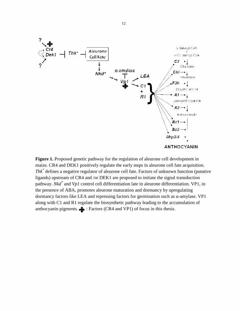
Figure 1. Proposed genetic pathway for the regulation of aleurone cell development in maize. CR4 and DEK1 positively regulate the early steps in aleurone cell fate acquisition. Thk* defines a negative regulator of aleurone cell fate. Factors of unknown function (putative ligands) upstream of CR4 and /or DEK1 are proposed to initiate the signal transduction pathway. Nkd* and Vp1 control cell differentiation late in aleurone differentiation. VP1, in the presence of ABA, promotes aleurone maturation and dormancy by upregulating dormancy factors like LEA and repressing factors for germination such as α-amylase. VP1 along with C1 and R1 regulate the biosynthetic pathway leading to the accumulation of anthocyanin pigments. : Factors (CR4 and VP1) of focus in this thesis.

13
REFERENCES Arenas-Huertero, F., Arroyo, A., Zhou, L., Sheen, J. and Leon, P. (2000) Analysis of
Arabidopsis glucose insensitive mutants, gin5 and gin6, reveals a central role of the
plant hormone ABA in the regulation of plant vegetative development by sugar.
Genes Dev, 14, 2085-2096.
Becraft, P.W. (2001) Cell fate specification in the cereal endosperm. Semin Cell Dev Biol,
12, 387-394.
Becraft, P.W. (2002) Receptor kinase signaling in plant development. Annu Rev Cell Dev
Biol, 18, 163-192.
Becraft, P.W. and Asuncion-Crabb, Y. (2000) Positional cues specify and maintain
aleurone cell fate in maize endosperm development. Development, 127, 4039-4048.
Becraft, P.W., Stinard, P.S. and McCarty, D.R. (1996) CRINKLY4: A TNFR-like
receptor kinase involved in maize epidermal differentiation. Science, 273, 1406-1409.
Bethke, P.C., Libourel, I.G., Aoyama, N., Chung, Y.Y., Still, D.W. and Jones, R.L.
(2007) The Arabidopsis aleurone layer responds to nitric oxide, gibberellin, and
abscisic acid and is sufficient and necessary for seed dormancy. Plant Physiol, 143,
1173-1188.
Busk, P.K., Jensen, A.B. and Pages, M. (1997) Regulatory elements in vivo in the promoter
of the abscisic acid responsive gene rab17 from maize. Plant J, 11, 1285-1295.
Busk, P.K. and Pages, M. (1998) Regulation of abscisic acid-induced transcription. Plant
Mol Biol, 37, 425-435.

14
Cao, X., Costa, L.M., Biderre-Petit, C., Kbhaya, B., Dey, N., Perez, P., McCarty, D.R.,
Gutierrez-Marcos, J.F. and Becraft, P.W. (2007) Abscisic acid and stress signals
induce Viviparous1 expression in seed and vegetative tissues of maize. Plant Physiol,
143, 720-731.
Cao, X., Li, K., Suh, S.G., Guo, T. and Becraft, P.W. (2005) Molecular analysis of the
CRINKLY4 gene family in Arabidopsis thaliana. Planta, 220, 645-657.
Cone, K.C., Burr, F.A. and Burr, B. (1986) Molecular analysis of the maize anthocyanin
regulatory locus C1. Proc Natl Acad Sci U S A, 83, 9631-9635.
Cordain, L. (1999) Cereal grains: humanity's double-edged sword. World Rev Nutr Diet, 84,
19-73.
Dumas, C. and Mogensen, H.L. (1993) Gametes and Fertilization: Maize as a Model
System for Experimental Embryogenesis in Flowering Plants. Plant Cell, 5, 1337-
1348.
Dure, L., 3rd (1993) A repeating 11-mer amino acid motif and plant desiccation. Plant J, 3,
363-369.
Finch-Savage, W.E. and Leubner-Metzger, G. (2006) Seed dormancy and the control of
germination. New Phytol, 171, 501-523.
Finkelstein, R.R., Gampala, S.S. and Rock, C.D. (2002) Abscisic acid signaling in seeds
and seedlings. Plant Cell, 14 Suppl, S15-45.
Gavazzi, G., Dolfini, S., Allegra, D., Castiglioni, P., Todesco, G. and Hoxha, M. (1997)
Dap (Defective aleurone pigmentation) mutations affect maize aleurone development.
Mol Gen Genet, 256, 223-230.

15
Gifford, M.L., Dean, S. and Ingram, G.C. (2003) The Arabidopsis ACR4 gene plays a role
in cell layer organisation during ovule integument and sepal margin development.
Development, 130, 4249-4258.
Gifford, M.L., Robertson, F.C., Soares, D.C. and Ingram, G.C. (2005) ARABIDOPSIS
CRINKLY4 function, internalization, and turnover are dependent on the extracellular
crinkly repeat domain. Plant Cell, 17, 1154-1166.
Hobo, T., Kowyama, Y. and Hattori, T. (1999) A bZIP factor, TRAB1, interacts with VP1
and mediates abscisic acid-induced transcription. Proc Natl Acad Sci U S A, 96,
15348-15353.
Hoecker, U., Vasil, I.K. and McCarty, D.R. (1995) Integrated control of seed maturation
and germination programs by activator and repressor functions of Viviparous-1 of
maize. Genes Dev, 9, 2459-2469.
Hoecker, U., Vasil, I.K. and McCarty, D.R. (1999) Signaling from the embryo conditions
Vp1-mediated repression of alpha-amylase genes in the aleurone of developing maize
seeds. Plant J, 19, 371-377.
Jin, P., Guo, T. and Becraft, P.W. (2000) The maize CR4 receptor-like kinase mediates a
growth factor-like differentiation response. Genesis, 27, 104-116.
Jones, R.L. (1969) The Effect of Ultracentrifugation on Fine Structure and alpha-Amylase
Production in Barley Aleurone Cells. Plant Physiol, 44, 1428-1438.
Lid, S.E., Gruis, D., Jung, R., Lorentzen, J.A., Ananiev, E., Chamberlin, M., Niu, X.,
Meeley, R., Nichols, S. and Olsen, O.A. (2002) The defective kernel 1 (dek1) gene
required for aleurone cell development in the endosperm of maize grains encodes a

16
membrane protein of the calpain gene superfamily. Proc Natl Acad Sci U S A, 99,
5460-5465.
Ludwig, S.R. and Wessler, S.R. (1990) Maize R gene family: tissue-specific helix-loop-
helix proteins. Cell, 62, 849-851.
McCarty, D.R., Carson, C.B., Stinard, P.S. and Robertson, D.S. (1989) Molecular
Analysis of viviparous-1: An Abscisic Acid-Insensitive Mutant of Maize. Plant Cell,
1, 523-532.
McCarty, D.R., Hattori, T., Carson, C.B., Vasil, V., Lazar, M. and Vasil, I.K. (1991) The
Viviparous-1 developmental gene of maize encodes a novel transcriptional activator.
Cell, 66, 895-905.
Niu, X., Helentjaris, T. and Bate, N.J. (2002) Maize ABI4 binds coupling element1 in
abscisic acid and sugar response genes. Plant Cell, 14, 2565-2575.
Offler, C.E., McCurdy, D.W., Patrick, J.W. and Talbot, M.J. (2003) Transfer cells: cells
specialized for a special purpose. Annu Rev Plant Biol, 54, 431-454.
Olsen, O.A. (2001) Endosperm development: Cellularization and Cell Fate Specification.
Annu Rev Plant Physiol Plant Mol Biol, 52, 233-267.
Pilu, R., Panzeri, D., Gavazzi, G., Rasmussen, S.K., Consonni, G. and Nielsen, E. (2003)
Phenotypic, genetic and molecular characterization of a maize low phytic acid mutant
(lpa241). Theor Appl Genet, 107, 980-987.
Rook, F., Corke, F., Card, R., Munz, G., Smith, C. and Bevan, M.W. (2001) Impaired
sucrose-induction mutants reveal the modulation of sugar-induced starch biosynthetic
gene expression by abscisic acid signalling. Plant J, 26, 421-433.

17
Shen, Q. and Ho, T.H. (1995) Functional dissection of an abscisic acid (ABA)-inducible
gene reveals two independent ABA-responsive complexes each containing a G-box
and a novel cis-acting element. Plant Cell, 7, 295-307.
Shen, Q., Zhang, P. and Ho, T.H. (1996) Modular nature of abscisic acid (ABA) response
complexes: composite promoter units that are necessary and sufficient for ABA
induction of gene expression in barley. Plant Cell, 8, 1107-1119.
Shen, Q.J., Casaretto, J.A., Zhang, P. and Ho, T.H. (2004) Functional definition of ABA-
response complexes: the promoter units necessary and sufficient for ABA induction
of gene expression in barley (Hordeum vulgare L.). Plant Mol Biol, 54, 111-124.
Singh, K.B. (1998) Transcriptional regulation in plants: the importance of combinatorial
control. Plant Physiol, 118, 1111-1120.
Suzuki, M., Kao, C.Y. and McCarty, D.R. (1997) The conserved B3 domain of
VIVIPAROUS1 has a cooperative DNA binding activity. Plant Cell, 9, 799-807.
Talbot, M.J., Offler, C.E. and McCurdy, D.W. (2002) Transfer cell wall architecture: a
contribution towards understanding localized wall deposition. Protoplasma, 219, 197-
209.
Tanaka, H., Watanabe, M., Watanabe, D., Tanaka, T., Machida, C. and Machida, Y.
(2002) ACR4, a putative receptor kinase gene of Arabidopsis thaliana, that is
expressed in the outer cell layers of embryos and plants, is involved in proper
embryogenesis. Plant Cell Physiol, 43, 419-428.
Thompson, R.D., Hueros, G., Becker, H. and Maitz, M. (2001) Development and
functions of seed transfer cells. Plant Sci, 160, 775-783.

18
Watanabe, M., Tanaka, H., Watanabe, D., Machida, C. and Machida, Y. (2004) The
ACR4 receptor-like kinase is required for surface formation of epidermis-related
tissues in Arabidopsis thaliana. Plant J, 39, 298-308.
Weber, H., Borisjuk, L. and Wobus, U. (2005) Molecular physiology of legume seed
development. Annu Rev Plant Biol, 56, 253-279.
Yamaguchi-Shinozaki, K. and Shinozaki, K. (2005) Organization of cis-acting regulatory
elements in osmotic- and cold-stress-responsive promoters. Trends Plant Sci, 10, 88-
94.
19
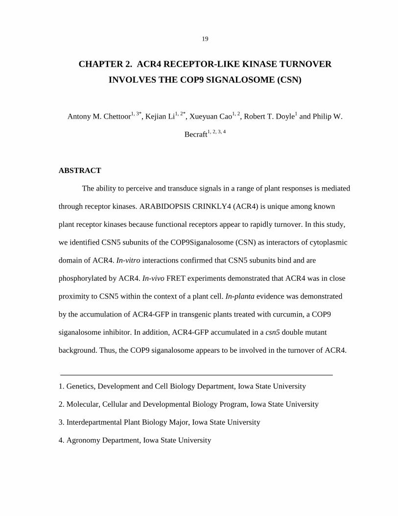
CHAPTER 2. ACR4 RECEPTOR-LIKE KINASE TURNOVER
INVOLVES THE COP9 SIGNALOSOME (CSN)
Antony M. Chettoor1, 3*, Kejian Li1, 2*, Xueyuan Cao1, 2, Robert T. Doyle1 and Philip W.
Becraft1, 2, 3, 4
ABSTRACT
The ability to perceive and transduce signals in a range of plant responses is mediated
through receptor kinases. ARABIDOPSIS CRINKLY4 (ACR4) is unique among known
plant receptor kinases because functional receptors appear to rapidly turnover. In this study,
we identified CSN5 subunits of the COP9Siganalosome (CSN) as interactors of cytoplasmic
domain of ACR4. In-vitro interactions confirmed that CSN5 subunits bind and are
phosphorylated by ACR4. In-vivo FRET experiments demonstrated that ACR4 was in close
proximity to CSN5 within the context of a plant cell. In-planta evidence was demonstrated
by the accumulation of ACR4-GFP in transgenic plants treated with curcumin, a COP9
siganalosome inhibitor. In addition, ACR4-GFP accumulated in a csn5 double mutant
background. Thus, the COP9 siganalosome appears to be involved in the turnover of ACR4.
1. Genetics, Development and Cell Biology Department, Iowa State University
2. Molecular, Cellular and Developmental Biology Program, Iowa State University
3. Interdepartmental Plant Biology Major, Iowa State University
4. Agronomy Department, Iowa State University

20
INTRODUCTION
Cells communicate with each other and with the environment by signaling through
cell surface receptors. One class of cell surface receptors called receptor kinases is defined
structurally by the presence of an extracellular domain that often communicates to the
outside by binding a ligand, a membrane spanning domain and a cytoplasmic kinase domain.
Plant receptor-like kinases (RLKs) are involved in hormonal response pathways, cell
differentiation, plant growth and development, self-incompatibility, and pathogen recognition
(Afzal et al. 2008, Becraft 2002, Hematy and Hofte 2008, Morris and Walker 2003,
Tichtinsky et al. 2003)
The maize (Zea mays) CRINKLY4 (CR4) gene encodes a receptor-like kinase that
controls a variety of cell differentiation responses (Becraft et al. 2001, Becraft et al. 1996, Jin
et al. 2000). Mutants in the maize cr4 gene disrupt aleurone cell fate specification causing
peripheral endosperm cells to be replaced by starch cells. The mutant plants also appear short
with leaves crinkled and fused (Becraft et al. 1996). CR4 encodes a 901-amino acid RLK
with an extracellular domain containing seven copies of a 39-amino acid repeat termed
“crinkly repeats” that are predicted to fold into a β-propeller structure and a domain that
resembles the extracellular domain of mammalian tumor necrosis factor receptor (TNFR).
The intracellular domain consists of a serine/threonine kinase domain, a 40-amino acid
juxtamembrane region and a 116-amino acid carboxyl-terminal domain of unknown function
(Becraft et al. 1996). In the Arabidopsis genome, out of the 417 or more predicted RLKs,
five are predicted to encode CR4-related genes. One of the five, designated as ACR4
contains all the features of the maize CR4 and is believed to be the Arabidopsis ortholog
(Cao et al. 2005, Gifford et al. 2003, Tanaka et al. 2002, Watanabe et al. 2004). ACR4

21
mutants unlike CR4, do not exhibit any gross defects in plant morphology, but show
abnormalities in seed morphology and seed set. ACR4 is required during normal
development of ovule, integument and sepal margins (Gifford et al. 2003). Subtle changes in
cell shape and cuticle formation indicate that ACR4 is required for normal leaf differentiation
(Watanabe et al. 2004). In roots, ACR4 regulates both apical root and lateral root initiation
by controlling the number and plane of formative divisions during organogenesis (De Smet et
al. 2008).
Stereotypical receptor kinase mediated signaling in animals involves ligand binding,
receptor oligomerization, phosphorylation of the cytoplasmic domain and recruitment of
interactors that bind phosphorylated residues of the receptor (Schlessinger and Ullrich 1992,
Ullrich et al. 1984). The epidermal growth factor (EGF) and its cognate receptor epidermal
growth factor receptor (EGFR) is the best characterized ligand/receptor signaling pathway.
Upon ligand binding, the EGF/EGFR complex is endocytosed and then the receptor either
recycled or targeted for degradation h (Jiang and Sorkin 2003, Maxfield and McGraw 2004,
Sorkin and Goh 2008, van der Knaap et al. 1999, Wang and Moran 1996, Wiley et al. 1991).
In contrast, knowledge about receptor signaling in plants is limited. Of the ligand-
receptor pairs known in plants, BRASSINOSTEROID INSENSITIVE 1(BRI1) which binds
brassinosteroid (BR) is the best characterized (Gendron and Wang 2007, Kinoshita et al.
2005). Geldner et al (2007) found functional BRI1/BAK1 (BRI1- associated kinase 1)
complexes in endosomes whereas nonfunctional BRI1/BKI1 (BRI1 kinase inhibitor1)
complexes were restricted to the plasmamembrane. BRI1 trafficking is constitutive and the
endosomal pool is functional in signal transduction (Geldner et al. 2007).

22
Turnover of receptors and other membrane proteins depend on two processes, namely
endocytosis and/or ubiquitin- mediated protein degradation. Monoubiquitination serves as a
signal for endocytosis, whereas polyubiquitination has been associated mainly with targeting
substrates to the proteosome (Haglund et al. 2003). Ubiquitination of proteins is catalyzed by
the action of an E1 ubiquitin activating enzyme, E2 ubiquitin conjugating enzymes and E3
ubiquitin ligase. E3 ubiquitin ligase can function as a single peptide (Mdm2 and XIAP) or as
a multiple component complex (Skp1-Cullin-F-box protein [SCF]) (Bosu and Kipreos 2008).
In multiple component E3 ligases, the Cullin subunit is the scaffold which links a substrate-
binding adaptor with an E2-binding RING. The adaptor protein links the substrate
recognition protein to the cullin. Covalent attachment of the ubiquitin-like protein NEDD8 to
the cullin subunit stimulates E3 ligase ubiquitination activity (Bosu and Kipreos 2008,
Hotton and Callis 2008).
The COP9 signalosome (CSN) is a conserved protein complex that functions in the
ubiquitin-proteosome pathway. The CSN was originally identified by mutants that exhibited
constitutive photomorphogenesis and pigmented seed-coats, but has been shown to be
present in many organisms and perform diverse functions (Wei and Deng 1992). The best
characterized biochemical activity that can be ascribed directly to CSN is the isopeptidase
activity that removes the NEDD8 attachment of the culllin subunit of the cullin-E3 ligases
(Cope et al. 2002). The CSN complex is made up of eight subunits designated CSN1- CSN8.
The CSN5 subunit is unique in that it harbors the catalytic center for deneddylation activity
and can also stably exist independently of the CSN holocomplex in-vivo (Wei and Deng
2003). CSN5 is encoded by a single gene in most organisms, except in Arabidopsis it is
encoded by two highly homologous, partially redundant genes CSN5A and CSN5B

23
(Gusmaroli et al. 2004). The complete loss of CSN5B does not result in any obvious
developmental defect, whereas the loss of CSN5A triggers a number of pleiotrophic
developmental defects. Double null homozygous mutants invariably die at the seedling stage,
virtually identical to the null alleles of the cop/det/fus mutants, and do not express detectable
CSN5 proteins (Gusmaroli et al. 2007).
ACR4 localizes to the plasmamembrane and endosomal vesicles similar to BRI1
RLK (Geldner et al. 2007, Gifford et al. 2005, Gusmaroli et al. 2004). Using lines expressing
ACR4-GFP fusions under native promoters, Gifford and colleagues observed florescence in
the plasmamembrane and endosomal vesicles. Thus, it is possible that ACR4 receptor
turnover is regulated by ubiquitin- mediated protein degradation. The aim of this study was
to indentify intracellular components capable of interacting with the cytoplasmic domain of
ACR4. Among the interacting proteins identified was CSN5 subunit. We show that ACR4
associates with CSN5 in plant cells and that the CSN is involved in the turnover of ACR4.
RESULTS
Identification of proteins that interact with the cytoplasmic domain of ACR4
To identify other components of the ACR4 signal transduction system, we performed
a yeast two-hybrid screen to identify putative interactors with the ACR4 cytoplasmic domain.
The region encoding the ACR4 cytoplasmic domain was cloned into the bait vector pGBD-
C1 (James et al. 1996) to produce an in-frame fusion with the GAL4 DNA-binding domain
(pGBD-CR4). This construct was used to screen an Arabidopsis 3 day-old seedling cDNA
library in the yeast YRG2 strain. From screening approximately one million yeast
transformants, ninety four colonies grew on the SD/-Trp/-Leu/-His selective medium

24
supplemented with 5mM 3-AT and turned blue in the colony-lift filter assay for lacZ
expression. The relative strength of these interactions was measured using the ONPG (o-
nitrophenyl β-D-galactopyranoside) liquid culture assay for β-galactosidase activity (data not
shown). The fourteen clones that showed the strongest interactions were chosen for further
study. Sequence analysis revealed that these represented six independent clones. One gene
was not expressed in the same tissues as ACR4 and was not pursued further. None of these
prey clones showed positive interactions in combination with the empty pGBD-C1 vector,
indicating the interactions required the ACR4 cytoplasmic domain (data not shown).
Two of the clones indentified as CSN5A (COP9SIGNALOSOME 5A subunit;
At1g22920) and CSN5B (COP9SIGNALOSOME 5B subunit; At1g71230) were studied
further because of the known role of the COP9 SIGNALOSOME (CSN) in protein turnover.
CSN5A and CSN5B encode two closely related proteins that function as subunits of the CSN
(Dohmann et al. 2005, Gusmaroli et al. 2004, Jin et al. 2000, Kwok et al. 1998). CSN5
proteins appear to be ubiquitously expressed in various Arabidopsis organs, including
seedlings, siliques, flowers, leaves, stems and roots, with higher expression in floral tissues
and lower expression in siliques and leaves (Kwok et al. 1998). ACR4 is expressed in various
tissues with the strongest expression in shoot apical meristems (SAM) and flower buds. The
overlapping expression patterns allow the possibility that CSN5 proteins interact with ACR4
in the plant. The more extensive expression pattern of CSN5 is consistent with the CSN5
proteins functioning in additional pathways, such as photomorphogenesis.

25
The interactions between ACR4 and CSN5B do not require ACR4 kinase activity
To verify the interactions between ACR4 and these identified proteins, an in vitro
pull-down assay was carried out using E.coli expressed proteins. The cytoplasmic domain of
ACR4 was cloned into a pET-32a (+) expression vector to create a fusion protein (ACR4K)
tagged with the thioredoxin/6X His/S-tag (THS). The THS alone was also expressed in E.
coli and used as a negative control in the pull-down assay. The same regions of the
interacting proteins as those identified in the yeast two-hybrid screen were cloned into
pGEX-5X1 (Amersham Pharmacia Biotech) to make GST fusion proteins, and a c-myc tag
was added to the carboxyl end of each protein. ACR4K was incubated with each putative
interacting protein, pulled down using TALON resin (Clontech) to bind the 6X His tag and
the presence of the interacting proteins detected using anti-myc antibody. As shown in Figure
1, none of the fusion proteins interact with the control THS tag, while CSN5B binds with
ACR4K. However, CSN5A did not show a detectable level of binding under these
conditions.
Ligand binding typically induces phosphorylation of the cytoplasmic domain of
receptor kinases, and interactions with downstream factors are often phosphorylation
dependent. In this way, signal transduction components are recruited to an activated receptor
complex. To test whether the interactions between the cytoplasmic domain of ACR4 and the
interacting proteins require ACR4 kinase activity, an in vitro binding assay was carried out
using a kinase-dead ACR4. The essential Lys540 of ACR4 kinase domain was replaced by
Ala to create an inactive kinase (ACR4KM). ACR4KM does not autophosphorylate,
indicating that the substitution abolished kinase activity (Cao et al. 2005). As shown in
Figure 1, CSN5B was pulled down with the kinase-dead ACR4 at about the same level as

26
with the active ACR4. Thus, the interactions between ACR4 and CSN5B not require ACR4
kinase activity. Like with the kinase active ACR4, CSN5A did not show binding with the
kinase-dead ACR4.
Phosphorylation of the interacting proteins by ACR4
A common mechanism in the regulation of signal transduction systems is for receptor
kinases to regulate downstream components through phosphorylation. Previous work has
shown that ACR4 is an active serine/threonine kinase (Cao et al. 2005, Gifford et al. 2003).
To test whether ACR4 can phosphorylate the interacting proteins, we conducted a
phosphorylation assay. Because the sizes of these interacting proteins were similar to the two
strong ACR4K autophosphorylation bands, we covalently bound ACR4K to NHS-activated
sepharose beads. The interacting proteins were mixed with the ACR4K-coupled sepharose in
kinase buffer containing [γ-32] P-ATP. After incubation, the sepharose was pelleted by
centrifugation and the supernatants applied to an SDS-PAGE gel shown in Figure 2A and
2B. The pellets were resuspended in SDS loading buffer, boiled, and the supernatants
containing the released interacting proteins loaded onto another SDS-PAGE gel (Figure 2C,
D). ACR4K did not appear in either fraction because it remained covalently bound to the
sepharose.
The inactive kinase ACR4KM, which can be phosphorylated by the active kinase
ACR4K (Cao et al. 2005), was used as a substrate in the same kinase assay as a positive
control. As shown in Figure 2C, CSN5A and CSN5B show a single clear phosphorylated
band by ACR4K. None of the interacting proteins showed phosphorylation by ACR4KM
(data not shown), indicating that phosphorylation of the interacting proteins was catalyzed by

27
ACR4K. Thus, CSN5A and CSN5B can be phosphorylated in vitro by ACR4K indicating
that they are potential substrates of ACR4.
Binding to ACR4 is independent of the phosphorylation state of the interactors
Phospho-regulation of downstream factors is critical in many receptor kinase signal
transduction systems. Phosphorylation might regulate activity, turnover, cellular
translocation or further protein associations. As such, it is conceivable that phosphorylation
of the interacting proteins could promote dissociation with ACR4. To test this, we compared
the supernatants and pellet fractions from the phosphorylation assay. ACR4-bound proteins
appeared in the pellet fraction, while unbound proteins appeared in the supernatants. As
shown in Figure 2C, the phosphorylated CSN5B remained bound to ACR4, indicating it had
not dissociated after phosphorylation. In contrast, part of the phospyorylated CSN5A
appeared in the supernatant, suggesting that phosphorylation of CSN5A might cause some
disruption of the interaction, allowing part of the phosphorylated proteins to dissociate from
ACR4K, or that CSN5A binding to ACR4 is weak regardless of the phosphorylation status.
The carboxyl-terminal domain of ACR4 interacts with both CSN5A and CSN5B
proteins
The cytoplasmic domain of ACR4 consists of three domains: the juxtamembrane
region, the kinase domain, and a 116 amino acid carboxyl-terminal domain of unknown
function. The complete cytoplasmic domain was used as bait in the two-hybrid screen. To
test which domains interact with CSN5A and CSN5B, we separately cloned the kinase
domain (BD-KD) and the carboxyl-terminal domain (BD-CT) into the yeast two-hybrid bait
vector pGBD-C1 (James et al. 1996). The results are shown in Table 1. CSN5A showed

28
interactions with both BD-KD and BD-CT, whereas CSN5B interacted only with BD-CT but
not with BD-KD. The interactions were confirmed by lacZ reporter gene expression in a
colony-lift filter assay (Table 1). Thus, the kinase domain or the carboxyl-terminal domains
of the ACR4 are sufficient for the interactions with CSN5A, whereas the carboxyl-terminal
domain of ACR4, but not the kinase domain, is necessary and sufficient, for the interactions
with CSN5B.
It was surprising that CSN5A interacted with both the kinase and carboxyl-terminal
domains. We searched for motifs present in both the kinase and carboxyl-terminal domains
that could be responsible for these interactions. As shown in Figure 3, a potential motif was
found in both domains. The motif consists of SS(X)9G(X)6DE(X)2K(X)3A(X)5EE(X)3A
(where X stands for any amino acid) where the relative spacing of SS, G, DE, K, A and EE
are conserved. There is also an SEN before SS, and an SA between SS and G with variable
spacing. This opens up the intriguing possibility that this motif could be responsible for the
interactions with the proteins that recognized both the kinase and carboxyl-terminal domains
of ACR4.
Förster resonance energy transfer (FRET) indicates ACR4 interacts with CSN5A and
CSN5B in vivo
FRET donor fluorescence recovery following acceptor photobleaching was used to
test the interactions between CSN5A, CSN5B and ACR4 within the context of plant cells.
The donor fusion protein ACR4-CFP was co-expressed with each acceptor fusion CSN5A-
YFP and CSN5B-YFP in onion epidermal cells. FRET was quantified as donor dequenching
after photobleaching of the acceptor (Miyawaki and Tsein, 2000). For each donor and

29
acceptor fusion pair, FRET occurrence is shown by the difference in donor signal intensity
between acceptor pre-bleach and post-bleach. The basis for this is that energy that had been
transferred from the donor to the acceptor before acceptor photobleaching will be emitted as
increased donor fluorescence following acceptor bleaching As controls, ACR4-CFP was
expressed alone, co-expressed with YFP, and YFP was expressed alone in onion epidermal
cells. As seen in Figure 4C and G, FRET did occur for ACR4-CFP/CSN5A-YFP and ACR4-
CFP/CSN5B-YFP fusion pairs as shown by the difference in CFP signal intensity between
acceptor prebleach and post bleach. FRET was not observed in the case of ACR4-CFP/YFP
pairs that served as negative controls (Figure 4K). Onion epidermal cells transiently
transformed with the ACR4-CFP or YFP alone did not result in any observable FRET
(Figure 4O and S). Thus, FRET confirms that ACR4 is in close proximity to both CSN5A
and CSN5B within a plant cell.
Curcumin treatment, a CSN inhibitor prevents ACR4 turnover
Sequence analyses indicate that CSN5A and CSN5B share 86 and 88% identity at the
nucleotide (cDNA) and amino acid levels, respectively. The fact that yeast two hybrid screen
identified both CSN5A and CNS5B subunits of the CSN complex to interact with ACR4
suggests a role for the CSN-ubiquitin proteosome system. Since ACR4 protein has b
(Gifford et al., 2005), we hypothesized CSN might be involved in ACR4 turnover. This was
tested pharmacologically using the CSN inhibitor curcumin. Curcumin (diferuloylmethane) is
a natural polyphenolic compound extracted from the spice turmeric (Curcuma longa) that has
been reported to have antioxidant, anti-inflammatory, and antiproliferative properties (Dutta
et al. 2005, Quiles et al. 2002). It has also been well documented in mammalian systems that

30
curcumin is an inhibitor of the COP9 signalosome through the inhibition of its associated
kinases, such as CKII and PKD (Chaudhary and Hruska 2003, Uhle et al. 2003). To test
whether CSN was involved in ACR4 turnover, we treated ACR4-GFP seedlings with 10µM
curcumin and monitored GFP accumulation in roots using confocal microscopy. Mock
treatments of ACR4-GFP and wild type plants, or curcumin treated wild type plants, were
used as controls. Confocal image stacks across the whole root were collected from all
samples and signal intensities quantified. The results are shown in Figure 5. Curcumin
treatment of ACR4-GFP plants is significantly different from ACR4-GFP plants treated with
DMSO alone. The increase in fluorescence intensity of the ACR4-GFP plants treated with
curcumin was significant compared to wild type curcumin treatment (p<0.001) (Figure 5E).
Wild type controls showed no difference in the presence or absence of curcumin. Thus, the
results suggest that inhibition of the COP9 signalosome by curcumin inhibited ACR4 protein
turnover.
ACR4 accumulates in csn5a-1 csn5b-1 double mutants
A known biochemical role attributed to CSN is a metalloprotease activity responsible
for the deneddylation of cullins, core components of the multiple component RING types of
ubiquitin E3 ligases (Lyapina et al. 2001, Zhou et al. 2001). To test for a role of the COP9
signalosome in ACR4 turnover, csn5a-1/+; csn5b-1/ csn5b-1 plants were crossed to ACR4-
GFP plants and the F2 progeny seedlings were screened for phenotypes similar to
cop/det/fus. Figure 6 shows that the ACR4-GFP csn5a-1 csn5b-1 plants produced more GFP
fluorescence than ACR4-GFP controls. Only background fluorescence was detected in
csn5a-1 csn5b double mutant seedlings and wild type seedlings. Thus, ACR4-GFP

31
accumulated to higher levels in the absence of CSN5 function, supporting the role of the
CSN5 subunits of the COP9 signalosome in the turnover of ACR4.
DISCUSSION
ACR4 interactions with CSN5
ACR4, a developmental RLK, is structurally different from the predominantly LRR
type of RLKs characterized in plants. Identification of ligands and other components of the
ACR4 signal transduction pathway is critical to our understanding of receptor signaling in
plants. We utilized the yeast-two hybrid approach to identify interactors with the ACR4
cytoplasmic domain and recovered CSN5A and CSN5B. The fact that both isoforms of
CSN5 were picked up in the screen suggests that these proteins are possible bonafide
interactors of ACR4.
ACR4 shows the highest level of expression in the shoot apical meristem and organ
primordia (Cao et al. 2004, Gifford et al. 2003, Tanaka et al. 2002). The CSN5 genes are
ubiquitously expressed in various tissues with high expression in floral and root tissues. The
expression profile of CSN5 coincides with regions of highest ACR4 expression, consistent
with the possibility that these proteins could interact with ACR4 in the plant.
The in vitro interaction of CSN5B was confirmed by pull-down (Figures 1, 2). The
interaction did not require ACR4 kinase activity as the interaction occurred even with a
kinase inactive mutant form of ACR4. In contrast, the pull-downs with CSN5A were only
successful in the phosphorylation assay. The basis for the discrepancy between the two pull
down experiments is not clear but one possibility is that phosphorylation is required to
stabilize the interaction, since ATP was present in the kinase assay and in yeast cells but was

32
not supplied for the pull down reactions. In mammalian EGFR signaling, in keratinocytes
upon UV irradiation EGFR activation results in tyrosine phosphorylation of β-catenin. β-
catenin, a cadherin protein complex subunit relocates from the membrane to the nucleus and
regulates gene expression (Zhang et al. 2001). Experiments were conducted to test the ability
of the ACR4 kinase to phosphorylate CSN5A and CSN5B, which could result in the
recruitment of additional factors to a signaling complex or potentially could cause
dissociation as a prelude to subcellular translocation or other response of the interacting
protein. Our results showed that ACR4K could phosphorylate CSN5A and CSN5B in vitro.
In the case of CSN5B, the phosphorylated protein remained bound to ACR4, while
phosphorylated CSN5A was observed in both the bound and unbound fractions. At this point
it is unclear whether phosphorylation destabilized the interaction or whether the dissociated
CSN5A reflects an intrinsically weak interaction. The basis of the observed differences
between the two CSN5 isoforms and their functional implications need further testing.
FRET experiments confirmed the interactions of both CSN5 isoforms with ACR4 in
onion epidermal cells. The absences of FRET in the ACR4CFP/YFP and ACR4CFP along
with the robustness of the acceptor photobleaching method confirm the authenticity of the
ACR4/CSN5 interactions. CSN5 has been found to be present as monomers, small
complexes and large holocomplexes with the other seven CSN subunits of the CSN complex
in-vivo (Wei and Deng 2003). The locations of these varied forms of CSN5 complexes have
not been characterized to date. The CSN5 subunits are cytoplasmic and nuclear localized,
while ACR4 is localized to plasmamembrane and endosomes (Gifford et al. 2005, Kwok et
al. 1998, Watanabe et al. 2004). This suggests the interactions occur either at the

33
plasmamembrane and/or in the cytoplasm. Taken together, it is possible that ACR4 is
processed prior to interaction with CSN5 subunits. This possibility remains to be tested.
ACR4 receptor turnover
The lack of detectable functional ACR4 is attributed to its rapid turnover. It has been
proposed that ACR4 in Arabidopsis root cells was present in two distinct subcellular
compartments: protein export bodies or secretory vesicles and a population of internalized
vesicles or endosomes (Gifford et al. 2005, Wang and Moran 1996). Furthermore,
internalization and turnover for ACR4 appear to be linked, operating through endosomes. In
maize, CRINKLY4 (CR4), DEFECTIVE KERNEL1 (DEK1) and SUPERNUMERARY
ALEURONE LAYERS1 (SAL1), involved in aluerone cell fate specification, colocalize in
endosomal compartments (Tian et al. 2007). Taken together these studies suggest that the
endosomal internalization and turnover of ACR4 are possibly linked.
ACR4 shares some similarities with the LRR type BRI1 receptor kinase. Both
colocalize with the endocytic tracer FM4-64 and show sensitivity to brefeldin A (BFA)
treatment. Thus, ACR4 might represent another example of a receptor exhibiting constitutive
endocytosis. However, the ligand for ACR4 has not been identified and therefore it is
possible that continuous ligand stimulation results in endosomal localization. The ACR4
receptors differ from BRI1 receptor in protein stability. It is possible that the interaction of
ACR4 with CSN5 of the CSN complex might result in rapid turnover. The most well
characterized function of CSN is the regulation of protein degradation which involves the
removal of NEDD8 from cullin-based E3 ubiquitin ligase. Two experiments support the role
CSN in ACR4 receptor turnover. (1) Arabidopsis plants expressing ACR4-GFP fusion
protein treated with curcumin, a specific inhibitor of the CSN complex, displayed increased

34
amounts of ACR4-GFP accumulation. (2) In-planta accumulation of ACR4-GFP fusion
protein in csn5a csn5b double mutant background. It is likely that the CSN functions in
ubiquitin-mediated protein degradation of the receptor, resulting in the rapid turnover of the
receptor and the corresponding inability to detect functional ACR4 protein. In the case of
BRI1 the absence of rapid turnover and/or more receptor recycling accounts for the detection
of full length BRI1.
There are several examples in animal systems where the CSN5 subunit functions in
receptor turnover or membrane protein regulation. , CSN5 coimmunoprecipitates with the
nuclear estrogen receptor α (ERα) and overexpression of CSN5 results in ligand-induced
ERα degradation (Callige et al. 2005). CSN5 can also bind to the misfolded cystic fibrosis
transmembrane conductance regulator (CFTR), a membrane protein, and target it to the
degradative pathway (Tanguy et al. 2008). In COS7 cells, CSN5 interacts with, and inhibits
the activity of, the membrane localized cardiac L-type Ca2+ channel (Kameda et al. 2006)
In summary, the CSN5 subunit of the COP9 signalosome complex appears critical to
the rapid turnover of the ACR4 receptor. The fact that the CSN5 interactions identified here
have not been reported in other RLK studies suggests the signal transduction pathway for
ACR4 could be novel. Future work will examine the function of CSN-mediated ACR4
turnover in signaling and decipher in greater detail the ACR4 signal transduction pathway.
METHODS
Plant Materials and Growth Conditions
The wild-type Arabidopsis thaliana plants used in this study are Columbia-0 ecotype.
The CSN5 T-DNA insertion lines SALK_063436 (csn5a-1) and SALK_007134 (csn5b-1)
35
were identified in the SIGNAL database and obtained from ABRC (Alonso et al. 2003).
csn5a-1, csn5b-1 single mutants and csn5a-1 csn5b-1 double mutants have been
characterized as null CSN5 mutants (Gusmaroli et al. 2007). The Arabidopsis seeds were
surface sterilized and the plants grown on solid 1× MS medium supplemented with 1%
sucrose or on a mixture of 1:1 soil: vermiculite under long-day conditions (16 h light/8 h
dark) in a controlled environment chamber at 22°C.
Construction of ACR4-GFP Transgenics
A 1.38 kb promoter region upstream of the ACR4 start codon was amplified from
pBFScX by PCR (Cao et al., 2005). The PCR fragment was cloned into SmaI/BamHI site of
the vector pEZS-NL (a gift from D. Ehrhardt, Dept of Plant Biology, Stanford) resulting in
pBFSPEZ. The ACR4 ORF was amplified and cloned into pBFSPEZ as an Eco47III/BamHI
fragment resulting in pACR4EZ. The ACR4-eGFP chimera was cut out as a SacI/SalI
fragment and cloned into binary vector pCAMBIA2300 to obtain a translational fusion of the
ACR4 and eGFP coding sequence driven by the ACR4 native promoter. The construct was
introduced into Agrobacterium tumefacians strain C58C1. Plants were transformed by the
floral-dip method modified from the vacuum-infiltration method (Bechtold and Pelletier
1998). Seeds from the infiltrated plants were harvested, surface-sterilized and sown on MS
plates supplemented with 30 µg ml-1 hygromycin. Hygromycin-resistant seedling (T1) were
transplanted into soil, grown to maturity and seeds harvested.
Yeast two-hybrid library screening
pGBD-C1 (James et al. 1996), a GAL4 DNA-binding domain vector, was used to
construct the bait plasmid. The DNA fragment encoding the cytoplasmic domain of ACR4

36
was inserted to generate an in-frame fusion between the DNA-binding domain of GAL4 and
the cytoplasmic domain of ACR4, including amino acids 458-895 (pGBD-ACR4). An
Arabidopsis λACT cDNA library from 3 day-old seedlings was obtained from the
Arabidopsis Biological Resource Center (stock number CD4-22) of the Ohio State University
and used as the prey in the yeast two hybrid screening. The library was converted to plasmid
and the plasmid DNA prepared using a Qiagen plasmid Maxi kit.
The yeast host strain YRG2 (Stratagene) was transformed with plasmid DNA from
the library. Approximately 1 million transformants were plated on synthetic dextrose (SD)
minimal medium with 5mM 3-amino-1,2,4-triazole (3-AT) and lacking Trp, Leu and His.
Those that grew were subjected to the colony-lift β-galactosidase filter assay for lacZ
expression. The positive colonies were then streaked on the SD/-Leu plates with 0.5g/l 5-
FAA (5-fluoroanthranilic acid) after subculture three times in liquid SD/-Leu media to rescue
the library (prey) plasmid. The library plasmids were isolated and to verify positive
interactions, the library plasmid was retransformed back into yeast YRG2 strain in
combination with either pGBD-ACR4, or empty pGBD-C1 vector, and plated on SD/-His/-
Trp/-Leu containing 5mM 3-AT.
DNA sequence analysis
The cDNA inserts of the positive colonies were sequenced at the DNA Facility at
Iowa State University. DNA sequences were used in database searches by BLAST (Altschul
et al. 1997). Cellular localizations of encoded proteins were predicted using PSORT (Nakai
and Horton 1999) (http://psort.nibb.ac.jp) and SignalP (Nielsen et al. 1997)
(www.cbs.dtu.dk/services/SignalP).

37
Protein expression
The cytoplasmic domain of ACR4 (Arg458-Phe895) was cloned in the pET-32a (+)
vector (Novagen) with Trx/6X His/S-Tag (THS) and expressed in E. coli. Expression of this
protein, ACR4K, was induced in 1mM IPTG (Sigma) and proteins were purified from the
cell lysate using TALON purification kit (Clontech). As a control, the THS tag was
expressed and purified from the empty pET-32a (+) vector using the same procedure as
above. A kinase-dead ACR4 (ACR4KM) was also expressed in E. coli by substituting the
essential Lys540 with Ala. The mutation was introduced by PCR where the AAG to GCG
codon change was incorporated by primer mismatch.
Pull-down assays
Approximately 5µg of purified ACR4K, ACR4KM, or THS (the fusion peptide
consisting of thioredoxin-, S- and 6X His tags encoded by the empty PET vector) protein was
combined with approximately 5µg purified CSN5A and CSN5B protein in 200µl binding
buffer [50mM Tris pH 8.8, 300mM NaCl, 50mM sodium phosphate, and DOC (1.5% for
interactions with CSN5B and phosphatase, 7% for interactions with CSN5A, CIRK1 and
CIRK2)]. 10µl TALON resin (Clontech) was added with continued rocking at room
temperature for 45 additional minutes. Beads were then pelleted by brief centrifugation and
washed 6 times with 200µl binding buffer. 20µl 1x SDS loading buffer was added to each
reaction and the resuspended beads were boiled for 5 minutes. After brief centrifugation, the
supernatants were loaded on a 10% SDS-PAGE gel, electrophoresed and transferred to
nitrocellulose membrane. The membrane was immunoblotted with the anti-myc primary
antibody (Invitrogen, 1:5,000 dilution) and then with the goat anti-mouse IgG secondary
38
antibody conjugated with horseradish peroxidase (Pierce, 1:10,000). Finally, the membrane
was incubated with the ECL western blotting detection reagents and analysis system
(Amersham Bioscience), and exposed to Kodak X-Omat film.
Kinase assays
ACR4K purified protein was coupled to NHS-activated Sepharose 4 Fast Flow
(Pharmacia Biotech) according to the manufacturers recommendations. Approximately 3µg
NHS-activated sepharose-bound ACR4K was combined with approximately 5µg CSN5A,
CSN5B, or ACR4KM in 60µl kinase buffer (50mM Tris-HCl [pH 7.5], 5mM MgCl2, and
5mM MnCl2). 1µl [γ-32] P-ATP was added to each and the reaction was incubated at room
temperature for 45 minutes. After brief centrifugation, the supernatants were loaded on a
10% SDS-PAGE gel. 20µl of 1x SDS loading buffer was added to the remaining beads and
the resuspended beads were boiled for 5 minutes. After brief centrifugation, the supernatants
from the pelleted beads were also loaded on a 10% SDS-PAGE gel. The gels were then
stained with Coomassie blue, dried, and subjected to phosphoimaging (Molecular Dynamics,
Model 400A) to detect the phosphoproteins.
FRET detection by confocal microscopy
ACR4 ORF was cloned into pECFP (BD Biosciences) as a BamHI and KpnI
fragment amplified from genomic DNA obtained using the following primers: ACR4FRETF
and ACR4FRETB ( Table 2), resulting in pACFP. The ACR4CFP fragment from pACFP
was introduced into the vector pJ4GFP-XB (Igarashi et al. 2001) as BamHI/NotI fragment so
that the chimeric gene replaced the original GFP, resulting in pJACFP. CSN5A and CSN5B

39
were amplified using cDNA’s U87212 and U13455 obtained from ABRC with the following
primers: CSN5A (CSN5AYFPF and CSN5AYFPB; Table 2) and CSN5B (CSN5BYFPF and
CSN5BYFPB; Table 2). The CSN5A ORF was cloned into pEYFP (BD Biosciences) as a
SalI and BamHI fragment, the CSN5B ORF was cloned into pEYFP as a SalI and AgeI
fragment resulting in pC5AYFP and pC5BYFP respectively. CSN5AYFP and CSN5BYFP
fragments from pC5AYFP and pC5BYFP were introduced into the vector pJ4GFP-XB as
Sal1 and Not1 fragments resulting in pJC5AYFP and pJC5BYFP and respectively.
Cells from onion epidermis were placed on agar plates containing half-strength
Murashige and Skoog (MS) salts and were transformed biolistically. The epidermal tissues
were incubated for 20hrs at room temperature prior to analysis. Onion epidermal cells were
examined for the localization of the CFP/YFP fused proteins using a fluorescence
microscope (Prairie Technologies) under a filter set (all filters from Omega Optical) for CFP:
Excitation filter of 440/21nm, a dichoric beam splitter of 455nm and emission filter of
480/30nm. YFP was viewed under a filter set with an excitation filter of 500/25nm, a
dichroic beam splitter of 525nm, and an emission filter of 545/35nm. Images were captured
using a cooled CCD camera (Hamamatsu Photonics) and controlled by Prairie View (Priarie
Technologies). CFP and YFP derived fluorescence were recorded, averaged and used for
corrections. To quantify the efficiency of FRET in absolute terms, we selectively
photobleached the acceptor fluorophore and measured the dequenching of the donor
fluorescence (Miyawaki and Tsien 2000). FRET efficiency E is given by: E = 1 − (Fda/Fd),
where Fda is the donor emission when both donor and acceptor are present, and Fd is donor
emission in the absence of acceptor, (i.e. after photobleach). Three successive digital images
(with appropriate background subtractions) were acquired: Pre-bleached donor, Pre-bleached

40
acceptor and post-bleached donor. We routinely photobleached YFP by illuminating the cells
for 3–5 min with no neutral density filters. This treatment had no effect on CFP in the
absence of YFP, as shown in Figure 6.
Curcumin experiments
ACR4-GFP young seedlings were incubated in 10µM curcumin (C 1386, Sigma-
Aldrich) at 22°C. The working curcumin solution was made by diluting a 10 mM DMSO
stock 1:1000 in water. Control roots were incubated for the same period of time in a 1:1000
dilution of DMSO in water and observed under a laser confocal microscope (Prairie
Technologies). Confocal image stacks were collected and then processed using Metamorph
6.0 (Molecular Devices). Statistical two-way analysis of variance was performed using the
SAS system (SAS Institute). Tukey’s studentized range (honestly significant difference) test
was used to determine significant differences among genotypes and treatments. An α level of
0.05 was used as the criterion of statistical significance.
ACKNOWLEDGEMENTS
The authors thank the Becraft lab for discussions and critical reading of this
manuscript. We are grateful to the Arabidopsis Biological Resource Center, Ohio State
University, for supplying the cDNA library used in the yeast two-hybrid screen. This work
was funded by grant DE-FG02-98ER20303 from the U.S. Department of Energy and IBN-
9604426 from the U.S. National Science Foundation.

41
REFERENCES
Afzal, A.J., Wood, A.J. and Lightfoot, D.A. (2008) Plant receptor-like serine threonine
kinases: roles in signaling and plant defense. Mol Plant Microbe Interact, 21, 507-
517.
Alonso, J.M., Stepanova, A.N., Leisse, T.J., Kim, C.J., Chen, H., Shinn, P., Stevenson,
D.K., Zimmerman, J., Barajas, P., Cheuk, R., Gadrinab, C., Heller, C., Jeske, A.,
Koesema, E., Meyers, C.C., Parker, H., Prednis, L., Ansari, Y., Choy, N., Deen,
H., Geralt, M., Hazari, N., Hom, E., Karnes, M., Mulholland, C., Ndubaku, R.,
Schmidt, I., Guzman, P., Aguilar-Henonin, L., Schmid, M., Weigel, D., Carter,
D.E., Marchand, T., Risseeuw, E., Brogden, D., Zeko, A., Crosby, W.L., Berry,
C.C. and Ecker, J.R. (2003) Genome-wide insertional mutagenesis of Arabidopsis
thaliana. Science, 301, 653-657.
Altschul, S.F., Madden, T.L., Schaffer, A.A., Zhang, J., Zhang, Z., Miller, W. and
Lipman, D.J. (1997) Gapped BLAST and PSI-BLAST: a new generation of protein
database search programs. Nucleic Acids Res, 25, 3389-3402.
Bechtold, N. and Pelletier, G. (1998) In planta Agrobacterium-mediated transformation of
adult Arabidopsis thaliana plants by vacuum infiltration. Methods Mol Biol, 82, 259-
266.
Becraft, P.W. (2002) Receptor kinase signaling in plant development. Annu Rev Cell Dev
Biol, 18, 163-192.
Becraft, P.W., Kang, S.H. and Suh, S.G. (2001) The maize CRINKLY4 receptor kinase
controls a cell-autonomous differentiation response. Plant Physiol, 127, 486-496.
42
Becraft, P.W., Stinard, P.S. and McCarty, D.R. (1996) CRINKLY4: A TNFR-like
receptor kinase involved in maize epidermal differentiation. Science, 273, 1406-1409.
Bosu, D.R. and Kipreos, E.T. (2008) Cullin-RING ubiquitin ligases: global regulation and
activation cycles. Cell Div, 3, 7.
Callige, M., Kieffer, I. and Richard-Foy, H. (2005) CSN5/Jab1 is involved in ligand-
dependent degradation of estrogen receptor α by the proteasome. Mol Cell Biol, 25,
4349-4358.
Cao, X., Li, K., Suh, S.G., Guo, T. and Becraft, P.W. (2005) Molecular analysis of the
CRINKLY4 gene family in Arabidopsis thaliana. Planta, 220, 645-657.
Chaudhary, L.R. and Hruska, K.A. (2003) Inhibition of cell survival signal protein kinase
B/Akt by curcumin in human prostate cancer cells. J Cell Biochem, 89, 1-5.
Cope, G.A., Suh, G.S., Aravind, L., Schwarz, S.E., Zipursky, S.L., Koonin, E.V. and
Deshaies, R.J. (2002) Role of predicted metalloprotease motif of Jab1/Csn5 in
cleavage of Nedd8 from Cul1. Science, 298, 608-611.
De Smet, I., Vassileva, V., De Rybel, B., Levesque, M.P., Grunewald, W., Van Damme,
D., Van Noorden, G., Naudts, M., Van Isterdael, G., De Clercq, R., Wang, J.Y.,
Meuli, N., Vanneste, S., Friml, J., Hilson, P., Jurgens, G., Ingram, G.C., Inze, D.,
Benfey, P.N. and Beeckman, T. (2008) Receptor-like kinase ACR4 restricts
formative cell divisions in the Arabidopsis root. Science, 322, 594-597
Dohmann, E.M., Kuhnle, C. and Schwechheimer, C. (2005) Loss of the CONSTITUTIVE
PHOTOMORPHOGENIC9 signalosome subunit 5 is sufficient to cause the
cop/det/fus mutant phenotype in Arabidopsis. Plant Cell, 17, 1967-1978.

43
Dutta, S., Padhye, S., Priyadarsini, K.I. and Newton, C. (2005) Antioxidant and
antiproliferative activity of curcumin semicarbazone. Bioorg Med Chem Lett, 15,
2738-2744.
Geldner, N., Hyman, D.L., Wang, X., Schumacher, K. and Chory, J. (2007) Endosomal
signaling of plant steroid receptor kinase BRI1. Genes Dev, 21, 1598-1602.
Gendron, J.M. and Wang, Z.Y. (2007) Multiple mechanisms modulate brassinosteroid
signaling. Curr Opin Plant Biol, 10, 436-441.
Gifford, M.L., Dean, S. and Ingram, G.C. (2003) The Arabidopsis ACR4 gene plays a role
in cell layer organisation during ovule integument and sepal margin development.
Development, 130, 4249-4258.
Gifford, M.L., Robertson, F.C., Soares, D.C. and Ingram, G.C. (2005) ARABIDOPSIS
CRINKLY4 function, internalization, and turnover are dependent on the extracellular
crinkly repeat domain. Plant Cell, 17, 1154-1166.
Gusmaroli, G., Feng, S. and Deng, X.W. (2004) The Arabidopsis CSN5A and CSN5B
subunits are present in distinct COP9 signalosome complexes, and mutations in their
JAMM domains exhibit differential dominant negative effects on development. Plant
Cell, 16, 2984-3001.
Gusmaroli, G., Figueroa, P., Serino, G. and Deng, X.W. (2007) Role of the MPN subunits
in COP9 signalosome assembly and activity, and their regulatory interaction with
Arabidopsis Cullin3-based E3 ligases. Plant Cell, 19, 564-581.
Haglund, K., Di Fiore, P.P. and Dikic, I. (2003) Distinct monoubiquitin signals in receptor
endocytosis. Trends Biochem Sci, 28, 598-603.

44
Hematy, K. and Hofte, H. (2008) Novel receptor kinases involved in growth regulation.
Curr Opin Plant Biol, 11, 321-328.
Hotton, S.K. and Callis, J. (2008) Regulation of cullin RING ligases. Annu Rev Plant Biol,
59, 467-489.
Igarashi, D., Ishida, S., Fukazawa, J. and Takahashi, Y. (2001) 14-3-3 proteins regulate
intracellular localization of the bZIP transcriptional activator RSG. Plant Cell, 13,
2483-2497.
James, P., Halladay, J. and Craig, E.A. (1996) Genomic libraries and a host strain
designed for highly efficient two-hybrid selection in yeast. Genetics, 144, 1425-1436.
Jiang, X. and Sorkin, A. (2003) Epidermal growth factor receptor internalization through
clathrin-coated pits requires Cbl RING finger and proline-rich domains but not
receptor polyubiquitylation. Traffic, 4, 529-543.
Jin, P., Guo, T. and Becraft, P.W. (2000) The maize CR4 receptor-like kinase mediates a
growth factor-like differentiation response. Genesis, 27, 104-116.
Kameda, K., Fukao, M., Kobayashi, T., Tsutsuura, M., Nagashima, M., Yamada, Y.,
Yamashita, T. and Tohse, N. (2006) CSN5/Jab1 inhibits cardiac L-type Ca2+
channel activity through protein-protein interactions. Journal of Molecular and
Cellular Cardiology, 40, 562.
Kinoshita, T., Cano-Delgado, A., Seto, H., Hiranuma, S., Fujioka, S., Yoshida, S. and
Chory, J. (2005) Binding of brassinosteroids to the extracellular domain of plant
receptor kinase BRI1. Nature, 433, 167-171.
Kwok, S.F., Solano, R., Tsuge, T., Chamovitz, D.A., Ecker, J.R., Matsui, M. and Deng,
X.W. (1998) Arabidopsis homologs of a c-Jun coactivator are present both in
45
monomeric form and in the COP9 complex, and their abundance is differentially
affected by the pleiotropic cop/det/fus mutations. Plant Cell, 10, 1779-1790.
Lyapina, S., Cope, G., Shevchenko, A., Serino, G., Tsuge, T., Zhou, C., Wolf, D.A., Wei,
N. and Deshaies, R.J. (2001) Promotion of NEDD-CUL1 conjugate cleavage by
COP9 signalosome. Science, 292, 1382-1385.
Maxfield, F.R. and McGraw, T.E. (2004) Endocytic recycling. Nat Rev Mol Cell Biol, 5,
121-132.
Miyawaki, A. and Tsien, R.Y. (2000) Monitoring protein conformations and interactions by
fluorescence resonance energy transfer between mutants of green fluorescent protein.
Methods Enzymol, 327, 472-500.
Morris, E.R. and Walker, J.C. (2003) Receptor-like protein kinases: the keys to response.
Curr Opin Plant Biol, 6, 339-342.
Nakai, K. and Horton, P. (1999) PSORT: a program for detecting sorting signals in proteins
and predicting their subcellular localization. Trends Biochem Sci, 24, 34-36.
Nielsen, H., Engelbrecht, J., Brunak, S. and von Heijne, G. (1997) Identification of
prokaryotic and eukaryotic signal peptides and prediction of their cleavage sites.
Protein Eng, 10, 1-6.
Quiles, J.L., Mesa, M.D., Ramirez-Tortosa, C.L., Aguilera, C.M., Battino, M., Gil, A.
and Ramirez-Tortosa, M.C. (2002) Curcuma longa extract supplementation reduces
oxidative stress and attenuates aortic fatty streak development in rabbits. Arterioscler
Thromb Vasc Biol, 22, 1225-1231.
Schlessinger, J. and Ullrich, A. (1992) Growth factor signaling by receptor tyrosine
kinases. Neuron, 9, 383-391.

46
Sorkin, A. and Goh, L.K. (2008) Endocytosis and intracellular trafficking of ErbBs. Exp
Cell Res, 314, 3093-3106.
Tanaka, H., Watanabe, M., Watanabe, D., Tanaka, T., Machida, C. and Machida, Y.
(2002) ACR4, a putative receptor kinase gene of Arabidopsis thaliana, that is
expressed in the outer cell layers of embryos and plants, is involved in proper
embryogenesis. Plant Cell Physiol, 43, 419-428.
Tanguy, G., Drevillon, L., Arous, N., Hasnain, A., Hinzpeter, A., Fritsch, J., Goossens,
M. and Fanen, P. (2008) CSN5 binds to misfolded CFTR and promotes its
degradation. Biochim Biophys Acta, 1783, 1189-1199.
Tian, Q., Olsen, L., Sun, B., Lid, S.E., Brown, R.C., Lemmon, B.E., Fosnes, K., Gruis,
D.F., Opsahl-Sorteberg, H.G., Otegui, M.S. and Olsen, O.A. (2007) Subcellular
localization and functional domain studies of DEFECTIVE KERNEL1 in maize and
Arabidopsis suggest a model for aleurone cell fate specification involving
CRINKLY4 and SUPERNUMERARY ALEURONE LAYER1. Plant Cell, 19, 3127-
3145.
Tichtinsky, G., Vanoosthuyse, V., Cock, J.M. and Gaude, T. (2003) Making inroads into
plant receptor kinase signaling pathways. Trends Plant Sci, 8, 231-237.
Uhle, S., Medalia, O., Waldron, R., Dumdey, R., Henklein, P., Bech-Otschir, D., Huang,
X., Berse, M., Sperling, J., Schade, R. and Dubiel, W. (2003) Protein kinase CK2
and protein kinase D are associated with the COP9 signalosome. EMBO J, 22, 1302-
1312.
Ullrich, A., Coussens, L., Hayflick, J.S., Dull, T.J., Gray, A., Tam, A.W., Lee, J.,
Yarden, Y., Libermann, T.A., Schlessinger, J. and et al. (1984) Human epidermal

47
growth factor receptor cDNA sequence and aberrant expression of the amplified gene
in A431 epidermoid carcinoma cells. Nature, 309, 418-425.
van der Knaap, E., Song, W.Y., Ruan, D.L., Sauter, M., Ronald, P.C. and Kende, H.
(1999) Expression of a gibberellin-induced leucine-rich repeat receptor-like protein
kinase in deepwater rice and its interaction with kinase-associated protein
phosphatase. Plant Physiol, 120, 559-570.
Wang, Z. and Moran, M.F. (1996) Requirement for the adapter protein GRB2 in EGF
receptor endocytosis. Science, 272, 1935-1939.
Watanabe, M., Tanaka, H., Watanabe, D., Machida, C. and Machida, Y. (2004) The
ACR4 receptor-like kinase is required for surface formation of epidermis-related
tissues in Arabidopsis thaliana. Plant J, 39, 298-308.
Wei, N. and Deng, X.W. (1992) COP9: a new genetic locus involved in light-regulated
development and gene expression in arabidopsis. Plant Cell, 4, 1507-1518.
Wei, N. and Deng, X.W. (2003) The COP9 signalosome. Annu Rev Cell Dev Biol, 19, 261-
286.
Wiley, H.S., Herbst, J.J., Walsh, B.J., Lauffenburger, D.A., Rosenfeld, M.G. and Gill,
G.N. (1991) The role of tyrosine kinase activity in endocytosis, compartmentation,
and down-regulation of the epidermal growth factor receptor. J Biol Chem, 266,
11083-11094.
Zhang, Y., Dong, Z., Bode, A.M., Ma, W.Y. and Chen, N. (2001) Induction of EGFR-
dependent and EGFR-independent signaling pathways by ultraviolet A irradiation.
DNA Cell Biol, 20, 769-779.

48
Zhou, C., Seibert, V., Geyer, R., Rhee, E., Lyapina, S., Cope, G., Deshaies, R.J. and
Wolf, D.A. (2001) The fission yeast COP9/signalosome is involved in cullin
modification by ubiquitin-related Ned8p. BMC Biochem, 2, 7.
FIGURE LEGENDS
Figure 1. The cytoplasmic domain of ACR4 binds to the interacting proteins in a pull-
down assay and the interactions do not require ACR4 kinase activity.
Each interacting protein was mixed with the THS tag encoded by the empty pET-32a (+)
vector (THS), the kinase-active ACR4 cytoplasmic domain (ACR4K) or the kinase-inactive
ACR4 cytoplasmic domain (ACR4KM). THS, ACR4K or ACR4KM were pulled-down with
TALON affinity resin, and the myc-tagged interacting proteins were detected by immunoblot
with anti-myc antibody.
Figure 2. Phosphorylation of the interacting proteins by ACR4K.
ACR4K-coupled sepharose was mixed with each interacting protein or the kinase-inactive
ACR4KM in kinase buffer containing α-32P-ATP.
(A, B) After centrifugation, supernatants were run on SDS-PAGE.
(C, D) Pellets containing the sepharose coupled ACR4K were boiled in SDS loading buffer
and the bound proteins separated by SDS-PAGE.
(A, C) Autoradiographs.
(B, D) Coomassie blue stained gels.

49
Figure 3. A sequence motif found repeated in the kinase domain and carboxy terminal
domain of ACR4.
Figure 4. ACR4 interacts with CSN5A and CSN5B in FRET Assays
Onion epidermal cells were transiently transformed with the ACR4 donor (CFP fusion) and
interactor acceptors (YFP fusions). FRET was detected by the acceptor photobleaching
technique (Miyawaki and Tsein, 2000).
(A, B, C, D) ACR4CFP+CSN5AYFP FRET pairs.
(E, F, G, H) ACR4CFP+CSN5BYFP FRET pairs.
(I, J, K, L) ACR4CFP+YFP FRET pairs.
(M, N, O, P) ACR4CFP control.
(Q, R, S, T) YFP control
(A, E, I, M, Q) Pre-bleach donor images.
(B, F, J, N, R) Post-bleach donor.
(C, G, K, O, S) FRET intensity difference profiles.
(D, H, L, P, T) Pre-bleach acceptor. Scale bar 25µm.
Figure 5. ACR4 accumulates with Curcumin, a COP9 signalosome inhibitor
ACR4-GFP transgenic and wild type Columbia seedlings were treated with 10µM curcumin
or mock treated with DMSO and the sub-apical region of roots observed. Confocal image
stacks were collected.
(A) Representative mid plane image of control Columbia roots with DMSO
(B) Representative mid plane image of Columbia roots with curcumin

50
(C) Representative mid plane image of ACR4-GFP roots with DMSO
(D) Representative mid plane image of ACR4-GFP roots with curcumin
(E) Fluorescence signal intensities was quantified (Metamorph) from image stacks and tested
for statistical significance. Error bars indicate Standard error (n=10). **: Indicates significant
increase in fluorescence intensity for curcumin treated ACR4-GFP roots compared to ACR4-
GFP with DMSO and curcumin treated wild type Columbia. (p< 0.001). (Scale bar = 25µm
for A-D).
Figure 6. ACR4-GFP accumulates in csn5 mutants
An ACR4-GFP transgene was crossed into a csn5a-1; csn5b-1 double null mutant line and
segregating progeny were screened for cop/det/fus phenotype.
(A, B) ACR4-GFP; csn5a-1; csn5b-1 root.
(C, D) ACR4-GFP wild type root.
(E, F) csn5a-1; csn5b-1 root
(G, H) Col wild type root.
(A, C, E, G) Confocal fluorescence images.
(B, D, F, H) Bright field images. Scale bars are 25µm.

51
Table 1. CSN5A and CSN5B interactions with different cytoplasmic domains of ACR4
BD-KD BD-CTD
Sample -LTH SD β-Gal Activity -LTH SD β-Gal Activity
CSN5B - - + +
CSN5A + + + +
BD-KD: Yeast-two hybrid screen using kinase domain; BD-CTD: Yeast-two hybrid screen
using carboxyl terminal domain; -LTH SD: synthetic dextrose (SD) minimal medium with
5mM 3AT and lacking Leu, Trp, His; β-Gal Activity: colony-lift β-galactosidase filter assay
for lacZ expression.

52
Table 2. Primers used for PCR
Primer Sequence (5’-3’) Strand
CSN5B-5 GCAGGAATTCGAAGGTTCCTCGTCAG Forward
CSN5B-3 GACGGTCGACTAAGATCCTCCTCAGAAATGAGCTTTTGCTCCGATGTAATCATGGGCTCT Reverse
CSN5A-5 GCAGGAATTCAGCATTCTAACAGTAGACTCAC Forward
CSN5A-3 GACGGTCGACTAAGATCCTCCTCAGAAATGAGCTTTTGCTCATATGTAATCATAGGGTCTG Reverse
BD-KD-5 CTGAGGATCCAGATTGAGGAATTGTAGGT Forward
BD-KD-3 AGCCCTGCAGCATTGATGGTCTATCTTTC Reverse
BD-CT-5 GTCGGGATCCGATAAAGTGACAACAGCATTGGAA Forward
BD-CT-3 CGCACTGCAGGAAATTATGATGCAAGAAC Reverse
CSN5AYFPF GCTAGTCGACATGGAAGGTTCCTCGTCAGCCATC Forward
CSN5AYFPB ACTGGGATCCGATGGTAATCATGGGCTCTGGATC Reverse
CSN5BYFPF GCATCCCGGGGAATGGAGGGTTCGTCGTC Forward
CSN5BYFPR GCATCCCGGGATGTAATCATAGGGTCTGGATCCGA Reverse
ACR4FRETF ATGCGGATCCATGAGAATGTTCGAAACGAG Forward
ACR4FRETB AGCAGGTACCAAATTATGATGCAAGAACAAG Reverse

53
Figure 1

54
Figure 2
Kinase 1 LRNCRCSENDTR............SS...KDSAFTK COOH 1 GSKRSGSENTEFRGGSWITFPSVTSSQRRKSSA... kinase 22 DNGKIRPDLDELQKRRRARVFTYEELEKAADGFKEE COOH 34 SEGDVAEEEDEGRKQQEALRSLEEEIGPASPGQSLF Figure 3

55
Figure 4

56
Figure 5

57
Figure 6

58
CHAPTER 3. CE1LBP1, A NOVEL PROTEIN BINDS THE CE1L
ELEMENT OF THE MAIZE VP1 PROMOTER
Antony Chettoor1, 3 and Philip W. Becraft1, 2, 3
ABSTRACT
The plant hormone abscisic acid and the transcriptional activator VIVIPAROUS1
(VP1) regulate the transcription of overlapping gene sets during embryo development.
Significant progress has been made in elucidating the mechanisms by which VP1 activates
transcription, including via the ABA-responsive element (ABRE). However, regulation of
Vp1 is not well understood. Sequence analysis of the Vp1 promoter identified an ACGT
containing ABRE and an element resembling a CE1 element (CE1L). CE1LBP1, a novel
protein that binds the CE1L element of the Vp1 promoter, was isolated in a yeast one-hybrid
screen. CE1LBP1 homologs are present in all angiosperms examined, and appear to be a
single copy in maize. Using a zinc binding colorimetric assay, we demonstrate that
CE1LBP1 binds zinc. Electrophoretic mobility shift assays (EMSAs) show that recombinant
CE1LBP1 protein specifically binds to the CE1L element. Antibodies specific to CE1LBP1
1Department of Genetics, Development and Cell Biology, Iowa State University, Ames, IA 50011, USA; 2Department of Agronomy, Iowa State University, Ames, IA 50011, USA; 3Plant Biology Program, Iowa State University, Ames, IA 50011, USA.

59
identified CE1LBP1 as part of a DNA/protein complex in supershift EMSAs using nuclear
proteins extracted from maize embryos. Identification of the maize CE1LBP1 as a possible
trans-acting factor of the Vp1 CE1L element provides a new link in our understanding of
transcriptional regulation during seed maturation and dormancy.
INTRODUCTION
Seed development is a crucial process in the life cycle of higher plants, providing the
link between two distinct sporophytic generations. According to several plant models, this
process can be divided into two stages, morphogenesis and maturation (Gutierrez et al. 2007,
Weber et al. 2005, West and Harada 1993). The morphogenesis phase involves the
establishment of an embryo and endosperm by the process of double fertilization. The
maturation phase is marked by the arrest of embryo growth, the accumulation of storage
compounds, the onset of desiccation tolerance in embryo tissues, finally leading to seed
quiescence and usually dormancy. In maize and cereal grains, aleurone, the outermost layer
of the endosperm also undergoes the maturation process and remains viable in the dry seed.
Seed dormancy is an adaptive trait that promotes the survival of plants in hostile
environments (Bewley 1997). In important cereal crops like wheat, dormancy affects yield
quality by preventing pre-harvest sprouting (Gubler et al. 2005).
Seed dormancy and germination are known to be regulated by several hormones;
ethylene, gibberellin, nitrous oxide and brassinosteroids promote seed germination, while
abscisic acid seed (ABA) mediates seed dormancy (Finch-Savage and Leubner-Metzger
2006, Finkelstein et al. 2002, Koornneef et al. 2002, Nambara and Marion-Poll 2003). In

60
addition, ABA mediates plant responses to various environmental stresses, such as drought,
salt, and cold stress (Ingram and Bartels 1996). ABA induces the expression of many
maturation-associated genes, such as Em and the late embryogenesis abundant (Lea) genes
(Busk and Pages 1998, Marcotte et al. 1989). ABA deficient or insensitive mutants fail to
undergo quiescence and precociously germinate, accompanied by decreased expression of
maturation genes (Bewley 1997, Koornneef et al. 2002). Of the several ABA-insensitive
loci identified to date, three ABI3, ABI4 and ABI5 encode B3, AP2 and bZIP-type
transcription factors respectively (Finkelstein and Lynch 2000, Finkelstein et al. 1998,
Giraudat et al. 1992, Lopez-Molina and Chua 2000, Lopez-Molina 2000).
Abscisic acid response mutants have been useful to dissect ABA signal transduction
pathways (Kucera 2005). The viviparous1 (vp1) and ABA-insensitive (abi3) genes are
required for the ABA response associated with late embryogenesis of maize (McCarty, et al.
1989, McCarty et al. 1991) and Arabidopsis (Giraudat et al. 1992), respectively. Maize vp1
mutant kernels are ABA insensitive, undergo precocious germination on the ear, fail to
withstand desiccation, and lack anthocyanin pigmentation. (McCarty et al.1989; McCarty et
al. 1991). Functional complementation of abi3 mutants by Vp1, and sequence similarities
suggests that Vp1 and Abi3 are functionally orthologous genes (Giraudat et al. 1992, Suzuki
et al. 2001). VIVIPAROUS1 (VP1) is a B3 domain -containing transcription factor that is
central to the regulation of seed maturation in maize (McCarty et al. 1991). VP1/ABI3
homologues are present in most seed plant species (Bobb et al. 1995, Carrari et al. 2001,
Hattori et al. 1995, Hollung et al. 1997, Shiota et al. 1998). This indicates that the regulation
through VP1/ABI3 is well conserved and is a general theme in the plant kingdom.

61
Functional analysis of ABA-regulated gene promoters has identified the motifs
mediating ABA response (Busk et al. 1997, Hattori et al. 1995, Marcotte et al. 1989, Shen
et al. 1996, Straub et al. 1994). Most of these studies identified ABA-responsive elements
(ABRE) or G-box-like elements with an ACGT core sequence as important cis elements for
ABA- induced gene expression (Busk et al. 1997, Hobo et al. 1999a, Shen and Ho 1995,
Shen et al. 1996). The ABRE requires a second element to form a functional ABRC
complex (ABRC). The coupling element (CE) is one such element (Shen et al. 1996).
Promoters of the barley genes HVA22 and HVA1 contain two types of the coupling elements,
CE1 or CE3, that are necessary for activation by ABA (Shen and Ho 1995, Shen et al. 1996).
The ABREs and CEs together form ABA-responsive complexes (ABRC1 and ABRC3). The
CE1 (ABRC1) and CE3 (ABRC3) are different in sequence and position relative to the
ABRE, but both elements are rich in cytosines and guanines (Shen et al. 2004).
Biochemical approaches revealed that ABI3 binds to the RY/Sph motif, ABI4 to CE1-like
element and ABI5 to ACGT- containing ABRE element (Carles and Koornneef 2002, Kim et
al. 2002, Kim 2002, Monke et al. 2004, Monke and H.; Conrad 2004, Niu, Helentjaris and
Bate 2002). Furthermore, ABI3 and ABI5, or their rice orthologs, VP1 and TRAB1, have
been shown to physically interact (Hobo et al. 1999b, Nakamura 2001). ABI3 and ABI5
bind ABRE elements in response to ABA to regulate gene expression in the maturating seed.
Although considerable information is available on the functions of the Vp1 and ABI3
genes, less is understood about their regulation. Several genes like LEAFY COTYLEDON1
(LEC1), LEC2 and FUSCA3 that regulate the transcription of the Arabidopsis ABI3 gene are
known, but the molecular mechanisms by which this regulation occurs are unknown (Kagaya
et al. 2005, To et al. 2006). Maize Vp1 expression is induced by ABA and osmoticum during

62
the mid-maturation embryogenesis phase (Bobb et al., 1995; Hoecker et al., 1995, Cao et al.,
2007). A 958 bp (base pair) promoter region of maize Vp1 was sufficient for the reproduction
of the endogenous Vp1 expression pattern when fused to β-glucoronidase in transgenic
maize. The Vp1 promoter contains ABRE and CE1-like (CE1L) elements that bind to embryo
nuclear proteins (Cao et al. 2007), which represent potential transcripiton factors that
regulate seed maturation. In this study, we describe the identification of CE1LBP1, a novel
protein that binds the CE1L element of the Vp1 promoter. We demonstrate the in-vitro and
ex-planta interaction of CE1LBP1 with CE1L element through gel shift and super-shift
assays.
RESULTS
Isolation and analysis of CE1LBP1
To better understand the transcriptional regulation of the Vp1 and identify upstream
candidates in the seed maturation and dormancy process, we performed a yeast one-hybrid
screen to isolate cDNA clones that encode proteins, which bind to CE1L (Figure 2). The
double-reporter yeast strain containing three 28 bp repeats of CE1L upstream of the His3 or
lacZ reporters, failed to grow on selective medium. A cDNA library was constructed from 7
to 20 DAP(days after pollination) embryos using lambda HybriZapII (Stratagene) which
fused the cDNA fragments to the yeast GAL4 activation domain in the pGAD424 vector.
The reporter yeast strain was transformed with the cDNA library and plated on selective
medium. Approximately 3.7 x 10⁶ yeast transformants were screened and selected colonies
were subsequently re-grown and assayed for ß-galactosidase activity using an X-gal filter

63
assay. 16 clones grew on His selection and showed increased lacZ activity. The isolated
cDNA inserts were sequenced and analyzed by BLAST (Altschul and Lipman, 1990). One
clone, designated CE1LBPpar grew on His selection and was Lac Z positive (Figure. 3A). It
was a partial clone of the sequence GI:85540419 (designated as CE1LBP1 in this study)
which encodes a 279 amino acid protein (Figure 3B). CE1LBP1 maps to the chromosome
4L (bin 9) (MaizeGDB, http://www.maizegdb.org). The encoded protein is predicted to be
nuclear localized (Hoglund et al. 2006) and does not contain any known protein domains.
Phylogenetic relationships of the CE1LBP1related proteins
A BLAST search of NCBI databases identified CE1LBP1 homologs in Rice,
Sorghum and Arabidopsis (Altschul et.al., 1990). To address the relationships among the
CE1LBP1 related proteins, a multiple sequence alignment was conducted using ClustalW
(Figure 4A). Protein sequence alignment of the orthologs showed that Sorghum CE1LBP1
had the highest conservation to CE1LBP1 with 73% amino acid identity, followed by Rice
CE1LBP1( japonica) at 40% amino acid identity, Rice CE1LBP1( indica) at 22% amino acid
identity and Arabidopsis CE1LBP1 at 18% amino acid identity. The Phylogenetic
relationships of the CE1LBP1 proteins are shown in Figure 4B. Importantly, these analyses
indicate that CE1LBP1 appears to be a single copy gene in maize and the other species
analyzed.
CE1LBP1 binds to CEIL element in EMSA gel shift assays
CE1LBP1 was isolated from the yeast- one hybrid screen by its ability to bind the
CE1L element of the VP1 promoter. To test whether CE1LBP1 binds the CE1L element in
vitro, recombinant CE1LBP1 was used in an EMSA. The full length CE1LBP1 was

64
expressed as a GST fusion and purified from E. coli RosettaTM cells (Invitrogen). When the
28bp CE1L oligonucleotide was used as a probe, a major retarded band was observed using
CE1LBP1-GST protein, indicating that CE1LBP1 bound to CE1L element (Figure 5). The
unlabeled CEIL oligonucleotide competed away the retarded band, indicating that the
binding was specific (Figure 5). No retarded band was observed using GST alone (Figure 5).
This confirms that CE1LBP1 can bind the CE1L element of the Vp1 promoter in vitro.
Transiently expressed CE1LBP1-GFP is nuclear localized
To study the in vivo subcellular distribution of CE1LBP1, we expressed a
translational fusion between green fluorescent protein (GFP) and CE1LBP1 in onion
epidermal cells (Figure 6). As shown in Figure 6C, the CE1LBP1-GFP fusion protein was
detected exclusively in the nucleus, indicating that it is targeted to the nucleus. As expected,
GFP alone did not localize to a specific compartment and was detected in the cytoplasm and
nucleus (Figure 6A).
CE1LBP1 binds zinc
Sequence analysis of CE1LBP1 did not identify any known protein domains. The
primary amino acid sequence of CE1LBP1 contains a number of cysteines and histidines
suggestive of a zinc finger protein, but does not appear to be a canonical zinc finger protein.
CE1LBP1 could therefore be a noncanonical zinc finger protein or other type of zinc binding
protein. To determine whether CE1LBP1 binds zinc, acolorimetric zinc binding assay using
4-(2- pyridylazo) resorcinol (PAR) was performed on recombinant protein (Hunt et al., 1984;
Modrof et al., 2003). When an EDTA treatment followed the Zinc incubation of GST or
CE1LPB1-GST, only trace amounts of zinc ions were detected (Figure 7). However when

65
Zinc incubation followed the EDTA treatment, 3.3 ±0.7mol of Zn2+ per mol of CE1LBP1-
GST was bound (mean ± sd). In the case of CE1LBP1par-GST 0.55±0.06 mol of Zn2+ per
mol of CE1LBP1-GST was bound (average± sd). For GST, only trace amounts of bound zinc
was detected.
Nuclear CE1LBP1 binds CE1L
A previous study performed using 20 DAP embryo nuclear protein extracts identified
a specific binding activity to the 28-bp CE1L oligonucleotide probe (Cao et al. 2007). To test
the presence of CE1LBP1 in the nuclear proteins forming the specific complex on the CEIL
region of Vp1 promoter, a supershift EMSA was performed. The two independent mice
polyclonal anti-CELBP1 antisera (pN and pR) were raised and both recognized recombinant
CELBP1 and embryo nuclear proteins specifically whereas the control serum did not (data
not shown). Nuclear protein extracts from 20 DAP embryos were incubated with the labeled
CE1L oligonucleotide in the presence of anti-CE1LBP1 antibodies ( pN and pR). As
previously reported, nuclear extracts interacted specifically with the probe. Decreased
mobility of the complex was observed in the presence of the anti-CE1LBP1 antibodies, but
not in the presence of control antibodies from pre-immune serum (Figure 8). This incidates
that the antibodies recognized CE1LBP1 in the nuclear proteins binding to the CE1L
oligonucleotide. To confirm the specificity of the interaction of the embryo nuclear proteins
to the 28-bp CEIL oligonucleotide, EMSA was repeated and the major retarded band
outcompeted by unlabeled CEIL competitor. These results indicate that CE1LBP1 present in
nuclear protein extracts from maturation stage embryos specifically bound the CE1L probe.

66
DISCUSSION
VP1 along with ABA and other factors promote seed maturation and dormancy
(McCarty 1995). Post transcriptional and posttranslational regulation has been reported for
VP1/ABI3 (Bassel et al. 2006, Brady 2003, Zhang et al. 2005). However, transcriptional
regulation of VP1/ABI3 has not been extensively studied. Vp1 promoter analysis defined a
potential ABRC, consisting of an ACGT core ABRE and a CE1-like motif (Cao et al. 2007).
EMSA confirmed that the ABRE and CE1L specifically bound embryo nuclear proteins. In
this study, we identify and characterize CE1LBP1, a novel maize DNA-binding factor that
binds to the CE1L cis- element of the Vp1 promoter. CE1LBP1 was indentified using the
CE1L element of Vp1 in a one-hybrid screen of a 7-20 DAP embryo library (Figure 2).
It is unclear whether the CE1L element of the Vp1 promoter in fact functions as a
coupling element in ABA regulated transcription. The only transfactor known to bind CE1
elements is ZmABI4 (Niu et al. 2002). ZmABI4, like the Arabidopsis homolog ABI4 is an
APETALA2 (AP2)-like transcription factor that regulates ABA and sugar response genes
(Finkelstein et al. 1998). The CE1 element found in sugar-regulated genes, which binds
ZmABI4, and the archetypical CE1of Hva22 share a six nucleotide motif (CCACCG),
however CACCG appear to be critical for ZmABI4 binding. The CE1L element of Vp1
shares only the CCACC motif with both CE1of Hva22 and the CE1 elements that bind
ZmABI4. The distance between the ABRE and CE1L of Vp1 is 50bp. Distances in multiples
of 10bp between CE1and ABREs appear to be critical for ABRC function, but 50 bp is
greater than has been previously reported. Furthermore, mutagenesis of core CE1L residues
previously reported to be essential for binding or function of CE1 elements did not abrogate

67
binding (Cao et al., 2007). Taken together, it is possible that the ABRC present in the Vp1
promoter represents a different transacting factor binding site.
The CE1L element of Vp1 and CEILBP1 have many of the hallmarks of a trans factor
binding site and a trans factor respectively. Gel shift assays using recombinant CE1LBP1
showed specific binding to CE1L.A CE1LBP1-GFP fusion was targeted to the nucleus
(Figure 6), consistent with its function as a DNA-binding protein. Supershift experiments
using anti-CE1LBP1 polyclonal antibodies demonstrated that CE1LBP1 was present in
embryo nuclear protein extracts and could bind a CE1L oligonucleotide. Overall, these
experiments indicate that CE1LBP1 is a nuclear protein that is capable of binding to the
CE1L element of Vp1 in a sequence-specific manner.
CE1LBP1 is devoid of any known functional protein domains. A striking
characteristic of the amino acid sequence is the presence of a number of cysteines and
histidines (Figure 3). Although CE1LBP1 is not predicted to be a canonical zinc finger
protein; the sequence suggested the possibility that CE1LBP1 could bind Zn++, which was
demonstrated with a. colorimetric Zn++ binding assay (Figure.7).
A Zn++ cofactor is associated with several known transcription factors. p53 is a
widely distributed tumor suppressor protein that acts as a sequence-specific transcriptional
regulator in response to DNA damage (Prives and Hall 1999, Vousden and Lu 2002). Proper
conformation of the p53 DNA binding domain is necessary for DNA binding and
transactivation of target genes. Zn++ is coordinated to C176 and H179 of the L2 loop and to
C238 and C242 of the L3 loops of the protein. Zn++ coordination appears necessary for
transcriptional activation and removal of Zn++ reduces the DNA-binding specificity (Hainaut
and Milner 1993, Meplan et al. 2000, Pavletich et al. 1993, Verhaegh et al. 1998) .

68
Interestingly, when p53 and CE1LBP1 sequences are compared, the four residues critical to
Zn++ coordination are completely conserved (data not shown). This suggests the possibility
that CE1LBP1 could similarly be a Zn++ binding transcription factor.
The developmental expression pattern of CE1LBP1 is as yet unknown. However,
based on transcriptome metadata, the putative Arabidopsis ortholog (At 3g63040; Figure 4)
is specifically expressed in the maturation phase of seed development. Similarly, the rice
ortholog is annotated as embryo-specific. In addition, the expression profile of At3g63040
overlaps with At2g30470 (ABI3) (Nakabayashi et al. 2005), except that ABI3 expression is
also seen in vegetative tissues (Winter et al. 2007). Thus it is likely that maize CE1LBP1
expression is also seed specific, consistent with a potential function controlling Vp1
expression during seed maturation. It is also relevant that ZmABI4, which binds CE1, is also
specifically expressed during the maturation phase of maize seed development (Niu et al.
2002).
ABI3/VP1 expression has been examined in various species to establish whether it is
correlated with seed dormancy. A study utilizing various wild oat (Avena fatua) germplasm
with varying degrees of embryo dormancy revealed an increased abundance of AfABI3
transcripts in cultivars with increased embryo dormancy (Jones et al. 1997). However, a
similar study using Arabidopsis mutants with various degrees of dormancy did not reveal a
correlation between the abundance of AtABI3 transcripts and dormancy (Baumbusch et al.
2004). The fact that dormancy is a complex trait whose control involves strong interactions
with many genes and the environment, makes utilizing Quantitative Trait Locus (QTL)
analysis a tool of choice. QTL analysis in wheat (Osa et al. 2003) and sorghum (Carrari et
al.2001) did not identify Vp1 as a locus regulating this trait. A major component of the

69
observed genetic variation for pre-harvest sprouting in wheat appears to be the level of seed
dormancy. Therefore identifying QTLs associated with pre-harvest sprouting could provide a
better understanding of seed maturation and dormancy. Two QTLs for seed dormancy in
wheat, designated as QPhs.ocs-3A.1 and QPhs.ocs-3A.2, were identified on the short and
long arms on chromosome 3A, respectively. More of the variation for high dormancy
associated with chromosome 3A is ascribable to QPhs.ocs-1 on the short arm than to the
QTL on the long arm (Osa et al. 2003). The closest wheat ortholog of CE1LBP1, represented
as an EST BJ293947, maps to the short arm of chromosome 3A in addition to 3B and 3D
chromosomes. Furthermore, data from the wheat 61k Affimetrix Microarray suggests
elevated expression around 22 DAP, coinciding with seed maturation and seed dormancy.
Further studies will address the enticing possibility that CE1LBP1 might play an important
role in the transcriptional regulation of VP1 and other genes associated with seed maturation
or dormancy in maize and other species.
MATERIALS AND METHODS
Plant materials
The embryos used in electrophoretic mobility shift assay (EMSA) assays and
supershifts were collected from B73 or W22 grown in the field, frozen in liquid N2 and
stored in a -50°C freezer.
Yeast one-hybrid screen The 28 bp oligonucleotide containing the CE1L cis-element from the Vp1 promoter
was synthesized (Cao et al.2007), attaching EcoRI and XbaI adaptors at its 5´ and 3´ ends.

70
Three tandem repeats of the oligonucleotide were ligated together and cloned into the
corresponding restriction enzyme sites of the pHISi and pUC19 plasmids. With EcoRI and
SalI restriction sites, the insert containing three tandem copies from pUC19 was cloned into
the pLacZi plasmid. The described pLacZi and pHISi plasmid constructs were sequentially
transformed into the yeast strain YM4271 to obtain the double-reporter yeast strain. A cDNA
library was constructed in the lambda HybriZapII vector (Stratagene) from 7 to 20 DAP
embryos, creating fusions of the cDNA fragments to the yeast GAL4 activation domain in
the pGAD424 vector. The cDNAs were transformed into the dual-reporter yeast strain and
the transformants selected on –HIS selection media. Transformants that grow in the absence
of histidine and in the presence of 40 mM 3-AT were selected and tested for activation of the
lacZ reporter. The colony- lift filter assay was carried out according to standard protocol
(Breeden and Nasmyth 1985).
Sequence and phylogenetic analysis
Positive yeast 1-hybrid clones were sequenced and used in BLAST searches (Altschul
et al. 1990) and the predicted subcellular localizations of the encoded proteins determined by
MultiLoc/TargetLoc (Hoglund et al. 2006). The sequences of the CE1LBP1 homologs were
obtained from NCBI (Maize: GI195607718, Oryza sativa (japonica): GI 115486027, Oryza
sativa (indica): GI 85699976, Sorghum: Sb05g022640 and Arabidopsis: At3g63040).
MegAlign from DNASTAR (Madison, WI) was used to calculate percent identity between
CE1LBP1, sorghum CE1LBP1, rice CE1LBP1’s (japonica and indica) and
ArabidopsisCE1LBP1. Sequence alignments were carried out using ClustalW2 and MEGA4

71
was used to develop phylogenetic relationship using the neighbor- joining method (Kumar et
al. 2008).
Constructions of CE1LBP1-GFP and CE1LBP1-GST fusions
For construction of the35S:: CE1LBP1-GFP reporter plasmid, the CE1LBP1 cDNA
was amplified by polymerase chain reaction (PCR) with the 5’ primer CE1LPB1GFPF 5’-
ACGTTCTAGATGAGGCGGTGCCGC and the 3’ primer CE1LPB1GFPB 5’-
TGACGGATCCCACCACCTCAAGGATGG. The CE1LBP1 cDNA PCR fragment was
digested with Xba1 and BamH1 and cloned into the pJ4GFP-XB (Igarashi et al. 2001).
For construction of the full length CE1LBP1-GST plasmid, the CE1LBP1 cDNA was
amplified by polymerase chain reaction (PCR) with the 5’ primer CE1LPB1GSTF 5’-
TAGCGAATTCATGAGGCGGTGCCGC and the 3’ primer CE1LPB1GSTB 5’-
GTCACTCGAGTCACACCACCTCAAGGATGG. The CE1LBP1 cDNA PCR fragment
was digested with EcoR1 and Xho1 and cloned into the pGEX-5X-1. Both constructs were
sequenced and confirmed to be free of mutations.
Transient expression and visualization of CE1LBP1-GFP in onion epidermal cells
Onion (Allium cepa) transformation was performed essentially as described
previously (Varagona et al. 1992). Inner epidermal peels of white onions were placed inside-
up on half-strength MS. Onion peels were bombarded using the PDS-1000/He system
(DuPont) at 1100 p.s.i. with DNA-coated 1µm gold particles (Bio-Rad). The epidermal
tissues were incubated for 20hrs at room temperature prior to analysis. Onion epidermal
tissues were mounted in water and viewed under a Nikon inverted confocal microscope. GFP

72
was visualized using an excitation wavelength of 488 nm and a bandpass 510- to 525-nm
emission filter. Under the conditions used, only small amounts of chlorophyll
autofluorescence were visualized in untransformed plant tissue.
Expression and purification of recombinant CE1LBP1-GST protein
The recombinant full length CE1LBP1-GST plasmids were transformed into E. coli
RosettaTM cells (Invitrogen). 1mM IPTG was used to induce the expression of these GST-
fusion proteins and the proteins were purified from the cell lysate using Glutathione
SepharoseTM 4B according to the manufacturers protocol (Amersham Bioscience). The
fusion proteins were eluted from the Glutathione Sepharose beads and stored at -80 °C.
Generation of polyclonal anti-CE1LBP1 antibody
Mouse anti-CE1LBP1 sera was raised against recombinant CE1LBP1 at the
Hybridoma Facilty of Iowa State University. Briefly, BALB/c female adult mice were
injected subcutaneously with 50 µg of purified recombinant protein. Fifteen days later, the
mice were boosted with the same inoculum and 1 month after the initial inoculation, a third
boost was administered by intraperitoneally injecting ∼50 µg of CE1LBP1 protein/mouse.
Fifteen days after this final boost, mice were exanimated by bleeding out, and serum was
stored at −80°C.Serum prior to the initial injection of recombinant CE1LBP1 served as the
control serum.
Electrophoretic mobility shift assay (EMSA) and EMSA Supershift assays
The nuclear protein extracts were prepared from 20 DAP embryos by first using a glycerol
based method for isolation of nuclei (Dorweiler et al. 2000) followed by isolation of nuclear

73
proteins in protein isolation buffer (10mM Tis-HCl, 1 mM DTT, 0.4 M NaCl (pH 7.5)
(Escobar et al. 2001). DNA probes were generated by filling in the 3´ end overhangs of
complementary oligonucleotides with 32P-dCTP (PerkinElmer) and the Klenow fragment of
DNA polymerase I (Promega). Labeled probes were subsequently purified using a QIAquick
nucleotide removal kit (Qiagen). Binding reactions (25 µL) containing 0.4 picomole of
radiolabeled probe, 50 ng of sonicated pBlueScript II SK (Stratagene) and recombinant
proteins were incubated in 1 X binding buffer (10 mM Tris-HCl pH 7.4, 50 mM NaCl, 2 mM
EDTA, 2.5 mM DTT, 1.25 µg BSA, 0.05 % v/v NP-40, 10 % glycerol) at room temperature
for 30 min..Electrophoresis was conducted on a non-denaturing 4.5 % polyacrylamide gel in
0.5 x TBE (45 mM Tris-borate, 0.5 mM EDTA pH 8.0) buffer with constant 70 V for 4 hr at
4 °C. The gels were vacuum dried for 2 hr at 70 °C, and autoradiographed. For the
competition assays, unlabeled competitors were incubated with recombinant proteins in 1 X
binding buffer on ice for 10 min. After adding the radioactive probe, the binding reactions
were further incubated for 30 min at room temperature before electrophoresis was performed,
as described. For supershift reactions, 1 µl of CE1LBP1 polyclonal mouse antiserum was
added 30 min after addition of the radiolabeled probe with an additional 30 min of incubation
at 4°C.
CE1LBP1 Zn++ binding assay
The colorimetric Assay for Zn++ binding using 4-(2- pyridylazo) resorcinol (PAR) is a
modified protocol first developed by Hunt and coworkers (Hunt et al. 1984, Modrof et al.
2003). Briefly, CE1LBP1-GST protein complexes were washed with HSD (50mM HEPES-
KOH[ph 7.5], 200mM NaCl, 1mM dithiothretiol). Sequential treatment of the fusion protein

74
complexes with 1mM EDTA and 0.1mM Zinc acetate is carried out. As a control, a similar
treatment of the fusion protein with 0.1mM Zinc acetate and followed by 1mM EDTA is
carried out. Proteins are then digested using Proteinase K prior to the addition of 0.2mM 4-
(2- pyridylazo) resorcinol (PAR). The absorption at 490 nm was measured in a microplate
reader (Dynatech Laboratories, Chantilly, Va.). Zinc ions bound to the fusion protein was
determined by comparison with a Zinc acetate standard curve.
ACKNOWLEDGEMENTS
The authors thank the Becraft’s lab members for critical reading of the manuscript.
This research was supported by USDA-NRI (2006-01163).
REFERENCES
Altschul, S.F., Gish, W., Miller, W., Myers, E.W., and Lipman, D.J. (1990). Basic local
alignment search tool. J Mol Biol 215, 403-410.
Arenas-Huertero, F., Arroyo, A., Zhou, L., Sheen, J., and Leon, P. (2000). Analysis of
Arabidopsis glucose insensitive mutants, gin5 and gin6, reveals a central role of the
plant hormone ABA in the regulation of plant vegetative development by sugar.
Genes Dev 14, 2085-2096.
Bassel, G.W., Mullen, R.T., and Bewley, J.D. (2006). ABI3 expression ceases following,
but not during, germination of tomato and Arabidopsis seeds. J Exp Bot 57, 1291-
1297.

75
Baumbusch, L.O., Hughes, D.W., Galau, G.A., and Jakobsen, K.S. (2004). LEC1, FUS3,
ABI3 and Em expression reveals no correlation with dormancy in Arabidopsis. J Exp
Bot 55, 77-87.
Bethke, P.C., Libourel, I.G., Aoyama, N., Chung, Y.Y., Still, D.W., and Jones, R.L.
(2007). The Arabidopsis aleurone layer responds to nitric oxide, gibberellin, and
abscisic acid and is sufficient and necessary for seed dormancy. Plant Physiol 143,
1173-1188.
Bewley, J.D. (1997). Seed Germination and Dormancy. Plant Cell 9, 1055-1066.
Bobb, A.J., Eiben, H.G., and Bustos, M.M. (1995). PvAlf, an embryo-specific acidic
transcriptional activator enhances gene expression from phaseolin and
phytohemagglutinin promoters. Plant J 8, 331-343.
Brady, Sarkar, S.F., Bonetta, D. and McCourt, P. (2003). The ABSCISIC ACID
INSENSITIVE 3 (ABI3) gene is modulated by farnesylation and is involved in auxin
signaling and lateral root development in Arabidopsis. Plant journal 34, 67-75.
Breeden, L., and Nasmyth, K. (1985). Regulation of the yeast HO gene. Cold Spring Harb
Symp Quant Biol 50, 643-650.
Busk, P.K., and Pages, M. (1998). Regulation of abscisic acid-induced transcription. Plant
Mol Biol 37, 425-435.
Busk, P.K., Jensen, A.B., and Pages, M. (1997). Regulatory elements in vivo in the
promoter of the abscisic acid responsive gene rab17 from maize. Plant J 11, 1285-
1295.
Cao, X., Costa, L.M., Biderre-Petit, C., Kbhaya, B., Dey, N., Perez, P., McCarty, D.R.,
Gutierrez-Marcos, J.F., and Becraft, P.W. (2007). Abscisic acid and stress signals

76
induce Viviparous1 expression in seed and vegetative tissues of maize. Plant Physiol
143, 720-731.
Carles, C., Bies-Etheve, N., Aspart, L., Leon-Kloosterziel, K.M., Koornneef, M.,
Echeverria, M. and Delseny, M. (2002). Regulation of Arabidopsis thaliana Em
genes: role of ABI5.
Carrari, F., Perez-Flore, L., Lijavetzky, D., Enciso, S., Sanchez, R., Benech-Arnold, R.,
and Iusem, N. (2001). Cloning and expression of a sorghum gene with homology to
maize vp1. Its potential involvement in pre-harvest sprouting resistance. Plant Mol
Biol 45, 631-640.
Dorweiler, J.E., Carey, C.C., Kubo, K.M., Hollick, J.B., Kermicle, J.L., and Chandler,
V.L. (2000). mediator of paramutation1 is required for establishment and
maintenance of paramutation at multiple maize loci. Plant Cell 12, 2101-2118.
Escobar, C., Aristizábal, F., Navas, A., Campo, F.F.D., and and Fenoll, C. (2001).
Isolation of Active DNA-Binding Nuclear Proteins From Tomato Galls Induced by
Root-Knot Nematodes. Plant Mol Biol Rep. 19, 375a-375h.
Finch-Savage, W.E., and Leubner-Metzger, G. (2006). Seed dormancy and the control of
germination. New Phytol 171, 501-523.
Finkelstein, R.R., and Lynch, T.J. (2000). The Arabidopsis abscisic acid response gene
ABI5 encodes a basic leucine zipper transcription factor. Plant Cell 12, 599-609.
Finkelstein, R.R., Gampala, S.S., and Rock, C.D. (2002). Abscisic acid signaling in seeds
and seedlings. Plant Cell 14 Suppl, S15-45.

77
Finkelstein, R.R., Wang, M.L., Lynch, T.J., Rao, S., and Goodman, H.M. (1998). The
Arabidopsis abscisic acid response locus ABI4 encodes an APETALA 2 domain
protein. Plant Cell 10, 1043-1054.
Giraudat, J., Hauge, B.M., Valon, C., Smalle, J., Parcy, F., and Goodman, H.M. (1992).
Isolation of the Arabidopsis ABI3 gene by positional cloning. Plant Cell 4, 1251-
1261.
Gubler, F., Millar, A.A., and Jacobsen, J.V. (2005). Dormancy release, ABA and pre-
harvest sprouting. Curr Opin Plant Biol 8, 183-187.
Gutierrez, L., Van Wuytswinkel, O., Castelain, M., and Bellini, C. (2007). Combined
networks regulating seed maturation. Trends Plant Sci 12, 294-300.
Hainaut, P., and Milner, J. (1993). A structural role for metal ions in the "wild-type"
conformation of the tumor suppressor protein p53. Cancer Res 53, 1739-1742.
Hattori, T., Terada, T., and Hamasuna, S. (1995). Regulation of the Osem gene by
abscisic acid and the transcriptional activator VP1: analysis of cis-acting promoter
elements required for regulation by abscisic acid and VP1. Plant J 7, 913-925.
Hobo, T., Kowyama, Y., and Hattori, T. (1999a). A bZIP factor, TRAB1, interacts with
VP1 and mediates abscisic acid-induced transcription. Proc Natl Acad Sci U S A 96,
15348-15353.
Hobo, T., Asada, M., Kowyama, Y., and Hattori, T. (1999b). ACGT-containing abscisic
acid response element (ABRE) and coupling element 3 (CE3) are functionally
equivalent. Plant J 19, 679-689.

78
Hoglund, A., Donnes, P., Blum, T., Adolph, H.W., and Kohlbacher, O. (2006). MultiLoc:
prediction of protein subcellular localization using N-terminal targeting sequences,
sequence motifs and amino acid composition. Bioinformatics 22, 1158-1165.
Hollung, K., Espelund, M., Schou, K., and Jakobsen, K.S. (1997). Developmental, stress
and ABA modulation of mRNA levels for bZip transcription factors and Vp1 in
barley embryos and embryo-derived suspension cultures. Plant Mol Biol 35, 561-571.
Hunt, J.B., Neece, S.H., Schachman, H.K., and Ginsburg, A. (1984). Mercurial-promoted
Zn2+ release from Escherichia coli aspartate transcarbamoylase. J Biol Chem 259,
14793-14803.
Igarashi, D., Ishida, S., Fukazawa, J., and Takahashi, Y. (2001). 14-3-3 proteins regulate
intracellular localization of the bZIP transcriptional activator RSG. Plant Cell 13,
2483-2497.
Ingram, J., and Bartels, D. (1996). The Molecular Basis of Dehydration Tolerance in
Plants. Annu Rev Plant Physiol Plant Mol Biol 47, 377-403.
Jin, P., Guo, T., and Becraft, P.W. (2000). The maize CR4 receptor-like kinase mediates a
growth factor-like differentiation response. Genesis 27, 104-116.
Jones, H.D., Peters, N.C., and Holdsworth, M.J. (1997). Genotype and environment
interact to control dormancy and differential expression of the VIVIPAROUS 1
homologue in embryos of Avena fatua. Plant J 12, 911-920.
Jones, R.L. (1969). The Effect of Ultracentrifugation on Fine Structure and alpha-Amylase
Production in Barley Aleurone Cells. Plant Physiol 44, 1428-1438.
Kagaya, Y., Okuda, R., Ban, A., Toyoshima, R., Tsutsumida, K., Usui, H., Yamamoto,
A., and Hattori, T. (2005). Indirect ABA-dependent regulation of seed storage

79
protein genes by FUSCA3 transcription factor in Arabidopsis. Plant Cell Physiol 46,
300-311.
Kim, S.Y., Ma, J., Perret, P., Li, Z., and Thomas, T.L. (2002). Arabidopsis ABI5
subfamily members have distinct DNA-binding and transcriptional activities. Plant
Physiol 130, 688-697.
Koornneef, M., Bentsink, L., and Hilhorst, H. (2002). Seed dormancy and germination.
Curr Opin Plant Biol 5, 33-36.
Kumar, S., Nei, M., Dudley, J., and Tamura, K. (2008). MEGA: a biologist-centric
software for evolutionary analysis of DNA and protein sequences. Brief Bioinform 9,
299-306.
Kwok, S.F., Solano, R., Tsuge, T., Chamovitz, D.A., Ecker, J.R., Matsui, M., and Deng,
X.W. (1998). Arabidopsis homologs of a c-Jun coactivator are present both in
monomeric form and in the COP9 complex, and their abundance is differentially
affected by the pleiotropic cop/det/fus mutations. Plant Cell 10, 1779-1790.
Lopez-Molina, L., and Chua, N.H. (2000). A null mutation in a bZIP factor confers ABA-
insensitivity in Arabidopsis thaliana. Plant Cell Physiol 41, 541-547.
Marcotte, W.R., Jr., Russell, S.H., and Quatrano, R.S. (1989). Abscisic acid-responsive
sequences from the em gene of wheat. Plant Cell 1, 969-976.
McCarty, D.R. (1995). Genetic control and integration of maturation and germination
pathways in seed development. Annual review of plant physiology and plant
molecular biology 46, 71-93.

80
McCarty, D.R., Carson, C.B., Stinard, P.S., and Robertson, D.S. (1989). Molecular
Analysis of viviparous-1: An Abscisic Acid-Insensitive Mutant of Maize. Plant Cell
1, 523-532.
McCarty, D.R., Hattori, T., Carson, C.B., Vasil, V., Lazar, M., and Vasil, I.K. (1991).
The Viviparous-1 developmental gene of maize encodes a novel transcriptional
activator. Cell 66, 895-905.
Meplan, C., Richard, M.J., and Hainaut, P. (2000). Metalloregulation of the tumor
suppressor protein p53: zinc mediates the renaturation of p53 after exposure to metal
chelators in vitro and in intact cells. Oncogene 19, 5227-5236.
Modrof, J., Becker, S., and Muhlberger, E. (2003). Ebola virus transcription activator
VP30 is a zinc-binding protein. J Virol 77, 3334-3338.
Monke, G., Altschmied, L., Tewes, A., Reidt, W., Mock, H.P., Baumlein, H., and
Conrad, U. (2004). Seed-specific transcription factors ABI3 and FUS3: molecular
interaction with DNA. Planta 219, 158-166.
Nakabayashi, K., Okamoto, M., Koshiba, T., Kamiya, Y., and Nambara, E. (2005).
Genome-wide profiling of stored mRNA in Arabidopsis thaliana seed germination:
epigenetic and genetic regulation of transcription in seed. Plant J 41, 697-709.
Nakamura, S., Lynch, T.J. and Finkelstein, R.R. (2001). Physical interactions between
ABA response loci of Arabidopsis.
Nambara, E., and Marion-Poll, A. (2003). ABA action and interactions in seeds. Trends
Plant Sci 8, 213-217.
Niu, X., Helentjaris, T., and Bate, N.J. (2002). Maize ABI4 binds coupling element1 in
abscisic acid and sugar response genes. Plant Cell 14, 2565-2575.

81
Osa, M., Kato, K., Mori, M., Shindo, C., Torada, A., and Miura, H. (2003). Mapping
QTLs for seed dormancy and the Vp1 homologue on chromosome 3A in wheat.
Theor Appl Genet 106, 1491-1496.
Pavletich, N.P., Chambers, K.A., and Pabo, C.O. (1993). The DNA-binding domain of
p53 contains the four conserved regions and the major mutation hot spots. Genes Dev
7, 2556-2564.
Prives, C., and Hall, P.A. (1999). The p53 pathway. J Pathol 187, 112-126.
Shen, Q., and Ho, T.H. (1995). Functional dissection of an abscisic acid (ABA)-inducible
gene reveals two independent ABA-responsive complexes each containing a G-box
and a novel cis-acting element. Plant Cell 7, 295-307.
Shen, Q., Zhang, P., and Ho, T.H. (1996). Modular nature of abscisic acid (ABA) response
complexes: composite promoter units that are necessary and sufficient for ABA
induction of gene expression in barley. Plant Cell 8, 1107-1119.
Shen, Q.J., Casaretto, J.A., Zhang, P., and Ho, T.H. (2004). Functional definition of
ABA-response complexes: the promoter units necessary and sufficient for ABA
induction of gene expression in barley ( Hordeum vulgare L.). Plant Mol Biol 54,
111-124.
Shiota, H., Satoh, R., Watabe, K., Harada, H., and Kamada, H. (1998). C-ABI3, the
carrot homologue of the Arabidopsis ABI3, is expressed during both zygotic and
somatic embryogenesis and functions in the regulation of embryo-specific ABA-
inducible genes. Plant Cell Physiol 39, 1184-1193.
Straub, P.F., Shen, Q., and Ho, T.D. (1994). Structure and promoter analysis of an ABA-
and stress-regulated barley gene, HVA1. Plant Mol Biol 26, 617-630.

82
Suzuki, M., Kao, C.Y., Cocciolone, S., and McCarty, D.R. (2001). Maize VP1
complements Arabidopsis abi3 and confers a novel ABA/auxin interaction in roots.
Plant J 28, 409-418.
To, A., Valon, C., Savino, G., Guilleminot, J., Devic, M., Giraudat, J., and Parcy, F.
(2006). A network of local and redundant gene regulation governs Arabidopsis seed
maturation. Plant Cell 18, 1642-1651.
Varagona, M.J., Schmidt, R.J., and Raikhel, N.V. (1992). Nuclear localization signal(s)
required for nuclear targeting of the maize regulatory protein Opaque-2. Plant Cell 4,
1213-1227.
Verhaegh, G.W., Parat, M.O., Richard, M.J., and Hainaut, P. (1998). Modulation of p53
protein conformation and DNA-binding activity by intracellular chelation of zinc.
Mol Carcinog 21, 205-214.
Vousden, K.H., and Lu, X. (2002). Live or let die: the cell's response to p53. Nat Rev
Cancer 2, 594-604.
Weber, H., Borisjuk, L., and Wobus, U. (2005). Molecular physiology of legume seed
development. Annu Rev Plant Biol 56, 253-279.
West, M., and Harada, J.J. (1993). Embryogenesis in Higher Plants: An Overview. Plant
Cell 5, 1361-1369.
Winter, D., Vinegar, B., Nahal, H., Ammar, R., Wilson, G.V., and Provart, N.J. (2007).
An "electronic fluorescent pictograph" browser for exploring and analyzing large-
scale biological data sets. PLoS ONE 2, e718.

83
Zhang, X., Garreton, V., and Chua, N.H. (2005). The AIP2 E3 ligase acts as a novel
negative regulator of ABA signaling by promoting ABI3 degradation. Genes Dev 19,
1532-1543.
FIGURE LEGENDS
Figure1. The predicted features of the 958bp Vp1 promoter. The Vp1, OsVP1and SbVp1
promoter sequences were subjected to ConSite analysis and analyzed by a PLACE database
search to identify cis-acting regulatory elements. The conserved regions between Vp1 and
OsVP1 are gray shaded and labeled as PE1 to PE4 and ABRE. 28bp CEIL region is
underlined and shown in bold. The predicted binding sites by PLACE search are in bold
italics. The DNA sequences used in EMSA are underlined.
Figure 2. Isolation of CE1LBP1cDNA in a yeast one-hybrid assay. Target–reporter
constructs containing three copies of the CE1L element regulating the expression of HIS or
lacZ were integrated into the YM4271 yeast genome to generate strain CEIL reporter. A
cDNA library from7-20 DAP embryos was ligated into a yeast expression vector to generate
a library of plasmids encoding embryo cDNA–GAL4 activation domain fusion proteins. This
library was transformed into CEIL reporter. Clones encoding putative DNA-binding cDNAs
were then isolated by their lacZ activity and their ability to grow on selective synthetic drop-
out (SD) medium (SD –Leu/–His + 30mM 3-AT). The clone encoding partial
CE1BP1demonstrated strong lacZ expression in filter lift assays using X-Gal (visible as blue
staining), while CEIL reporter showed no background lacZ expression. The control strain

84
used in the screen lacked the CEIL element inserted upstream of HIS3 or lacZ reporter genes.
YPAD (complete media); -HIS (selective media) and LacZ (β-galactosidase assay).
Figure 3. CE1LBP1 and CELBPpar sequences. cDNA (EL01N0323B09) was used to predict
the CEILBP1 ORF. (3A) Nucleotide and (3B) deduced amino acid sequence of the CE1LBP1
ORF. CELPpar, the partial clone isolated from the yeast- one hybrid screen is shown in bold.
Figure 4. Comparison and phylogeny of CE1LBP1 with close homologs. (A) Alignment of
deduced amino acid sequence of maize CE1LBP1: GI195607718, Oryza sativa (japonica):
GI 115486027, Oryza sativa (indica): GI 85699976, sorghum: Sb05g022640 and
Arabidopsis: At3g63040. Single amino acid residues were aligned, and the consensus
sequence is presented underneath as asterisks. (B). Phylogeny was inferred using the
neighbor-joining method. The percentage of replicate trees in which the associated taxa
clustered together in the bootstrap test (1000 replicates) are shown next to the branches.
Phylogenetic analyses were conducted in MEGA4
Figure 5. CE1LBP1 binds specifically to the CE1L element in EMSA. Each DNA-protein
binding reaction contained 0.4 picomole 32P labeled CEIL with or without 1 µg recombinant
CE1LBP1 protein. CEIL bound to CE1LBP1 recombinant protein causing one major retarded
band. The retarded band could be competed away with 100X unlabeled CEIL. Recombinant
CELPB1 was expressed as GST fusion protein and used in the gel retardation assay. Open
arrow: retarded band; closed arrow: free probe.

85
Figure 6. CE1LBP1 is a nuclear localized protein. The intracellular localization of CE1LBP1
was investigated by transient assay of a GFP fusion protein in onion epidermal cells. (A,B)
As control, GFP alone was expressed in onion cells. GFP localizes to the cytoplasm and
nucleus. (C,D) CE1LBP1–GFP was mainly localized to the nucleus. Confocal fluorescent
images (A and C) and bright field images (B and D) of the same cells. Arrows indicate
positions of onion epidermal cell nuclei. The bar in (B) represents 50 µm for all images.
Figure 7. CE1LBP1 binds zinc. Binding of zinc ions by GST and CE1LBP1was measured by
a colorimetric assay. CE1LBP1-GST and CELBPpar -GST were immobilized on GST beads
and treated with EDTA (1mM) followed by zinc acetate (0.1mM) or vice versa. The eluted
CE1LBP1-GST and CELBPpar –GST was digested with proteinase K and treated with 4-(2-
pyridylazo) resorcinol (PAR). GST was used as a control. PAR forms a complex with Zn++
that was monitored by an increased absorption at 490nm. The average and standard deviation
of 5 replicates are reported.
Figure 8. CE1LPB1 nuclear protein binds specifically to the CE1L element of Vp1. 20 DAP
embryo nuclear proteins bound to CE1L causing a retarded band, which could be competed
away by 100X unlabeled CE1L. Supershift assays were performed with embryo nuclear
protein extracts and polyclonal mouse antiserum raised to CE1LBP1 (N and R). The
supershifted bands indicate the antibodies recognized the CE1LBP1 protein in a complex
with the CEIL oligonucleotide. Preimmune serum (PI) was used as a negative control. Dark
lined arrow: supershifted band; open arrow: retarded band; closed arrow: free probe.

86
-891 GTCGACACCATATTTAAGAACGCCAGTCATCTTTTTTTGTAAATGCACAG -841 GTGGGAAAATGCTAAAAGAACATAAGTACATAGAGCCAAAGATGAAGTAA -791 GAGGAGGACCGCAGATTAATAATAAAAAGCCCGAGGAGTTTGCAAAGGAA -741 CTCCTACTACTAGTACAATCCACCCATCTAAGTATACTACACGCAAAGTT -691 CCCAAATAGAAGGGTTCCACTTCCACGTCGTCGTGCCCGCCTTCAAGTCC -641 CCGAATCACCCACACCGTGTAGGTTCCGCGCGTGCGCGTAGAGAAACTCC -591 TCTGCTTCGTAGACAAGCCAGTGGCACCATCCTGGAGCTTCCGTCCGTGG -541 GATCACGCAGAGACTGCATCGGACGGTCAGGAATCGTCCCAACACTCGGG -491 ATTCAGTCGGCGCGCCGGCGACACGGACATCCTACGGCGCCGAAGCAGCT -441 ACGCGCACGCTGTTTTCTCCACGCACGGGCTCGCGGGGCCCACGCCTCAT -391 TGACTGACCAAAAACATGGGCGCTCTCTCGTTTCCGTTTCTGGAGCAGTC -341 AAATTAGAACGCGGCGAAGAGGCGAGACCTGAAAACACACAGACACTGAC -291 ATGTTGGACAGCATCGCTCGGCCCATGCCATAGACGGGTCATGCACAGAG -241 AAACCTAATCAGTAATAGTTTGTGATACCTCTATGACAAAGTGGGTCCAT -191 TATCTTCAGCCCCACGTCTCCGAGAGAGGTATAGGGTGCGCGCCAGGGGT -141 GAGAAGTATGGTGGCCGAGGAGAGGCTGGCGTGGGCGGTGGAGCGCTCCC -91 CTCGGTCGCCACGTGTGTCACGCAGCCGCATCTCGGTTCCCTCCTCTCTC -41 TCTCTCTTCTTCTCCCTCT CCACCTCCTGCTTGCAC TGTTCGTCTCCTCA +10 GTAACTGCACCCACCTCCACACCTACACCCGAGAGGCGGCGGCGGCAGAC +60 ACAGACAC
Figure 1
CE1L
ABRE/
+1 PE1
PE2
PE3
PE4

87
YPAD -HIS LacZ
Figure 2
YM4271
CE1L Reporter
Vector control
CE1LBP1

88
3A
ATGAGGCGGTGCCGCCGCCGCGGACTGGCCGTCGACATGGAGCAAGAGTACGGTCGCGG
CAGCGAGAGCGCGGGCAAGTGCCGGGAGAGGTGCGAGCGCCACCACCGCGGGGACTGG
TGGGAGAGGCAGCGCTGCCTGATGGACTGCAAGAGCCGGGAGCGGGAGGAGGAGGGCG
GCAGCGGCGGCCGGCGCGGCGAGGAGGAGGAGGACAGCGGCGACCGCTGCCCTTGCGA
GAAGCAGTGCGAGGGCCACCACGACCGGGAAAGCAGGCAGCAGTGCGTCCAGGCTTGC
GAGCGCCGGCGACAGCAGCGCGGCGGCAGCCGTGACTCCAACGTCGACGAGGACGACA
GCGACCGGCGGTGCCAGATGAAATGCCGGCGCCACAGCGATCGGCAGGCGAGGCAGT
GGTGCGTGCAGCGCTGCGAGCGTAAGCAACAGGAGGAGGACGCCGCCGCCG ACGA
CGACAACAGCGGCCGGTGCCAGAAGAGATGCCAGCACCACAGCGATCGGCA GGCG
AGGCAGTGGTGCGTGCAGCGCTGCGAGCGTAAGCAACAGGAGGACGCCGCC GCCG
ACGACGACGAGAACAGCGACCGGTGCCAGAAAAGATGCCAGCAGCACAGCG ACTG
GATGAAAAGGCTGCGGTGCATGCAACGGTGCGGGCGACAGGAGGAGGGCGG CGCA
CGCGACGACGCTGACGACGACGCCAGCCACGGCGACCGGTGCGAGAAGAAG TGCC
AGCAGCACCACCGCGACTACGACAAGCAGCAGCAGTGCGTGCGCGACTGCC GCCG
CGGCCGTGGCTGGGAAACGGTGGCCGGCGCCATCCTTGAGGTGGTGTGA
3B
MRRCRRRGLAVDMEQEYGRGSESAGKCRERCERHHRGDWWERQRCLMDCKSREREEEGG
SGGRRGEEEEDSGDRCPCEKQCEGHHDRESRQQCVQACERRRQQRGGSRDSNVDEDDSDR
RCQMKCRRHSDRQARQWCVQRCERKQQEEDAAADDDNSGRCQKRCQHHSDRQARQWC
VQRCERKQQEDAAADDDENSDRCQKRCQQHSDWMKRLRCMQRCGRQEEGGAR DDA
DDDASHGDRCEKKCQQHHRDYDKQQQCVRDCRRGRGWETVAGAILEVV
Figure 3
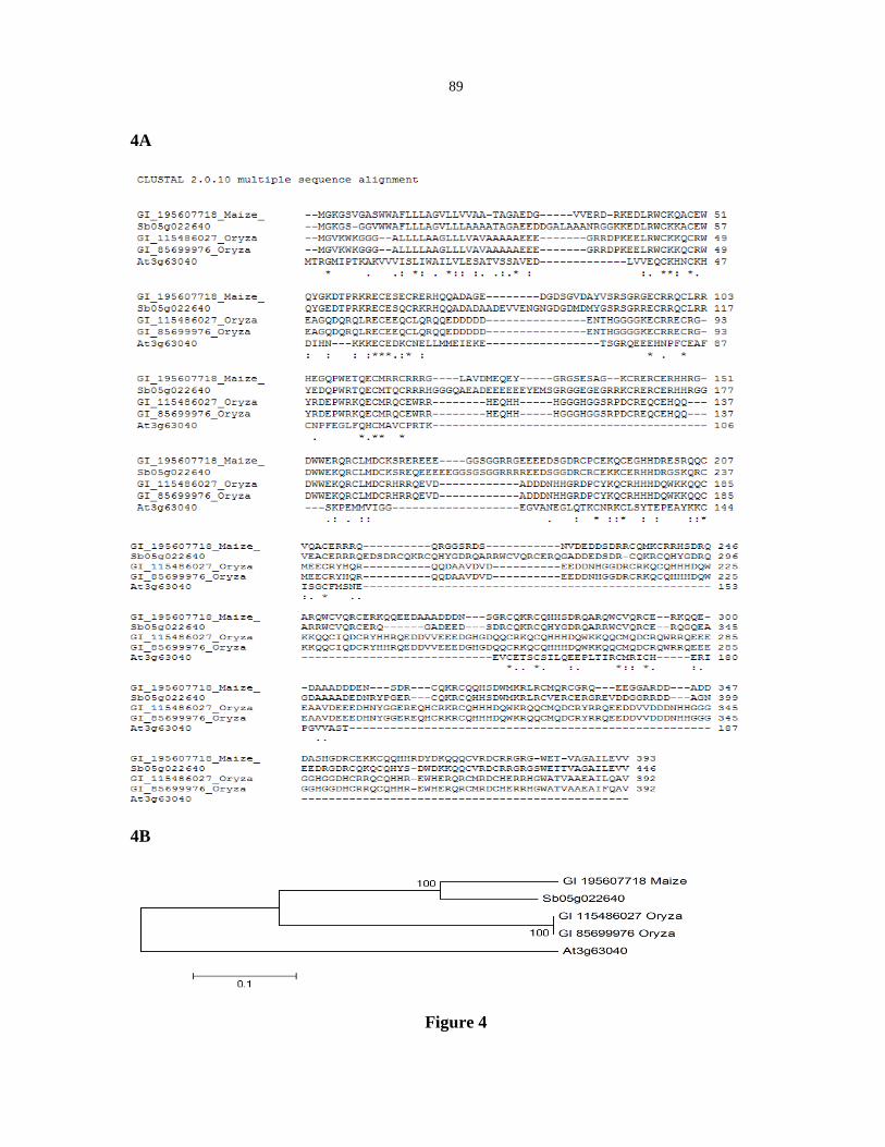
89
4A
4B
Figure 4

90
Competitor _ _ CE1LBP1 _ CE1BP1
Protein _ GST GST CE1LBP1 CE1LBP1
Figure 5

91
Figure 6
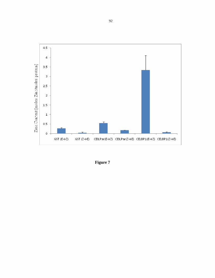
92
Figure 7
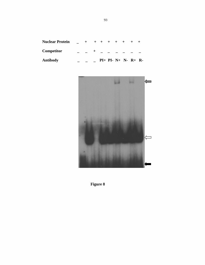
93
Nuclear Protein _ + + + + + + + +
Competitor _ _ + _ _ _ _ _ _
Antibody _ _ _ PI+ PI- N+ N- R+ R-
Figure 8

94
CHAPTER 4. GENERAL CONCLUSIONS
CONCLUSIONS
Genetic studies suggest that a hierarchical system regulates the acquisition of
aleurone cell fate and subsequent differentiation of aleurone. In this thesis, I reported two
studies, which will help to understand endosperm cell fate specification and differentiation;
one involved in early and the other in late differentiation. CR4 is critical for early aleurone
cell fate specification in maize and active forms of the Arabidopsis CR4 ortholog, ACR4,
are rapidly turned over. We present data that suggests the CSN5 subunit of the COP9
signalosome (CSN) is involved in the rapid turnover of ACR4. Vp1 is the most upstream
known transcriptional regulator in the control of anthocyanin production in the aleurone. In
the second part of this thesis, I report the identification and characterization of CE1LBP1, a
putative transcription factor that binds to the CE1-like element of the Vp1 promoter.
The cr4 gene encodes a receptor-like kinase with a tumor necrosis factor receptor
(TNFR)-like motif in the extracellular domain. Disruption of aleurone cell fate in cr4 mutants
suggests that CR4 could function in the perception and transduction of the positional cues
that specify aleurone cell fate (Becraft et al. 1996). In this thesis, I describe the
characterization of proteins that interact with ACR4 and might regulate the ACR4 signal
transduction pathway. A yeast two-hybrid screen with the ACR4 cytoplasmic domain
resulted in the identification of CSN5A and CSN5B, two Arabidopsis CSN5 subunits of the
CSN complex. The interactions were confirmed with in-vitro pull downs and ACR4 was
capable of phosphorylating the CSN5 proteins. In-vivo FRET experiments confirmed that

95
ACR4 was in close proximity to CSN5A and CSN5B within the context of a plant cell. The
interactions with CSN5 subunits suggested the CSN might be involved in ACR4 protein
turnover. Treatment of plants with curcumin, a CSN inhibitor, resulted in the accumulation of
ACR4-GFP in roots. ACR4-GFP also accumulated in csn5a-1 csn5b-1double mutants,
confirming the role of CSN5 in ACR4 turnover.
It is been more than a decade since the identification of CR4 in Maize. However,
compared to other RLK’s in plants, progress in characterization of CR4 signal components
and transduction has been limited. The lack of detectable levels of receptor protein has been
the limitation in the utilization of biochemical, molecular and cell biology approaches
(Gifford et al. 2005). The work presented in this thesis is significant in identification of CSN
as a factor involved in ACR4 turnover. The major function of the CSN is ascribed to
regulating ubiquitination of protein substrates to trigger the protein degradation pathway.
This study for the first time reports that a RLK is in contact with the CSN subunit
(CSN5).The interaction between a CSN subunit has not been demonstrated in well
characterized RLK’s like BRI1 or FLS2 (Kinoshita et al. 2005, Robatzek et al. 2006). I
hypothesize that this interaction between the CSN and ACR4 could be responsible for the
undetectable levels of ACR4 receptor protein, whereas BRI1 and FLS2, which apparently
don’t interact with the CSN, are readily detectable.
In this study, I show that curcumin, a CSN complex inhibitor that has been used
extensively in animal systems, can also be used in plants (Chaudhary and Hruska 2003, Uhle
et al. 2003). The use of the inhibitor results in accumulation of ACR4 presumably by
inhibiting turnover. The slowing down of receptor turnover could provide an opportunity to
biochemically characterize interactors that bind to the receptor. Identification of the ligands

96
for the receptor is critical to understanding the signal transduction pathway initiated at the
membrane and transduced into the cell resulting in cellular responses. Based on the
extracellular domain of ACR4, the ligand is proposed to be proteinacious in nature. In the
case of mammalian EGF/EGFR receptor interaction, EGF is bound to the EGFR at the
membrane, internalized and finally degraded (Hanover et al. 1984). It is possible that the
putative ligand of ACR4 induces receptor internalization and degradation. The use of
curcumin could possibly slow down receptor turnover and thereby provide an opportunity to
detect the ligand bound to the receptor. Experiment design would involve treating seedlings
with curcumin, followed by protein crosslinking, pull down of receptor-ligand complexes
and detection of the ligand using mass-spectroscopy.
To define the role of CSN5 subunit interactions with ACR4, it is required to map the
binding sites on the cytoplasmic domain of ACR4. Two processes that could modulate the
receptor signaling include phosphorylation and ubiquitination. ACR4 being a
serine/threonine kinase, phosphorylation occurs on serine and threonine residues, while
ubiquitination occurs on lysine residues. In-vitro and in-vivo interaction assays of mutant
versions of ACR4 with CSN5 would provide an insight into amino acid residues critical to
the interaction. The results from these experiments would guide in-planta experiments to
address the functional implication of ACR4 turnover in the context of aleurone cell fate. I
hypothesize that stable trangenics expressing ACR4 versions with mutations in key
cytoplasmic residues could impair receptor turnover. Furthermore, introduction of the
cognate versions of CR4 into maize could address the functional role of CR4 receptor
turnover in aleurone cell fate specification. The completion of these proposed experiments on

97
ACR4 / CR4 receptor turnover would advance our knowledge of this important class of plant
RLKs.
In the second study, I focus on VP1, which is expressed in the embryo and aleurone,
and plays an important regulatory role in late embryogenesis and seed maturation in Zea
mays (McCarty et al. 1989). It also regulates the expression of C1, a transcription factor
regulating anthocyanin genes in aleurone cells (Suzuki et al. 1997). In many plant species,
the inception of dormancy, which is a part of the seed maturation process, is mediated by
abscisic acid (ABA) and VP1 (Giraudat et al. 1992, McCarty et al. 1991). Significant
progress has been made in elucidating the mechanism of ABA and VP1 regulated gene
expression, including the characterization of an ABA-responsive element (ABRE) (Busk and
Pages 1997, Hobo et al. 1999a, Shen et al. 1996). However, the mechanism of Vp1
regulation has not been well understood. In this thesis, I describe a trans factor that interacts
with a CE1-like cis element of the Vp1 promoter. CE1LBP1 was isolated from a one-hybrid
screen. Structure-function predictions of CE1LBP1 did not identify any known protein
domains; however CE1LBP1 was confirmed to bind zinc using a colorimetric zinc binding
assay. CE1LBP1-GFP fusions localize to the nucleus, consistent with a role of CE1LBP1 as a
transcriptional factor. Electrophoretic mobility shift assays with recombinant CE1LBP1 and
a CE1-like oligonucleotide confirmed the binding specificity of CE1LBP1 to the.CE1-like
element. Super shift assays with embryo nuclear extract and antibodies raised against
recombinant CE1LBP1 demonstrated that CE1LBP1 is part of an embryo DNA/protein
complex and therefore could function as a trans factor that regulates Vp1 expression via the
CE1-like cis element of the promoter.

98
The identification of CE1LBP1 as putative CE1L transacting factor is significant for a
number of reasons. (1) CEILBP1 is only the second known CE1L transfactor other than
ZmABI4. (2) CEILBP1 has no domains of known function, unlike the APETALA2 (AP2)
domain present in ZmABI4. (3) Dormancy levels in small grains correlate with Vp1
expression but QTL’s do not map to Vp1 locus suggesting that factors that regulate Vp1
expression could also regulate dormancy levels. Thus, it is possible that CELPB1 could
regulate seed dormancy.
A putative ABRC1 was identified in the Vp1 promoter (Cao et al. 2007), however
several items raise the question of whether the CE1-like element actually functions as a
coupling element. Although the spacing between the ABRE and CE1L elements fit the
“multiple of 10 rule”, 50 nucleotides is further than the 20 nucleotides found in the well
characterized ABRC1 in HVA22 (Shen et al. 2004, (Shen and Ho 1995). In addition,
mutagenesis of the CE1L element did not abrogate binding of nuclear proteins as would be
expected from published studies (Cao et al. 2007, Shen et al. 2004, Niu et al. 2002). Thus,
this element in the Vp1 promoter might not function as a coupling element and could have a
function unrelated to ABA responses. In the present study, I focused on identifying
CE1LBP1 as a nuclear protein that binds the CE1L element of the Vp1 promoter during
embryo maturation. I propose using RT-PCR to characterize the developmental profile of
CE1LBP1 expression through the maturation process. Characterization of the CE1LBP1
binding site on the Vp1 promoter has not yet been completed. To carry out an unbiased
consenses DNA-biniding motif of CE1LBP1, I propose the systematic evolution of ligands
by exponential enrichment (SELEX) method. This experiment could define the element and
help clarify the nature of CE1-like element present in Vp1 promoter. To address the

99
functional role of CE1LBP1, databases of publically available mutant collection were
scanned. Due to the lack of any available mutants, reverse genetic approaches like RNA
interference (RNAi) and/or targeting induced local lesions in genomes (TILLING) will be
required to target CE1LBP1. CE1LBP1 is predicted to be a single copy gene in maize, which
should allow targeting by the above methods.
The one-hybrid screen used in this study used an embryo specific library to identify
CE1LBP1. In cereals and maize in particular, the aleurone along with the embryo undergo
maturation and dormancy (Bewley 1997). It will be interesting to characterize the role of
CE1LBP1 in aleurone cells, and to determine the similarities and differences in Vp1
regulation between embryos and aleurone cells.
CE1LBP1 can bind Zn++ and does not contain any domain of known function. p53,
the tumor suppressor protein, binds Zn++ through a highly conserved quartet of three
cysteines and one histidine and functions as a transcription factor (Hainaut and Milner 1993,
Pavletich et al. 1993, Prives and Hall 1999, Verhaegh et al. 1998). CE1LBP1 shares
complete conservation of these four residues with p53. It is hypothesized that the conserved
amino acid quartet of CE1LBP1 might also be critical in binding Zn++. To test that
possibility, in vitro Zn++ binding assays of mutant CE1LBP1 proteins that harbor mutations
in the four amino acids of the conserved quartet could be carried out.
Taken together, these two studies attempt to fill in some of the gaps in our
understanding of aleurone cell fate and differentiation. From the first study, CSN5 of the
CSN complex functions in CR4 regulation and from the second, CE1LBP1 can be placed
upstream of Vp1.

100
REFERENCES
Becraft, P.W., Stinard, P.S. and McCarty, D.R. (1996) CRINKLY4: A TNFR-like
receptor kinase involved in maize epidermal differentiation. Science, 273, 1406-1409.
Bewley, J.D. (1997) Seed Germination and Dormancy. Plant Cell, 9, 1055-1066.
Busk, P.K. and Pages, M. (1997) Protein binding to the abscisic acid-responsive element is
independent of VIVIPAROUS1 in vivo. Plant Cell, 9, 2261-2270.
Cao, X., Costa, L.M., Biderre-Petit, C., Kbhaya, B., Dey, N., Perez, P., McCarty, D.R.,
Gutierrez-Marcos, J.F. and Becraft, P.W. (2007) Abscisic acid and stress signals
induce Viviparous1 expression in seed and vegetative tissues of maize. Plant Physiol,
143, 720-731.
Chaudhary, L.R. and Hruska, K.A. (2003) Inhibition of cell survival signal protein kinase
B/Akt by curcumin in human prostate cancer cells. J Cell Biochem, 89, 1-5.
Gifford, M.L., Robertson, F.C., Soares, D.C. and Ingram, G.C. (2005) ARABIDOPSIS
CRINKLY4 function, internalization, and turnover are dependent on the extracellular
crinkly repeat domain. Plant Cell, 17, 1154-1166.
Giraudat, J., Hauge, B.M., Valon, C., Smalle, J., Parcy, F. and Goodman, H.M. (1992)
Isolation of the Arabidopsis ABI3 gene by positional cloning. Plant Cell, 4, 1251-
1261.
Hainaut, P. and Milner, J. (1993) A structural role for metal ions in the "wild-type"
conformation of the tumor suppressor protein p53. Cancer Res, 53, 1739-1742.
Hanover, J.A., Willingham, M.C. and Pastan, I. (1984) Kinetics of transit of transferrin
and epidermal growth factor through clathrin-coated membranes. Cell, 39, 283-293.

101
Hobo, T., Asada, M., Kowyama, Y. and Hattori, T. (1999) ACGT-containing abscisic acid
response element (ABRE) and coupling element 3 (CE3) are functionally equivalent.
Plant J, 19, 679-689.
Kinoshita, T., Cano-Delgado, A., Seto, H., Hiranuma, S., Fujioka, S., Yoshida, S. and
Chory, J. (2005) Binding of brassinosteroids to the extracellular domain of plant
receptor kinase BRI1. Nature, 433, 167-171.
McCarty, D.R., Carson, C.B., Stinard, P.S. and Robertson, D.S. (1989) Molecular
Analysis of viviparous-1: An Abscisic Acid-Insensitive Mutant of Maize. Plant Cell,
1, 523-532.
McCarty, D.R., Hattori, T., Carson, C.B., Vasil, V., Lazar, M. and Vasil, I.K. (1991) The
Viviparous-1 developmental gene of maize encodes a novel transcriptional activator.
Cell, 66, 895-905.
Pavletich, N.P., Chambers, K.A. and Pabo, C.O. (1993) The DNA-binding domain of p53
contains the four conserved regions and the major mutation hot spots. Genes Dev, 7,
2556-2564.
Prives, C. and Hall, P.A. (1999) The p53 pathway. J Pathol, 187, 112-126.
Robatzek, S., Chinchilla, D. and Boller, T. (2006) Ligand-induced endocytosis of the
pattern recognition receptor FLS2 in Arabidopsis. Genes Dev, 20, 537-542.
Shen, Q. and Ho, T.H. (1995) Functional dissection of an abscisic acid (ABA)-inducible
gene reveals two independent ABA-responsive complexes each containing a G-box
and a novel cis-acting element. Plant Cell, 7, 295-307.

102
Shen, Q., Zhang, P. and Ho, T.H. (1996) Modular nature of abscisic acid (ABA) response
complexes: composite promoter units that are necessary and sufficient for ABA
induction of gene expression in barley. Plant Cell, 8, 1107-1119.
Shen, Q.J., Casaretto, J.A., Zhang, P., and Ho, T.H. (2004). Functional definition of
ABA-response complexes: the promoter units necessary and sufficient for ABA
induction of gene expression in barley (Hordeum vulgare L.). Plant Mol Biol 54, 111-
124.
Suzuki, M., Kao, C.Y. and McCarty, D.R. (1997) The conserved B3 domain of
VIVIPAROUS1 has a cooperative DNA binding activity. Plant Cell, 9, 799-807.
Uhle, S., Medalia, O., Waldron, R., Dumdey, R., Henklein, P., Bech-Otschir, D., Huang,
X., Berse, M., Sperling, J., Schade, R. and Dubiel, W. (2003) Protein kinase CK2
and protein kinase D are associated with the COP9 signalosome. EMBO J, 22, 1302-
1312.
Verhaegh, G.W., Parat, M.O., Richard, M.J. and Hainaut, P. (1998) Modulation of p53
protein conformation and DNA-binding activity by intracellular chelation of zinc.
Mol Carcinog, 21, 205-214.

103
ACKNOWLEDGEMENTS
I would like to express my deep sense of gratitude to Dr. Philip W. Becraft for his
guidance, patience and encouragement shown to me throughout my graduate studies. The
many discussions that we have had during these years, not only were thought provoking but
also instilled in me a greater appreciation of plant biology in particular and the sciences in
general. I would like give my special thanks to my committee members, Dr David Hannapel,
Dr Diane Bassham, Dr Marit Nilsen-Hamilton and Dr David Oliver. Their encouragement
has been greatly appreciated.
During these years, the whole Becraft lab both past and present, especially Dr.
Nrisingha Dey, Dr. Xueyuan Cao, Kejian Li, Joonbae Seo, Gibum Yi and Gokhan Kir gave
continuous support. I would also like to thank Dr Robert Doyle for the support and
discussions that we have had.
I would like to express my deep sense of gratitude to my parents for the numerous
sacrifices that they have made in order for me to pursue my graduate study. I would like to
give special thanks to my wife, Bindu Joseph, and my daughter, Maria Chettoor, for their
love, support and encouragement.
I would also like to thank each and everyone not mentioned here who have in some
way made it possible for me to finish this thesis.