regulation of 22s dynein by a 29-kd light chain
TRANSCRIPT
Regulation of 22S Dynein by a 29-kD Light Chain Kur t Barkalow, Toshikazu H a m a s a k i , and Peter Satir
Department of Anatomy and Structural Biology, Albert Einstein College of Medicine, Bronx, New York 10461
Abstract. Previously, a 29-kD axonemal polypeptide (p29) that copurifies with 22S dynein has been shown to be phosphorylated in a cAMP- and Ca2+-sensitive manner, consistent with a role for this molecule in the signal transduction cascade leading to fast forward swimming in Paramecium tetraurelia (Hamasaki, T., K. Barkalow, J. Richmond, and E Satir. 1991. Proc. Natl. Acad. Sci. USA. 88:7912-7922). This study demonstrates the nature of the relationship between p29 and 22S dynein. Chaotropic agents can be used to separate p29 fractions from 22S dynein. When ex- tracted p29 is exchanged into physiological buffers, it regains the ability to recombine with 22S dynein with an apparent dissociation constant of 25 riM; no recom- bination is seen with 14S dynein or with unrelated control proteins, p29 from Paramecium will also recombine with Tetrahymena 22 but not 14S dynein.
After chymotryptic digestion of 22S dynein, p29 pref- erentially binds to a single-headed fragment, homolo- gous to the t~ H chain of Tetrahymena 22S dynein. 22S dynein treated in vitro by Paramecium protein ki- nase A in the presence of cAMP and ATP to phos- phorylate p29 translocates bovine brain microtubules significantly (1.53 x; p < 0.001) faster than before phosphorylation. Similarly, 22S dynein reconstituted in vitro with thiophosphorylated p29 translocates microtubules significantly (1.31x; p < 0.001) faster than controls reconstituted with nonthiophosphorylated p29. p29 is the only moiety thiophosphorylated in the reconstituted dynein. We conclude that p29 functions as a 22S dynein regulatory light chain in that it alone is sufficient to control the rate of microtubule translo- cation by changes in its phosphorylation state.
I N Paramecium tetraurelia (Bonini and Nelson, 1988; Satir et al., 1993), as in many ciliated cells (Stommel and Stephens, 1985; Sanderson et al., 1992), ciliary
beat and cell swimming are regulated by second messengers. In particular, the membrane-permeable cAMP-analogue, monobutyryl cAMP, and the phosphodiesterase-inhibitor, 3'-isobutyl-l-methylxanthine, increase forward swimming speed of living Paramecium (Bonini et al., 1986). An in- crease in CAMP levels occurs in conjunction with a mem- brane hyperpolarization that leads to fast forward swimming in Paramecium (Bonini et al., 1986) and adenylate cyclase activity seems to be tightly coupled to, or part of, a K + channel in the cell membrane (Sehultz et al., 1992). Further, cAMP has been shown to increase the forward swimming speed of permeabilized demembranated ceils (Bonini and Nelson, 1988; Hamasaki et al., 1991).
Likely candidates for the effector molecules in this cAMP cascade probably are parts of, or interact strongly with, the dynein arms. Brokaw and Kamiya (1987) have shown that in Chlamydomonas reinhardtii the outer dynein arm seems to be a primary regulator of beat frequency. Changes in beat frequency are measured directly in mussel gill and other cilia as a response to an increase in intracellular cAMP (Mura-
Address all correspondence to Dr. Peler Satir, Dept. of Anatomy and Struc- tural Biology, Albert Einstein College of Medicine, 1300 Morris Ave., Bronx, NY 10461.
kami, 1987). In the absence of changes in beat form, beat frequency is proportional to the rate at which microtubules slide relative to one another, which is determined by axone- mal dynein, presumably the outer arm (Satir et al., 1993).
In Paramecium, the isolated outer dynein arm is a three- headed molecule with a sedimentation coefficient of 22S (Larsen et al., 1991; Beckwith and Asai, 1993; Walczak et al., 1993). A 29-kD axonemal polypeptide (p29) ~ that co- purifies with 22S dynein (Bonini and Nelson, 1990; Hama- saki et al., 1991) has been shown to be phosphorylated in a cAMP-dependent and Ca~+-sensitive manner in Paramecium (Hamasaki et al., 1989). Axonemal polypeptides of similar molecular weight have been identified by virtue of their cAMP-dependent phosphorylation in mussel gill cilia (Ste- phens and Prior, 1992), in Tetrahymena (Chilcote and John- son, 1990) and in ovine respiratory cilia (Salathe et al., 1993). In Paramecium, experimental phosphorylation of p29 is achieved within isolated axonemes via an endogenous kinase. In in vitro microtubule mobility assays, phosphory- lated p29 induces 22S dynein to translocate bovine brain mi- crotubules 1.4 times faster than controls (Hamasaki et al., 1991). However, many axonemal proteins are phosphorylated (Table I) and minor quantitative changes in phosphorylation in proteins other than p29 could influence these findings.
1. Abbreviations used in this paper: H, heavy; p29, 29-kD axonemal poly- pepfide; PKA, cAMP-dependent protein kinase.
© The Rockefeller University Press, 0021-9525/94/08/727/9 $2.00 The Journal of Cell Biology, Volume 126, Number 3, August 1994 727-735 727
Dow
nloaded from http://rupress.org/jcb/article-pdf/126/3/727/1261374/727.pdf by guest on 12 January 2022

Table L Phosphorylation of p29 Correlates with Translocation Velocity
Sample type Experimentally phosphorylated bands 22S dynein preparation KD
Axonemal treatment* S H 79 72 68 54 49 47 29 23 No cAMP ± - + ± :i: + - + - + + cAMP + - + + + + - + + + + c A M P + Ca 2+ + - + ± ± + - + - ±
Dynein treatment PKA control . . . . . . . . . . PKA - + . . . . + - + -
Dynein recombination No addition . . . . . . . . . . +p29 fraction . . . . . . . . . . +pp29 fraction . . . . . . . . + -
Translocation velocity + SD Versus
n control
1.81 Jr 0.41 (296) 1.12§ 2.53 + 0.55 (340) 1.57' 1.92 + 0 . 5 6 (210) 1.12§
1.61 + 0.79 (29) 1.00 2.48 + 1.08 (44) 1.53,
1.05 + 0.41 (53) 0.65 1.60 :t= 0.68 (59) 1.00§ 2.10 + 0.78 (47) 1.31,
This table compares the phosphorylated polypeptides formed in various preparations of 22S dynein used in microtubule translocation measurements in this study with those demonstrated by Hamasaki et al. (1991). Phosphorylated bands present in kinase only sample are omitted. When p29 is phosphorylated in a cAMP- dependent manner, either before extraction of 22S dynein from the axonerne or in vitro with PKA, translocation velocity compared to a control dynein incubated in cAMP and ATP without PKA in vitro is increased. A comparable increase is seen when unphosphorylatcd 22S dynein is reconstituted with phosphorylated p29 (pp29). Other phosphorylated polypeptides vary in each preparation. These may have modulatery effects on translocation. S, sky chain; H, dynein H chain. * Hamasaki et al., 1991. * p < 0.001 versus PKA control. § p > 0.05 versus PKA control, no significant difference.
Analysis of Triton X-100-permeabilized cells, under condi- tions where p29 is phosphorylated but cAMP is removed, demonstrates that swimming proceeds at nearly twice the speed of control cells.
These previous studies strongly suggest that p29 or its equivalent plays an important role in the regulation of the outer dynein arm. In this study we provide additional evi- dence to confirm this hypothesis. This has led to a further understanding of the construction of the outer dynein arm of Paramecium and the role of p29 as a regulatory light chain of 22S dynein. From a crude mixture of Paramecium dyneins we have isolated a fraction containing p29, but lacking the dynein heavy (H) chains. Using this fraction, we document how I)29 can specifically reassociate with 22 but not 14S dynein of Paramecium, and that this reassociation extends to the outer arm dynein of another ciliate genus, Tetrahymena. We are able to localize the p29-binding site for reassociation to a specific H chain of these dyneins, most likely the o~ H chain.
We further exploit our ability to repopulate 22S dynein with p29 to examine the ability of thiophosphorylated p29 to regulate the activity of reconstituted dynein in vitro, and we also extend the axonemal phosphorylation studies to the in vitro translocation system, using a Paramecium cAMP- dependent protein kinase (PICA). In both of these different types of experiments, the number of phosphorylated constit- uents is more limited than previously, and in the re, constitu- tion experiment only p29 is experimentally thiophosphor- ylated. Microtubule translocation velocity increases in a manner comparable to that reported by Hamasaki et al. (1991), which implies that p29 phosphorylation is critical in determining changes in translocation velocity in this assay, possibly by altering the mechanochemical properties of outer arm dynein.
Materials and Methods
Axoneme Isolation Paramecium tetraurelia are cultured axenieally and harvested as described
previously (Hamasaki et al., 1989, 1991). The harvested Paramecium are deciLiated by calcium shock; the cilia are isolated, demembranated, and purified (Hamasaki et al., 1989). ~trahymena are grown axenicly (Bar- kalow et al., 1994) and deciliated by addition of 0.25 mg/ml dibucaine (Ciba-Geigy, Summit, NJ). Complete deciliation occurs within 10-15 min. From this point in the isolation, handling of Paramecium and Tetrahymena samples is identical. Methods follow Hamasaki et al. (1989, 1991).
Axonemal Phosphorylation and Isolation of Crude Dynein
The axonemes are used directly for dynein exlraction when unphosphory- lated dynein is required or for thiophosphorylation (used as a stable coun- terpart of phosphorylation). Paramec/um axonemes are thiophosphorylated on ice for 30 rain in 2 ml of axoneme buffer containing 10/~M "y-[S]ATP (Boehringer Mannheim, Indianapolis, IN) in the presence or absence of 20 ~M cAMP (Boehringer Mannheim), and/or 1 mCi 7-[35S]ATP (NEN, Boston, MA or Amersham Corp., Arlington Heights, IL) at a total protein concentration of ,x, 3 mg/ml. The thiophosphorylated axonemes are repel- leted and washed with 5 ml axoneme buffer at 12,000 g at 4°C for 10 rain.
In all cases, for dynein extraction the washed pellet is resnspended in ax- onerne buffer containing 0.6 M KCI and allowed to stand on ice for 30 min. The salt extract is centrifuged at 12,000 g at 4°C for 10 min. The superna- tant, containing the extracted dynein, is collected and centrifuged at 100,000 g at 4°C for 30 min to remove large particulates. The supernatant is concentrated in a Centficon 30 or 100 (Amicon, Beverly, MA) by cen- trifugation at 5,000 or 1,000 g, respectively, at 4°C to a volume ~100/d, and washed with 0.5 ml axoneme buffer containing 0.6 M KCI. In the case of the Centricon 100, all proteins or protein complexes less than ,,080 kD flow through a filter. The Centricon retentate is enriched in dynein and will be referred to as crude dynein.
Purification of Unphosphorylated Dyneins and Phosphorylation In Vitro
Paramecium or Tetrahymena crude dynein from unphosphorylated axo- heroes is loaded onto an 11.6 ml, 5-30% linear sucrose gradient in axoneme buffer; no more than 200 ~,1 is loaded onto a single gradient. The gradients are centrifuged in L5-50 centrifuge (Beckman Instrs., Inc., Fullerton, CA) and an SW41 Ti rotor for 15 h at 35,000 RPM and 4°C, and fractionated into 0.5-ml fractions. Dynein is detected by ATPase activity (Hayashi and Takahashi, 1979; Murphy and Riley, 1962) and SDS-polyacrylamide gel ¢lectrophoresis (modified after Laemmli, 1970). Protein concentration is determined by the BCA protein assay using BSA as a standard (Pierce, Rockford, IL). In these gradients two peaks of dynein ATPase are found at 14 and 22S. Fractions containing these dyneins are pooled, frozen, and stored at -80°C for later use in the teconstimtion and translocation assays.
The Journal of Cell Biology, Volume 126, 1994 728
Dow
nloaded from http://rupress.org/jcb/article-pdf/126/3/727/1261374/727.pdf by guest on 12 January 2022

In some experiments, a partially purified cyclic AMP-dependent PKA isolated from Paramecium (Hochestrasser and Nelson, 1989; kindly provided by C. Walczak and D. Nelson, University of Wisconsin, Madison, WI), has been used to phosphorylate 22S dynein in vitro (C. Walczak and D. Nelson, personal communication). Unphosphorylated 22S dynein (,,00.3 /~g) was incubated with PKA ('~10 U), in the presence of 5/~M cAMP and 0.1 mM ATP (including 30 ~tCi 3,-[32p]ATP) in 10 mM Mg-acetate and 50 mM MOPS, pH ~5, at room temperature for 10 rain. Controls omitted ei- ther the dynein or the PICA. The reaction was terminated by the addition of an equal volume of cold 30% trichioroacetic acid and allowed to stand for 30 min on ice. The samples were centrifuged in a microfuge, and resuspended in SDS sample buffer with 1 M Tris, pH 8.0, and incubated for 3 rain at 80°C. Samples are analyzed by SDS-PAGE and autoradiogra- phy using X-OMAT AR scientific imaging film (Eastman g.~lak, Roch- ester, NY).
p29 Isolation In general, p29 has been isolated from thiophosphorylated crude dy- nein. When prepared from axonemes that have been treated either with y-[35S]ATP or 7-[S]ATP in the absence of cAMP, p29 is unlabeled and not experimentally phosphorylated (Hamasaki et al., 1991); when prepared from axonemes treated with 7-[3SS]ATP and cAMP, p29 is thiophosphory- lated and labeled; when prepared from axonemas treated with -/-[S]ATP and cAMP, p29 is thiophosphorylated and unlabeled.
To determine the original percent dissociation of p29 from dynein, la- beled axonemes are treated with 0.6 M KC1 and the resulting crude dynein is allowed to stand on ice for 30 rain. The sample is loaded onto a Centricon 100 and centrifuged at 1,000 g at 4°C until the volume of the retentate is ~100/~1. An additional 0.5 mi of axoneme buffer containing 0.6 M KCI is added to wash the retentate. FOr measurements the Centricon 100 flow- through is concentrated in a Centricon 10 (Cutoff •10 kD). p29 does not flow through the Centricon 10. The retentates of the Centricon 100 and 10 are repetitively concentrated to <100 Id and washed with axoneme buffer. The initial experiments were done in 200/~1, with a protein concentration of 1.5 mg/ml. In individual retentates and flowthroughs, p29 is detected by SDS-PAGE employing an acrylamide gradient of 5-15 %, usually followed by phosphorimnging using a Molecular Dynamics Phosphorimager (Sun- nyvale, CA) and/or autoradiography. Percent p29 in the flowthrough is the amount in the Centricon 100 flowthrough, determined quantitatively using the Phnsphorimager, divided by amount in Centricon 100 (flowthrongh plus retentate) × 100. Based on competition experiments, we assume the distri- bution of label is indicative of the distribution of total p29. Additional chaotropic agents tested were 1.2 M KCI with and without 10 mM DTT, and 4 M urea.
The 1.2 M KC1 treatment yields a Centricon 10 retentate containing a significant amount of p29 in an enriched fraction lacking the dynein H chains. This fraction, which is used routinely, will be referred to as the p29 fraction. It is routinely obtained starting with crude dynein, 1.5 mg/mi, in a volume of 2 mi in axoneme buffer containing 0.6 M KCI to which is added 2 ml of axoneme buffer containing 1.8 M KC1, processed as above. The Cen- tricon 10 retentate is washed to a final volume of *500/tl , a final KC1 con- centration of '~50 mM, and a final protein concentration of 1 mg/ml. FOr some experiments, the p29 fraction has been further purified by column chromatography. The p29 fraction is loaded onto a Superdex 75 column (Pharmacia LKB Biotechnolngy, Piscataway, NJ) pre-equllibrated in axo- neme buffer. 0.5-mi fractions are collected and assayed by scintillation counting, SDS-PAGE (proteins detected by silver staining as described by Merrill et al., 1984), and autoradiography. The Superdex 75 column is calibrated using chromatographic standards (Mr 68000, 45,000, 25,000, and 12,500) (Boehringer Mannheim, Mannheim, Germany).
Tests of Association of p29 to 22S Dynein and Other Proteins Reconstimtion of purified Paramecium 22S dynein with p29 was tested by sucrose density gradient purification. Unphosphorylated 22S dynein was incubated with a labeled p29 fraction at room temperature for 2 h, subse- quentiy loaded onto a sucrose gradient and repurified. Gradient fractions have been analyzed by ATPase activity, SDS-PAGE, autoradiography and scintillation counting. As a control, purified Paramecium 14S dynein was substituted for 22S dynein.
To test the ability of p29 to associate with 22S dynein or other moieties more generally, a force filtration procedure similar to that described above has been employed. After incubation, the sample is loaded on a Centricon 100 to recover the retentate in a final volume of 40 tzl. The flowthrough is
concentrated in a Centricon 10 to 40 /A. The samples are analyzed separately by SDS-PAGE and autoradiography.
For the quantitative analysis of reconsfitution, one constituent (p29 or 22S dynein) is maintained at a constant concentration in a series of aliquots, while the concentration of the other constituent is systematically increased. Reconsfitution is performed at room temperature for a period of 2 h (based on a time course analysis of reconstitution that indicates saturation at >30 min). Controls are for volume, and for protein concentration when appro- priate; all samples are treated identically throughout. Samples are collected using the above Centricon procedure. To improve recovery of all fractions, in some experiments the Centricons used are pretreated overnight at room temperature with 1% nonfat instant ndlk (Carnation, Los Angeles, CA) with 0.02 % NAN3. Before use, the Centricons are extensively washed with dis- tilled water. Retentate and flowthrough are analyzed by SDS-PAGE and quantitatively analyzed using a Phosphorimager. The re.association of p29, at varying concentrations, with limiting amounts of 22S dynein was also analyzed using a Molecular Dynamics densitometer by scanning a 5-15% acrylamide gel stained with Coomassie brilliant blue 11250 and destalned using a low (10%) methanol-7.5 % acetic acid solution, which preserves p29 staining.
Chymotryptic Digestion of 22S Dynein Chymotryptic digestion of 22S dynein has been performed essentially as de- scribed by Toyoshima (1987a, b). Digestion is achieved at a chymotryp- sin/22S dynein ratio of 1:100, by weight, at room temperature for 5 and 30 min. The digestion is stopped with 1 mM PMSE The resulting fragments are separated by sucrose density gradient centrifugation and analyzed as above.
The structural characteristics of the peak fractions of the dynein frag- ments are assayed by negative stain electron microscopy, using a carbon- coated mica method described in Barkalow et al. (1994).
Microtubule Translocation Assay The functional effects of reconstitution are assayed by in vitro microtubule translocation as described previously (Vale and Toyoshima, 1988, 1989; Hamasaki et al., 1991). In a field selected for measurement, all microtu- bules that produce consistent movement of over 30 sec are measured. Quali- tatively, percent mobility does not vary with treatment. All 22S dyneins used in these assays are initially unphosphorylated. Recunstimtion experi- ments compare dyneins from retentates obtained using the force filtration recombination of unlabeled 22S dynein and a p29 fraction obtained by 1.2 M extraction of crude dynein from axonemes thiophosphorylated in the presence of 3,-[S]ATP with versus without cAMP.
For the in vitro phosphorylation experiments, unphosphorylated 22S dynein is perinsed into the translocation chamber and motility is recorded. The translocation chamber is then washed with gliding buffer followed by phosphorylation buffer. Then phosphorylation buffer containing 1 mM ATP, with or without 10/~M cAMP or PKA ('~10 U), is perfused into the chamber and incubated at room temperature for 10 rain. This buffer is then replaced with gliding buffer. Motility of the treated dynein is analyzed within 10 rain of phosphorylation.
Results
When thiophosphorylated crude dynein is isolated from axo- nemes in 0.6 M KC1, 28% of p29 as dissociated from dynein H chains, based on quantitative analysis of the radiolabeled p29 as discussed in the Materials and Methods. When this mixture is further subjected to high salt (1.2 M KCI), an ex- tract is obtained in which a total of 60% of p29 is dis- sociated. Addition of a reducing agent (10 mM DTT) to the mixture does not increase the dissociation. Dissociation is slightly enhanced (71%) when 4 M urea is used in lieu of the 1.2 M KCI. The final high salt extract (using the 1.2 M KC1 treatment) is subjected to Centricon force filtration steps to separate the dissociated p29 from the H chains and to obtain proteins of Mr > 10 < 100 kD in a filtrate, p29 retains its label throughout and is a principal labeled band of the filtrate (Fig. 1, p29 fraction).
Barkalow et al. Dynein Light Chain 729
Dow
nloaded from http://rupress.org/jcb/article-pdf/126/3/727/1261374/727.pdf by guest on 12 January 2022
B
0.3
0.2- /-i O
o.1-
14S Dynein + p29 fraction 25o0
5 10 15 20 Fraction run"ber
22,5 Dynein + p29 fract~ 250O
lOO ~
.... ; o , 1 o
5 10 15 20 Fraction
C
0.3
0.2-
Ci (~
0.1-
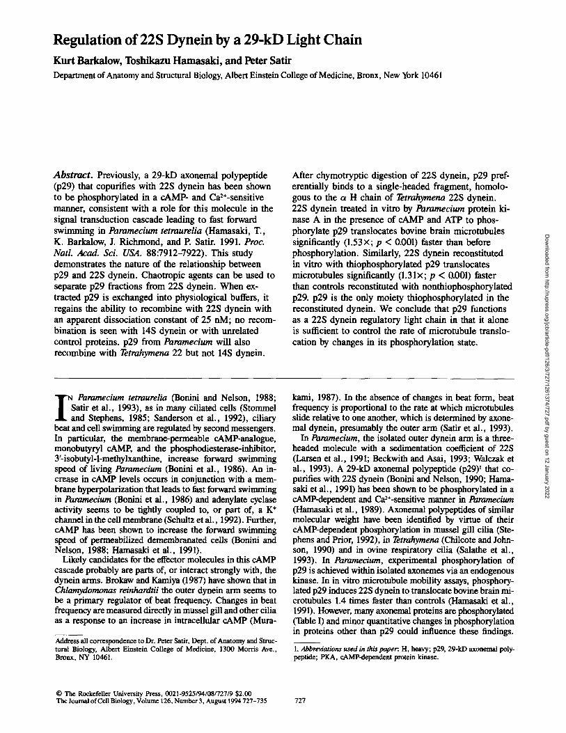
Figure 1. Recombination of p29 fractions with Paramecium dyneins. (.4) p29 fraction from thiophosphorylated labeled axo- acmes. Coomassie blue-stained lane from a 5-15 % linear gradient SDS-PAGE (gel) and corresponding autoradiogram (arg) are shown. (B) Unphosphorylated 14S dynein recombined with the la- beled p29 fraction. Sucrose density gradient fractions (right, gel and arg of fraction 9). (C) Unphosphorylated 22S dynein recom- bined with the labeled p29 (right, gel and arg of fraction 15). A peak of radioactivity corresponding to labeled p29 cosediments with 22S dynein. 60 #g of 22 and 14S dynein and 50 #g of p29 fraction are used. Samples were not concentrated prior to SDS- PAGE. Indicators for all Figs. p29, arrow; dynein H chain region, arrowhead; lines, Mr standards (from top) 66, 45, 36, 32, and 24 kD (some gels have a sixth standard at 20.1 kD).
Reconstitution of Dyneins with p29
When placed in physiological buffers, the dissociated p29 will reassociate with 22S dynein. To demonstrate reassocia- tion, we purified unlabeled crude dynein by sucrose density centrifugation. Based on ATPase activity, fractions contain- ing 14S and 22S dynein were obtained and incubated for 2 h with the labeled p29 fraction before a second sucrose gra- dient centrifugation of each dynein was performed. Fig. 1 shows that although most of the label is found unassociated with either dynein, a significant peak of p29 radioactivity reassociates and cosediments with 22S dynein, p29 is the only radiolabeled band to reassociate in this way. In contrast, very little, if any, p29 radioactivity cosediments with 14S dynein.
In further experiments, this reconstitution assay has been simplified. The dynein to be tested for reassociation is in- cubated with the labeled p29 fraction and subjected to force filtration in a Centricon 100, through which the dynein H chains and bound proteins do not pass. The amount of label that remains with the dynein, after extensive washing, is taken as a measure of reconstitution. The results of this assay with 22 and 14S dynein, BSA and no added proteins (buffer
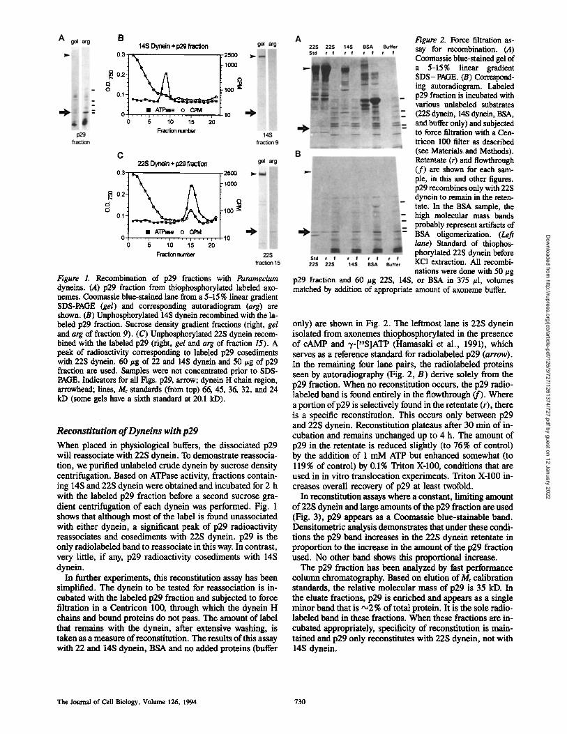
Figure 2. Force filtration as- say for recombination. (A) Coomassie blue-stained gel of a 5-15% linear gradient SDS-PAGE. (B) Correspond- ing autoradiogram. Labeled p29 fraction is incubated with various unlabeled substrates (22S dynein, 14S dynein, BSA, and buffer only) and subjected to force filtration with a Cen- triton 100 filter as described (see Materials and Methods). Retentate (r) and flowthrough (f) are shown for each sam- ple, in this and other figures. p29 recombines only with 22S dynein to remain in the reten- tate. In the BSA sample, the high molecular mass bands probably represent artifacts of BSA oligomerization. (Left lane) Standard of thiophos- phorylated 22S dynein before KC1 extraction. All recombi- nations were done with 50 #g
p29 fraction and 60 #g 22S, 14S, or BSA in 375 #1, volumes matched by addition of appropriate amount of axoneme buffer.
only) axe shown in Fig. 2. The leftmost lane is 22S dynein isolated from axonemes thiophosphorylated in the presence of cAMP and -t-[35S]ATP (Hamasaki et al., 1991), which serves as a reference standard for radiolabeled p29 (arrow). In the remaining four lane pairs, the radiolabeled proteins seen by autoradiography (Fig. 2, B) derive solely from the p29 fraction. When no re, constitution occurs, the p29 radio- labeled band is found entirely in the flowthrough (f). Where a portion of p29 is selectively found in the retentate (r), there is a specific re, constitution. This occurs only between p29 and 22S dynein. Reconstitution plateaus after 30 min of in- cubation and remains unchanged up to 4 h. The amount of p29 in the retentate is reduced slightly (to 76 % of control) by the addition of 1 mM ATP but enhanced somewhat (to 119% of control) by 0.1% Triton X-100, conditions that are used in in vitro translocation experiments. Triton X-100 in- creases overall recovery of p29 at least twofold.
In reconstitution assays where a constant, limiting amount of 22S dynein and large amounts of the p29 fraction are used (Fig. 3), p29 appears as a Coomassie blue-stainable band. Densitometric analysis demonstrates that under these condi- tions the p29 band increases in the 22S dynein retentate in proportion to the increase in the amount of the p29 fraction used. No other band shows this proportional increase.
The p29 fraction has been analyzed by fast performance column chromatography. Based on elution of Mr calibration standards, the relative molecular mass of p29 is 35 kD. In the eluate fractions, p29 is enriched and appears as a single minor band that is ",,2 % of total protein. It is the sole radio- labeled band in these fractions. When these fractions are in- cubated appropriately, specificity of reconstitution is main- tained and p29 only reconstitutes with 22S dynein, not with 14S dynein.
The Journal of Cell Biology, Volume 126, 1994 730
Dow
nloaded from http://rupress.org/jcb/article-pdf/126/3/727/1261374/727.pdf by guest on 12 January 2022
A
p29 friction (Pg):
96
36
Y
4
°:I.. 30 40 50 60 70 80 90 100
p~ fractk~ (.g)
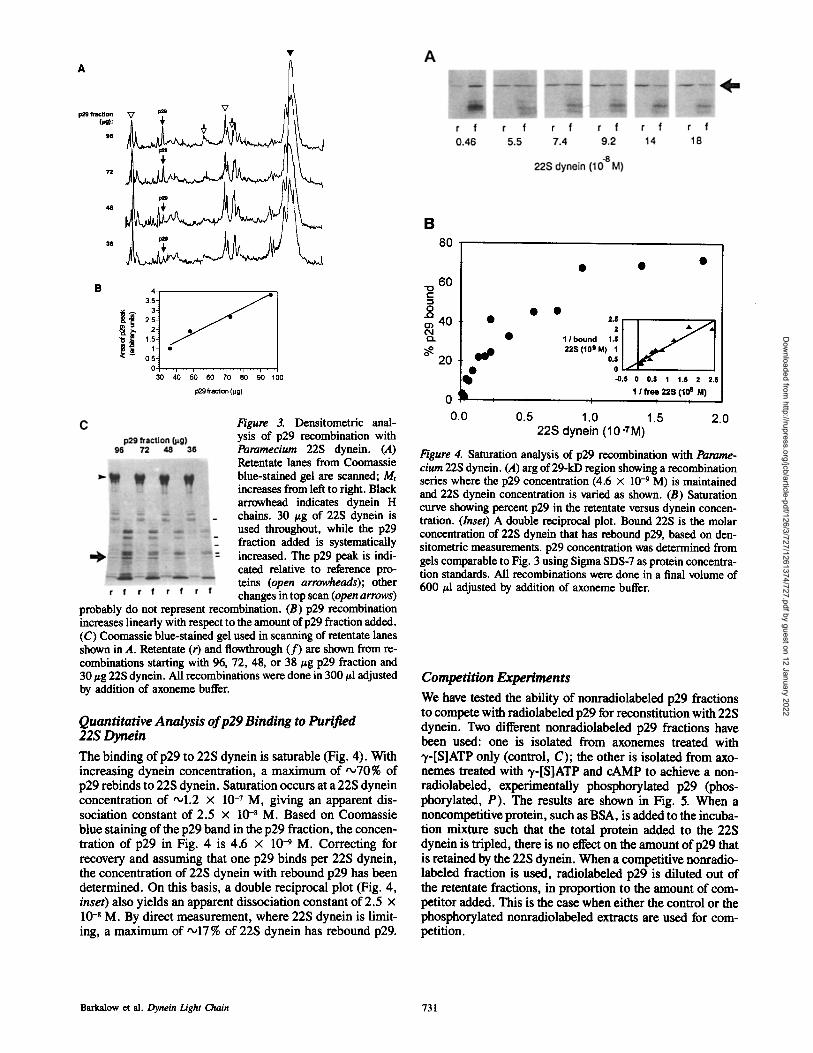
Figure 3. Densitometric anal- ysis of p29 recombination with Paramecium 22S dynein. (A) Retentate lanes from Coomassie blue-stained gel are scanned; Mr increases from left to right. Black arrowhead indicates dynein H chains. 30 #g of 22S dynein is used throughout, while the p29 fraction added is systematically increased. The p29 peak is indi- cated relative to reference pro- reins (open arrowheads); other changes in top scan (open arrows)
probably do not represent recombination. (B) p29 recombination increases linearly with respect to the amount of p29 fraction added. (C) Coomassie blue-stained gel used in scanning of retentate lanes shown in A. Retentate (r) and flowthrough (f) are shown from re- combinations starting with 96, 72, 48, or 38/~g p29 fraction and 30 #g 22S dynein. All recombinations were done in 300 #1 adjusted by addition of axoneme buffer.
Quantitative Analysis of p29 Binding to Purified 22S Dynein The binding of p29 to 22S dynein is saturable (Fig. 4). With increasing dynein concentration, a maximum of ,~70% of p29 rebinds to 22S dynein. Saturation occurs at a 22S dynein concentration of ,ol.2 x 10 -7 M, giving an apparent dis- sociation constant of 2.5 x 10 -8 M. Based on Coomassie blue staining of the p29 band in the p29 fraction, the concen- tration of p29 in Fig. 4 is 4.6 x 10⁻⁹ M. Correcting for recovery and assuming that one p29 binds per 22S dynein, the concentration of 22S dynein with rebound p29 has been determined. On this basis, a double reciprocal plot (Fig. 4, inset) also yields an apparent dissociation constant of 2.5 x 10 -8 M. By direct measurement, where 22S dynein is limit- ing, a maximum of '~17 % of 22S dynein has rebound p29.
B 8O
• • •
6O t""
o • • 40 • ,.s
¢'~ • 1 I bound 1.$
o~ 20 ~ 22s (10 9 M) 1 0.5 ! /
• 0 • L$ 0 0.8 1 1.8 2 2.8
0 ~ , 1/free~ 22S(10 s, M)
0.0 0.5 1.0 1.5 2.0 22S dynein (10 -TM)
Figure 4. Saturation analysis of p29 recombination with Parame- cium 22S dynein. (A) arg of 29-kD region showing a recombination series where the p29 concentration (4.6 x 10 -9 M) is maintained and 22S dynein concentration is varied as shown. (B) Saturation curve showing percent p29 in the retentate versus dynein concen- tration. (Inset) A double reciprocal plot. Bound 22S is the molar concentration of 22S dynein that has rebound p29, based on den- sitometric measurements, p29 concentration was determined from gels comparable to Fig. 3 using Sigma SDS-7 as protein concentra- tion standards. All recombinations were done in a final volume of 600 #1 adjusted by addition of axoneme buffer.
Competition Experiments We have tested the ability of nonradiolabeled p29 fractions to compete with radiolabeled p29 for re.constitution with 22S dynein. Two different nonradiolabeled p29 fractions have been used: one is isolated from axonemes treated with "r-[S]ATP only (control, C); the other is isolated from axo- nemes treated with 7-[S]ATP and cAMP to achieve a non- radiolabeled, experimentally phosphorylated p29 (phos- phorylated, P). The results are shown in Fig. 5. When a noncompetitive protein, such as BSA, is added to the incuba- tion mixture such that the total protein added to the 22S dynein is tripled, there is no effect on the amount of p29 that is retained by the 22S dynein. When a competitive nonradio- labeled fraction is used, radiolabeled p29 is diluted out of the retentate fractions, in proportion to the amount of com- petitor added. This is the case when either the control or the phosphorylated nonradiolabeled extracts are used for com- petition.
Barkalow et al. Dynein I-z'ght Chain 731
Dow
nloaded from http://rupress.org/jcb/article-pdf/126/3/727/1261374/727.pdf by guest on 12 January 2022

Figure 5. Competition experi- ments. Aliquots of labeled p29 fraction (12/zg each) are recombined with unphosphor- ylated 22S dynein (38 #g) in the presence of a competitor
for recombination volumes were adjusted with axoneme buffer to 400/A. The arg of the 29-kD region is shown. Competitors are: buffer alone (22S); 24 #g BSA (+BSA); 24 or 120/~g of unlabeled, unphosphorylated p29 fraction (2x or 10x C); 24 or 120/~g unla- beled, thiophosphorylated p29 fraction, (2 or 10x P). Both un- phosphorylated and phosphorylated unlabeled p29 fractions com- pete for the labeled p29 equally well.
Recombination of Paramecium p29 with Tetrahymena Dynein Recombination of Paramecium p29 has been tested with Tetrahymena dyneins (Fig. 6). p29 reassociates specifically with Tetrahymena 22S dynein to about the same extent as with Paramecium 22S dynein. Tetrahymena 145 dynein does not bind p29. In preliminary experiments, neither Chla- mydomonas 12 nor 18S outer arm dynein bind p29.
Recombination of p29 with Tetrahymena 22S Dynein Fragments When Tetrahymena 22S dynein is treated with chymotrypsin according to the procedure of Toyoshima (1987a,b), one- headed (12-14S), and two-headed (18-205) fragments are
Figure 6. Recombination of 7ktrahymena dyneins and dy- nein fragments with Parame- c/urn p29. (Top) Tetrahymena 22S dynein is equally compe- tent as Paramecium 22S dy- nein for recombination with Paramecium p29. In each re- constitution the same amount of labeled p29 fraction is used. Tetrahymena 14S dy- nein does not recombine with p29. Coomassie blne-stained gel, 5-15% linear gradient SDS-PAGE, with correspond- ing arg of 29-kD region. (Bot- tom) Recombination of p29 with chymotryptic fragments of /~trahymena 22S dynein. The ~ t ~ n a one-headed fragment (1H) preferentially recombines with Paramecium p29, as compared to the two- headed fragment (2//). Arg of 29-kD region. Quantitative
structural analysis of one- and two-headed fractions demonstrates that one-beaded fraction is nearly homogeneous. One-headed frac- tion: 89% one-headed, 4% two-headed, 0% three-headed, 7% structure is obscured (n = 28). Two-headed fraction: 17% one- headed, 71% two-headed, 9% three-headed, 3% structure is ob- muted (n = 105). All recombinations done in 300/zl with 30/zg 22 and 14S dyneins and 20/~g p29 fraction. For chymotrypsin di- gest 100 #g 22S is digested and run on sucrose gradient. Peak one- and two-headed fractions were used (400 pl) and recombined with 25/zg p29 fraction.
obtained. We tested the ability of unlabeled proteolytically generated fragments of Tetrahymena dynein to re.associate with labeled p29. p29 reassociates preferentially with the 12-14S single-headed dynein (Fig. 6).
Control of In Vitro Microtubule Sliding Velocity by p29 Phosphorylation An in vitro microtubule translocation assay has been used to test for the functional capacity of the reassociated p29 to modulate the rate at which 22S dynein translocates brain microtubules. As a control, unphosphorylated Paramecium 22S dynein has been isolated and, without further treatment, in vitro translocation has been measured as in Hamasaki et al. (1991). The velocity of translocation ranges from about 0.5-3.3/zms-', with an average of 1.61 + 0.79/~ms -~ (Table I, Fig. 7). Microtubules have been washed out and the bed of dynein has then been treated with cAMP and ATE with or without PKA. Micrombules are reintroduced and translo- cation rate rerneasured. In the absence of PKA, velocity does not increase; in fact the average velocity is slightly slowed (1.3 + 0.59 ~ms -', n = 32) ( p versus control: 0.1 > p > 0.05). After PKA perfusion, the translocation velocity in- creases by 1.53x to 2.48 + 1.08/zms -1 (p < 0.001), with 19% of the microtubules having velocities >3.3 /~ms -t. Comparison between the untreated and PKA-treated 22S dynein reveals that several dynein components have been phosphorylated by the PKA treatment, including the dynein H chains, a ,050 kD protein and p29 (Fig. 7).
22S dynein has also been tested for its ability to translocate microtubules after reconstitution with various p29 fractions. Without reconstimtion, this dynein, which has been exten- sively washed in the Centricon, produced microtubule trans- location velocities that were more sluggish than usual, but addition of a control p29 fraction where the p29 was not thiophosphorylated increased translocation velocity to con- trol levels (Table I). Compared to reconstitution with control p29 fractions, reconstitution with thiophosphorylated p29 increases translocation rate by 1.31x to 2.1 + 0.78/zms -~ (p < 0.001) (Table I) with 10% of the microtubules being translocated at velocities >3,3/zms-~ (Fig. 7). These prepa- rations have no differences in H chain phosphorylation. Moving on the dynein substratum reconstituted with the thiophosphorylated p29, a few microtubules translocate at velocities up to 10 #ms -1 for short intervals (not shown).
Discussion
The results presented here clarify and further our understand- ing of the relationship and interaction of p29 and 22S dynein. In isolated 225 dynein, p29 has been found to be substoi- chiometric in the past (Travis and Nelson, 1988; Hamasaki et al., 1991), presumably because of the loss of "028% of p29 during isolation of crude dynein with 0.6 M KC1. We as- sume that this corresponds to the unpopulated sites of iso- lated 22S dynein, i.e., the 17 % or so of the dynein molecules that can rebind p29. The molar ratio of p29/22$ dynein would then be ,00.6, suggesting that, in the intact axoneme, p29 is a component of a large proportion, perhaps all, of the outer arms, a conclusion consistent with that of Walczak and Nelson (1994). p29 is not associated with 22S dynein via a covalent linkage, since it is extracted by chaotropic agents
The Journal of Cell Biology, Volume 126, 1994 732
Dow
nloaded from http://rupress.org/jcb/article-pdf/126/3/727/1261374/727.pdf by guest on 12 January 2022

A lO
o . . . . 2 . . . . 4 . . . . Velocity [ I~m s "1]
B
14 m
12
.g 8
° I 0 - ' . . . . . . . " " " " ;
0 2 4 Vel~:~b/[g m s "~ 1
Figure 7. Effect of p29 phosphorylation on microtubule transloca- tion in vitro. (A,/eft) Histogram shows distribution of microtubule translocation velocities by Paramecium 22S dynein before treat- ment with PICA (open bars) and after treatment (black bars). (Right) After PKA treatment, p29, dynein H chain(s), and a •50- kD protein in the 22S dynein substratum are phosphorylated. Arg after SDS-PAGE using a 5-15 % gel. (d) 22S dynein incubated with cAMP and ),-[3sS]ATP; no radiolabeled bands are found; (d+k) 22S dynein incubated with cAMP, T-[3sS]ATP and PKA; (k) ki- nase only incubated with cAMP and "y-psS]ATP (autophosphory- lated PKA, *). (B,/eft) Histogram showing distribution of microtu- bule velocities by Paramecium 22S dynein recombined with unlabeled p29 (open bars) versus thiophosphorylated p29 (black bars). (Right) after recombination, p29 is the only thiophosphory- lated protein present in the 22S dynein substratum. Arg of retentate fractions of 22s dynein recombined with labeled p29 fractions thio- phosphorylated in the absence ( - , unlabeled I)29) or presence (+, thiophosphorylated p29) of cAMP. Recombinations were done in 130/zl with 30/~g 22S dynein and 30/~g p29 fraction.
alone, and in the presence of a reducing agent no effect on the interaction with 22S dynein is seen. The association is fairly strong, with an apparent dissociation constant of 25 nM. Even after treatment with 4 M urea a small population of p29 remains associated with the H chains.
22S dynein treated with urea or 1.2 M KCI retains less p29 than the standard 22S dynein. High resolution negative stain techniques demonstrate that the 22S dynein extracted using 1.2 M KCI can be recognized as a three headed structure, al- though its appearance is altered. The three-headed molecule, in general, appears more unfolded, perhaps due to an un- raveling. In fields scored for one-, two-, and three-headed structures this 1.2 M KCI extracted dynein has significantly more (2.5 times) single headed molecules. These observa- tions suggest that the 22S dynein bouquet structure is per- turbed by the 1.2 M KC1 treatment in comparison to standard
dynein preparations. In vitro microtubule translocatlon as- says demonstrate that this 1.2 M KC1 extracted 22S dynein translocates microtubules significantly slower (0.72 + 0.23 #ms-I; p < 0.001) than matched controls using our standard 22S dynein. These results raised concerns about the struc- tural and functional integrity of the 1.2 M KC1 extracted 22S dynein and our ability to compare it directly to the standard 22S dynein phosphorylated in situ before extraction. There- fore, we selected the standard (0.6 M KCI extracted) dynein instead in reconstimtion assays.
The specificity of the 22S dynein-p29 interaction is demonstrated by a competition experiment. The amount of competing p29 directly affects the amount of radiolabeled p29 that binds to the 22S dynein. The addition of a non- dynein protein such as BSA has no effect on the binding. Densitometric analysis of retentates where p29 can be visualized by Coomassie staining demonstrates that only the p29 band varies proportionately in the retentate, when in- creasing amounts of p29 fraction are recombined with a con- stant amount of 225 dynein. When p29 is further enriched on a sizing column, it elutes with an Mr of 35 kD, suggest- ing that it is not complexed with a protein greater than •10 kD, if any, although specificity of re, association is main- tained. These results strongly suggest that p29 is binding to 22S dynein as a single entity, rather than associating via an accessory factor in the p29 fraction.
The ability of the Paramecium 1)29 to associate specifically with/ktrahymena 22 but not 14S dynein demonstrates the similarity of the axonemal dyneins of these two genera and suggests that 1)29 or its homologues may be more widely used as a cAMP-dependent regulator of outer arm dynein. After chymotryptic digestion, p29 preferentially binds to a 12-14S single-headed dynein fragment. This single-headed fragment from 22S dynein is different from the usual 14S dynein, a single-headed dynein to which p29 does not bind. If 14S dynein is in part a "g H chain breakdown product of 22S dynein, as suggested by Walczak et al. (1993) and Beck- with and Asai (1993), this H chain is unlikely to be the site of p29 association.
Proteolysis of Tetrahymena 22S dynein, as described by Toyoshima (1987a,b), has shown that the single-headed frag- ment contains polypeptides uniquely originating from the in- tact ot H chain. The small amount of binding to the 18-20S- fraction could be the result of contamination by one- and/or three-headed structures or it might indicate a weaker associ- ation of p29 with the two-headed molecule. It seems likely that p29 could preferentially be associated with the homo- logue of the o~ H chain in Paramecium 22S dynein. Appar- ently the Paramec/um p29 does not associate with Chlamydo- monas 12 or 18S outer ann dynein, but cAMP-dependent effects are different in the two organisms.
With the idea in mind that p29 should be considered a reg- ulatory light chain, perhaps specific to the ot H chain, of cer- tain outer arm dyneins, we have attempted to clarify the functional role of p29. Hamasaki et al. (1991) reported that cAMP-dependent phosphorylation of p29 prior to 22S dy- nein isolation resulted in a 1.4 times increase in microtubule translocation rate measured in an in vitro assay. In this study, we have shown that 22S dynein reconstituted with thiophos- phorylated p29 translocates microtubules significantly (1.31 times, p < 0.001) faster than controls reconstituted with frac- tions where p29 is not experimentally phosphorylated. Fur-
Barkalow et al. Dynein Light Chain 733
Dow
nloaded from http://rupress.org/jcb/article-pdf/126/3/727/1261374/727.pdf by guest on 12 January 2022

ther, when the field of unphosphorylated 22S dynein used for translocation is phosphorylated by a Paramecium PICA in the presence of cAMP and ATP, the identical dynein molecules translocate microtubules 1.53 times faster than in cAMP and ATP alone. As is also true for Tetrahymena (Chilcote and Johnson, 1990) both the dynein H chains and p29 become phosphorylated in such in vitro incubations (C. Walczak and D. Nelson, personal communication; Fig. 7). However, in the reconstituted dynein, p29 is the only moiety phosphory- lated in a cAMP-dependent manner (Table I). Therefore, the ability of phosphorylated p29 to modulate 22S dynein activ- ity is functional after reconstitution. The common denomi- nator in the PKA experiments, the reconstitution experiments and previous experiments with axonemally phosphorylated dyneins is that cAMP-dependent phosphorylation of p29 re- suits in a significant increase in microtubule translocation rate by 22S dynein. This confirms the hypothesis that p29 alone is sufficient to control the rate of microtubule translo- cation by changes in its phosphorylation state. In vivo, H chain or other phosphorylation events could, of course, con- tribute to changes in microtubnle sliding rates and some of the variability seen in vitro could be due to changes in dynein constituents other than p29. Nevertheless, our results make it probable that p29 is the molecule in the ciliary axoneme that is directly in the cAMP-dependent signal transduction cascade.
The relatively small percentage increase in translocation rate seen after phosphorylation of p29 in these assays may reflect the relatively small percentage of 22S dynein mole- cules that are reconstituted with a phosphorylated p29. In the course of several seconds" translocation at the density of 22S dynein used in our chamber, a 10-#m-long microtubule would encounter a few, perhaps only one or two appropri- ately phosphorylated, reconstituted dyneins. Hamasaki et al. (1991) suggest that these dyneins could act as pacemakers for the increase in translocation rate. This suggestion gains some plausibility because it has been demonstrated that a very few special motors (in this case, kinesin) can override the force generation of a considerable number of dyneins (Vale et al., 1992), but perhaps other, more complicated, ex- planations are necessary. For example, upon reconstitution, the phosphorylated p29 could act catalytically (C. Walczak and D. Nelson, personal communication) to increase phos- phorylation in some other dynein constituent.
It remains unclear how p29, a dynein regulatory light chain, in its phosphorylated state modulates the 22S dynein mechanochemical cycle. Two principal mechanisms can be envisioned. The mechanochemistry of dynein could be changed such that each step, i.e., the distance a microtubnle is moved in each ATPase hydrolysis cycle, could be in- creased. This would require substantial changes in the dynein conformation. It also would likely require a high de- gree of cooperativity between adjacent arms so as to avoid steric difficulties. Alternatively, p29 in its phosphorylated state could decrease cycle time, i.e., the duration of a single mechanocbemieal cycle, thus increasing the number of steps per unit time. There is some precedence for this latter mech- anism. Phosphorylation of myosin light chain is thought to decrease the cycle time of smooth muscle and nonmuscle myosins by accelerating the rate limiting step, which is prod- uct release (Butler et al., 1989, 1990). This could potentially be tested for dynein reconstituted with thiophosphory- lated p29.
We thank Dr. C. Walczak and Dr. D. Nelson, University of Wisconsin, Madison, WI, for kindly providing the PICA and Dr. W. Sale, Emory University, Atlanta, GA, for Chlamydomonas dynein. We thank Dr. B. Satir, Dr. K. Krauter, and the Imaging Facility at Albert Einstein College of Medicine for use of instrumentation. We thank Dr. M. Brenowitz for useful discussion and S. Erfani for laboratory assistance. A portion of this work will be submitted as a thesis CK. Barkalow) in the Sue Golding Gradu- ate Division. K. Barkalow is a predoctoral fellow on National Institutes of Health CA09475; T. Hamasaki is supported by the American Heart Associ- ation, New York affiliate.
Received for publication 13 December 1993 and in revised form 24 May 1994.
References
Barkalow, D., J. Avolio, M. E. J. Holwlll, T. Hamasaki, and P. Satir. 1994. Structural and geometrical constraints on outer arm dynein in situ. Cell Motil. C~ytoskeleton. 27:299-312.
Brokaw, C. J., and R. Kamiya. 1987. Bending patterns of Chlamydomonas flagella IV. Mutants with defects in inner and outer dynein arms indicate differences in dynein arm function. Cell Motil. Cytoskeleton. 8:68-75.
Beckwith, S. M., and D. J. Asai. 1993. Ciliary dynein of Paramecium tetraure- l/a: photolysis maps of the three heavy chains. Cell Motil. Cytoskeleton. 24:29-38.
Bonini, N. M., M. C. Gustin, and D. L. Nelson. 1986. Regulation of ciliary motility by membrane potential in Paramecium. Cell Motil. Cytoskeleton. 6:256-272.
Bouini, N. M., and D. L. Nelson. 1988. Differential regulation of Paramecium ciliary motility by cAMP and cGMP. J. Cell Biol. 106:1615-1624.
Bonini, N. M., and D. L. Nelson. 1990. Phosphoproteins associated with cyclic nncleotide stimulation of ciliary motility in Paramecium. J. Cell Sci. 95: 219-230.
Butler, T. M., D. S. Pacifico, and M. J. Seigman. 1989. ADP release from my- osin in permcabilized smooth muscle. Am. J. Physiol. 256 (Cell Physiol. 13):C59-C66.
Butler, T. M., M. J. Sie~m~, S. U. Mooers, and S. R. Narayan. 1990. Myosin-product complex in the resting state and during relaxation of smooth muscle. Am. J. Physiol. 258 (Cell Physiol. 27):C1092-C1099.
Chilcote, T. J., and K. A. Johnson. 1990. Phosphorylation of Tetrahymena 22S dynein. J. Biol Chem. 265:17257-17266.
Hamasaki, T., T. J. Murtangh, B. H. Satir, and P. Satir. 1989. In vitro phos- phorylation of Paramecium axouemes and permeabilized cells. Cell Motil. Cytosheleton. 12:1-11.
Hamaseki, T., K. Berkalow, J. Richmond, and P. Satir. 1991. cAMP- stimulated phosphorylatiou of an axonemal polypeptide that copurifies with the 22S dynein arm regulates microtubule translecatiou velocity and swim- nfing speed in Paramecium. Proc. Natl. Acad. Sci. USA. 88:7918-7922.
Hayashi, M., and M. Takahashi. 1979. Ciliary adenosine tripbosphatase from a slow swimming mutant in Paramecium c~;dotum. J. Biol. Chem. 254: 11561-11565.
Hochestrasser, M., and D. Nelson. 1989. Cyclic AMP-dependent protein ki- nase in Paramecium tetraurelia: its purification and production of moneclo- ual antibodies against both subunits. J. Biol. Chem. 264:14510-14518.
Laemmli, U. K. 1970. Cleavage of strncmral proteins during the assembly of the head of bacteriophage T4. Nature (Lond.). 227:680-685.
Larsen, J., K. Barkalow, T. Hamasaki, and P. Satir. 1991. Structural and func- tional characterization of Paramecium dynein: initial studies. J. Protozool. 38:55-61.
Merril, C. R., D. Goldman, and M. L. van Keuren. 1984. Gel protein stains: silver stain. Methods Enzymol. 104:441-447.
Murakami, A. 1987. Control of ciliary beat frequency in the gill of Mytillas-I. Activation of the lateral cilia by cAMP. Corap. Biochera. Physiol. 86C (2):273-279.
Murphy, J., and J. P. Riley. 1962. A modified single solution method for the determination of phosphate in natural waters. Anal Chim. Acta. 27:31-36.
Salathe, M., M. M. Pratt, and A. Wanner. 1993. Cyclic AMP-dependent pbos- phorylation of a 16 kD axonemal protein in ovine cilia isolated from small tissue pieces. Am. J. Respir. Cell Mol. Biol. 9:306-314.
Satir, P., K. Berkalow, and T. Hamasaki. 1993. The control of ciliary beat fre- quency. Trends Cell Biol. 3:409-412.
Sanderson, M. J., and E. R. Dirksen. 1992. Regulation of ciliary beat frequency in respiratory tract cilia. Chest. 101(suppl):69s-71s.
Schultz, J. E., S. Klumpp, R. Beoz, W. H. Schurhoff-Goeters, and A. Schmid. 1992. Regulation ofadenylyl cyclase from Paramecium by an intrinsic potas- sium conductance, Science (Wash. DC). 255:600-603.
Stepheus, R. E., and G. Prior. 1992. Dynein from serotonin-activated cilia and flagella: extraction characteristics and distinct sites of cAMP-dependent pro- tein phosphorylatiou. J. Cell Sci. 103:999-1012.
Stommel, E. W., and R. E. Stepheus. 1985. Cyclic AMP and calcium in the differential control of Mytillus gill cilia. J. Comp. Physiology. 157A: 451--459.
The Journal of Cell Biology, Volume 126, 1994 734
Dow
nloaded from http://rupress.org/jcb/article-pdf/126/3/727/1261374/727.pdf by guest on 12 January 2022
Toyoshima, Y. Y. 1987a. Chymotryptic digestion of Tetrahymena 22S Dynein I. Decomposition of the three-headed dynein to one- and two-headed parti- cles. J. Cell Biol. 105:887-895.
Toyoshima, Y. Y. 1987b. Chymotryptic digestion of Tetrahymena ciliary dynein II. Pathway of degradation of 22S dynein heavy chain. J. Cell Biol. 105:897-901.
Travis, S. M., and D. L. Nelson. 1988. Purification and properties of dyneins from Paramecium cilia. Biochem. Biophys. Acta. 966:73-83.
Vale, R. D., and Y. Y. Toyoshima. 1988. Rotation and translocafion of microtubules in vitro induced by dyneins from Tetrahymena cilia. Cell. 52:459-469.
Vale, R. D., and Y. Y. Toyoshima. 1989. Microtubule translocation properties of intact and proteolytically digested dyneins from Tetrahymena cilia. J. Cell Biol. 108:2327-2334.
Vale, R. D., F. Malik, and D. Brown. 1992. Directional instability of microtu- bule transport in the presence of kinesin and dynein, two opposite polarity motors. J. Cell Biol. 119:1589-1596.
Walczak, C. E., S. P. Marchesi-Ragona, and D. L. Nelson. 1993. Immunologi- cal comparison of 22S, 19S and 12S dyneins from Paramecium cilia. Cell Motil. Cytoskeleton. 24:17-28.
Walczak, C. E., and D. L. Nelson. 1994. Regulation of dynein-driven motility in cilia and flagella. Cell Motil. Cytoskeleton. 27:101-107.
Barkalow et al. Dynein Light Chain 735
Dow
nloaded from http://rupress.org/jcb/article-pdf/126/3/727/1261374/727.pdf by guest on 12 January 2022