regulate aplnr expression by sponging hsa-mir-483-3p in
TRANSCRIPT
Page 1/17
Hsa_circ_0123190 acts as a competitive endogenous RNA toregulate APLNR expression by sponging hsa-miR-483-3p inlupus nephritisChunyi Zhang
Zhengzhou University First A�liated HospitalCongcong Gao
Zhengzhou University First A�liated HospitalXueqi Di
Zhengzhou University First A�liated HospitalSiwan Cui
Zhengzhou University First A�liated HospitalWenfang Liang
Zhengzhou University First A�liated HospitalWenbo Sun
Zhengzhou University First A�liated HospitalMenghui Yao
Zhengzhou University First A�liated HospitalShengyun Liu
Zhengzhou University First A�liated HospitalZhaohui Zheng ( [email protected] )
Zhengzhou University First A�liated Hospital https://orcid.org/0000-0003-3817-0171
Research article
Keywords: circRNAs, microRNAs, competitive endogenous RNAs, lupus nephritis, systemic lupus erythematosus
Posted Date: May 10th, 2020
DOI: https://doi.org/10.21203/rs.3.rs-26778/v1
License: This work is licensed under a Creative Commons Attribution 4.0 International License. Read Full License
Version of Record: A version of this preprint was published on January 13th, 2021. See the published version athttps://doi.org/10.1186/s13075-020-02404-8.

Page 2/17
Abstract
BackgroundLupus nephritis (LN) is one of the most severe complications of systemic lupus erythematosus (SLE). CircularRNAs(circRNAs) can act as competitive endogenous RNAs (ceRNAs) to regulate gene transcription, which is involved inmechanism of many diseases. However, the role of circRNA in lupus nephritis has been rarely reported. In this study, we aim toinvestigate the clinical value of circRNAs and explore the mechanism of circRNA involvement in the pathogenesis of LN.
MethodsRenal tissues from three untreated LN patients and three normal controls (NCs) were used to identify differently expressedcircRNAs by next-generation sequencing (NGS). Validated assays were used by quantitative reverse transcription polymerasechain reaction (qRT-PCR). The interactions between circRNA and miRNA, or miRNA and mRNA were further determined byluciferase reporter assay. The extent of renal �brosis between the two groups was assessed by Masson-trichome staining andimmunohistochemistry (IHC) staining.
Results159 circRNAs were signi�cantly dysregulated in LN patients compared with NCs. The expression of hsa_circ_0123190 wassigni�cantly decreased in renal tissues of patients with LN (P = 0.014). Bio-informatic analysis and luciferase reporter assayillustrated that hsa_circ_0123190 can act as a sponge for hsa-miR-483-3p, which was also validated to interact with APLNR.APLNR mRNA expression was related with chronicity index (CI) of LN (P = 0.033, R2 = 0.452). Moreover, the �brotic-relatedprotein, transforming growth factor-β1 (TGF-β1), which was regulated by APLNR, was more pronounced in the LN group (P = 0.018).
ConclusionHsa_circ_0123190 may function as a ceRNA to regulate APLNR expression by sponging hsa-miR-483-3p in LN.
1. BackgroundLupus nephritis (LN) is one of the most common and severe complications of systemic lupus erythematosus (SLE), andseriously affects the quality of life and prognosis of SLE patients. About 80% of children and 60% of adults are susceptible,and up to 30% of patients could progress to end-stage renal disease(ESRD) [1]. Previous studies investigated that 491 casesof LN patients in central plains of China with 5-year, 10-year, 15-year and 20-year survival rates of 88%, 77%, 53% ,and 46%respectively, and found that renal failure was the main cause of death [2]. The loss of labor in patients with LN who haveadvanced to end-stage renal disease places an enormous economic burden on society and families. Therefore, exploring thepathogenesis of LN is important and urgent.
Circular RNA (circRNA) is a new type of non-coding RNA, which produced from precursor mRNA back-splicing by covalentlyclosed, single-stranded RNA circles at the junction site of 3’5’-phosphodiester bond [3]. CircRNA expression is more stable tobe detected because of its closed circular structure. Due to the cell type-speci�c or tissue-speci�c manner [4, 5], circRNAs areinvolved in the pathogenesis of various human diseases. CircRNAs are rich in microRNA binding sites, acting as miRNAsponges and regulating gene transcription, which is called competitive endogenous RNA (ceRNA) molecules [6, 7]. In thestudies on renal diseases, Wang et al. reported that androgen receptor enhanced migration and invasion of renal transparentcell carcinoma by inhibiting the expression of circHTAT1 regulating miR-195-5p/29a-3p/29c-3p [8]. In addition, circRNA
Page 3/17
ZNF609 was found to regulate fork head box P4 (FOXP4) expression by targeting miR-138-5p in renal carcinoma[9].However,the role and mechanism of circRNAs in LN had been rarely reported.
Renal �brosis is a common pathological feature of progressive LN, which is closely related to ESRD[10, 11].Apelin and itsreceptor (apelin receptor, APLNR) can be widely distributed in heart, lung, pancreas, kidney and other tissues [12]. Numerousevidences indicate that apelin and APLNR play a key role in various kidney diseases, such as renal �brosis, renalischemia/reperfusion injury, polycystic kidney disease and diabetic nephropathy[13].Particularly, apelin and APLNR couldinhibit the deposition of extracellular matrix (ECM) and attenuate renal �brosis by acting on TGF-β [14]. In the present study,we �rstly established the circRNAs expression pro�le in kidney tissues of patients with LN. And then, we further explored thathsa_circ_0123190 was a novel biomarker of peripheral blood for LN and could act as a sponge for hsa-miR-483-3p to regulateAPLNR expression involved in renal �brosis in LN.
2. Methods
2.1 Subjects and samplesA total of 10 LN patients with renal biopsy were enrolled in this study between May 2018 and December 2018 from theDepartment of Rheumatology and Immunology of the First A�liated Hospital of Zhengzhou University. Five patients withrenal tumor were from the Urology Department at the same hospital. Ten peripheral blood samples were collected from thevolunteers as healthy controls (HCs). All patients and volunteers were female, between 18 to 60 years old. The following wereexclusion criteria: (1) patients with serious infection within one month before admission; (2) patients with malignant tumors;(3) patients with other autoimmune diseases; (4) patients with pregnancy; and (5) patients with eGFR lower than30 mL/min/1.73 m2.
Kidney tissues from LN patients were obtained from renal biopsies before the treatment with steroid and/orimmunosuppressant. Renal normal controls (NCs) were kidney tissues at least 5 cm from the edge of tumor from patientswith renal cancer, and then con�rmed to be normal histological morphology under microscopy. All fresh tissues were stored inRNAlater® Solution (Thermo Fisher Scienti�c, CA, USA) and then frozen in -80℃ until RNA extraction. All peripheral bloodsamples (2 mL) were drawn from the median cubital vein with an PAXgene Blood RNA Tube (Qiagen, Hilden, Germany).
2.2 NGS pro�ling analysisTotal RNA was extracted from the frozen renal tissues with using Trizol LS reagent (Invitrogen, CA, USA). Total RNA from freshperipheral blood samples are isolated by PAXgene Blood RNA Kit (Qiagen, Hilden, Germany). Total RNA was quanti�ed andquali�ed by an Agilent 2100 Bioanalyzer (Agilent Technologies, CA, USA), NanoDrop™ 2000 spectrophotometer (Thermo FisherScienti�c, CA, USA) and 1% agarose gel. The criteria of total RNA was used for subsequent library preparation: (1) the value ofOD260/280 was between1.8 ~ 2.2 and OD260/230 was above 2.0;(2) the value of RIN was above seven.
NGS library preparation was performed by Genesky Biotechnologies Inc (Shanghai, China). The rRNA was depleted from totalRNA by Ribo-Zero™ rRNA removal kit (Human/Mouse/Rat) (Illumina, CA, USA) before building the RNA-seq library. Afterpuri�cation, divalent cations at higher temperatures were applied for making small pieces of fragments of the residual RNAfractions. The reverse transcription of all the cleaved RNA fragments was used to construct the complementary DNA (cDNA)library with TruSeq Stranded Total RNA Library Prep Kit (Illumina, CA, USA) according to the manufacturer’s instructions. Thelibrary quality was evaluated with Agilent 2100 Bioanalyzer (Agilent Technologies, CA, USA). RNA libraries were denatured assingle-stranded DNA molecules. Finally, sequencing was carried out using a 2 × 150 base paired-end con�guration withIllumina Hiseq 2500 (Illumina, CA, USA).
2.3 GO and KEGG pathway analysisThe predicted functions of the differentially expressed circRNAs between LN and NC were conducted by gene ontology (GO,http://geneontology.org) and Kyoto Encyclopedia of Genes and Genomes (KEGG, http://www.kegg.jp) analysis. Hierarchical
Page 4/17
clustering of the differentially expressed circRNA according to three categories, the biological process (BP), cellularcomponent (CC), and molecular function (MF) was used by GO analysis. Pathway analysis of circRNA was performed byKEGG database.
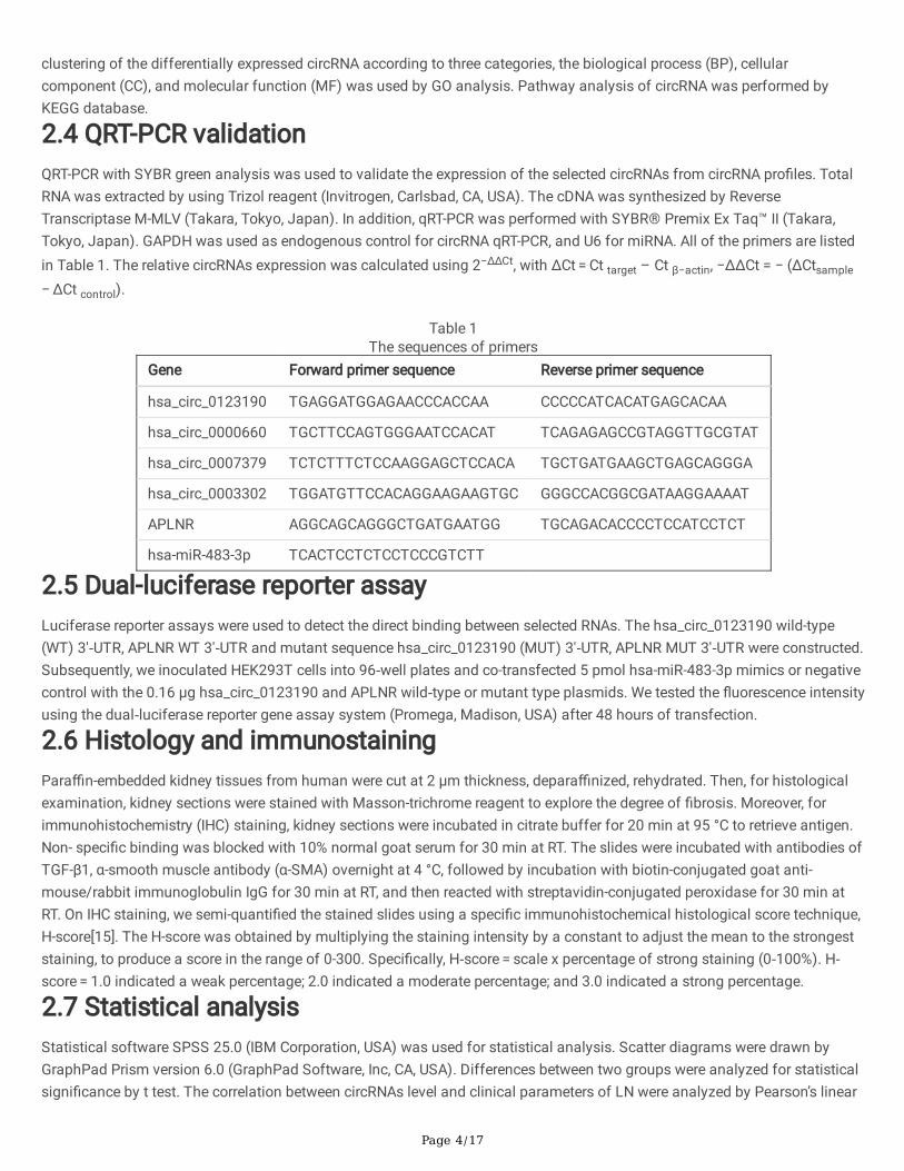
2.4 QRT-PCR validationQRT-PCR with SYBR green analysis was used to validate the expression of the selected circRNAs from circRNA pro�les. TotalRNA was extracted by using Trizol reagent (Invitrogen, Carlsbad, CA, USA). The cDNA was synthesized by ReverseTranscriptase M-MLV (Takara, Tokyo, Japan). In addition, qRT-PCR was performed with SYBR® Premix Ex Taq™ II (Takara,Tokyo, Japan). GAPDH was used as endogenous control for circRNA qRT-PCR, and U6 for miRNA. All of the primers are listedin Table 1. The relative circRNAs expression was calculated using 2−ΔΔCt, with ΔCt = Ct target – Ct β−actin, −ΔΔCt = − (ΔCtsample − ΔCt control).
Table 1The sequences of primers
Gene Forward primer sequence Reverse primer sequence
hsa_circ_0123190 TGAGGATGGAGAACCCACCAA CCCCCATCACATGAGCACAA
hsa_circ_0000660 TGCTTCCAGTGGGAATCCACAT TCAGAGAGCCGTAGGTTGCGTAT
hsa_circ_0007379 TCTCTTTCTCCAAGGAGCTCCACA TGCTGATGAAGCTGAGCAGGGA
hsa_circ_0003302 TGGATGTTCCACAGGAAGAAGTGC GGGCCACGGCGATAAGGAAAAT
APLNR AGGCAGCAGGGCTGATGAATGG TGCAGACACCCCTCCATCCTCT
hsa-miR-483-3p TCACTCCTCTCCTCCCGTCTT
2.5 Dual-luciferase reporter assayLuciferase reporter assays were used to detect the direct binding between selected RNAs. The hsa_circ_0123190 wild-type(WT) 3′‐UTR, APLNR WT 3′‐UTR and mutant sequence hsa_circ_0123190 (MUT) 3′‐UTR, APLNR MUT 3′‐UTR were constructed.Subsequently, we inoculated HEK293T cells into 96‐well plates and co-transfected 5 pmol hsa-miR-483-3p mimics or negativecontrol with the 0.16 µg hsa_circ_0123190 and APLNR wild‐type or mutant type plasmids. We tested the �uorescence intensityusing the dual‐luciferase reporter gene assay system (Promega, Madison, USA) after 48 hours of transfection.
2.6 Histology and immunostainingPara�n-embedded kidney tissues from human were cut at 2 µm thickness, depara�nized, rehydrated. Then, for histologicalexamination, kidney sections were stained with Masson-trichrome reagent to explore the degree of �brosis. Moreover, forimmunohistochemistry (IHC) staining, kidney sections were incubated in citrate buffer for 20 min at 95 °C to retrieve antigen.Non- speci�c binding was blocked with 10% normal goat serum for 30 min at RT. The slides were incubated with antibodies ofTGF-β1, α-smooth muscle antibody (α-SMA) overnight at 4 °C, followed by incubation with biotin-conjugated goat anti-mouse/rabbit immunoglobulin IgG for 30 min at RT, and then reacted with streptavidin-conjugated peroxidase for 30 min atRT. On IHC staining, we semi-quanti�ed the stained slides using a speci�c immunohistochemical histological score technique,H-score[15]. The H-score was obtained by multiplying the staining intensity by a constant to adjust the mean to the strongeststaining, to produce a score in the range of 0-300. Speci�cally, H‐score = scale x percentage of strong staining (0‐100%). H‐score = 1.0 indicated a weak percentage; 2.0 indicated a moderate percentage; and 3.0 indicated a strong percentage.
2.7 Statistical analysisStatistical software SPSS 25.0 (IBM Corporation, USA) was used for statistical analysis. Scatter diagrams were drawn byGraphPad Prism version 6.0 (GraphPad Software, Inc, CA, USA). Differences between two groups were analyzed for statisticalsigni�cance by t test. The correlation between circRNAs level and clinical parameters of LN were analyzed by Pearson’s linear
Page 5/17
correlation. ROC curves were performed, and the speci�city and sensitivity of predictive power was assessed by area undercurve (AUC). A P value < 0.05 was considered as statistically signi�cant.
3. Results
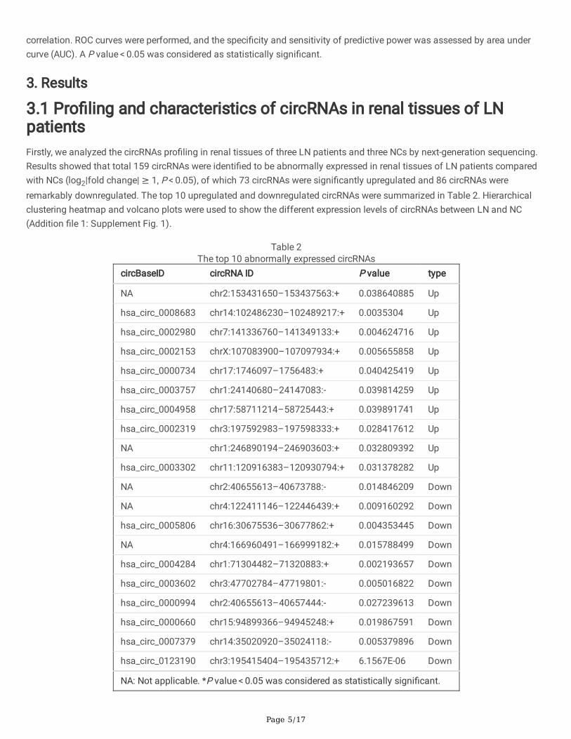
3.1 Pro�ling and characteristics of circRNAs in renal tissues of LNpatientsFirstly, we analyzed the circRNAs pro�ling in renal tissues of three LN patients and three NCs by next-generation sequencing.Results showed that total 159 circRNAs were identi�ed to be abnormally expressed in renal tissues of LN patients comparedwith NCs (log2|fold change| ≥ 1, P < 0.05), of which 73 circRNAs were signi�cantly upregulated and 86 circRNAs wereremarkably downregulated. The top 10 upregulated and downregulated circRNAs were summarized in Table 2. Hierarchicalclustering heatmap and volcano plots were used to show the different expression levels of circRNAs between LN and NC(Addition �le 1: Supplement Fig. 1).
Table 2The top 10 abnormally expressed circRNAs
circBaseID circRNA ID P value type
NA chr2:153431650–153437563:+ 0.038640885 Up
hsa_circ_0008683 chr14:102486230–102489217:+ 0.0035304 Up
hsa_circ_0002980 chr7:141336760–141349133:+ 0.004624716 Up
hsa_circ_0002153 chrX:107083900–107097934:+ 0.005655858 Up
hsa_circ_0000734 chr17:1746097–1756483:+ 0.040425419 Up
hsa_circ_0003757 chr1:24140680–24147083:- 0.039814259 Up
hsa_circ_0004958 chr17:58711214–58725443:+ 0.039891741 Up
hsa_circ_0002319 chr3:197592983–197598333:+ 0.028417612 Up
NA chr1:246890194–246903603:+ 0.032809392 Up
hsa_circ_0003302 chr11:120916383–120930794:+ 0.031378282 Up
NA chr2:40655613–40673788:- 0.014846209 Down
NA chr4:122411146–122446439:+ 0.009160292 Down
hsa_circ_0005806 chr16:30675536–30677862:+ 0.004353445 Down
NA chr4:166960491–166999182:+ 0.015788499 Down
hsa_circ_0004284 chr1:71304482–71320883:+ 0.002193657 Down
hsa_circ_0003602 chr3:47702784–47719801:- 0.005016822 Down
hsa_circ_0000994 chr2:40655613–40657444:- 0.027239613 Down
hsa_circ_0000660 chr15:94899366–94945248:+ 0.019867591 Down
hsa_circ_0007379 chr14:35020920–35024118:- 0.005379896 Down
hsa_circ_0123190 chr3:195415404–195435712:+ 6.1567E-06 Down
NA: Not applicable. *P value < 0.05 was considered as statistically signi�cant.
Page 6/17
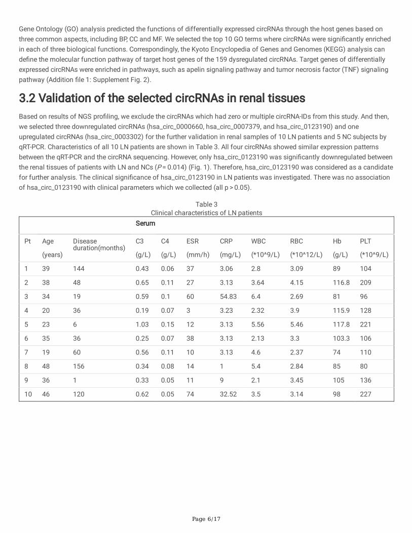
Gene Ontology (GO) analysis predicted the functions of differentially expressed circRNAs through the host genes based onthree common aspects, including BP, CC and MF. We selected the top 10 GO terms where circRNAs were signi�cantly enrichedin each of three biological functions. Correspondingly, the Kyoto Encyclopedia of Genes and Genomes (KEGG) analysis cande�ne the molecular function pathway of target host genes of the 159 dysregulated circRNAs. Target genes of differentiallyexpressed circRNAs were enriched in pathways, such as apelin signaling pathway and tumor necrosis factor (TNF) signalingpathway (Addition �le 1: Supplement Fig. 2).
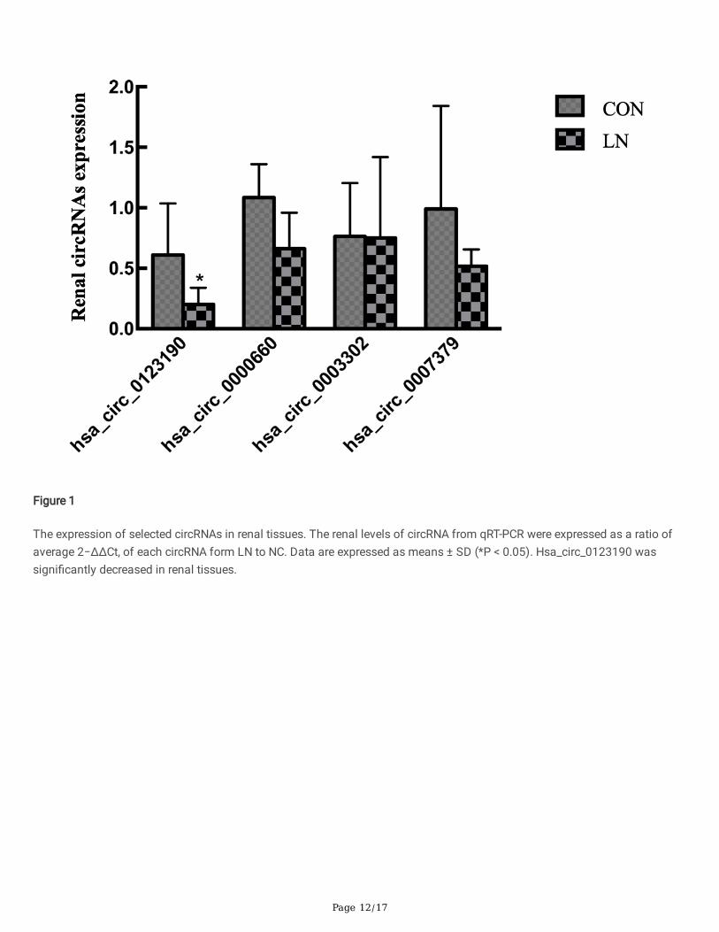
3.2 Validation of the selected circRNAs in renal tissuesBased on results of NGS pro�ling, we exclude the circRNAs which had zero or multiple circRNA-IDs from this study. And then,we selected three downregulated circRNAs (hsa_circ_0000660, hsa_circ_0007379, and hsa_circ_0123190) and oneupregulated circRNAs (hsa_circ_0003302) for the further validation in renal samples of 10 LN patients and 5 NC subjects byqRT-PCR. Characteristics of all 10 LN patients are shown in Table 3. All four circRNAs showed similar expression patternsbetween the qRT-PCR and the circRNA sequencing. However, only hsa_circ_0123190 was signi�cantly downregulated betweenthe renal tissues of patients with LN and NCs (P = 0.014) (Fig. 1). Therefore, hsa_circ_0123190 was considered as a candidatefor further analysis. The clinical signi�cance of hsa_circ_0123190 in LN patients was investigated. There was no associationof hsa_circ_0123190 with clinical parameters which we collected (all p > 0.05).
Table 3Clinical characteristics of LN patients
Serum
Pt Age
(years)
Diseaseduration(months)
C3
(g/L)
C4
(g/L)
ESR
(mm/h)
CRP
(mg/L)
WBC
(*10^9/L)
RBC
(*10^12/L)
Hb
(g/L)
PLT
(*10^9/L)
1 39 144 0.43 0.06 37 3.06 2.8 3.09 89 104
2 38 48 0.65 0.11 27 3.13 3.64 4.15 116.8 209
3 34 19 0.59 0.1 60 54.83 6.4 2.69 81 96
4 20 36 0.19 0.07 3 3.23 2.32 3.9 115.9 128
5 23 6 1.03 0.15 12 3.13 5.56 5.46 117.8 221
6 35 36 0.25 0.07 38 3.13 2.13 3.3 103.3 106
7 19 60 0.56 0.11 10 3.13 4.6 2.37 74 110
8 48 156 0.34 0.08 14 1 5.4 2.84 85 80
9 36 1 0.33 0.05 11 9 2.1 3.45 105 136
10 46 120 0.62 0.05 74 32.52 3.5 3.14 98 227
Page 7/17
Table 3Continued
Serum Urine Renal biopsy SLEDAI
(score)Pt Creatinine
(umol/L)
Urea
(mmol/L)
eGFR
(mL/min
/1.73 m2)
UTP
(g/24 h)
RBC
(/uL)
WBC
(/uL)
Classi�cation AI
(score)
CI
(score)
1 59 5.03 111.223 8.3 95 56 IV-S(A) + V 9 0 9
2 63 4.50 107.899 2.52 26 4 III-(A) 8 1 8
3 130 12.44 46.224 6.03 252 94 IV-G(A) + V 19 3 11
4 51 3.85 133.345 2.07 50 0 III-(A/C) 6 1 12
5 58 7.11 109.314 2.03 50 0 III-(A) + V 0 0 6
6 135 12.40 38.299 6.25 92 22 IV-G(A/C) 10 2 4
7 77 4.90 96.472 2.82 350 12 IV-S(A) 9 0 8
8 56 10.36 98.682 6.39 45 22 IV-G(A) 15 0 5
9 69 4.31 98.028 2.35 184 50 IV-S(A) 9 0 4
10 59 6.91 93.006 1.4 37 28 IV-S(A) 7 0 4
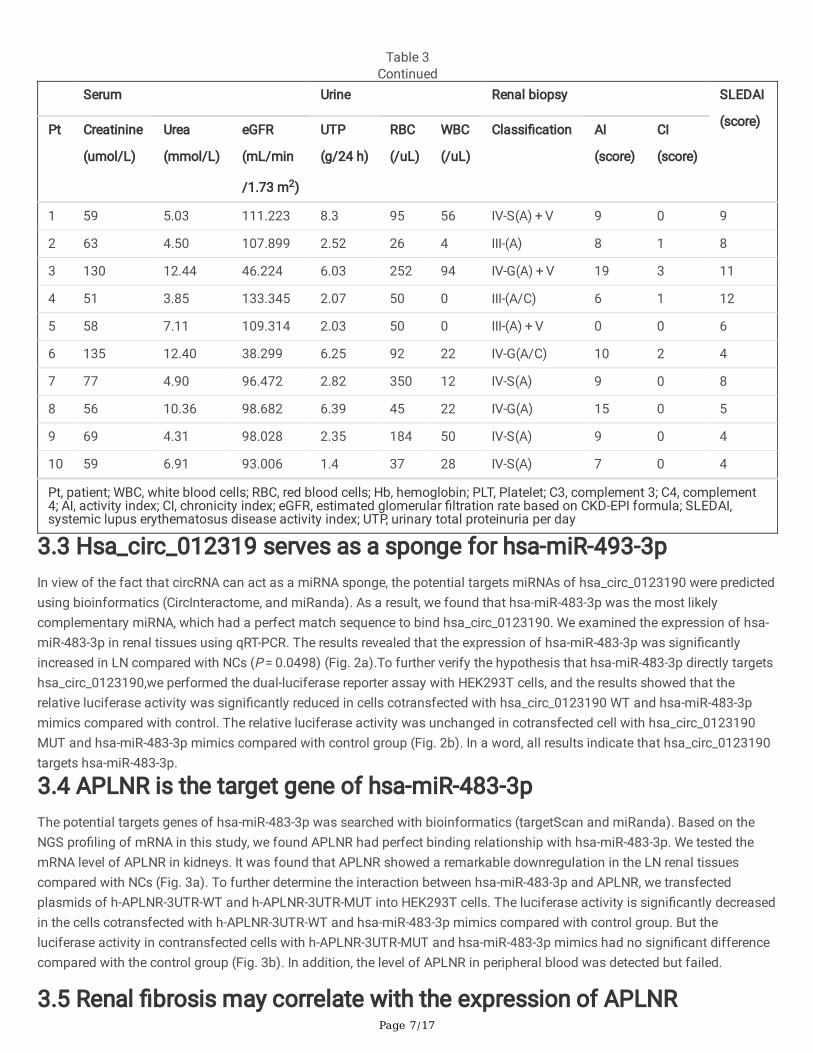
Pt, patient; WBC, white blood cells; RBC, red blood cells; Hb, hemoglobin; PLT, Platelet; C3, complement 3; C4, complement4; AI, activity index; CI, chronicity index; eGFR, estimated glomerular �ltration rate based on CKD-EPI formula; SLEDAI,systemic lupus erythematosus disease activity index; UTP, urinary total proteinuria per day
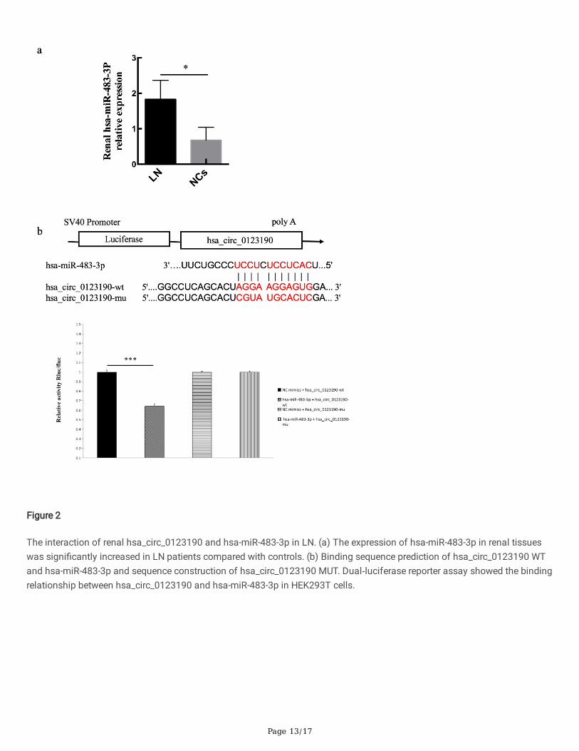
3.3 Hsa_circ_012319 serves as a sponge for hsa-miR-493-3pIn view of the fact that circRNA can act as a miRNA sponge, the potential targets miRNAs of hsa_circ_0123190 were predictedusing bioinformatics (CircInteractome, and miRanda). As a result, we found that hsa-miR-483-3p was the most likelycomplementary miRNA, which had a perfect match sequence to bind hsa_circ_0123190. We examined the expression of hsa-miR-483-3p in renal tissues using qRT-PCR. The results revealed that the expression of hsa-miR-483-3p was signi�cantlyincreased in LN compared with NCs (P = 0.0498) (Fig. 2a).To further verify the hypothesis that hsa-miR-483-3p directly targetshsa_circ_0123190,we performed the dual-luciferase reporter assay with HEK293T cells, and the results showed that therelative luciferase activity was signi�cantly reduced in cells cotransfected with hsa_circ_0123190 WT and hsa-miR-483-3pmimics compared with control. The relative luciferase activity was unchanged in cotransfected cell with hsa_circ_0123190MUT and hsa-miR-483-3p mimics compared with control group (Fig. 2b). In a word, all results indicate that hsa_circ_0123190targets hsa-miR-483-3p.
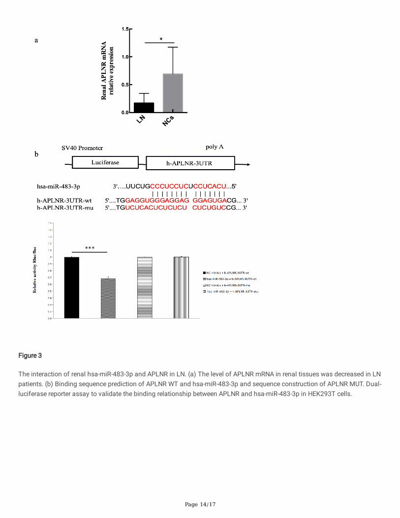
3.4 APLNR is the target gene of hsa-miR-483-3pThe potential targets genes of hsa-miR-483-3p was searched with bioinformatics (targetScan and miRanda). Based on theNGS pro�ling of mRNA in this study, we found APLNR had perfect binding relationship with hsa-miR-483-3p. We tested themRNA level of APLNR in kidneys. It was found that APLNR showed a remarkable downregulation in the LN renal tissuescompared with NCs (Fig. 3a). To further determine the interaction between hsa-miR-483-3p and APLNR, we transfectedplasmids of h-APLNR-3UTR-WT and h-APLNR-3UTR-MUT into HEK293T cells. The luciferase activity is signi�cantly decreasedin the cells cotransfected with h-APLNR-3UTR-WT and hsa-miR-483-3p mimics compared with control group. But theluciferase activity in contransfected cells with h-APLNR-3UTR-MUT and hsa-miR-483-3p mimics had no signi�cant differencecompared with the control group (Fig. 3b). In addition, the level of APLNR in peripheral blood was detected but failed.
3.5 Renal �brosis may correlate with the expression of APLNR
Page 8/17
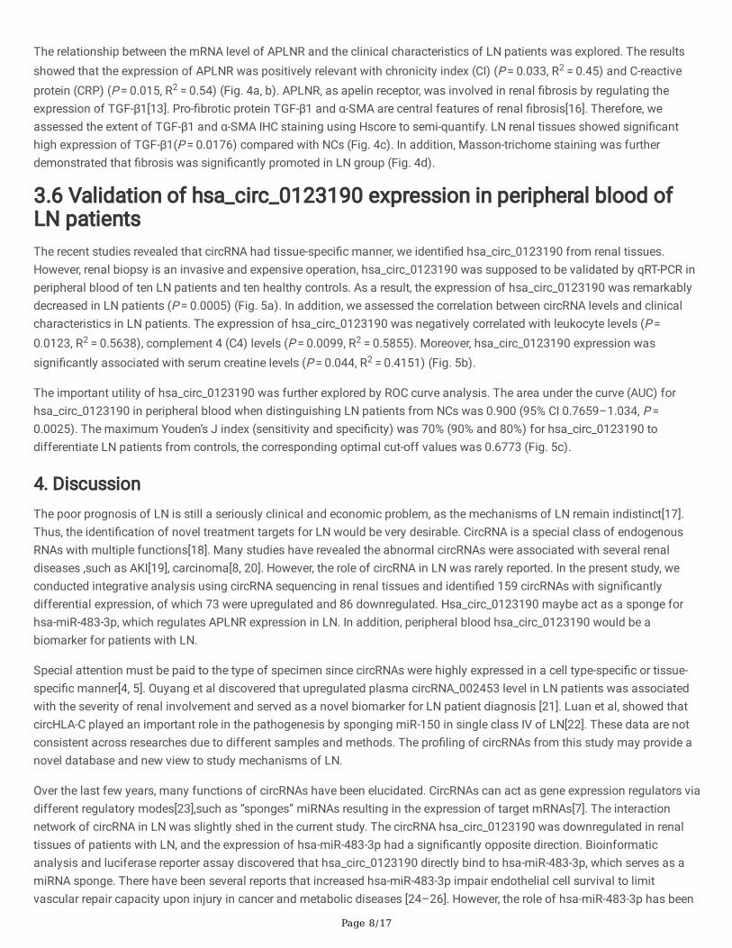
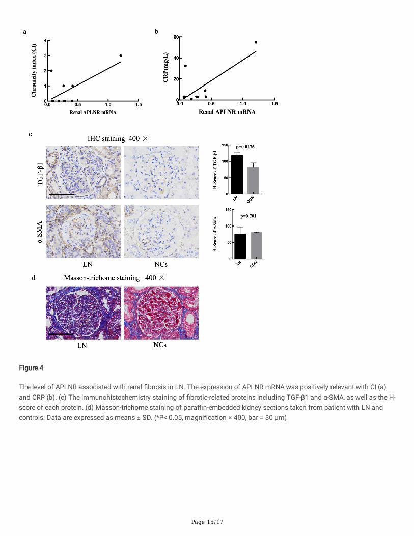
The relationship between the mRNA level of APLNR and the clinical characteristics of LN patients was explored. The resultsshowed that the expression of APLNR was positively relevant with chronicity index (CI) (P = 0.033, R2 = 0.45) and C-reactiveprotein (CRP) (P = 0.015, R2 = 0.54) (Fig. 4a, b). APLNR, as apelin receptor, was involved in renal �brosis by regulating theexpression of TGF-β1[13]. Pro-�brotic protein TGF-β1 and α-SMA are central features of renal �brosis[16]. Therefore, weassessed the extent of TGF-β1 and α-SMA IHC staining using Hscore to semi-quantify. LN renal tissues showed signi�canthigh expression of TGF-β1(P = 0.0176) compared with NCs (Fig. 4c). In addition, Masson-trichome staining was furtherdemonstrated that �brosis was signi�cantly promoted in LN group (Fig. 4d).
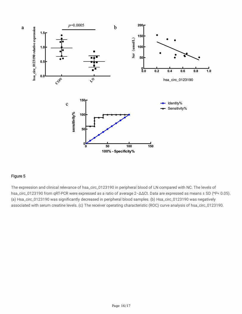
3.6 Validation of hsa_circ_0123190 expression in peripheral blood ofLN patientsThe recent studies revealed that circRNA had tissue-speci�c manner, we identi�ed hsa_circ_0123190 from renal tissues.However, renal biopsy is an invasive and expensive operation, hsa_circ_0123190 was supposed to be validated by qRT-PCR inperipheral blood of ten LN patients and ten healthy controls. As a result, the expression of hsa_circ_0123190 was remarkablydecreased in LN patients (P = 0.0005) (Fig. 5a). In addition, we assessed the correlation between circRNA levels and clinicalcharacteristics in LN patients. The expression of hsa_circ_0123190 was negatively correlated with leukocyte levels (P = 0.0123, R2 = 0.5638), complement 4 (C4) levels (P = 0.0099, R2 = 0.5855). Moreover, hsa_circ_0123190 expression wassigni�cantly associated with serum creatine levels (P = 0.044, R2 = 0.4151) (Fig. 5b).
The important utility of hsa_circ_0123190 was further explored by ROC curve analysis. The area under the curve (AUC) forhsa_circ_0123190 in peripheral blood when distinguishing LN patients from NCs was 0.900 (95% CI 0.7659–1.034, P = 0.0025). The maximum Youden’s J index (sensitivity and speci�city) was 70% (90% and 80%) for hsa_circ_0123190 todifferentiate LN patients from controls, the corresponding optimal cut-off values was 0.6773 (Fig. 5c).
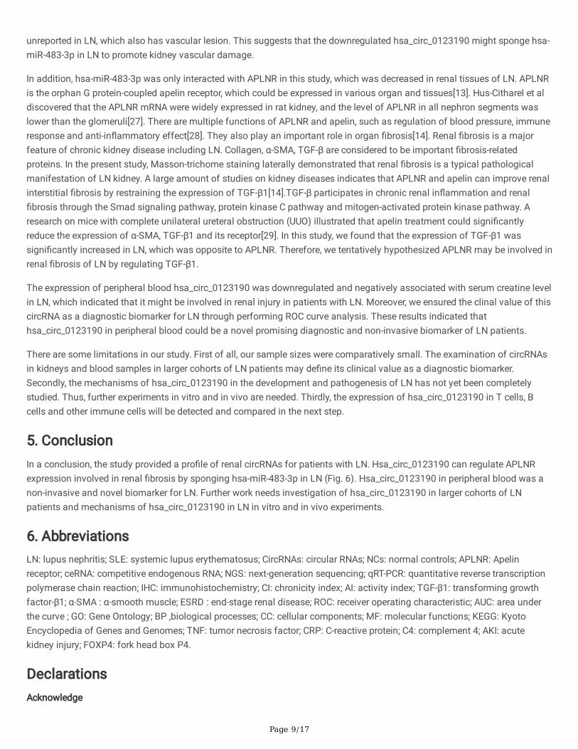
4. DiscussionThe poor prognosis of LN is still a seriously clinical and economic problem, as the mechanisms of LN remain indistinct[17].Thus, the identi�cation of novel treatment targets for LN would be very desirable. CircRNA is a special class of endogenousRNAs with multiple functions[18]. Many studies have revealed the abnormal circRNAs were associated with several renaldiseases ,such as AKI[19], carcinoma[8, 20]. However, the role of circRNA in LN was rarely reported. In the present study, weconducted integrative analysis using circRNA sequencing in renal tissues and identi�ed 159 circRNAs with signi�cantlydifferential expression, of which 73 were upregulated and 86 downregulated. Hsa_circ_0123190 maybe act as a sponge forhsa-miR-483-3p, which regulates APLNR expression in LN. In addition, peripheral blood hsa_circ_0123190 would be abiomarker for patients with LN.
Special attention must be paid to the type of specimen since circRNAs were highly expressed in a cell type-speci�c or tissue-speci�c manner[4, 5]. Ouyang et al discovered that upregulated plasma circRNA_002453 level in LN patients was associatedwith the severity of renal involvement and served as a novel biomarker for LN patient diagnosis [21]. Luan et al, showed thatcircHLA-C played an important role in the pathogenesis by sponging miR-150 in single class IV of LN[22]. These data are notconsistent across researches due to different samples and methods. The pro�ling of circRNAs from this study may provide anovel database and new view to study mechanisms of LN.
Over the last few years, many functions of circRNAs have been elucidated. CircRNAs can act as gene expression regulators viadifferent regulatory modes[23],such as “sponges” miRNAs resulting in the expression of target mRNAs[7]. The interactionnetwork of circRNA in LN was slightly shed in the current study. The circRNA hsa_circ_0123190 was downregulated in renaltissues of patients with LN, and the expression of hsa-miR-483-3p had a signi�cantly opposite direction. Bioinformaticanalysis and luciferase reporter assay discovered that hsa_circ_0123190 directly bind to hsa-miR-483-3p, which serves as amiRNA sponge. There have been several reports that increased hsa-miR-483-3p impair endothelial cell survival to limitvascular repair capacity upon injury in cancer and metabolic diseases [24–26]. However, the role of hsa-miR-483-3p has been
Page 9/17
unreported in LN, which also has vascular lesion. This suggests that the downregulated hsa_circ_0123190 might sponge hsa-miR-483-3p in LN to promote kidney vascular damage.
In addition, hsa-miR-483-3p was only interacted with APLNR in this study, which was decreased in renal tissues of LN. APLNRis the orphan G protein-coupled apelin receptor, which could be expressed in various organ and tissues[13]. Hus-Citharel et aldiscovered that the APLNR mRNA were widely expressed in rat kidney, and the level of APLNR in all nephron segments waslower than the glomeruli[27]. There are multiple functions of APLNR and apelin, such as regulation of blood pressure, immuneresponse and anti-in�ammatory effect[28]. They also play an important role in organ �brosis[14]. Renal �brosis is a majorfeature of chronic kidney disease including LN. Collagen, α-SMA, TGF-β are considered to be important �brosis-relatedproteins. In the present study, Masson-trichome staining laterally demonstrated that renal �brosis is a typical pathologicalmanifestation of LN kidney. A large amount of studies on kidney diseases indicates that APLNR and apelin can improve renalinterstitial �brosis by restraining the expression of TGF-β1[14].TGF-β participates in chronic renal in�ammation and renal�brosis through the Smad signaling pathway, protein kinase C pathway and mitogen-activated protein kinase pathway. Aresearch on mice with complete unilateral ureteral obstruction (UUO) illustrated that apelin treatment could signi�cantlyreduce the expression of α-SMA, TGF-β1 and its receptor[29]. In this study, we found that the expression of TGF-β1 wassigni�cantly increased in LN, which was opposite to APLNR. Therefore, we tentatively hypothesized APLNR may be involved inrenal �brosis of LN by regulating TGF-β1.
The expression of peripheral blood hsa_circ_0123190 was downregulated and negatively associated with serum creatine levelin LN, which indicated that it might be involved in renal injury in patients with LN. Moreover, we ensured the clinal value of thiscircRNA as a diagnostic biomarker for LN through performing ROC curve analysis. These results indicated thathsa_circ_0123190 in peripheral blood could be a novel promising diagnostic and non-invasive biomarker of LN patients.
There are some limitations in our study. First of all, our sample sizes were comparatively small. The examination of circRNAsin kidneys and blood samples in larger cohorts of LN patients may de�ne its clinical value as a diagnostic biomarker.Secondly, the mechanisms of hsa_circ_0123190 in the development and pathogenesis of LN has not yet been completelystudied. Thus, further experiments in vitro and in vivo are needed. Thirdly, the expression of hsa_circ_0123190 in T cells, Bcells and other immune cells will be detected and compared in the next step.
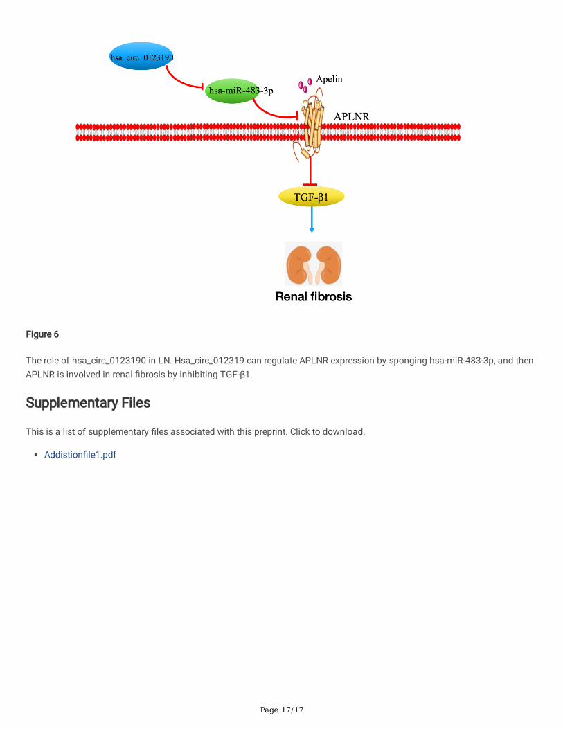
5. ConclusionIn a conclusion, the study provided a pro�le of renal circRNAs for patients with LN. Hsa_circ_0123190 can regulate APLNRexpression involved in renal �brosis by sponging hsa-miR-483-3p in LN (Fig. 6). Hsa_circ_0123190 in peripheral blood was anon-invasive and novel biomarker for LN. Further work needs investigation of hsa_circ_0123190 in larger cohorts of LNpatients and mechanisms of hsa_circ_0123190 in LN in vitro and in vivo experiments.
6. AbbreviationsLN: lupus nephritis; SLE: systemic lupus erythematosus; CircRNAs: circular RNAs; NCs: normal controls; APLNR: Apelinreceptor; ceRNA: competitive endogenous RNA; NGS: next-generation sequencing; qRT-PCR: quantitative reverse transcriptionpolymerase chain reaction; IHC: immunohistochemistry; CI: chronicity index; AI: activity index; TGF-β1: transforming growthfactor-β1; α-SMA : α-smooth muscle; ESRD : end-stage renal disease; ROC: receiver operating characteristic; AUC: area underthe curve ; GO: Gene Ontology; BP ,biological processes; CC: cellular components; MF: molecular functions; KEGG: KyotoEncyclopedia of Genes and Genomes; TNF: tumor necrosis factor; CRP: C-reactive protein; C4: complement 4; AKI: acutekidney injury; FOXP4: fork head box P4.
DeclarationsAcknowledge
Page 10/17
We thank the technical assistance and expertise of Genesky Biotechnologies Inc. (Shanghai, China) for their perfect technicalassistance.
Funding
This work was supported by the Natural Science Foundation of Henan Province of China [ grant number 162300410273].
Availability of data and materials
The datasets analyzed during the current study are available from the corresponding author on reasonable request.
Authors’ contributions
All authors have contributed su�ciently to the project. C.Z. participated in kidney sample collection, clinical data collection,data analysis, and manuscript writing. Z.Z. designed and conducted the whole experiment and �nalized the manuscript. D.D.,W.S. and W.L. participated in data collection. M.Y. and Q.W. collected kidney tissues from patients. C.G. participated in thedesign of experiments.
Ethics approval and consent to participate
The study was approved by the Ethics Committee of the First A�liated Hospital of Zhengzhou University (2018-KY-22). Allpatients provided written informed consent.
Consent for publication
Not applicable.
Competing interests
The authors declare no competing interests.
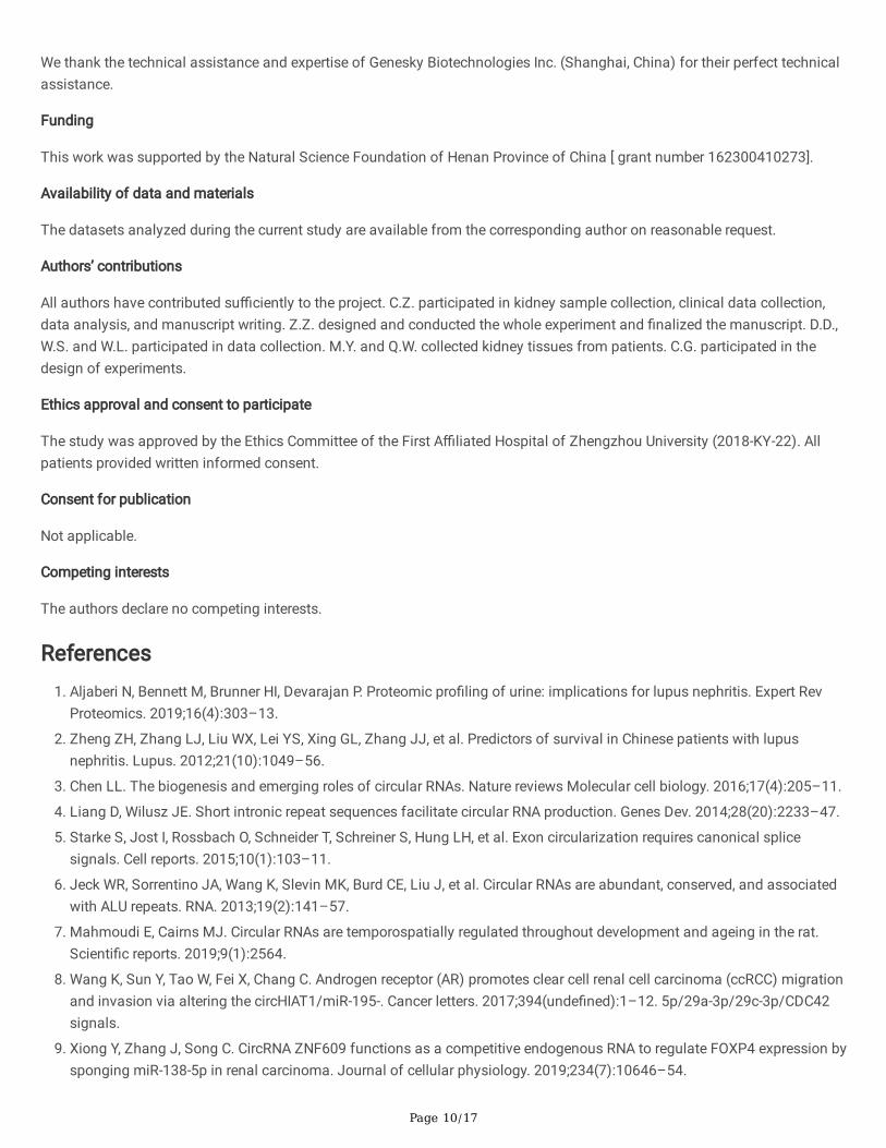
References1. Aljaberi N, Bennett M, Brunner HI, Devarajan P. Proteomic pro�ling of urine: implications for lupus nephritis. Expert Rev
Proteomics. 2019;16(4):303–13.
2. Zheng ZH, Zhang LJ, Liu WX, Lei YS, Xing GL, Zhang JJ, et al. Predictors of survival in Chinese patients with lupusnephritis. Lupus. 2012;21(10):1049–56.
3. Chen LL. The biogenesis and emerging roles of circular RNAs. Nature reviews Molecular cell biology. 2016;17(4):205–11.
4. Liang D, Wilusz JE. Short intronic repeat sequences facilitate circular RNA production. Genes Dev. 2014;28(20):2233–47.
5. Starke S, Jost I, Rossbach O, Schneider T, Schreiner S, Hung LH, et al. Exon circularization requires canonical splicesignals. Cell reports. 2015;10(1):103–11.
�. Jeck WR, Sorrentino JA, Wang K, Slevin MK, Burd CE, Liu J, et al. Circular RNAs are abundant, conserved, and associatedwith ALU repeats. RNA. 2013;19(2):141–57.
7. Mahmoudi E, Cairns MJ. Circular RNAs are temporospatially regulated throughout development and ageing in the rat.Scienti�c reports. 2019;9(1):2564.
�. Wang K, Sun Y, Tao W, Fei X, Chang C. Androgen receptor (AR) promotes clear cell renal cell carcinoma (ccRCC) migrationand invasion via altering the circHIAT1/miR-195-. Cancer letters. 2017;394(unde�ned):1–12. 5p/29a-3p/29c-3p/CDC42signals.
9. Xiong Y, Zhang J, Song C. CircRNA ZNF609 functions as a competitive endogenous RNA to regulate FOXP4 expression bysponging miR-138-5p in renal carcinoma. Journal of cellular physiology. 2019;234(7):10646–54.
Page 11/17
10. Austin HA, Muenz LR, Joyce KM, Antonovych TT, Balow JE. Diffuse proliferative lupus nephritis: identi�cation of speci�cpathologic features affecting renal outcome. Kidney international. 1984;25(4):689–95.
11. Zhou D, Liu Y. Renal �brosis in 2015: Understanding the mechanisms of kidney �brosis. Nature reviews Nephrology.2016;12(2):68–70.
12. Antushevich H, Wójcik M. Review. Apelin in disease. Clin Chim Acta. 2018;483:241–8.
13. Huang Z, Wu L, Chen L. Apelin/APJ system: A novel potential therapy target for kidney disease. Journal of cellularphysiology. 2018;233(5):3892–900.
14. Huang S, Chen L, Lu L, Li L. The apelin-APJ axis: A novel potential therapeutic target for organ �brosis. Clin Chim Acta.2016;456(unde�ned):81–8.
15. Batu ED, Erden A, Seyhoğlu E, Kilic L, Büyükasık Y, Karadag O, et al. Assessment of the HScore for reactivehaemophagocytic syndrome in patients with rheumatic diseases. Scand J Rheumatol. 2017;46(1):44–8.
1�. Djudjaj S, Boor P. Cellular and molecular mechanisms of kidney �brosis. Mol Aspects Med. 2019;65:16–36.
17. Anders HJ, Saxena R, Zhao MH, Parodis I, Salmon JE, Mohan C. Lupus nephritis. Nature reviews Disease primers.2020;6(1):7.
1�. Kristensen LS, Andersen MS, Stagsted LVW, Ebbesen KK, Hansen TB, Kjems J. The biogenesis, biology andcharacterization of circular RNAs. Nature reviews Genetics. 2019;20(11):675–91.
19. Kölling M, Seeger H, Haddad G, Kistler A, Nowak A, Faulhaber-Walter R, et al. The Circular RNA Predicts Survival inCritically Ill Patients With Acute Kidney Injury. Kidney international reports. 2018;3(5):1144–52.
20. Jin C, Shi L, Li Z, Liu W, Zhao B, Qiu Y, et al. Circ_0039569 promotes renal cell carcinoma growth and metastasis byregulating miR-34a-5p/CCL22. American journal of translational research. 2019;11(8):4935–45.
21. Ouyang Q, Huang Q, Jiang Z, Zhao J, Shi GP, Yang M. Using plasma circRNA_002453 as a novel biomarker in thediagnosis of lupus nephritis. Molecular immunology. 2018;101(unde�ned):531–8.
22. Luan J, Jiao C, Kong W, Fu J, Qu W, Chen Y, et al. circHLA-C Plays an Important Role in Lupus Nephritis by Sponging miR-150. Molecular therapy Nucleic acids. 2018;10(unde�ned):245–53.
23. Han B, Chao J, Yao H. Circular RNA and its mechanisms in disease: From the bench to the clinic. Pharmacol Ther.2018;187(unde�ned):31–44.
24. Kuschnerus K, Straessler ET, Müller MF, Lüscher TF, Landmesser U, Kränkel N. Increased Expression of miR-483-3pImpairs the Vascular Response to Injury in Type 2 Diabetes. Diabetes. 2019;68(2):349–60.
25. Abue M, Yokoyama M, Shibuya R, Tamai K, Yamaguchi K, Sato I, et al. Circulating miR-483-3p and miR-21 is highlyexpressed in plasma of pancreatic cancer. Int J Oncol. 2015;46(2):539–47.
2�. Pepe F, Visone R, Veronese A. The Glucose-Regulated In�uences Key Signaling Pathways in Cancer. Cancers.2018;10(6):unde�ned.
27. Hus-Citharel A, Bouby N, Frugière A, Bodineau L, Gasc JM, Llorens-Cortes C. Effect of apelin on glomerular hemodynamicfunction in the rat kidney. Kidney international. 2008;74(4):486–94.
2�. Wu L, Chen L, Li L. Apelin/APJ system: A novel promising therapy target for pathological angiogenesis. Clin Chim Acta.2017;466:78–84.
29. Wang LY, Diao ZL, Zhang DL, Zheng JF, Zhang QD, Ding JX, et al. The regulatory peptide apelin: a novel inhibitor of renalinterstitial �brosis. Amino Acids. 2014;46(12):2693–704.
Figures
Page 12/17
Figure 1
The expression of selected circRNAs in renal tissues. The renal levels of circRNA from qRT-PCR were expressed as a ratio ofaverage 2−ΔΔCt, of each circRNA form LN to NC. Data are expressed as means ± SD (*P < 0.05). Hsa_circ_0123190 wassigni�cantly decreased in renal tissues.
Page 13/17
Figure 2
The interaction of renal hsa_circ_0123190 and hsa-miR-483-3p in LN. (a) The expression of hsa-miR-483-3p in renal tissueswas signi�cantly increased in LN patients compared with controls. (b) Binding sequence prediction of hsa_circ_0123190 WTand hsa-miR-483-3p and sequence construction of hsa_circ_0123190 MUT. Dual‐luciferase reporter assay showed the bindingrelationship between hsa_circ_0123190 and hsa-miR-483-3p in HEK293T cells.
Page 14/17
Figure 3
The interaction of renal hsa-miR-483-3p and APLNR in LN. (a) The level of APLNR mRNA in renal tissues was decreased in LNpatients. (b) Binding sequence prediction of APLNR WT and hsa-miR-483-3p and sequence construction of APLNR MUT. Dual‐luciferase reporter assay to validate the binding relationship between APLNR and hsa-miR-483-3p in HEK293T cells.
Page 15/17
Figure 4
The level of APLNR associated with renal �brosis in LN. The expression of APLNR mRNA was positively relevant with CI (a)and CRP (b). (c) The immunohistochemistry staining of �brotic-related proteins including TGF-β1 and α-SMA, as well as the H-score of each protein. (d) Masson-trichome staining of para�n-embedded kidney sections taken from patient with LN andcontrols. Data are expressed as means ± SD. (*P< 0.05, magni�cation × 400, bar = 30 μm)
Page 16/17
Figure 5
The expression and clinical relevance of hsa_circ_0123190 in peripheral blood of LN compared with NC. The levels ofhsa_circ_0123190 from qRT-PCR were expressed as a ratio of average 2−ΔΔCt. Data are expressed as means ± SD (*P< 0.05).(a) Hsa_circ_0123190 was signi�cantly decreased in peripheral blood samples. (b) Hsa_circ_0123190 was negativelyassociated with serum creatine levels. (c) The receiver operating characteristic (ROC) curve analysis of hsa_circ_0123190.
Page 17/17
Figure 6
The role of hsa_circ_0123190 in LN. Hsa_circ_012319 can regulate APLNR expression by sponging hsa-miR-483-3p, and thenAPLNR is involved in renal �brosis by inhibiting TGF-β1.
Supplementary Files
This is a list of supplementary �les associated with this preprint. Click to download.
Addistion�le1.pdf