refining the role for adult stem cells as cancer cells of...
TRANSCRIPT

Refining the role for adult stem cells ascancer cells of originAndrew C. White and William E. Lowry
Eli and Edythe Broad Center for Regenerative Medicine, UCLA, Los Angeles, LA, USA
Jonsson Comprehensive Cancer Center, UCLA, Los Angeles, LA, USA
Molecular Biology Institute, UCLA, Los Angeles, LA, USA
Department of Molecular, Cell and Developmental Biology, UCLA, Los Angeles, LA, USA
Review
Significant progress has been made to identify the cellsat the foundation of tumorigenesis, the cancer cell oforigin (CCO). The majority of data points towards resi-dent adult stem cells (ASCs) or primitive progenitors asthe CCO for those cancers studied, highlighting theimportance of stem cells not only as propagators butalso as initiators of cancer. Recent data suggest tumorinitiation at the CCOs can be regulated through bothintrinsic and extrinsic signals and that the identity of theCCOs and their propensity to initiate tumorigenesis iscontext dependent. In this review, we summarize someof the recent findings regarding CCOs and solid tumorinitiation and highlight its relation with bona fide humancancer.
Decoding the cell of origin in cancerCancer is a complex disease due to the wide variety ofcellular and molecular mechanisms associated with itsinitiation and progression. It is accepted that cancer cellsdivide and proliferate uncontrollably because of the accu-mulation of somatic mutations in normal tissue, whichconfers a selective growth advantage in the mutated prog-eny [1]. However, the cells that make up a tumor areheterogeneous; often making it difficult to determine theCCO, which is the normal cell that acquires the mutationalload necessary to first initiate cancerous proliferation.Furthermore, since cancer is a transformative process,the cells composing advanced cancers may no longer con-tain morphological or molecular characteristics of the CCO[2]. The identity of the CCO could be critical to the genera-tion of more effective treatments and preventative strate-gies. If CCOs can be identified and targeted specifically, itwould be possible to stop cancer before it has a chance toundergo expansion. Molecular or physiological attributesspecific to CCOs could be exploited to slow or block pro-gression, thus avoiding treatments that simply kill divid-ing cells. This has led to significant recent efforts to defineCCOs for all types of cancers, and numerous lines ofevidence point towards ASCs as possible CCOs [3].
It is worth noting that CCOs are likely different fromcancer stem cells. CCOs are the first cells to initiate a
0962-8924/
� 2014 Elsevier Ltd. All rights reserved. http://dx.doi.org/10.1016/j.tcb.2014.08.008
Corresponding author: Lowry, W.E. ([email protected]).Keywords: cancer cells of origin; genetic hits; tumor initiation; stem cell niche.
tumor, but cancer stem cells exist within a growing tumorand are defined by their ability to propagate tumors whenserially transplanted [4]. Although cancer stem cells havemany properties and gene expression patterns similar toASCs, it is not clear whether there is a direct relationbetween the CCOs and cancer stem cells. It is possible andprobable that cancer stem cells evolve from cells other thanCCOs after tumor initiation. Cancer stem cells are coveredelsewhere in several important reviews [4–6]. Here, wefocus on CCOs and discuss the intrinsic and extrinsicmechanisms that regulate their ability to initiate variouscancers.
ASCs and CCOs: is there a link?ASCs make for a compelling target of tumorigenesis be-cause of several basic properties. First, they are long lived,and thus capable of persisting long enough to accumulateDNA damage. Second, ASCs in general are multipotent(sometimes unipotent), and this could explain the variabil-ity of cell types found within most tumors. Third, ASCs,while normally quiescent, do have significant self-renewalpotential, which could be critical for tumor expansion.ASCs are also capable of giving rise to a limited numberof cell types [7–11]. For example, intestinal stem cells areable to differentiate into all the various secretory cell typesof the villus, but not brain or muscle cells. Finally, experi-mental evidence from lineage tracing suggests that ASCsmay in fact be the CCOs in various solid tumors [3]. How-ever, numerous exceptions to this theory have also beenidentified, where various environmental insults appear toinfluence the nature of CCOs, and these are also discussedbelow.
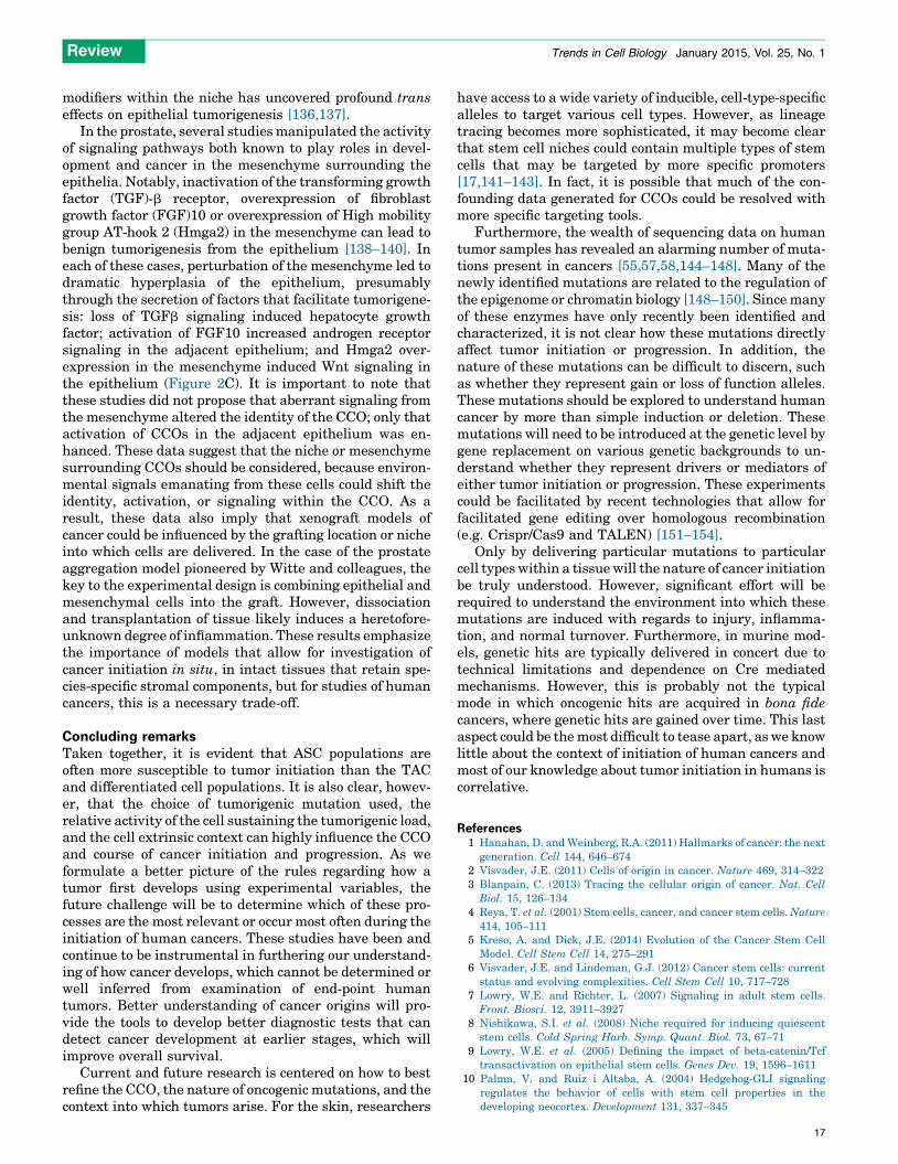
ASCs are found in many of the major adult organs and areessential for tissue homeostasis as well as regeneration inresponse to injury [12–17]. Most ASCs were discovered onthe basis of their relative quiescence and their ability toreconstitute differentiated cell lineages of the tissue ororgan in which they reside [8,18–22] (Figure 1). Either uponactivation by natural turnover/cycling or in the case ofregeneration due to injury, ASCs give rise to multilineagerestricted progenitors or, as they are often called, transitamplifying cells (TACs) (Figure 1). These cells divide rapidlyand then differentiate to generate the bulk of cells requiredfor tissue turnover or regeneration. Due to their rapiddivision, TACs are also targeted by chemotherapeutics thatact on cell division pathways to kill cancer cells, most
Trends in Cell Biology, January 2015, Vol. 25, No. 1 11

ASC
TACs
Differen�ated cells
TRENDS in Cell Biology
Figure 1. Typical ASC hierarchy. Within most mature tissues, a hierarchy exists
where cell turnover is controlled first by a self-renewing ASC. These relatively
quiescent cells give rise to TACs. TACs go through several rounds of division and
then immediately differentiate to form the various specialized cells of the tissue.
The delicate balance of cell fate decisions summarized in this figure are typically
controlled by well-known signaling pathways (such as Wnt, Tgf, Bmp, Shh, and
Fgf) acting through both autocrine and paracrine mechanisms. Abbreviations:
ASC, adult stem cell; TAC, transient amplifying cell.
Review Trends in Cell Biology January 2015, Vol. 25, No. 1
obviously manifested as loss of hair and intestinal cells. Inmost cases, TACs quickly give rise to terminally differenti-ated cells that then perform the basic functions of the tissueor organ [23]. This type of hierarchy is present in mosttissues, although tissues such as the epidermis and intestineexperience tissue turnover and stem cell cycling with higherfrequency [24]. While the identity of ASCs has not beenconfirmed in all tissues, most tissues are thought to possessthem, with a few notable and controversial exceptions (liverand pancreas). These tissues are thought to regenerate bydedifferentiation of a differentiated cell type back to aproliferative state. However, this is thought to only happenin cases of regeneration in response to tissue injury; anexample being the liver, where mature hepatocytes revertto a proliferative state in response to hepatectomy [25–30]. Cellular hierarchies based on developmental potential(ability to make more differentiated progeny) exist in alltissues, with stem cells and terminally differentiated cells atopposite ends of the spectrum.
ASCs appear to be regulated by intrinsic and extrinsicmechanisms. ASCs are intrinsically distinguished fromtheir progeny on the basis of epigenetic, transcriptional,and potentially metabolic modes of regulation [14,31–34]. Dysregulation of these intrinsic factors such as theintroduction of oncogenic mutations can result in cancerinitiation [35]. Moreover, the extrinsic environment inwhich ASCs reside also regulates their identity andactivity. ASCs live in specialized niches, which are oftenmade up of several different cell types, frequently fromdifferent embryonic germ layers [19,36]. ASCs send andreceive signals from their niche, such as growth factorsignaling, extracellular matrix association, and mechan-ical regulation. Disruption of this signaling crosstalk orchanges in the makeup of the niche can affect various
12
aspects of ASC homeostasis such as the induction of ASCproliferation. Many of the pathways important for ASC toniche crosstalk are pathways also often aberrantly regu-lated in human cancer [9,37–39]. Below, we provideevidence for the role of ASCs as the CCOs of epithelialcancers including skin, intestinal, and prostate cancer.
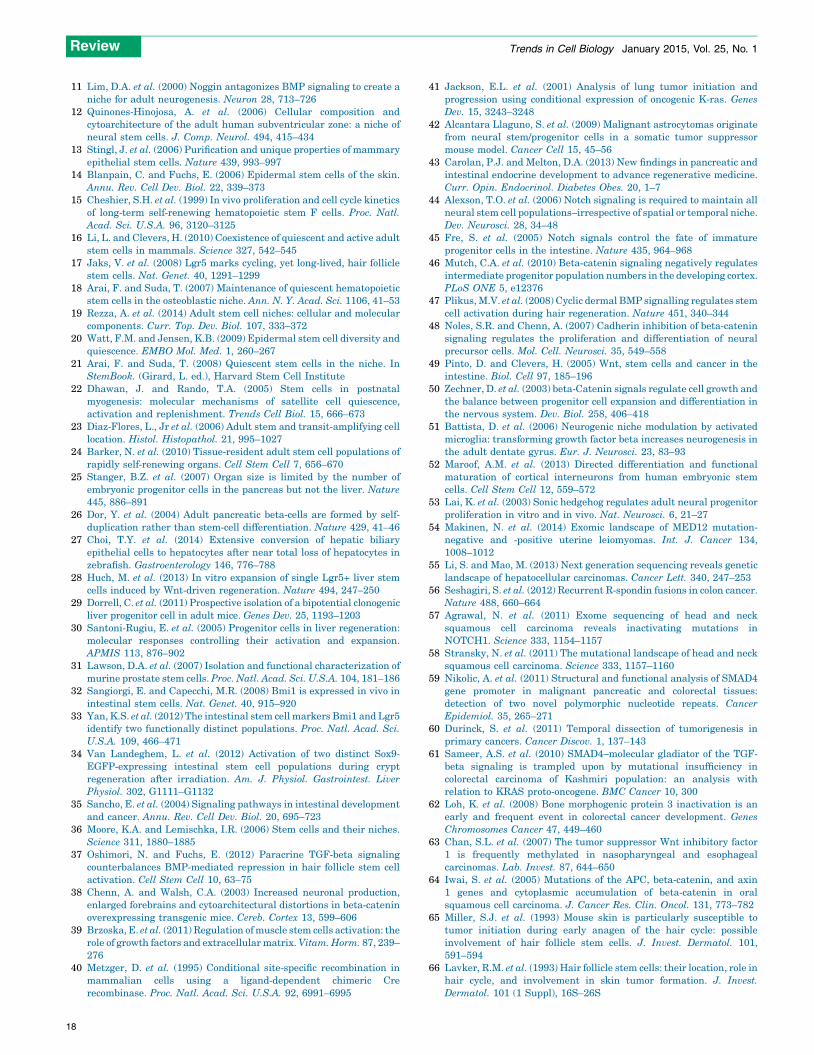
Developmental hierarchy and cancer initiationTumors are heterogeneous and can display distinct pheno-typic profiles such as morphology, gene expression, andproliferation. It has been assumed that the final morphol-ogy of the cells within a tumor can determine the CCO;however, cancer cells undergo a myriad of changes duringtumor initiation to progression, suggesting that the finaltumor cell may bear little resemblance to the CCO. There-fore, a priori, several scenarios are possible for tumorinitiation (Figure 2A). With this realization, new lineagetracing methods have sought to uncover the origin ofcancer from many tissues. Cell-type-specific promotersdriving inducible Cre recombinase alleles has allowedfor the prospective introduction of oncogenes or removalof tumor suppressors in postnatal murine models in intacttissue. These models are preferable to in vitro models orreconstitution/xenograft models as they contain the appro-priate organization of the tissue and the presence of thenative stromal, immune, lymphatic, nervous, and vascularsystems. Taking advantage of lineage tracing mechanisms(CreER/CrePR) [40] and knock-in alleles [41] of oncogenesor floxed tumor suppressors [42], one can now initiateoncogenesis from particular cell types within an adulttissue by injection of an estrogen/progesterone antagonist.These experiments have suggested that pathological, ret-rospective studies on existing tumor tissue from human ormouse could be misleading when trying to identify theCCO.
The simplest interpretation of the data produced bythese new prospective approaches is that ASCs are morelikely to serve as CCOs in many cancers [3], such as those ofthe skin, prostate, intestine, and brain. Since ASCs arecontinuously available to maintain tissue homeostasis andto repopulate cellular compartments lost during injury intissue, it has been speculated that only ASCs are present inthe tissue for a sufficient length of time to accumulate thenecessary genetic mutations for tumorigenic transforma-tion and cancer initiation (Figure 2). Below, we discuss thecurrent understanding of the CCOs of these cancers, whichrepresent a variety of solid tumors from well-describedtissues with defined hierarchies of differentiation poten-tial. We propose that the CCO is context dependent andcan change depending on intrinsic (genetic mutation andcell of origin) and extrinsic (homeostasis or injury/inflam-mation) stimuli.
Intrinsic factors influence CCOsThe developmental origins for each hierarchy could yieldinsight into the mechanisms by which tumors arise fromASCs, because the same dominant signaling pathwaysthat specify cell fate also play important roles in ASChomeostasis [7,35]. Indeed, developmental pathways in-cluding Wnt, Tgfb, Bmp, Shh, Fgf, and Notch signaling,have all been implicated in the development of epithelial

Benign tumor Malignant tumor
Malignant tumor
(A) (B)
(C)
Infla
mm
a�on
/inju
ry
TGF-βR
Fgf10
Androgenreceptor
↑ Wnt↑ HGF
Hmga2
↑TRENDS in Cell Biology
Figure 2. Tumor initiation scenarios and factors that can affect them. (A) Based on the existing literature, there are several scenarios by which tumor initiation could occur in
the cell types of the stem cell hierarchy. Retrospective pathological studies have suggested that differentiated cells can initiate cancers, while prospective approaches to the
study of cancer initiation using molecular genetics suggest that either stem or transit-amplifying cells are more relevant. (B) Data in various tissues proposes models
whereby tumor initiation in unperturbed tissue follows scenario A. However, upon induction of dramatic changes to the microenvironment of tumor initiation, CCOs do not
necessarily follow a typical stem cell hierarchy. There are several examples where terminally specified cells can dedifferentiate to create a cell that adopts stem cell
properties and is thus able to become a CCO. (C) Changes to the signaling microenvironment can also affect tumor initiation. The same signaling pathways that are known
to drive development of tissues are also implicated in tumor initiation and progression. For example, in the prostate, when stromal Tgfb signaling is reduced, HGF is
induced in the epithelium, leading to proliferation. In addition, upregulated expression of Fgf10 and Hmga2 in the meschyme can lead to increased androgen receptor
signaling and Wnt signaling in the adjacent epithelium (respectively), again leading to abnormal epithelial proliferation. These cases are demonstrative of
microenvironmental aberrations that can potentially enhance or induce epithelial cancer. Abbreviations: CCO, cancer cell of origin; Fgf10, fibroblast growth factor 10;
HGF, hepatocyte growth factor; Hmga2, high mobility group AT-hook 2.
Review Trends in Cell Biology January 2015, Vol. 25, No. 1
tissues, and for many, also in the homeostasis and propor-tion of ASCs and their progeny [9,37,43–53]. Gain or loss offunction in these pathways often disrupts the balancebetween ASCs and their progeny and can act as driversof tumor initiation. ASCs from epithelial tissue sharesimilar regulatory schemes and routes to tumor initiation,therefore, it could be that each of them also shares defensemechanisms to prevent aberrant growth, and that lessonslearned in one could be applicable to all. The degree towhich authentic tumor initiation is caused by an imbalanceof these pathways to maintain homeostasis versus moredramatic genetic alterations (activation of oncogenes, lossof tumor suppressors) has only been explored experimen-tally in murine models. However, correlative evidence fromgenome sequencing in human tumors suggests the possi-bility that disruption of these pathways could lead to
excess proliferation that is then exacerbated by oncogeneexpression or loss of tumor suppressors [54–64].
We discuss several examples of how the accumulation ofoncogenic mutations and aberrant signaling of develop-mental pathways can promote tumor formation in a cell-type-dependent manner. Furthermore, we discuss theemerging concept of stem cell quiescence as a barrier totumorigenesis suggesting intrinsic cell cycle dependentchanges may also regulate tumor initiation.
Oncogenic mutations in ASCs initiate cutaneous
squamous cell carcinoma (SCC)
Conflicting retrospective pathological studies and experi-mental evidence have made it difficult to define the CCO ofcutaneous SCC. Since it is pathologically defined by thepresence of squamous cells, or terminally differentiated
13

Review Trends in Cell Biology January 2015, Vol. 25, No. 1
cells from the interfollicular epidermis, and not from thehair follicle, it was assumed that SCC arose from differen-tiated cells of the interfollicular epidermis, and not fromthe ASC population nor from hair follicles. By contrast,experimental evidence implicated cells of the hair follicle inSCC initiation [65–67]: the rate of tumor formation wasaffected by depilation, or hair removal [68]; and deletion ofa hair follicle stem cell specific gene (CD34) affected tumorinitiation [69].
Therefore, the question remained, does oncogenic mu-tation in ASCs or TACs of the hair follicle result in SCC?First, it is important to point out the convention thatit takes multiple genetic hits to create bona fide cancer[70–72]. In murine models, the consensus is 2–3 hits, whilein human settings, it is thought that at least three hits arerequired to transform cells. Of course, genetic background(sensitizing alleles [73]), injury, and inflammation can allaffect the number of hits that lead to individual tumors, butregardless, cells do implement defense mechanisms toavoid transformation. Loss of defense mechanisms suchas tumor suppressors alone is insufficient to drive cancer,and typically, a proliferative stimulus is also required[74]. Recent lineage tracing revealed that the expressionof oncogenic Kras (KrasG12D) and the conditional deletionof tumor suppressor p53 in hair follicle ASCs (through theuse of the K15-CrePR allele) resulted in the initiation oftumors, while no tumors were observed in its descendentTACs (through the use of the Shh–CreER allele), whichwere also exposed to the same oncogenic mutation[75,76]. Furthermore, the coupling of Dimethylbenzan-thracene/tetradecanoylphobol-acetate (DMBA/TPA) chem-ical carcinogenesis to lineage tracing demonstrated that
Table 1. Experimental protocols and results for two well-explored
Tumor type Cre driver Cell type Oncogenes/tumor
suppressors lineage trac
BCC K14Cre-ER
ShhCre-ER
K15Cre-ER
Rosa-LSL-SmoM2
K15Cre-PR
K14Cre-ER
Ptch1+/1 +
Ionizing Radiation+
LSY-YFP
+p53 KO
K15Cre-PR
Lgr5Cre-ER
K5Cre-ER
Rosa-LSL-rtTA
tetO-Gli2deltaN
K15Cre-PR Rosa-LSL-SmoM2 +
wounding
K5Cre-tTA
K5Cre-PR
Lgr5Cre-ER
TRE-Gli1
Ptch1 flox/flox
Ptch1 flox/flox
Intestinal
Adenoma
Cd133Cre-ER Activated b-catenin
Lgr5Cre-ER
AH-Cre
APC KO
Xbp1sCre-ER Activated
b-catenin and
nuclear factor of kappa
light polypeptide gene
enhancer in B cells
inhibitor, alpha (IkB-a) K
KrasG12D
14
the vast majority of SCC tumors derived from hair follicleASCs, even when all cells of the epidermis received acarcinogenic insult [77]. Together, these data suggestnot only that SCC can arise from the hair follicle, but thatthe cells with the greatest developmental capacity, in thiscase ASCs, can serve as CCOs.
Mutation of developmental pathways in ASCs results in
basal cell carcinoma (BCC)
BCC is often characterized by aberrant Sonic Hedgehog(SHH) signaling. Indeed, the loss of SHH pathway compo-nent Patched (Ptch1) is found in 30–60% of human BCCs,while activated Smoothened (Smo) is found in 10–20% ofhuman BCCs [78]. SHH signaling has been observed toregulate ASCs and is involved in the maintenance of tissuehomeostasis. The binding of SHH to the receptor Ptch1results in its repression and activation of Smo, whichpromotes the nuclear translocation of Gli transcriptionfactors, consequently increasing the self-renewal capacityof some ASCs [10].
BCC was thought to derive from the hair follicle due itscellular similarity to the hair follicle [78]. However, usingan inducible Cre allele, driven by either a basal keratino-cyte promoter (Keratin 14), a hair follicle TAC promoter(Shh), or a hair follicle stem cell promoter (Keratin 15) todrive expression of activated Smo, it was found that cellsadjacent to the follicle but within the interfollicular epi-dermis were the CCOs (Table 1) [79]. Coupled with carefultemporal analysis, it was demonstrated that BCC tumorsfirst initiate at the junction between follicles and theinterfollicular epidermis. By contrast, another group sug-gested that hair follicle stem cells are the CCO for BCC in a
CCO models
er
CCO Refs
Interfollicular epidermis [79]
HFSCs
HFSCs and interfollicular epidermis
[80]
HFSCs = nodular BCC
Interfollicular epidermis = superficial BCC
[81]
HFSCs [126]
Interfollicular epidermis
HFSCs only when wounded
[124]
Crypt stem cells [87]
Crypt stem cells [82]
O or
Differentiated cells via dedifferentiation [132]

Review Trends in Cell Biology January 2015, Vol. 25, No. 1
model that used heterozygous loss of function in Ptch1 viaeither the Keratin 15 or Keratin 14 Cre and ionizingradiation as a mutagen [80]. However, the additional lossof p53 in the interfollicular epidermis facilitated initiationof BCC from this site. Additionally, a recent finding sug-gested that both interfollicular and hair follicle stem cellscould give rise to BCC using a variety of lineage drivers(K15, Lgr5, K5, and K14) and a different oncogenic allele[81]. A constitutively active allele of the glioma-associatedoncogene homolog 2 (Gli2) transcription factor, whichmimics the activation of the Shh pathway, led to theformation of two different BCC subtypes from either theinterfollicular epidermis or the hair follicle [81]. While it ispossible that the differences lay in the sensitivity of variouscell types to the dose of pathway activation, these results,coupled with those from SCC experiments demonstratethat mutation in an appropriate cell of origin can lead tospecific cancer subtypes. Whether the same is true formurine models of tumor initiation in other tissues is notyet clear, as so far, most models have only shown oneparticular cell type to be the cell of origin in a given tissue.
Two populations of CCOs in the intestine
Recent lineage tracing experiments in the small intestinehave produced a variety of models for the identity of ASCs.There appear to be at least two candidates for ASCs in theintestinal crypt; one at the base between Paneth cells,called the crypt base columnar cells (CBCs), and the otherroughly four cells up from the bottom of the crypt, namedthe +4 cells. The CBCs are marked by Lgr5, whereas the +4cells are labeled by Bmi1. A variety of lineage tracing andlineage ablation protocols have been used to resolve thestem cell hierarchy in these crypts (Table 1) [32,33,82–91]. It seems that the Lgr5/CD133+ CBCs are important innormal homeostasis of the villus, dividing once a day,whereas the +4 Bmi1+ cells are a reserve stem cell popula-tion that can replenish the CBCs after injury [33,90]. Fur-thermore, ASCs residing in the base of the crypt give rise toa TAC population found higher up in the crypt, which inturn gives rise to a variety of differentiated cell types thatpopulate the villus. With regard to tumorigenesis, bothLgr5/CD133+ CBCs and Bmi1+ +4 cells are capable ofacting as CCOs in adenoma initiation, when Apc is condi-tionally deleted, whereas TACs residing further up thevillus appear to be less sensitive to transformation, at leastin undisturbed intestinal epithelium [33,82,87,92,93]. Asadenomatous polyopsis coli (APC) is a critical scaffold forcomponents of the Wnt signaling pathway, this is alsofurther evidence where cancer can exploit a developmentalsignaling or cell fate pathway to drive transformation [94].
Prostate cancer: luminal or basal origin?
Models for both murine and human prostate cancer haveproduced conflicting conclusions within the field as towhether the CCO is of basal or luminal origin. Debatehas arisen as to whether the stem cells of the prostate resideineither thebasalor luminalpopulations [31,95–101]. Usinga broader range of lineage tracing alleles, it was suggestedthat a multipotent population arises from the basal popula-tion, while separate unipotent progenitors populate theneuroendocrine and luminal pools [102]. The lack of a
consensus on the identity of ASCs of the prostate has alsoclouded the interpretation of CCO studies for the prostate.
Similar to the discrepancies observed for SCC/BCC,much of the debate regarding CCOs for prostate cancercenters on the fact that prostate tumors typically adopt amorphology consistent with a luminal origin, while experi-mental data often point towards a basal source for CCOs.Human prostatic epithelial transplantation studies, whichdo not include a native stromal and immune component,indicated a basal CCO with MYC, AKT or ERG as onco-genic drivers. By contrast, genetically modified mousemodels that used Pten deletion implicated both basaland luminal cells as CCOs, depending on the targetingalleles and tumorigenic strategies used [100–103]. In ad-dition, one study showed that initiation from human basalcells generates transformed luminal-like cells that are ableto propagate the tumor [95]. Together, these results sug-gest that the identity of the CCO for prostate cancer couldbe dependent on cellular, genetic, and environmental con-texts, and further work will be needed to address whetherdifferences exist between human and mouse models sys-tems or whether the differences are caused by nonequiva-lent cell-intrinsic and cell-extrinsic stimuli.
Heterogeneity of tumor initiators and tumor phenotypes
The experimental models described here have proven toyield important insights into tumor initiation and CCOs.However, there are technical limitations to these modelsthat ignore the heterogeneity of bona fide cancer initiation.Tumors are thought to be initiated in a clonal fashion as aresult of mutations in individual cells surrounded by rela-tively normal cells, therefore, it is postulated that theheterogeneity observed in many tumors is due to eithersubtle variations across seemingly homogenous CCO popu-lations; the multipotency of a single CCO; the adaptation toselective pressures within the tumor during progression, orby the resulting inflammation that typically accompaniestumor formation (discussed below). New data also suggestsanother possibility: interactions between individual tu-mor-initiating clones lead to diverse phenotypes[104]. The conditional or inducible Cre alleles used inthe experimental designs described above allow for target-ing of oncogenic hits to individual cells, but it is difficult tothen observe the various events associated with tumorprogression. In addition, if observations are not made untilhigh grade tumors are formed, it is difficult to determinewhether the tumors observed arose clonally or from severaladjacent cells. Therefore, these models cannot easily iden-tify the source of tumor heterogeneity. Alternatively, tu-mor initiation followed by careful lineage tracing canreveal the existence of specialized tumor-propagating cells,or cancer stem cells [105,106]. In addition, this type ofprotocol has also shown that tumor-propagating cells tendto expand over time in at least one malignant cancer model[107]. As murine models of cancer initiation increase insophistication, it is likely that some combination of clonallineage trace coupled with bar-coding could reveal thesource of tumor heterogeneity. For now, the heterogeneityof phenotypes observed in many murine models could be atechnical limitation that creates confounding interpreta-tions and unnecessary controversies.
15

Review Trends in Cell Biology January 2015, Vol. 25, No. 1
Intrinsic quiescence of ASCs as a tumor suppressor
Another key aspect of ASC biology that could potentiallyinfluence their propensity to initiate tumors relates to theirinherent quiescence. In the hair follicle, ASCs go throughsynchronized waves of proliferation at the start of hair cyclesto regenerate the bottom of the follicle and make a new hairshaft [47,108–110]. They then return to quiescence until thenext hair cycle, which can take several weeks. As a result,ASCs in the hair follicle are only proliferative for briefperiods. Recent work showed that when these cells arequiescent, they are unable to respond to the induction ofthe oncogene Ras and/or removal of the tumor suppressorp53 [111]. These data demonstrate that cellular quiescencesuppresses tumor growth, which is dependent on the tumorsuppressor Pten, as deletion of Pten alone had no effect onthe hair cycle. Activation of the hair cycle by normal mecha-nisms can act as an additional tumor-promoting insult toASCs that have already accumulated genetic hits. The haircycle is controlled by a variety of signaling pathways thatconverge on the stem cell niche. Some of these signals arelocal (Wnt, BMP, Fgf, Tgfb), while others are systemic(hormones) [108,110,112]. This quiescent state may helpexplain why sun-exposed skin does not continually generatenew tumors or papillomas despite the presence of Rasmutations, as regulation of these pathways serves to main-tain the appropriate activation state of this stem cell popu-lation (inhibitory: Bmp and Fgf18; stimulatory: Wnt andTgfb) [37,113–118].
These results also suggest that the regulatory mecha-nisms that affect the hair cycle could, as a result, affecttumor initiation [47,108,110,118,119]. Furthermore, it ispossible that studies implicating particular genes or sig-naling pathways in tumor initiation such as Ras, couldhave indirectly impacted tumor initiation by affecting thehair cycle instead. Indeed, loss of Pten by itself did notaffect initiation or progression of the hair cycle, but insteadaffected the sensitivity of the ASCs to Ras activation[111]. Whether the dependence on hair cycle status isspecific for Ras-induced tumors remains unclear becausea wide variety of genetic hits have been implicated incutaneous and head and neck SCC [57,58]. Whether ASCsin other tissues are also similarly refractory to transfor-mation and tumor initiation due to tumor suppressionmediated by quiescence remains to be determined.
Extrinsic factors influence CCOWhile intrinsic factors such as the ones discussed abovecan determine the CCO of many epithelial cancers, recentevidence has suggested that extrinsic factors such as inju-ry, inflammation, and signals from the stem cell niche canalso reprogram cells to become CCOs. These findingssuggest other cells beyond ASCs can also act as CCOsunder varying conditions. We discuss the role of theseextrinsic factors on redefining the CCO of epithelial cancer.
Does inflammation or injury reprogram TACs or
differentiated cells to initiate tumors?
It is unclear whether acute or chronic inflammation is neces-sary for, or coincident to, tumor initiation (Figure 2B). Insupport of a role for inflammation, many cancers have beenalso associated with chronic inflammatory diseases or
16
wounds [120–123]. If inflammation is a necessary componentin tumor initiation, animal models that do not take inflam-mation into account might have fewer experimental variablesto contend with, but could also fail to accurately model diseaseas it occurs in human tissues.
As previously mentioned, cells of the interfollicularepidermis are considered the CCO for BCC [79]; however,several reports suggest that the CCO for BCC can changewhen skin is sensitized by inflammation or injury [124–126]. It has been known for some time that hair folliclestem cells (HFSCs) can migrate out of the follicle towardsinterfollicular wounds to facilitate healing [127,128]. Thisphenomenon can complicate the interpretation of lineagetracing experiments as it impairs the ability to identify theorigin of cells that reconstitute a wound site. Using strate-gies employing Smo activation, Ptch1 inactivation, or Gli1overexpression in the hair follicle stem cell population, itwas shown that injury can enable HFSCs to act as CCOsfor BCC [124,126]. When the Shh pathway is activatedduring wound healing, tumorigenesis is initiated, evenfrom HFSCs which are normally refractive to tumorigene-sis at lower doses of Shh. Similarly, differentiated endo-crine cells of the pancreas are able to change fate and serveas CCOs for pancreatic ductal adenocarcinoma when ex-perimental chronic pancreatic injury is induced [129]. Inthe intestine, inflammation and injury also disrupt thebasic hierarchy and expand the pool of possible CCOs[34,91,130–133]. In particular, a recent report demonstrat-ed that the activation of the pro-inflammatory nuclearfactor (NF)-kb pathway increased the relative dose ofoncogenic Wnt stimulation by stabilizing b-catenin, theobligatory signal transducer for the Wnt pathway. Thissignificantly enhanced downstream Wnt signaling, caus-ing robust dedifferentiation of terminally differentiatedvillus cells expressing markers of stem cells, which wereable to generate intestinal adenomas [132]. This studysuggests inflammation could alter the CCOs by inducingcell identity changes and imparting stem cell character-istics on TACs or differentiated cells [132,134]. To supportthis finding, mice treated with infectious bacteria showedprostatic inflammation, which resulted in the differentia-tion of basal cells towards the luminal lineage [135].
The possibility that inflammation can reprogram cellfate could confound the ability to relate results from mu-rine models to actual human tumors, where inflammationis always associated with either tumor initiation or pro-gression. Whether authentic human CCOs are redefined byinflammation or injury will remain unclear until moresuitable models are generated.
Regulation of tumorigenesis by the stem cell niche
While much of the focus on the origins of epithelial tumorshas been on ASCs and cell intrinsic tumorigenic stimuli, itis important to consider the potential contribution of theniche where these cells reside (Figure 2C). It is possiblethat alterations in paracrine signaling from niche cellscould enhance, enable, or alter the CCO during tumorinitiation from ASCs. Indeed, recent studies confirm thatepithelial tumors can be generated through cell-extrinsicmeans via the microenvironment. Significantly, both gainand loss of function experiments for various signaling
Review Trends in Cell Biology January 2015, Vol. 25, No. 1
modifiers within the niche has uncovered profound transeffects on epithelial tumorigenesis [136,137].
In the prostate, several studies manipulated the activityof signaling pathways both known to play roles in devel-opment and cancer in the mesenchyme surrounding theepithelia. Notably, inactivation of the transforming growthfactor (TGF)-b receptor, overexpression of fibroblastgrowth factor (FGF)10 or overexpression of High mobilitygroup AT-hook 2 (Hmga2) in the mesenchyme can lead tobenign tumorigenesis from the epithelium [138–140]. Ineach of these cases, perturbation of the mesenchyme led todramatic hyperplasia of the epithelium, presumablythrough the secretion of factors that facilitate tumorigene-sis: loss of TGFb signaling induced hepatocyte growthfactor; activation of FGF10 increased androgen receptorsignaling in the adjacent epithelium; and Hmga2 over-expression in the mesenchyme induced Wnt signaling inthe epithelium (Figure 2C). It is important to note thatthese studies did not propose that aberrant signaling fromthe mesenchyme altered the identity of the CCO; only thatactivation of CCOs in the adjacent epithelium was en-hanced. These data suggest that the niche or mesenchymesurrounding CCOs should be considered, because environ-mental signals emanating from these cells could shift theidentity, activation, or signaling within the CCO. As aresult, these data also imply that xenograft models ofcancer could be influenced by the grafting location or nicheinto which cells are delivered. In the case of the prostateaggregation model pioneered by Witte and colleagues, thekey to the experimental design is combining epithelial andmesenchymal cells into the graft. However, dissociationand transplantation of tissue likely induces a heretofore-unknown degree of inflammation. These results emphasizethe importance of models that allow for investigation ofcancer initiation in situ, in intact tissues that retain spe-cies-specific stromal components, but for studies of humancancers, this is a necessary trade-off.
Concluding remarksTaken together, it is evident that ASC populations areoften more susceptible to tumor initiation than the TACand differentiated cell populations. It is also clear, howev-er, that the choice of tumorigenic mutation used, therelative activity of the cell sustaining the tumorigenic load,and the cell extrinsic context can highly influence the CCOand course of cancer initiation and progression. As weformulate a better picture of the rules regarding how atumor first develops using experimental variables, thefuture challenge will be to determine which of these pro-cesses are the most relevant or occur most often during theinitiation of human cancers. These studies have been andcontinue to be instrumental in furthering our understand-ing of how cancer develops, which cannot be determined orwell inferred from examination of end-point humantumors. Better understanding of cancer origins will pro-vide the tools to develop better diagnostic tests that candetect cancer development at earlier stages, which willimprove overall survival.
Current and future research is centered on how to bestrefine the CCO, the nature of oncogenic mutations, and thecontext into which tumors arise. For the skin, researchers
have access to a wide variety of inducible, cell-type-specificalleles to target various cell types. However, as lineagetracing becomes more sophisticated, it may become clearthat stem cell niches could contain multiple types of stemcells that may be targeted by more specific promoters[17,141–143]. In fact, it is possible that much of the con-founding data generated for CCOs could be resolved withmore specific targeting tools.
Furthermore, the wealth of sequencing data on humantumor samples has revealed an alarming number of muta-tions present in cancers [55,57,58,144–148]. Many of thenewly identified mutations are related to the regulation ofthe epigenome or chromatin biology [148–150]. Since manyof these enzymes have only recently been identified andcharacterized, it is not clear how these mutations directlyaffect tumor initiation or progression. In addition, thenature of these mutations can be difficult to discern, suchas whether they represent gain or loss of function alleles.These mutations should be explored to understand humancancer by more than simple induction or deletion. Thesemutations will need to be introduced at the genetic level bygene replacement on various genetic backgrounds to un-derstand whether they represent drivers or mediators ofeither tumor initiation or progression. These experimentscould be facilitated by recent technologies that allow forfacilitated gene editing over homologous recombination(e.g. Crispr/Cas9 and TALEN) [151–154].
Only by delivering particular mutations to particularcell types within a tissue will the nature of cancer initiationbe truly understood. However, significant effort will berequired to understand the environment into which thesemutations are induced with regards to injury, inflamma-tion, and normal turnover. Furthermore, in murine mod-els, genetic hits are typically delivered in concert due totechnical limitations and dependence on Cre mediatedmechanisms. However, this is probably not the typicalmode in which oncogenic hits are acquired in bona fidecancers, where genetic hits are gained over time. This lastaspect could be the most difficult to tease apart, as we knowlittle about the context of initiation of human cancers andmost of our knowledge about tumor initiation in humans iscorrelative.
References1 Hanahan, D. and Weinberg, R.A. (2011) Hallmarks of cancer: the next
generation. Cell 144, 646–6742 Visvader, J.E. (2011) Cells of origin in cancer. Nature 469, 314–3223 Blanpain, C. (2013) Tracing the cellular origin of cancer. Nat. Cell
Biol. 15, 126–1344 Reya, T. et al. (2001) Stem cells, cancer, and cancer stem cells. Nature
414, 105–1115 Kreso, A. and Dick, J.E. (2014) Evolution of the Cancer Stem Cell
Model. Cell Stem Cell 14, 275–2916 Visvader, J.E. and Lindeman, G.J. (2012) Cancer stem cells: current
status and evolving complexities. Cell Stem Cell 10, 717–7287 Lowry, W.E. and Richter, L. (2007) Signaling in adult stem cells.
Front. Biosci. 12, 3911–39278 Nishikawa, S.I. et al. (2008) Niche required for inducing quiescent
stem cells. Cold Spring Harb. Symp. Quant. Biol. 73, 67–719 Lowry, W.E. et al. (2005) Defining the impact of beta-catenin/Tcf
transactivation on epithelial stem cells. Genes Dev. 19, 1596–161110 Palma, V. and Ruiz i Altaba, A. (2004) Hedgehog-GLI signaling
regulates the behavior of cells with stem cell properties in thedeveloping neocortex. Development 131, 337–345
17
Review Trends in Cell Biology January 2015, Vol. 25, No. 1
11 Lim, D.A. et al. (2000) Noggin antagonizes BMP signaling to create aniche for adult neurogenesis. Neuron 28, 713–726
12 Quinones-Hinojosa, A. et al. (2006) Cellular composition andcytoarchitecture of the adult human subventricular zone: a niche ofneural stem cells. J. Comp. Neurol. 494, 415–434
13 Stingl, J. et al. (2006) Purification and unique properties of mammaryepithelial stem cells. Nature 439, 993–997
14 Blanpain, C. and Fuchs, E. (2006) Epidermal stem cells of the skin.Annu. Rev. Cell Dev. Biol. 22, 339–373
15 Cheshier, S.H. et al. (1999) In vivo proliferation and cell cycle kineticsof long-term self-renewing hematopoietic stem F cells. Proc. Natl.Acad. Sci. U.S.A. 96, 3120–3125
16 Li, L. and Clevers, H. (2010) Coexistence of quiescent and active adultstem cells in mammals. Science 327, 542–545
17 Jaks, V. et al. (2008) Lgr5 marks cycling, yet long-lived, hair folliclestem cells. Nat. Genet. 40, 1291–1299
18 Arai, F. and Suda, T. (2007) Maintenance of quiescent hematopoieticstem cells in the osteoblastic niche. Ann. N. Y. Acad. Sci. 1106, 41–53
19 Rezza, A. et al. (2014) Adult stem cell niches: cellular and molecularcomponents. Curr. Top. Dev. Biol. 107, 333–372
20 Watt, F.M. and Jensen, K.B. (2009) Epidermal stem cell diversity andquiescence. EMBO Mol. Med. 1, 260–267
21 Arai, F. and Suda, T. (2008) Quiescent stem cells in the niche. InStemBook. (Girard, L. ed.), Harvard Stem Cell Institute
22 Dhawan, J. and Rando, T.A. (2005) Stem cells in postnatalmyogenesis: molecular mechanisms of satellite cell quiescence,activation and replenishment. Trends Cell Biol. 15, 666–673
23 Diaz-Flores, L., Jr et al. (2006) Adult stem and transit-amplifying celllocation. Histol. Histopathol. 21, 995–1027
24 Barker, N. et al. (2010) Tissue-resident adult stem cell populations ofrapidly self-renewing organs. Cell Stem Cell 7, 656–670
25 Stanger, B.Z. et al. (2007) Organ size is limited by the number ofembryonic progenitor cells in the pancreas but not the liver. Nature445, 886–891
26 Dor, Y. et al. (2004) Adult pancreatic beta-cells are formed by self-duplication rather than stem-cell differentiation. Nature 429, 41–46
27 Choi, T.Y. et al. (2014) Extensive conversion of hepatic biliaryepithelial cells to hepatocytes after near total loss of hepatocytes inzebrafish. Gastroenterology 146, 776–788
28 Huch, M. et al. (2013) In vitro expansion of single Lgr5+ liver stemcells induced by Wnt-driven regeneration. Nature 494, 247–250
29 Dorrell, C. et al. (2011) Prospective isolation of a bipotential clonogenicliver progenitor cell in adult mice. Genes Dev. 25, 1193–1203
30 Santoni-Rugiu, E. et al. (2005) Progenitor cells in liver regeneration:molecular responses controlling their activation and expansion.APMIS 113, 876–902
31 Lawson, D.A. et al. (2007) Isolation and functional characterization ofmurine prostate stem cells. Proc. Natl. Acad. Sci. U.S.A. 104, 181–186
32 Sangiorgi, E. and Capecchi, M.R. (2008) Bmi1 is expressed in vivo inintestinal stem cells. Nat. Genet. 40, 915–920
33 Yan, K.S. et al. (2012) The intestinal stem cell markers Bmi1 and Lgr5identify two functionally distinct populations. Proc. Natl. Acad. Sci.U.S.A. 109, 466–471
34 Van Landeghem, L. et al. (2012) Activation of two distinct Sox9-EGFP-expressing intestinal stem cell populations during cryptregeneration after irradiation. Am. J. Physiol. Gastrointest. LiverPhysiol. 302, G1111–G1132
35 Sancho, E. et al. (2004) Signaling pathways in intestinal developmentand cancer. Annu. Rev. Cell Dev. Biol. 20, 695–723
36 Moore, K.A. and Lemischka, I.R. (2006) Stem cells and their niches.Science 311, 1880–1885
37 Oshimori, N. and Fuchs, E. (2012) Paracrine TGF-beta signalingcounterbalances BMP-mediated repression in hair follicle stem cellactivation. Cell Stem Cell 10, 63–75
38 Chenn, A. and Walsh, C.A. (2003) Increased neuronal production,enlarged forebrains and cytoarchitectural distortions in beta-cateninoverexpressing transgenic mice. Cereb. Cortex 13, 599–606
39 Brzoska, E. et al. (2011) Regulation of muscle stem cells activation: therole of growth factors and extracellular matrix. Vitam. Horm. 87, 239–276
40 Metzger, D. et al. (1995) Conditional site-specific recombination inmammalian cells using a ligand-dependent chimeric Crerecombinase. Proc. Natl. Acad. Sci. U.S.A. 92, 6991–6995
18
41 Jackson, E.L. et al. (2001) Analysis of lung tumor initiation andprogression using conditional expression of oncogenic K-ras. GenesDev. 15, 3243–3248
42 Alcantara Llaguno, S. et al. (2009) Malignant astrocytomas originatefrom neural stem/progenitor cells in a somatic tumor suppressormouse model. Cancer Cell 15, 45–56
43 Carolan, P.J. and Melton, D.A. (2013) New findings in pancreatic andintestinal endocrine development to advance regenerative medicine.Curr. Opin. Endocrinol. Diabetes Obes. 20, 1–7
44 Alexson, T.O. et al. (2006) Notch signaling is required to maintain allneural stem cell populations–irrespective of spatial or temporal niche.Dev. Neurosci. 28, 34–48
45 Fre, S. et al. (2005) Notch signals control the fate of immatureprogenitor cells in the intestine. Nature 435, 964–968
46 Mutch, C.A. et al. (2010) Beta-catenin signaling negatively regulatesintermediate progenitor population numbers in the developing cortex.PLoS ONE 5, e12376
47 Plikus, M.V. et al. (2008) Cyclic dermal BMP signalling regulates stemcell activation during hair regeneration. Nature 451, 340–344
48 Noles, S.R. and Chenn, A. (2007) Cadherin inhibition of beta-cateninsignaling regulates the proliferation and differentiation of neuralprecursor cells. Mol. Cell. Neurosci. 35, 549–558
49 Pinto, D. and Clevers, H. (2005) Wnt, stem cells and cancer in theintestine. Biol. Cell 97, 185–196
50 Zechner, D. et al. (2003) beta-Catenin signals regulate cell growth andthe balance between progenitor cell expansion and differentiation inthe nervous system. Dev. Biol. 258, 406–418
51 Battista, D. et al. (2006) Neurogenic niche modulation by activatedmicroglia: transforming growth factor beta increases neurogenesis inthe adult dentate gyrus. Eur. J. Neurosci. 23, 83–93
52 Maroof, A.M. et al. (2013) Directed differentiation and functionalmaturation of cortical interneurons from human embryonic stemcells. Cell Stem Cell 12, 559–572
53 Lai, K. et al. (2003) Sonic hedgehog regulates adult neural progenitorproliferation in vitro and in vivo. Nat. Neurosci. 6, 21–27
54 Makinen, N. et al. (2014) Exomic landscape of MED12 mutation-negative and -positive uterine leiomyomas. Int. J. Cancer 134,1008–1012
55 Li, S. and Mao, M. (2013) Next generation sequencing reveals geneticlandscape of hepatocellular carcinomas. Cancer Lett. 340, 247–253
56 Seshagiri, S. et al. (2012) Recurrent R-spondin fusions in colon cancer.Nature 488, 660–664
57 Agrawal, N. et al. (2011) Exome sequencing of head and necksquamous cell carcinoma reveals inactivating mutations inNOTCH1. Science 333, 1154–1157
58 Stransky, N. et al. (2011) The mutational landscape of head and necksquamous cell carcinoma. Science 333, 1157–1160
59 Nikolic, A. et al. (2011) Structural and functional analysis of SMAD4gene promoter in malignant pancreatic and colorectal tissues:detection of two novel polymorphic nucleotide repeats. CancerEpidemiol. 35, 265–271
60 Durinck, S. et al. (2011) Temporal dissection of tumorigenesis inprimary cancers. Cancer Discov. 1, 137–143
61 Sameer, A.S. et al. (2010) SMAD4–molecular gladiator of the TGF-beta signaling is trampled upon by mutational insufficiency incolorectal carcinoma of Kashmiri population: an analysis withrelation to KRAS proto-oncogene. BMC Cancer 10, 300
62 Loh, K. et al. (2008) Bone morphogenic protein 3 inactivation is anearly and frequent event in colorectal cancer development. GenesChromosomes Cancer 47, 449–460
63 Chan, S.L. et al. (2007) The tumor suppressor Wnt inhibitory factor1 is frequently methylated in nasopharyngeal and esophagealcarcinomas. Lab. Invest. 87, 644–650
64 Iwai, S. et al. (2005) Mutations of the APC, beta-catenin, and axin1 genes and cytoplasmic accumulation of beta-catenin in oralsquamous cell carcinoma. J. Cancer Res. Clin. Oncol. 131, 773–782
65 Miller, S.J. et al. (1993) Mouse skin is particularly susceptible totumor initiation during early anagen of the hair cycle: possibleinvolvement of hair follicle stem cells. J. Invest. Dermatol. 101,591–594
66 Lavker, R.M. et al. (1993) Hair follicle stem cells: their location, role inhair cycle, and involvement in skin tumor formation. J. Invest.Dermatol. 101 (1 Suppl), 16S–26S

Review Trends in Cell Biology January 2015, Vol. 25, No. 1
67 Andreasen, E. and Borum, K. (1956) The influence of the mouse haircycle on 9, 10-dimethyl-1, 2-benzanthracene-induced skin tumors.Acta Pathol. Microbiol. Scand. Suppl. 39 (Suppl 111), 76–77
68 Morris, R.J. et al. (2000) Evidence that the epidermal targets ofcarcinogen action are found in the interfollicular epidermis ofinfundibulum as well as in the hair follicles. Cancer Res. 60, 226–229
69 Trempus, C.S. et al. (2007) CD34 expression by hair follicle stem cells isrequired for skin tumor development in mice. Cancer Res. 67, 4173–4181
70 Whitcomb, D. and Greer, J. (2009) Germ-line mutations, pancreaticinflammation, and pancreatic cancer. Clin. Gastroenterol. Hepatol. 7(11 Suppl), S29–S34
71 Ceol, C.J. et al. (2007) APC and colon cancer: two hits for one. Nat.Med. 13, 1286–1287
72 Komarova, N.L. and Wang, L. (2004) Initiation of colorectal cancer:where do the two hits hit? Cell Cycle 3, 1558–1565
73 Quigley, D.A. et al. (2009) Genetic architecture of mouse skininflammation and tumour susceptibility. Nature 458, 505–508
74 Mondal, S. et al. (1976) Two-stage chemical oncogenesis in cultures ofC3H/10T1/2 cells. Cancer Res. 36, 2254–2260
75 Lapouge, G. et al. (2011) Identifying the cellular origin of squamousskin tumors. Proc. Natl. Acad. Sci. U.S.A. 108, 7431–7436
76 White, A.C. et al. (2011) Defining the origins of Ras/p53-mediatedsquamous cell carcinoma. Proc. Natl. Acad. Sci. U.S.A. 108, 7425–7430
77 Li, S. et al. (2012) A keratin 15 containing stem cell population fromthe hair follicle contributes to squamous papilloma development inthe mouse. Mol. Carcinog. 52, 751–759
78 Donovan, J. (2009) Review of the hair follicle origin hypothesis forbasal cell carcinoma. Dermatol. Surg. 35, 1311–1323
79 Youssef, K.K. et al. (2010) Identification of the cell lineage at the originof basal cell carcinoma. Nat. Cell Biol. 12, 299–305
80 Wang, G.Y. et al. (2011) Basal cell carcinomas arise from hair folliclestem cells in Ptch1(+/-) mice. Cancer Cell 19, 114–124
81 Grachtchouk, M. et al. (2011) Basal cell carcinomas in mice arise fromhair follicle stem cells and multiple epithelial progenitor populations.J. Clin. Invest. 121, 1768–1781
82 Barker, N. et al. (2009) Crypt stem cells as the cells-of-origin ofintestinal cancer. Nature 457, 608–611
83 Walther, V. and Graham, T.A. (2014) Location, location, location! Thereality of life for an intestinal stem cell in the crypt. J. Pathol. 234, 1–4
84 Ritsma, L. et al. (2014) Intestinal crypt homeostasis revealed atsingle-stem-cell level by in vivo live imaging. Nature 507, 362–365
85 Powell, A.E. et al. (2012) The pan-ErbB negative regulator Lrig1 is anintestinal stem cell marker that functions as a tumor suppressor. Cell149, 146–158
86 May, R. et al. (2009) Doublecortin and CaM kinase-like-1 and leucine-rich-repeat-containing G-protein-coupled receptor mark quiescentand cycling intestinal stem cells, respectively. Stem Cells 27, 2571–2579
87 Zhu, L. et al. (2009) Prominin 1 marks intestinal stem cells that aresusceptible to neoplastic transformation. Nature 457, 603–607
88 Maria Cambuli, F. et al. (2013) Brief report: musashi1-eGFP mice, anew tool for differential isolation of the intestinal stem cellpopulations. Stem Cells 31, 2273–2278
89 Munoz, J. et al. (2012) The Lgr5 intestinal stem cell signature: robustexpression of proposed quiescent ‘+4’ cell markers. EMBO J. 31, 3079–3091
90 Tian, H. et al. (2011) A reserve stem cell population in small intestinerenders Lgr5-positive cells dispensable. Nature 478, 255–259
91 Takeda, N. et al. (2011) Interconversion between intestinal stem cellpopulations in distinct niches. Science 334, 1420–1424
92 Schepers, A.G. et al. (2012) Lineage tracing reveals Lgr5+ stem cellactivity in mouse intestinal adenomas. Science 337, 730–735
93 Simon, E. et al. (2012) The spatial distribution of LGR5+ cellscorrelates with gastric cancer progression. PLoS ONE 7, e35486
94 Alman, B.A. et al. (1997) Increased beta-catenin protein and somaticAPC mutations in sporadic aggressive fibromatoses (desmoidtumors). Am. J. Pathol. 151, 329–334
95 Stoyanova, T. et al. (2013) Prostate cancer originating in basal cellsprogresses to adenocarcinoma propagated by luminal-like cells. Proc.Natl. Acad. Sci. U.S.A. 110, 20111–20116
96 Garraway, I.P. et al. (2010) Human prostate sphere-forming cellsrepresent a subset of basal epithelial cells capable of glandularregeneration in vivo. Prostate 70, 491–501
97 Goldstein, A.S. et al. (2008) Trop2 identifies a subpopulation of murineand human prostate basal cells with stem cell characteristics. Proc.Natl. Acad. Sci. U.S.A. 105, 20882–20887
98 Xin, L. et al. (2007) Self-renewal and multilineage differentiation invitro from murine prostate stem cells. Stem Cells 25, 2760–2769
99 Xin, L. et al. (2005) The Sca-1 cell surface marker enriches for aprostate-regenerating cell subpopulation that can initiate prostatetumorigenesis. Proc. Natl. Acad. Sci. U.S.A. 102, 6942–6947
100 Wang, Z.A. et al. (2013) Lineage analysis of basal epithelial cellsreveals their unexpected plasticity and supports a cell-of-origin modelfor prostate cancer heterogeneity. Nat. Cell Biol. 15, 274–283
101 Wang, X. et al. (2009) A luminal epithelial stem cell that is a cell oforigin for prostate cancer. Nature 461, 495–500
102 Ousset, M. et al. (2012) Multipotent and unipotent progenitorscontribute to prostate postnatal development. Nat. Cell Biol. 14,1131–1138
103 Goldstein, A.S. et al. (2010) Identification of a cell of origin for humanprostate cancer. Science 329, 568–571
104 Marusyk, A. et al. (2014) Non-cell-autonomous driving of tumourgrowth supports sub-clonal heterogeneity. Nature http://dx.doi.org/10.1038/nature13556
105 Driessens, G. et al. (2012) Defining the mode of tumour growth byclonal analysis. Nature 488, 527–530
106 Chen, J. et al. (2012) A restricted cell population propagatesglioblastoma growth after chemotherapy. Nature 488, 522–526
107 Lapouge, G. et al. (2012) Skin squamous cell carcinoma propagatingcells increase with tumour progression and invasiveness. EMBO J.31, 4563–4575
108 Plikus, M.V. (2012) New activators and inhibitors in the hair cycleclock: targeting stem cells’ state of competence. J. Invest. Dermatol.132, 1321–1324
109 Alonso, L. and Fuchs, E. (2006) The hair cycle. J. Cell Sci. 119, 391–393
110 Paus, R. and Foitzik, K. (2004) In search of the ‘hair cycle clock’: aguided tour. Differentiation 72, 489–511
111 White, A.C. et al. (2014) Stem cell quiescence acts as a tumoursuppressor in squamous tumours. Nat. Cell Biol. 16, 99–107
112 Paus, R. et al. (1999) Chronobiology of the hair follicle: hunting the‘hair cycle clock’. J. Investig. Dermatol. Symp. Proc. 4, 338–345
113 Spencer, J.M. et al. (1995) Activated ras genes occur in human actinickeratoses, premalignant precursors to squamous cell carcinomas.Arch. Dermatol. 131, 796–800
114 Pierceall, W.E. et al. (1991) Ras gene mutation and amplification inhuman nonmelanoma skin cancers. Mol. Carcinog. 4, 196–202
115 Corominas, M. et al. (1991) ras activation in human tumors and inanimal model systems. Environ. Health Perspect. 93, 19–25
116 van der Schroeff, J.G. et al. (1990) Ras oncogene mutations in basalcell carcinomas and squamous cell carcinomas of human skin. J.Invest. Dermatol. 94, 423–425
117 Blanpain, C. et al. (2004) Self-renewal, multipotency, and theexistence of two cell populations within an epithelial stem cellniche. Cell 118, 635–648
118 Kimura-Ueki, M. et al. (2012) Hair Cycle Resting Phase Is Regulatedby Cyclic Epithelial FGF18 Signaling. J. Invest. Dermatol. 132, 1338–1345
119 Zhang, J. et al. (2006) BMP signaling inhibits hair follicle anageninduction by restricting epithelial stem/progenitor cell activation andexpansion. Stem Cells 24, 2826–2839
120 Cabodi, S. and Taverna, D. (2010) Interfering with inflammation: anew strategy to block breast cancer self-renewal and progression?Breast Cancer Res. 12, 305
121 Greer, J.B. and Whitcomb, D.C. (2009) Inflammation and pancreaticcancer: an evidence-based review. Curr. Opin. Pharmacol. 9, 411–418
122 Aggarwal, B.B. (2009) Inflammation, a silent killer in cancer is not sosilent! Curr. Opin. Pharmacol. 9, 347–350
123 Quante, M. and Wang, T.C. (2008) Inflammation and stem cells ingastrointestinal carcinogenesis. Physiology 23, 350–359
124 Kasper, M. et al. (2011) Wounding enhances epidermal tumorigenesisby recruiting hair follicle keratinocytes. Proc. Natl. Acad. Sci. U.S.A.108, 4099–4104
125 Mancuso, M. et al. (2006) Hair cycle-dependent basal cell carcinomatumorigenesis in Ptc1neo67/+ mice exposed to radiation. Cancer Res.66, 6606–6614
19

Review Trends in Cell Biology January 2015, Vol. 25, No. 1
126 Wong, S.Y. and Reiter, J.F. (2011) From the Cover: Woundingmobilizes hair follicle stem cells to form tumors. Proc. Natl. Acad.Sci. U.S.A. 108, 4093–4098
127 Tumbar, T. et al. (2004) Defining the epithelial stem cell niche in skin.Science 303, 359–363
128 Ito, M. et al. (2005) Stem cells in the hair follicle bulge contribute towound repair but not to homeostasis of the epidermis. Nat. Med. 11,1351–1354
129 Gidekel Friedlander, S.Y. et al. (2009) Context-dependenttransformation of adult pancreatic cells by oncogenic K-Ras.Cancer Cell 16, 379–389
130 Davidson, L.A. et al. (2012) Alteration of colonic stem cell genesignatures during the regenerative response to injury. Biochim.Biophys. Acta 1822, 1600–1607
131 Westphalen, C.B. et al. (2014) Long-lived intestinal tuft cells serve ascolon cancer-initiating cells. J. Clin. Invest. 124, 1283–1295
132 Schwitalla, S. et al. (2013) Intestinal tumorigenesis initiated bydedifferentiation and acquisition of stem-cell-like properties. Cell152, 25–38
133 Humphries, A. et al. (2011) Stem cells and inflammation in theintestine. Recent Results Cancer Res. 185, 51–63
134 Song, I.Y. and Balmain, A. (2014) Cellular reprogramming in skincancer. Semin. Cancer Biol. http://dx.doi.org/10.1016/j.semcancer.2014.03.006
135 Kwon, O.J. et al. (2014) Prostatic inflammation enhances basal-to-luminal differentiation and accelerates initiation of prostate cancerwith a basal cell origin. Proc. Natl. Acad. Sci. U.S.A. 111, E592–E600
136 Guasch, G. et al. (2007) Loss of TGFbeta signaling destabilizeshomeostasis and promotes squamous cell carcinomas in stratifiedepithelia. Cancer Cell 12, 313–327
137 Nik, A.M. et al. (2013) Foxf2 in intestinal fibroblasts reduces numbersof Lgr5(+) stem cells and adenoma formation by inhibiting Wntsignaling. Gastroenterology 144, 1001–1011
138 Zong, Y. et al. (2012) Stromal epigenetic dysregulation is sufficient toinitiate mouse prostate cancer via paracrine Wnt signaling. Proc.Natl. Acad. Sci. U.S.A. 109, E3395–E3404
139 Memarzadeh, S. et al. (2007) Enhanced paracrine FGF10 expressionpromotes formation of multifocal prostate adenocarcinoma and anincrease in epithelial androgen receptor. Cancer Cell 12, 572–585
20
140 Bhowmick, N.A. et al. (2004) TGF-beta signaling in fibroblastsmodulates the oncogenic potential of adjacent epithelia. Science303, 848–851
141 Snippert, H.J. et al. (2010) Lgr6 marks stem cells in the hair folliclethat generate all cell lineages of the skin. Science 327, 1385–1389
142 Jensen, K.B. et al. (2009) Lrig1 expression defines a distinctmultipotent stem cell population in mammalian epidermis. CellStem Cell 4, 427–439
143 Morris, R.J. et al. (2004) Capturing and profiling adult hair folliclestem cells. Nat. Biotechnol. 22, 411–417
144 Kannan, K. et al. (2012) Whole-exome sequencing identifies ATRXmutation as a key molecular determinant in lower-grade glioma.Oncotarget 3, 1194–1203
145 Sanders, M.A. and Valk, P.J. (2013) The evolving molecular geneticlandscape in acute myeloid leukaemia. Curr. Opin. Hematol. 20, 79–85
146 Donley, N. and Thayer, M.J. (2013) DNA replication timing, genomestability and cancer: late and/or delayed DNA replication timing isassociated with increased genomic instability. Semin. Cancer Biol. 23,80–89
147 Huang, S. and Ingber, D.E. (2006) A non-genetic basis for cancerprogression and metastasis: self-organizing attractors in cellregulatory networks. Breast Dis. 26, 27–54
148 Cancer Genome Atlas Research Network (2012) Comprehensivegenomic characterization of squamous cell lung cancers. Nature,489, 519–25
149 Nakagawachi, T. et al. (2003) Silencing effect of CpG islandhypermethylation and histone modifications on O6-methylguanine-DNA methyltransferase (MGMT) gene expression in human cancer.Oncogene 22, 8835–8844
150 Pajtler, K.W. et al. (2013) The KDM1A histone demethylase is apromising new target for the epigenetic therapy ofmedulloblastoma. Acta Neuropathol. Commun. 1, 19
151 Yang, L. et al. (2014) CRISPR-Cas-Mediated Targeted GenomeEditing in Human Cells. Methods Mol. Biol. 1114, 245–267
152 Mali, P. et al. (2013) Cas9 as a versatile tool for engineering biology.Nat. Methods 10, 957–963
153 Esvelt, K.M. et al. (2013) Orthogonal Cas9 proteins for RNA-guidedgene regulation and editing. Nat. Methods 10, 1116–1121
154 Mali, P. et al. (2013) RNA-guided human genome engineering viaCas9. Science 339, 823–826