references possible human-to-dog transmission of sars-cov
TRANSCRIPT

Emerging Infectious Diseases • www.cdc.gov/eid • Vol. 27, No. 7, July 2021 1981
RESEARCH LETTERS
References 1. Anderson EL, Turnham P, Griffin JR, Clarke CC.
Consideration of the aerosol transmission for COVID-19 and public health. Risk Anal. 2020;40:902–7. https://doi.org/ 10.1111/risa.13500
2. Shi J, Wen Z, Zhong G, Yang H, Wang C, Huang B, et al. Susceptibility of ferrets, cats, dogs, and other domesticated animals to SARS-coronavirus 2. Science. 2020;368:1016–20. https://doi.org/10.1126/science.abb7015
3. Kim YI, Kim SG, Kim SM, Kim EH, Park SJ, Yu KM, et al. Infection and rapid transmission of SARS-CoV-2 in ferrets. Cell Host Microbe. 2020;27:704–709.e2. https://doi.org/ 10.1016/j.chom.2020.03.023
4. Sia SF, Yan LM, Chin AWH, Fung K, Choy KT, Wong AYL, et al. Pathogenesis and transmission of SARS-CoV-2 in golden hamsters. Nature. 2020;583:834–8. https://doi.org/ 10.1038/s41586-020-2342-5
5. Ma J, Qi X, Chen H, Li X, Zhang Z, Wang H, et al. Coronavirus disease 2019 patients in earlier stages exhaled millions of severe acute respiratory syndrome coronavirus 2 per hour. Clin Infect Dis. 2020;ciaa1283. https://doi.org/10.1093/cid/ciaa1283
6. Bischoff WE, Swett K, Leng I, Peters TR. Exposure to influenza virus aerosols during routine patient care. J Infect Dis. 2013;207:1037–46. https://doi.org/10.1093/infdis/jis773
7. Brankston G, Gitterman L, Hirji Z, Lemieux C, Gardam M. Transmission of influenza A in human beings. Lancet Infect Dis. 2007;7:257–65. https://doi.org/10.1016/ S1473-3099(07)70029-4
8. Zhou L, Yao M, Zhang X, Hu B, Li X, Chen H, et al. Breath-, air- and surface-borne SARS-CoV-2 in hospitals. J Aerosol Sci. 2021;152:105693. https://doi.org/10.1016/ j.jaerosci.2020.105693
9. Asadi S, Gaaloul Ben Hnia N, Barre RS, Wexler AS, Ristenpart WD, Bouvier NM. Influenza A virus is transmissible via aerosolized fomites. Nat Commun. 2020;11:4062. https://doi.org/10.1038/s41467-020-17888-w
Address for correspondence: Yuwei Gao, Military Veterinary Research Institute, 666 Liuying West Rd, Changchun, 130122, China; email:[email protected]
Possible Human-to-Dog Transmission of SARS-CoV-2, Italy, 2020
Nicola Decaro, Gabriele Vaccari, Alessio Lorusso, Eleonora Lorusso, Luca De Sabato, Edward I. Patterson, Ilaria Di Bartolo, Grant L. Hughes, Liana Teodori, Costantina Desario, Barbara Colitti, Dominga Ricci, Domenico Buonavoglia, Sergio Rosati, Vito Martella, Cesare Cammà, Umberto Agrimi, Gabriella EliaAuthor affiliations: University of Bari, Valenzano, Italy (N. Decaro, E. Lorusso, C. Desario, D. Buonavoglia, V. Martella, G. Elia); Istituto Superiore di Sanità, Rome, Italy (G. Vaccari, L. De Sabato, I. Di Bartolo, U. Agrimi); Istituto Zooprofilattico Sperimentale dell’Abruzzo e del Molise “G. Caporale,” Teramo, Italy (A. Lorusso, L. Teodori, C. Cammà); Liverpool School of Tropical Medicine, Liverpool, UK (E.I. Patterson, G.L. Hughes); University of Turin, Turin, Italy (B. Colitti, S. Rosati); Ambulatorio Veterinario Dott.ssa Ricci Dominga, Andria, Italy (D. Ricci)
DOI: https://doi.org/10.3201/eid2707.204959
Coronavirus disease (COVID-19), caused by infec-tion with severe acute respiratory syndrome coro-
navirus 2 (SARS-CoV-2), emerged in humans in Wu-han, China, in late December 2019, probably because of spillover from an unidentified animal host (1). Dogs and cats, to which some coronaviruses are endemic (2), are also susceptible to SARS-CoV-2 infection (3,4). Although the spread of SARS-CoV-2 is maintained mainly by human-to-human transmission, the epide-miologic implications of animal susceptibility remain uncertain (4). We characterized the full genome of a SARS-CoV-2 isolate detected in a dog.
A female poodle, who was 1.5 years of age, lived with 4 family members in Bitonto, Italy. All family members had signs and symptoms of COVID-19, the illness caused by SARS-CoV-2 infection. High tem-perature (37.5°C–38.5°C), coughing, anosmia, and ageusia developed in the mother, who was 54 years of age, on October 31, 2020. The woman tested posi-tive for SARS-CoV-2 by a rapid antigen test conduct-ed on November 3, 2020. The local health authority
We detected severe acute respiratory syndrome corona-virus 2 in an otherwise healthy poodle living with 4 family members who had coronavirus disease. We observed an-tibodies in serum samples taken from the dog, indicating seroconversion. Full-length genome sequencing showed that the canine and human viruses were identical, sug-gesting human-to-animal transmission.
1982 Emerging Infectious Diseases • www.cdc.gov/eid • Vol. 27, No. 7, July 2021
RESEARCH LETTERS
collected nasopharyngeal swab samples and used molecular testing to confirm SARS-CoV-2 infection in the woman’s husband and 2 daughters. Clinical signs in the other family members ranged from mild fatigue and high temperatures (37.5°C–37.8°C) in the daugh-ters to moderate respiratory signs and persistent high temperature (37.8°C–38.6°C) in the husband. This study was approved by the Ethics Committee of the Department of Veterinary Medicine at the University of Bari (approval no. 15/2020).
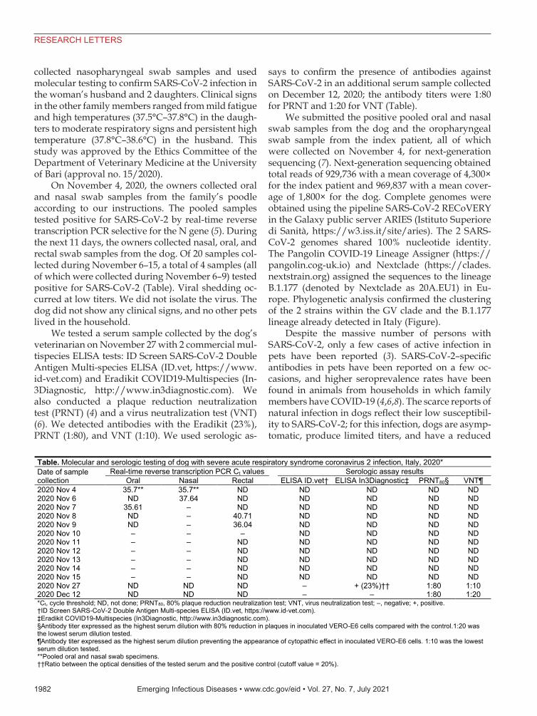
On November 4, 2020, the owners collected oral and nasal swab samples from the family’s poodle according to our instructions. The pooled samples tested positive for SARS-CoV-2 by real-time reverse transcription PCR selective for the N gene (5). During the next 11 days, the owners collected nasal, oral, and rectal swab samples from the dog. Of 20 samples col-lected during November 6–15, a total of 4 samples (all of which were collected during November 6–9) tested positive for SARS-CoV-2 (Table). Viral shedding oc-curred at low titers. We did not isolate the virus. The dog did not show any clinical signs, and no other pets lived in the household.
We tested a serum sample collected by the dog’s veterinarian on November 27 with 2 commercial mul-tispecies ELISA tests: ID Screen SARS-CoV-2 Double Antigen Multi-species ELISA (ID.vet, https://www.id-vet.com) and Eradikit COVID19-Multispecies (In-3Diagnostic, http://www.in3diagnostic.com). We also conducted a plaque reduction neutralization test (PRNT) (4) and a virus neutralization test (VNT) (6). We detected antibodies with the Eradikit (23%), PRNT (1:80), and VNT (1:10). We used serologic as-
says to confirm the presence of antibodies against SARS-CoV-2 in an additional serum sample collected on December 12, 2020; the antibody titers were 1:80 for PRNT and 1:20 for VNT (Table).
We submitted the positive pooled oral and nasal swab samples from the dog and the oropharyngeal swab sample from the index patient, all of which were collected on November 4, for next-generation sequencing (7). Next-generation sequencing obtained total reads of 929,736 with a mean coverage of 4,300× for the index patient and 969,837 with a mean cover-age of 1,800× for the dog. Complete genomes were obtained using the pipeline SARS-CoV-2 RECoVERY in the Galaxy public server ARIES (Istituto Superiore di Sanità, https://w3.iss.it/site/aries). The 2 SARS-CoV-2 genomes shared 100% nucleotide identity. The Pangolin COVID-19 Lineage Assigner (https://pangolin.cog-uk.io) and Nextclade (https://clades.nextstrain.org) assigned the sequences to the lineage B.1.177 (denoted by Nextclade as 20A.EU1) in Eu-rope. Phylogenetic analysis confirmed the clustering of the 2 strains within the GV clade and the B.1.177 lineage already detected in Italy (Figure).
Despite the massive number of persons with SARS-CoV-2, only a few cases of active infection in pets have been reported (3). SARS-CoV-2–specific antibodies in pets have been reported on a few oc-casions, and higher seroprevalence rates have been found in animals from households in which family members have COVID-19 (4,6,8). The scarce reports of natural infection in dogs reflect their low susceptibil-ity to SARS-CoV-2; for this infection, dogs are asymp-tomatic, produce limited titers, and have a reduced
Table. Molecular and serologic testing of dog with severe acute respiratory syndrome coronavirus 2 infection, Italy, 2020* Date of sample collection
Real-time reverse transcription PCR Ct values
Serologic assay results Oral Nasal Rectal ELISA ID.vet† ELISA In3Diagnostic‡ PRNT80§ VNT¶
2020 Nov 4 35.7** 35.7** ND ND ND ND ND 2020 Nov 6 ND 37.64 ND ND ND ND ND 2020 Nov 7 35.61 – ND ND ND ND ND 2020 Nov 8 ND – 40.71 ND ND ND ND 2020 Nov 9 ND – 36.04 ND ND ND ND 2020 Nov 10 – – – ND ND ND ND 2020 Nov 11 – – ND ND ND ND ND 2020 Nov 12 – – ND ND ND ND ND 2020 Nov 13 – – ND ND ND ND ND 2020 Nov 14 – – ND ND ND ND ND 2020 Nov 15 – – ND ND ND ND ND 2020 Nov 27 ND ND ND – + (23%)†† 1:80 1:10 2020 Dec 12 ND ND ND – – 1:80 1:20 *Ct, cycle threshold; ND, not done; PRNT80, 80% plaque reduction neutralization test; VNT, virus neutralization test; –, negative; +, positive. †ID Screen SARS-CoV-2 Double Antigen Multi-species ELISA (ID.vet, https://www.id-vet.com). ‡Eradikit COVID19-Multispecies (In3Diagnostic, http://www.in3diagnostic.com). §Antibody titer expressed as the highest serum dilution with 80% reduction in plaques in inoculated VERO-E6 cells compared with the control.1:20 was the lowest serum dilution tested. ¶Antibody titer expressed as the highest serum dilution preventing the appearance of cytopathic effect in inoculated VERO-E6 cells. 1:10 was the lowest serum dilution tested. **Pooled oral and nasal swab specimens. ††Ratio between the optical densities of the tested serum and the positive control (cutoff value = 20%).

Emerging Infectious Diseases • www.cdc.gov/eid • Vol. 27, No. 7, July 2021 1983
RESEARCH LETTERS
duration of viral shedding (9). Upon experimental infection, dogs shed SARS-CoV-2 at lower titers and for a shorter period than cats (10). Patterson et al. (4) found no actively infected dog or cat in a sampled population of 494 pets, including 67 dogs from house-holds in which family members had COVID-19; how-ever, SARS-CoV-2–specific antibodies were detected in a small proportion of pets (4). Delayed sampling of animals, caused by restrictions on human and animal movement during the pandemic, probably contribut-ed to the negative results of molecular testing in that study. The infected poodle we report was monitored after the identification of the index case in the family, enabling the detection of SARS-CoV-2 RNA in swab samples collected during the observational follow-up. Because the canine virus shared 100% nucleotide identity with the virus detected in the index case, we believe human-to-dog transmission of the virus prob-ably occurred in the household.
AcknowledgmentsWe are grateful to Maria Stella Lucente, Cristiana Catella, Carlo Armenise, and Arturo Gentile for their excellent technical assistance. We thank Marco Crescenzi, Manuela Marra, and Maria Carollo for the Next Generation Sequencing through Ion GeneStudio S5 System.
N.D. was supported by grants of Fondazione CARIPLO—Misura a sostegno dello sviluppo di collaborazioni per l’identificazione di terapie e sistemi di diagnostica, protezione e analisi per contrastare l’emergenza Coronavirus e altre emergenze virali del future, project “Genetic characterization of SARS-CoV2 and serological investigation in humans and pets to define cats and dogs role in the COVID-19 pandemic (COVIDinPET)”. A.L was supported by the Italian Ministry of Health Ricerca Corrente 2020 “PanCO: epidemiologia e patogenesi dei coronavirus umani ed animali” and Ricerca Strategica 2020 “Suscettibilità dei mammiferi a SARS-COV-2: rischi di zoonosi inversa e possibilità in medicina traslazionale.”
Figure. Maximum-likelihood tree comparing 108 strains of severe acute respiratory syndrome coronavirus 2 circulating among humans and canines. Tree shows 107 complete genomes downloaded from the GISAID database (https://www.gisaid.org) and the strains sequenced from an infected dog and family member in Italy (bold red text). The tree was built with IQ-TREE version 1.6.10 (http://www.iqtree.org) using the best fit model indicated by the Model Finder with 1,000 bootstrap replicates. Text at nodes indicates bootstrap values >70. Brackets to the right indicate clades. Scale bar indicates number of nucleotide substitutions per site.
1984 Emerging Infectious Diseases • www.cdc.gov/eid • Vol. 27, No. 7, July 2021
RESEARCH LETTERS
About the AuthorDr. Decaro is professor in the Department of Veterinary Medicine at the University of Bari in Valenzano, Italy. His research interests include the study of viral pathogens of dogs and cats, especially coronaviruses and parvoviruses.
References 1. Lorusso A, Calistri P, Petrini A, Savini G, Decaro N.
Novel coronavirus (SARS-CoV-2) epidemic: a veterinary perspective. Vet Ital. 2020;56:5–10. https://doi.org/10.12834/VetIt.2173.11599.1
2. Decaro N, Lorusso A. Novel human coronavirus (SARS-CoV-2): a lesson from animal coronaviruses. Vet Microbiol. 2020;244:108693. https://doi.org/10.1016/ j.vetmic.2020.108693
3. Bosco-Lauth AM, Hartwig AE, Porter SM, Gordy PW, Nehring M, Byas AD, et al. Experimental infection of domestic dogs and cats with SARS-CoV-2: pathogenesis, transmission, and response to reexposure in cats. Proc Natl Acad Sci U S A. 2020;117:26382–8. https://doi.org/10.1073/pnas.2013102117
4. Patterson EI, Elia G, Grassi A, Giordano A, Desario C, Medardo M, et al. Evidence of exposure to SARS-CoV-2 in cats and dogs from households in Italy. Nat Commun. 2020;11:6231. https://doi.org/10.1038/s41467-020-20097-0
5. Corman VM, Landt O, Kaiser M, Molenkamp R, Meijer A, Chu DK, et al. Detection of 2019 novel coronavirus (2019-nCoV) by real-time RT-PCR [Erratum in: Euro Surveill. 2020;25: 20200409c] [Erratum in: Euro Surveill. 2020;25:2007303]. Euro Surveill. 2020;25:2000045. https://doi.org/10.2807/1560-7917.ES.2020.25.3.2000045
6. Zhang Q, Zhang H, Gao J, Huang K, Yang Y, Hui X, et al. A serological survey of SARS-CoV-2 in cat in Wuhan. Emerg Microbes Infect. 2020;9:2013–9. https://doi.org/ 10.1080/22221751.2020.1817796
7. Di Giallonardo F, Duchene S, Puglia I, Curini V, Profeta F, Cammà C, et al. Genomic epidemiology of the first wave of SARS-CoV-2 in Italy. Viruses. 2020;12:1438. https://doi.org/10.3390/v12121438
8. Fritz M, Rosolen B, Krafft E, Becquart P, Elguero E, Vratskikh O, et al. High prevalence of SARS-CoV-2 antibodies in pets from COVID-19+ households. One Health. 2020;11:100192. https://doi.org/10.1016/ j.onehlt.2020.100192
9. Sit THC, Brackman CJ, Ip SM, Tam KWS, Law PYT, To EMW, et al. Infection of dogs with SARS-CoV-2. Nature. 2020;586:776–8. https://doi.org/10.1038/ s41586-020-2334-5
10. Shi J, Wen Z, Zhong G, Yang H, Wang C, Huang B, et al. Susceptibility of ferrets, cats, dogs, and other domesticated animals to SARS-coronavirus 2. Science. 2020;368:1016–20. https://doi.org/10.1126/science.abb7015
Address for correspondence: Nicola Decaro, Department of Veterinary Medicine, University of Bari, Strada provinciale per Casamassima Km 3, 70010 Valenzano (Bari), Italy; email: [email protected].
Postoperative Paenibacillus thiaminolyticus Wound Infection, Switzerland
Riccardo Di Micco, Matthias Schneider, Reto NüeschAuthor affiliations: Spital Schwyz, Schwyz, Switzerland (R. Di Micco, M. Schneider, R. Nüesch); University of Basel, Basel, Switzerland (R. Nüesch)
DOI: https://doi.org/10.3201/eid2707.203348
The genus Paenibacillus comprises a growing num-ber of species of rod-shaped, motile bacteria with
peritrichous flagella (1). Paenibacillus species share 89.6% similarity of 16S rDNA gene sequences and grow as nonpigmented colonies on tryptic soy agar (1). Best known as a nearly ubiquitous environmental bacteria, many Paenibacillus species are potential op-portunistic pathogens in humans (2). We report a case of isolated surgical site infection caused by P. thiami-nolyticus in an otherwise healthy patient.
A 33-year-old woman came to the emergency de-partment with a fever and reported having a painful and fluctuating abdominal wall mass for 3 days. She had undergone lipoabdominoplasty in a different hos-pital 7 days earlier. Laboratory tests showed anemia (hemoglobin 88 g/L, hematocrit 0.24 L/L) and isolated C-reactive protein elevation (117 mg/L). Computed to-mography of the abdomen demonstrated a fluid collec-tion in the abdominal wall measuring 22 × 9.5 × 5 cm. The patient was admitted for observation. Blood cul-tures performed at 38.5°C showed no bacterial growth.
Empirical intravenous antimicrobial drug ther-apy for suspected infected hematoma was initiated with amoxicillin/clavulanate (2.2 g 3×/d), accord-ing to local hospital guidelines. Under antimicrobial drug treatment, the patient’s fever resolved, but her abdominal pain persisted.
On day 3, we aspirated a sample of the fluid collec-tion in the abdominal wall for microbiological exami-nation. The aspirate was cultured on blood agar incu-bated at 35°C with 5% CO2 for 48 h; on MacConkey
Paenibacillus thiaminolyticus is a nonvirulent organ-ism found in human and ruminant microbiota. However, P. thiaminolyticus can act as an opportunistic pathogen in humans. We describe a case of abdominal wall he-matoma secondarily infected by P. thiaminolyticus. Our findings emphasize the risk for unusual Paenibacillus infections in otherwise healthy persons.