reduced acetylcholinesterase activity downregulates peripheral...
TRANSCRIPT

Indian Journal of Experimental Biology
Vol. 56, December 2018, pp. 859-874
Reduced acetylcholinesterase activity downregulates peripheral and central
inflammation during glucocorticoid resistance induced by chronic restraint
stress and systemic lipopolysaccharide challenge in male mice
Sayantika Mahanti, Arnab Majhi, Koyel Mukherjee & Biswadev Bishayi*
Department of Physiology, Immunology Laboratory, University of Calcutta, University Colleges of Science and Technology,
Kolkata-700 009, West Bengal, India
Received 07 December 2015; revised 29 January 2018
Stress sensitizes the neuroinflammatory response to immunogenic challenge and associated behavioral changes in
rodents. GlucocorticoidS (GCs) have been well known for their immunosuppressive and anti-inflammatory properties.
However, recent advances have uncovered situations wherein they have opposite effects, especially when activated immune
cells show resistance to circulating GCs. Under these circumstances, studying the role of the recently described ‘cholinergic
anti-inflammatory pathway’ was of considerable interest. In this study, we investigated the level of serum C-reactive protein
(CRP), cytokines and reactive oxygen and nitrogen species and antioxidant enzyme activities in the liver, brain and adrenal
gland following LPS administration in stressed mice. Hypothalamic acetyl cholinesterase (AChE) enzyme activity and the
expression of heat shock proteins 70 and 90, superoxide dismutase-1 and cycloxygenase-2 proteins in the hypothalamus
were estimated by immunobloting. Behavioural changes were observed on an elevated plus maze and in an open field. Our
results suggest that there exists a synergistic effect between inflammation and stress only when the stress exposure is acute
in nature. Immune activation following chronic stress, downregulated inflammation, in spite of the resistant endocrine
response to inflammation, via the newly described cholinergic anti-inflammatory pathway. Thus, it indicates that acute
immune activation during chronic stress may be beneficial for the host to maintain homeostasis.
Keywords: Antioxidant enzyme activities, Behaviour, C-reactive protein (CRP), Cholinergic anti-inflammatory pathway,
Cytokines, Homeostasis, Hypothalamic AChE activity, Hypothalamic-pituitary-adrenal (HPA) axis, Reactive oxygen and nitrogen species
The impact of systemic inflammation on the
pathogenesis of chronic brain diseases, such as
neurodegenerative diseases, is an emerging area of
biomedical research1. The mediators of systemic
inflammation have effects on both immune and
nervous systems, and are reported to be key players of
inflammation which may contribute to changes in the
brain during physiological and pathological processes.
The hypothalamic-pituitary-adrenal (HPA) axis has
long been known to play an important role in
regulation of stress induced changes in cellular and
biochemical factors of stress response and associated
changes in behaviour of humans and animals2.
Anomalies in the function of the HPA axis have been
described in patients with psychiatric illness, such as
depression, bipolar disorder, schizophrenia, cognitive
disorders including Alzheimer’s disease and others3.
Systemically administered lipopolysaccharide (LPS)
activates microglia, known to be the principal
inflammatory cells in the brain4. In addition,
inflammatory cells of the blood stream, activated by
systemically administered LPS, may enter the brain by
increasing the permeability of the blood-brain-barrier
(BBB) to infiltrating leukocytes5 and these pro-
inflammatory leukocytes participates in neuro-
inflammation and BBB disruption4,5
. Several studies
have reported that acute or chronic stress sensitizes the
neuroinflammatory response to both peripheral and
central immunologic challenges6. In particular, the effect
of chronic stress or chronic glucocorticoid (GC)
administration on the immune response in the brain is
surprisingly different from the classic picture of
suppression in the periphery7. Moreover, depending
upon the brain region, chronic exposure to GCs is often
not anti-inflammatory, and in greatest contrast to dogma,
can actually exacerbate various aspects of inflammation.
This is in stark contrast to the effects of high
concentrations of GCs in the periphery.
——————
*Correspondence:
Phone: +91 33 23508386 Ext: 225; Fax: +91 33 2351 9755
E-mail: [email protected] (BB);
[email protected] (SM)

INDIAN J EXP BIOL, DECEMBER 2018
860
After the discovery of the anti-inflammatory role
of the vagus nerve in an animal model of
endotoxemia8, the circuit for neural inhibition of
inflammation has been reported9. The autonomic
regulation of local and systemic inflammation through
the ‘cholinergic anti-inflammatory pathway’— a
mechanism consisting of the vagus nerve and its
major neurotransmitter, acetylcholine (ACh) as well
as its regulation by acetylcholinesterase (AChE) as a
means to attenuate inflammation8 has emerged to be
of considerable importance. AChE is an essential
hydrolytic enzyme in the cholinergic nervous system,
and is responsible for catalyzing the degradation of
ACh into acetate and choline10
. It has been reported
that inhibition of AChE activity by a drug
rivastigmine, upregulates cholinergic activity
thereby suppressing neuroinflammation (reduced
demyelination, microglia activation and axonal
damage). Increased cholinergic activity was also able
to decrease the production of pro-inflammatory
cytokines (TNF-α, IFN-γ and IL-6) without affecting
IL-10 production11
. Recently, it has been reported that
ACh producing natural killer (NK) cells was able to
attenuate inflammation in the CNS via modulating the
infiltration of monocytes/macrophages12
.
Hence, we hypothesized that since restraint stress mediated activation of the HPA axis potentiates neuroinflammation and neurodegeneration, a subsequent acute immunogenic systemic challenge following such exposure would either exacerbate the neuroinflammatory process going on or would lead to the triggering of neurohormonal anti-inflammatory pathway in order to maintain homeostasis. This study, thus would indirectly help us to understand the existence of possible link between the hormonal and neural control of inflammation at the periphery and at the CNS. This link may also throw light on the changes in cognitive behaviour that has been impaired due to stress. Moreover, whether activation of the cholinergic anti-inflammatory pathway when the GC concentration is high, particularly during GC resistance or insensitivity as observed during chronic stress, would be of any benefit for the host to maintain homeostasis needs to be elucidated.
Central and peripheral cytokine compartments are
integrated, but differentially regulated. Stress or
peripheral events such as systemic LPS administration
causing inflammation, can activate a cascade of
cytokines and related molecules such as nitric oxide
(NO), affecting outcomes such as behaviour13
. Stress
induces changes in expressive behavior and anxiety
like state, which are associated with oxidative
damage, hormone and neurotransmitter and cytokine
level14,15
. Moreover, it has been reported that restraint
stress increases iNOS, COX-2 expression and produces
an accumulation of lipid peroxidation products16
.
Activities of 3β hydroxy steroid dehydrogenase (3βHSD)
and 17β hydroxy steroid dehydrogenase (17βHSD)
enzymes play an important role in the regulation of
intracellular levels of biologically active steroid
hormones in the adrenal glands17
. Hence, the role of
these enzymes in production of the endogenous
corticosterone following acute and chronic restraint
stress and their alteration on acute exposure to LPS
following stress may also add some important
information related to the ongoing inflammatory
processes in the brain and at the periphery.
Typically, LPS stimulated leukocytes produce pro-
inflammatory cytokines, which trigger reactive
oxygen species (ROS) production in the tissues
through NADPH oxidase activation18
. Thus, the level
of antioxidant enzymes or their activity displays the
intracellular complex mechanisms of host defense.
Nitric oxide (NO) plays a complex role in free radical
mediated injury in the brain during exposure to
stressful condition and/or during immunogenic
stimulation19
. Production of NO correlates with the
level of expression of the iNOS gene which has been
directly correlated with production of COX-2 during
stress and inflammation which may be partly
regulated by the NO pathway20,21
. Heat shock proteins
(HSPs) are also synthesized in significant quantity to
protect eukaryotic cells from various insults during
periods of stress caused by infection, inflammation or
similar events22
. SOD-1 expression in the brain
correlates with the production of the enzyme
superoxide dismutase that helps to convert harmful
superoxide anion to hydrogen peroxide and its
subsequent degradation to water and oxygen by the
enzyme catalase.
As reduced locomotion is a common sickness
behaviour and an adaptive response to illness and
infection due to replicating pathogen or their
products23
, locomotor activity and exploration in the
open field test provides an excellent index for the
assessment of both the effects of and responses to
LPS treatment in animals exposed to short and long-
term stressors24
.
Therefore, in the present study, we tried to
understand the interaction between stress and

MAHANTI et al.: IMMUNOMODULATORY ROLE OF ACETYLCHOLINESTERASE DURING STRESS
861
neuroinflammatory markers evoked by LPS exposure
in mice and study the mechanisms that underlie the
stress-illness interaction. Further, this study may also
explain the anti-inflammatory role played by ACh
released in circulation due to peripheral activation of
afferent vagal nerve fibers by circulating pro-
inflammatory cytokines produced during stress and
inflammation.
Materials and Methods
Animals
All experiments involving animals were conducted
according to the protocols that had been approved by
the Institutional Animal Ethics Committee (IAEC),
Department of Physiology, University of Calcutta,
under the guidance of Committee for the purpose of
control and supervision of experiments on animal
(CPCSEA) [Approval Number: 820/04/ac/CPCSEA-
2010 dated: 16.11.2011], Ministry of Environment
and Forest, Govt. of India. This study did not involve
any invasive study using human subjects. Male
BALB/c (6-8 weeks) mice were obtained from a
registered breeder in our department and were used
for all studies. All animals were maintained and
utilized in accordance with recommendations from
the IAEC and were provided with food and water
ad libitum. Mice were housed 4-5 per cage and
maintained on a 12 h light:dark cycle (lights
on at 08.00 am) in a temperature controlled
room (22±2°C).
Restraint stress
In order to compare stress responses involving the
production of glucocorticoids hormone, which is
affected by various hormones, such as growth
hormone or sex hormones, 5-7 months old male mice
with a stable growth phase without sex cycle, were
selected. Due to inherent correlation of the circadian
rhythm with corticosterone production, mice were
exposed to restraint stress each day during a fixed
time period, from 09.00-15.00 h (6 h each day).
Restraint was applied in a separate room to eliminate
the possible effects of vocalizations or pheromones on
the control (non-stressed) mice. Non restrained mice
were left in their home cages in a noise-free
environment, with food and water during the restraint
period. Mice (n=6) per group were restrained each
day according to procedures described in previous
reports25
. Briefly, following the acclimation period,
individual animals were randomly assigned to six
groups. Restraint stress was performed in well
ventilated 50 mL polystyrene tubes and food and
water were not provided during the restraint period.
Restraint animals were also allowed to move freely in
their cages until the next restraint cycle. An restraint
period once was considered as acute stress where as
the similar period of stress for 3 weeks (21 days) on
each day was considered as chronic stress. Animals
consisted of six groups (n=6 for each group) which
were as follows: Group I: control (non-stressed);
Group II: acute stress; Group III: chronic stress;
Group IV: non-stressed LPS treated; Group V: acute
stress + LPS challenged; and Group VI: chronic stress
+ LPS challenged. LPS (derived from Escherichia
coli 055:B5; Sigma Chemical) at a dose of 250 µg/kg
body wt. of mice26
, was administered intravenously, a
single dose, 4 h after releasing the animals from the
stressed condition for acute stress group. Chronic
stressed animals were also challenged with a single,
intravenous dose of LPS 4h after completion of the
final stress period on the 21st day. 2h after LPS
challenge animals were observed for their behavioural
changes in an open-field and over an elevated plus-
maze and 24 h after LPS treatment animals were
sacrificed under ether anesthesia. Group I, II and III
were administered equal volume of sterile 0.9% saline
on that day. Non-stressed LPS treated group were
administered a single dose (250 µg/kg body weight)
of LPS (i.v.) via the tail vein and were also sacrificed
24 h post-LPS administration. Animals from all the
groups were sacrificed on the same day to avoid
inter-day variation in the tested parameters.
Behavioural activities in an open field and on an
elevated plus maze were recorded consecutively,
2 h after LPS challenge for all the LPS treated group
of animals.
Determination of level of corticosterone in serum
Blood samples were collected in anticoagulant free
tubes between 10:00 a.m. and 12:00 noon and
centrifuged at 1000 g for 10 min. Serum obtained was
distributed in separate tubes. Serum for cytokine
measurement were stored at 70°C until use and that
for determining the concentration of corticosterone
using a corticosterone EIA kit from Cayman
Chemical as per the manufacturer’s instructions, was
done on the same day of blood collection. For each
study, corticosterone levels were determined, in
duplicate; in a single run to avoid inter-assay
variability, and intra-assay variability was less than
10%. The minimum detectable limit for corticosterone
was 8.2 pg/mL.
INDIAN J EXP BIOL, DECEMBER 2018
862
Estimation of hydroxy steroid dehydrogenase (HSD) enzyme
activity
Adrenal glands were homogenized separately in
20% spectroscopic glycerol containing 5 mM
potassium phosphate and 1 mM ethylene diamine
tetra acetic acid (EDTA) at a tissue concentration of
100 mg/mL of homogenizing mixture. It was
centrifuged at 4°C at 10000×g for 30 min. 3ßHSD and
17ßHSD activities were estimated by methods as
described in earlier studies17
.
C-reactive protein (CRP) assay
Serum C-reactive protein (CRP) levels were
determined using a mouse high sensitive CRP ELISA
kit (GenWay Biotech, Inc, San Diego, CA) according
to the manufacturer’s protocol. Absorbance were
interpolated from a standard curve produced from CRP
standards (0-25 ng/mL) supplied within the kit. The
minimum detectable limit for CRP was 0.78 ng/mL.
Cytokine ELISA
Serum TNF-α, IFN-γ, IL-6 and IL-10
concentrations was estimated by Sandwich ELISA
and calculated based on the standard curve. Serum
cytokine levels were expressed in pg/mL of serum
analyzed, following manufacturer’s protocol. (Ray
Biotech, Inc. USA) For each study, cytokine levels
were determined, in duplicate, in a single run to avoid
inter-assay variability, and intra-assay variability was
lower than 10-12%. The minimum detectable dose of
the cytokines for IL-6, TNF-α, Il-10 and IFN-γ were
2, 60, 45and 5 pg/mL, respectively.
Acetyl cholinesterase (AChE) assay in brain tissue
On the day of sacrifice, stressed (acute and
chronic) and unstressed control mice were given mild
anesthesia with anaesthetic ether. The whole intact
brain was then removed carefully and placed in the
petri dish, over ice for 15 min. The brain was washed
with ice-chilled normal saline repeatedly to clean.
A 10% (w/v) homogenate of brain samples was
prepared first by homogenizing in a glass
homogenizer at a speed of 9500 rpm using a sodium
phosphate buffer (30 mM, pH 7.0). The assay of
AChE in the above mentioned supernatant was
performed by the modified Ellman’s method27
, using
acetylthiocholine iodide as substrate at a final
concentration of 1 mmol/L. A kinetic profile of the
enzyme activity was studied spectrophotometrically at
412 nm at an interval of 15 s. The assay for each
sample was run in duplicate and each experiment was
performed thrice. Protein was estimated in the range
0.01-0.1 mg/mL in the brain samples by the Folin–
Lowry method, using bovine serum albumin (BSA) as
standard at a concentration of 1 mg/mL.
Assay of antioxidant enzymes from liver, brain and adrenal
gland
The liver, brain and adrenal tissues were separately
homogenized in 10 volumes of 50 mM phosphate
buffer (pH 7.4) on ice for 30 s using a power driven
polytron homogenizer. The homogenate was
transferred into centrifuge tubes and centrifuged at
9000×g at 4°C for 20 min. The supernatant was
utilized to measure activity of antioxidant enzymes
and the amount of protein present.
Measurement of reduced glutathione level (GSH)
Reduced glutathione content (as acid soluble
sulfhydryl) was estimated by its reaction with DTNB
(Ellman’s reagent) following the method of Sedlak &
Lindsay28
. Values were expressed as nanomoles of
GSH per mg protein.
Catalase (CAT) activity
Decomposition of H2O2 due to CAT activity was
assayed by the decrease in absorbance of H2O2 at
240 nm. Catalase activity in cell free homogenate was
determined spectrophotometrically by measuring the
decrease in H2O2 concentration at 240 nm. At time
zero, 1.8 mL of each homogenate was combined with
0.2 mL of a phosphate buffer containing 10 mmol
H2O2. One mL of the mixture was immediately
added to a cuvette and placed into a
spectrophotometer. CAT activity was observed via
degradation of H2O2 as determined by a reduction in
UV light absorbance over time. Measurement of
absorbance was taken at 15 s interval after addition of
the homogenate to hydrogen peroxide buffer. Units of
CAT activity present in 1 mL of homogenate were
calculated29
.
Superoxide dismutase (SOD) activity
Tissue homogenate (100 µL) was mixed with 1.5 mL
of a Tris-EDTA-HCl buffer (pH 8.5), then 100 µL of
7.2 mM pyrogallol was added and the reaction
mixture was incubated at 25℃ for 10 min. The
reaction was terminated by the addition of 50 µL of
1M HCl and measured at 420 nm. One unit was
determined as the amount of enzyme that inhibited the
oxidation of pyrogallol by 50%. The activity was
expressed as U/mg protein29
.

MAHANTI et al.: IMMUNOMODULATORY ROLE OF ACETYLCHOLINESTERASE DURING STRESS
863
Estimation of nitric oxide (NO) production
The concentration of nitrite in the brain tissues was
measured as an index for NO production. Equal
weights of the brains of control, stressed and LPS
treated mice were homogenized in sterile PBS (1 mL).
Supernatants were collected, and analyzed for NO
production by modified Greiss method as described
earlier30
. Briefly, nitrate was converted to nitrites with
ß-nicotinamide adenine dinucleotide phosphate
(NADPH; 1.25 mg/mL) and nitrate reductase
followed by addition of the Griess reagent. The
reaction mixture was incubated at room temperature
(25℃) for 20 min followed by the addition of TCA.
Samples were centrifuged, clear supernatants were
collected, and optical density was recorded at 550 nm.
The amounts of NO produced were determined by
calibrating a standard curve using sodium nitrite. Determination of blood brain barrier (BBB) leakage
The permeability of the BBB was quantitatively
evaluated by detection of extravasated Evans blue
dye. Briefly, 2% Evans blue dye in saline was injected
intraperitoneally to 3 mice from each group, on the
day of sacrifice (24 h post LPS administration) and
after 4 h mice were deeply anesthetized with
Nembutal and transcardially perfused until colorless
perfusion fluid was obtained from the right atrium.
After decapitation, brain tissue was removed,
weighed, and homogenized. The supernatant was
obtained by centrifugation, and protein concentration
was determined. Evans blue intensity was determined
by a microplate reader at 550 nm. Calculations were
based on external standards dissolved in the same
solvent. The amount of extravasated Evans blue dye
was quantified as micrograms per milligram protein31
.
Brain IL-6 and TNF-α
Frozen brain samples (100 mg tissue/mL buffer)
were homogenized in cold lysis buffer (20 mM Tris,
0.25 M sucrose, 2 mM EDTA, 10 mM EGTA, 1%
Triton x-100) in the presence of 1 unit of protease
inhibitor cocktail/10 mL of lysis buffer. Homogenates
were centrifuged at 100000×g for 40 min. Supernatant
was collected, and protein levels determined using the
Pierce™ BCA Protein Assay Kit (Thermo Scientific.
USA). The levels of IL-6 and TNF-α in the brain were
measured with commercial enzyme-linked
immunosorbent assay (ELISA) kits from Ray Biotech,
Inc. For each study, cytokine levels were determined,
in duplicate, in a solo run to avoid inter-assay
variability, and intra-assay variability was less than
10-12%. The minimum detectable doses of cytokines
for IL-6 and TNF-α were 2 and 60 pg/mL,
respectively.
Expression of HSP 90, HSP 70, SOD1 and COX-2 in
hypothalamic tissue
Expression of cyclooxygenase-2 (COX-2), HSP
90, HSP 70, SOD1 in hypothalamic tissues was
determined by immunoblotting. Estimation of these
protein levels in the brain tissue was determined by
immunoblotting after estimating the protein level in
the tissue homogenates by the Bradford method. 20 µg
of each sample was electrophoresed on poly-
acrylamide gel and transferred onto a nitrocellulose
membrane. After blocking with 7% skimmed milk,
the blots were incubated overnight at 4°C with
primary antibodies against COX-2 (1:1000;
Chemicon, USA), HSP 90, HSP 70, SOD1 (1:1000;
Santa Cruz Biotechnology, USA). After extensive
washes with PBS-Tween, blots were incubated with
appropriate secondary antibodies conjugated with
peroxidase (Vector, Laboratories, USA). The blots
were again washed in PBS-Tween and processed for
development using chemiluminiscence reagent
(Millipore. USA). The images were captured and
analyzed using Chemigenius, bioimaging system
(Syngene, Cambridge, UK). The blots were then
stripped (30 min at 50°C in 62.5 mmole/L Tris-HCl,
pH 6.8, 2% sodium dodecylsulphate, and 100 mM
ß-mercaptoethanol) and reprobed with anti-ß tubulin
(Santa Cruz Biotechnology, USA) to determine
equivalent loading of samples29
.
Activity and exploratory behavior in open-field test
The open field was constructed as described
elsewhere29
. A central square was drawn in the middle
of the receptive field and was used to measure
exploration. The central square is used because some
mouse strains have high locomotor activity and cross
the lines of the test chamber many times during a test
session. The central square has ample space
surrounding it to give meaning to the central location
as being distinct from the outer locations. A digital
camera was fixed to the ceiling 2.1 m above the
apparatus and was utilized to measure line crosses.
Activity and exploratory behaviour of mice were
observed in the open field for a 5-min period, and the
following behavioural activities were scored: TNSCP,
total number of squares crossed in periphery; DTP,
distance traveled in the periphery; LRC, latency to
reach centre; DI, duration of restraint; TNR, total

INDIAN J EXP BIOL, DECEMBER 2018
864
number of rearing; URC, unable to reach the central
region of the open field; TNG, total number of
grooming; and TNF, total number of freezing.
Anxiety test-elevated plus maze (EPM)
One of the most popular tests of anxiety-like
behaviour in mice is the EPM, in which the decreased
number of entries or time spent in the open arms of
the EPM suggests the operation of anxiety-like
processes. The elevated plus maze was built
according to the instruction described elsewhere29
.
A camera was attached to the ceiling 2.1 m above the
apparatus to measure entries and duration in the
center, open and closed zones. We measured:
(A) TSC-total time spent at centre; (B) TSCA-the
time spent in the closed arms; (C) TSOA-time spent
in open arm; (D) NRCA- number of rearing in closed
arms; and (E) NGCA-number of grooming in closed
arms during the 5-min test period. An entry was
meant by all four paws in the arm. The elevated plus-
maze was thoroughly cleaned with 70% ethanol and
air dried following the testing of each animal to avoid
promising biasing effects due to odour clues left by
previous mice. The total number of open arms
entered, as well as the total number of closed arms
entered was used as indexes of general locomotor
activity.
Statistical analysis
All the data are represented as mean ± SD. The
results of the experiment were analyzed by a 3 × 2
analysis of variance (ANOVA), and post hoc
comparisons of mean were done by Tukey HSD
(honest significant difference) tests using the
statistical software Origin 8. Factors were identified
as follows: stress regimen, which was composed of
three levels (non-stressed, acute restraint stress and
chronic restraint stress), and systemic treatment,
consisted of two levels (intravenous vehicle or
intravenous LPS). Threshold for statistical
significance was set at α=0.05.
Results
Effect of LPS administration on the serum corticosterone
concentration following acute and chronic restraint stress
Serum corticosterone levels for non-stressed
(control), acute and chronic restraint stressed groups
administered either LPS or saline (vehicle) are shown
in Fig. 1(A). Low levels of corticosterone were
detected in the serum in the non-stressed saline
administered group whereas there was a significant
variation in the corticosterone level in acute and
chronic restraint stress groups, F (2, 30) = 114.52,
P <0.05. Administration of LPS was also able to
increase the serum corticosterone level in the non-
stressed group and the more importantly, the effect
was significantly more profound in the group of
animals exposed to stressor prior to LPS challenge,
F (1, 30) = 148.45, P <0.05. But, a two-way ANOVA
revealed no significant interaction between stress
condition and LPS administration on the increase in
serum corticosterone level, F (2, 30) = 1.24, p= 0.32.
Effects of LPS administration on steroidogenic enzyme
(3ßHSD and 17ßHSD) activities following acute and chronic
restraint stress in mouse adrenal gland
There was a significant effect of stress regimen on
3β hydroxy steroid dehydrogenase (3β HSD) and 17β
HSD level in the adrenal gland, F (2, 30) = 46.77,
P <0.05, and F (2, 30) = 92.66, P <0.05, respectively.
LPS administration produced significant effect on
these enzyme levels, F (1, 30) = 195.92, P<0.05, and
F (1, 30) = 165.47, P<0.05, respectively. However,
although we observed a significant interaction
Fig. 1 — Effect of LPS challenge on (A) the serum corticosterone concentration post exposure; (B) on steroidogenic enzyme (3βHSD)
activity; (C) on steroidogenic enzyme (17βHSD) activity; and (D) on the brain acetylcholine esterase (AChE) activity in acute and
chronic restraint stress. [Corticosterone level (pg/mL) in serum of acute and chronic stressed mice (n=6/group) treated with intravenous
LPS (250 µg/Kg) or saline have been expressed as mean ± SD. B & C: enzyme activity (Units/min.mg protein) in the adrenal gland. Bars
represents mean (n=6/group) ± standard deviations. *, Significant difference (P <0.05) compared to saline treated group; #, significant
difference (P <0.05) compared to non-stressed group and ^, significant difference (P <0.05) compared to acute stress group that had
received comparable LPS treatment]
MAHANTI et al.: IMMUNOMODULATORY ROLE OF ACETYLCHOLINESTERASE DURING STRESS
865
between the two factors in case of 3β HSD, F (2, 30)
= 9.15, P<0.05, but we failed to observe a significant
interaction between stress and LPS administration in
case of 17β HSD enzyme activity, F (2, 30) = 2.88,
P <0.095, see Fig. 1 (B) and (C).
Effect of LPS administration on the brain AChE activity in
acute and chronic restraint stressed mice
Acetylcholinesterase activity was significantly
influenced by restraint stress exposure, F (2, 30) =
16.50, P <0.05. AChE also varied significantly due to
treatment with LPS following stress exposure, F
(1, 30) = 100.70, P <0.05. A significant interaction
was observed (Fig. 1D) between duration of exposure
to stress and type of systemic treatment (Saline or
LPS) on brain AChE activity, F (2, 30) = 67.68,
P <0.05.
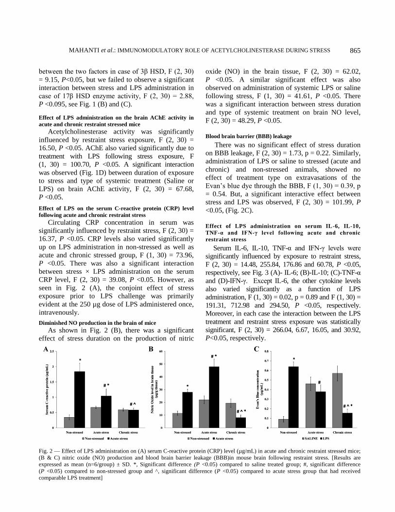
Effect of LPS on the serum C-reactive protein (CRP) level
following acute and chronic restraint stress
Circulating CRP concentration in serum was
significantly influenced by restraint stress, F (2, 30) =
16.37, P <0.05. CRP levels also varied significantly
up on LPS administration in non-stressed as well as
acute and chronic stressed group, F (1, 30) = 73.96,
P <0.05. There was also a significant interaction
between stress × LPS administration on the serum
CRP level, F (2, 30) = 39.08, P <0.05. However, as
seen in Fig. 2 (A), the conjoint effect of stress
exposure prior to LPS challenge was primarily
evident at the 250 µg dose of LPS administered once,
intravenously.
Diminished NO production in the brain of mice
As shown in Fig. 2 (B), there was a significant
effect of stress duration on the production of nitric
oxide (NO) in the brain tissue, F (2, 30) = 62.02,
P <0.05. A similar significant effect was also
observed on administration of systemic LPS or saline
following stress, F (1, 30) = 41.61, P <0.05. There
was a significant interaction between stress duration
and type of systemic treatment on brain NO level,
F (2, 30) = 48.29, P <0.05.
Blood brain barrier (BBB) leakage
There was no significant effect of stress duration
on BBB leakage, F (2, 30) = 1.73, p = 0.22. Similarly,
administration of LPS or saline to stressed (acute and
chronic) and non-stressed animals, showed no
effect of treatment type on extravasations of the
Evan’s blue dye through the BBB, F (1, 30) = 0.39, p
= 0.54. But, a significant interactive effect between
stress and LPS was observed, F (2, 30) = 101.99, P
<0.05, (Fig. 2C).
Effect of LPS administration on serum IL-6, IL-10,
TNF-α and IFN-γ level following acute and chronic
restraint stress
Serum IL-6, IL-10, TNF-α and IFN-γ levels were
significantly influenced by exposure to restraint stress,
F (2, 30) = 14.48, 255.84, 176.86 and 60.78, P <0.05,
respectively, see Fig. 3 (A)- IL-6; (B)-IL-10; (C)-TNF-α
and (D)-IFN-γ. Except IL-6, the other cytokine levels
also varied significantly as a function of LPS
administration, F (1, 30) = 0.02, p = 0.89 and F (1, 30) =
191.31, 712.98 and 294.50, P <0.05, respectively.
Moreover, in each case the interaction between the LPS
treatment and restraint stress exposure was statistically
significant, F (2, 30) = 266.04, 6.67, 16.05, and 30.92,
P<0.05, respectively.
Fig. 2 — Effect of LPS administration on (A) serum C-reactive protein (CRP) level (µg/mL) in acute and chronic restraint stressed mice;
(B & C) nitric oxide (NO) production and blood brain barrier leakage (BBB)in mouse brain following restraint stress. [Results are
expressed as mean (n=6/group) ± SD. *, Significant difference (P <0.05) compared to saline treated group; #, significant difference
(P <0.05) compared to non-stressed group and ^, significant difference (P <0.05) compared to acute stress group that had received
comparable LPS treatment]

INDIAN J EXP BIOL, DECEMBER 2018
866
Fig. 4 — Effect of LPS challenge on the brain proinflammatory
cytokine levels after exposure to restraint stress. (A) IL-6; and (B)
TNF-α levels in the brain were expressed as mean ± SD
(n=6/mice per group). *, Significant difference (P <0.05)
compared to saline treated group; #, significant difference
(P <0.05) compared to non-stressed group and ^, significant
difference (P <0.05) compared to acute stress group that had
received comparable LPS treatment.
Estimation of cytokines in the brain
A similar response in the production of IL-6,
Fig. 4 (A) and TNF-α (B) was observed in the brain
of the animals exposed to restraint stress and were
administered LPS following the stress exposure.
There was a significant influence of both, the stress
duration and the type of treatment, on the
production of these pro-inflammatory cytokines in
the brain, F (2, 30) = 38.19 and F (1, 30) = 99.77,
P <0.05. A significant interaction, F (2, 30) =
24.04, P <0.05, was also reflected when the stress
duration × LPS treatment was analyzed by a two-
way analysis of variance. Significant difference in
mean between the saline and LPS treated, the non-
stressed and the stressed group were confirmed by
Tukey’s HSD post hoc comparison of means.
Effect of LPS administration on the catalase, GSH and SOD
activity of liver, brain and adrenal tissues in acute and chronic
restraint stressed mice
There was a significant influence of stress
regimen on the catalase activity in liver (Fig. 5A),
Fig. 3 — Effect of systemic LPS challenge on the serum (A) IL-6; (B) IL-10: (C) TNF-α; and (D) IFN-γ concentration in restraint
stressed mice. [Values are expressed as mean ± SD (n=6/group). *, Significant difference (P <0.05) compared to saline treated group; #,
significant difference (P <0.05) compared to non-stressed group and ^, significant difference (P <0.05) compared to acute stress group
that had received comparable LPS treatment]

MAHANTI et al.: IMMUNOMODULATORY ROLE OF ACETYLCHOLINESTERASE DURING STRESS
867
brain (Fig. 5D) and adrenal gland (Fig. 5G), F (2, 30)
= 4.059, 371.05 and 17.83, P <0.05, respectively.
Excepting liver, F (1, 30) = 0.127; p = 0.72,
administration of saline or LPS also had significant
effect on brain and adrenal gland catalase activity, F
(1, 30) = 36.99 and 5.44, P <0.05, respectively. Most
importantly, there was a significant interaction of
stress x LPS on the catalase activity in all these
tissues, F (2, 30) = 88.19, 33.49 and 65.32, P <0.05,
respectively.
There was a significant influence of stress regimen
on the GSH content in liver (Fig. 5B), brain (Fig. 5E)
and adrenal gland (Fig. 5H), F (2, 30) = 158.08,
137.05 and 327.64, P <0.05, respectively. There was
also a significant influence of LPS administration on
the GSH content in liver, brain and adrenal gland, F (1, 30)
= 37.11, 226.87 and 712.64, P <0.05, respectively.
There was a significant interaction of stress × LPS on
the GSH content in all these tissues, F (2, 30) = 181.03,
143.58 and 310.41, P <0.05, respectively.
Significant variation in SOD activity due to stress
was observed in liver (Fig. 5C), brain (Fig. 5F) and
adrenal gland (Fig. 5I), F (2, 30) = 47.06, 35.25 and
75.27, P <0.05, respectively. The enzyme activity
varied significantly due to LPS or saline
administration in liver and brain, F (1, 30) = 54.23 and
25.19, respectively, P <0.05, but not in the adrenal
gland, F (2, 30) = 2.91, p=0.113. There was no
significant interaction of stress and LPS on the SOD
activity in the liver and brain, F (2, 30) = 3.20;
p = 0.076 and 0.598; p = 0.56, respectively, though
significant interaction was observed in the adrenal
gland, F (2, 30) = 10.84, P <0.05.
Expression of heat shock proteins (HSPs), SOD1 and COX-2
in hypothalamic tissue
Densitometric analysis of immunoblots (Fig. 6A),
normalized to equal protein content, showed a
significant influence in the expression of HSPs 90
(Fig. 6B) and 70 (Fig. 6C) in the hypothalamus due to
stress, F (2, 30) = 78.14 and 130.68, P <0.05,
respectively. There was a significant effect of LPS
administration on the expression of HSP 90, F (1, 30)
= 25.03, P <0.05; but not on HSP 70, F (1, 30) = 0.16,
p = 0.69. However, there was a significant interaction
of both the factors on the expression of both of the
HSPs tested in the hypothalamic tissue, F (2, 30) =
176.39 and 152.89, P <0.05, respectively.
Fig. 5 — Catalase (CAT) activity, GSH content and superoxide dismutase (SOD) activity, in
the liver (A, B and C); brain (D, E and F); and adrenal gland (G, H and I), respectively in
restraint stressed mice challenged with saline or LPS after exposure to stress. [All the data
represented are mean ± SD (n=6 mice per group). *, Significant difference (P <0.05)
compared to saline treated group; #, significant difference (P <0.05) compared to non-
stressed group and ^, significant difference (P <0.05) compared to acute stress group that
had received comparable LPS treatment]

INDIAN J EXP BIOL, DECEMBER 2018
868
There was a significant effect of stress regimen on
the expression of COX-2 (Fig. 6D), F (2, 30) = 10.02,
P <0.05; but there was no significant effect of
systemic LPS challenge on the expression of this
protein level in the hypothalamus, F (1, 30) = 0.09, p
= 0.77. Interestingly, there was a significant
interaction between the factors (stress × LPS) on the
expression of this protein, F (2, 30) = 100.61, P <0.05.
Interestingly, there was a significant effect of stress
and LPS administration on the expression of SOD-1
protein level (Fig. 6E) in the hypothalamus of mice
brain, F (2, 30) = 33.52 and F (1, 30) = 218.65, P
<0.05. A significant interaction was also found to
exist between these factors on the expression of this
protein level, F (2, 30) = 24.23, P <0.05.
Locomotor activity and exploratory behaviour
In the open field test it was observed that the total
number of squares crossed by the mice in the
periphery, along with distance traveled in periphery
was increased, whereas the total time of restraint in
periphery, total number of rearing and total number of
freezing was decreased, in the mice chronically
stressed and subsequently challenged with LPS when
compared with the stressed plus saline treated group,
indicating improvement of exploratory behavior when
stressed mice were treated with LPS (Table 1). The
effect of LPS administration on chronically stressed
mice showed a prominent increase in these locomotor
and behavioral activities compared to the acute
stressed group that had received comparable LPS
treatment. Moreover, the chronic stress plus LPS treated
animals were also able to reach the central region of
the open field, similar to the non-stressed saline treated
group of animals, compared to the non-stressed or
acute stress plus LPS challenged group (P <0.05). Anxiety test-elevated plus maze (EPM)
When tested in EPM, it was observed that the time
spent in central area increased significantly only in
case of acute stressed mice challenged with LPS when
compared to the acute stress plus saline treated group
or to the non-stressed LPS treated group, though no
significant changes in the behaviour was observed in
chronic condition. The time spent in closed arm
decreased significantly, time spent in the unsafe open
arms of the maze increased significantly, rearing
behaviour in closed arm was decreased in the group of
Fig. 6 — Expression of HSP90, HSP 70, SOD-1 and Cox-2 in the hypothalamus. [Expression of HSP 90, HSP 70, SOD-1 and Cox-2
were measured in terms of fold change among the different groups. (n=6/group). *, Significant difference (P <0.05) compared to saline
treated group; #, significant difference (P <0.05) compared to non-stressed group and ^, significant difference (P <0.05) compared to
acute stress group that had received comparable LPS treatment. Panel A: western blot for the proteins tested. Panel B: Fold change for
HSP 90, Panel C: Fold change for HSP 70, Panel D: Fold change for SOD-1, Panel E: Fold change for Cox-2. C= Control, AS= Acute
stress; CS = Chronic stress; LPS= non-stressed but LPS challenged only; ASL = Acute Stress + LPS challenged; CSL = Chronic Stress +
LPS challenged]

MAHANTI et al.: IMMUNOMODULATORY ROLE OF ACETYLCHOLINESTERASE DURING STRESS
869
mice who were stressed for long time and
subsequently challenged with LPS than the stressed
mice (acute or chronic) administered saline or than
the non-stressed LPS treated group (P <0.05),
indicating less anxiety in those group of mice and
improvement from depression like behavior when
stressed mice were treated with a single intravenous
dose of LPS (Table 2).
Discussion
Exposure to a single session of restraint stress or to
repeated stress for 3 weeks resulted in the
enhancement of serum corticosterone level and the
increase was significantly high in response to LPS,
administered 4h after cessation of the stress period.
Prior exposure to chronic restraint stress also
significantly increased the corticosterone level
compared to acute stress in response to systemic LPS
challenge. In addition, the activity of the enzymes 3β
and 17β HSD, in the adrenal gland also increased
significantly in both acute and chronic restraint stress
group in response to immune stimulation. These
results suggest that prior exposure to acute or chronic
restraint stress sensitizes the HPA axis to immune
activation that makes the subsequent response
greater7.
Acute phase protein like CRP has been considered
as a representative marker of the inflammatory
response32
and has been shown to protect mice from
lethal LPS challenge33
. From our study, it was
observed that LPS challenge or stressor exposure or
both increased the serum CRP level than the control
mice. Acute stress and/or LPS challenge also
augmented the inflammatory process. Moreover,
elevated corticosterone level following chronic stress
or following exposure to both (stressor plus immune
Table 1 — Effects of LPS administration on the locomotor activity IS mice in the open field test
Groups Locomotor activity Behavioral activity
TNSCP
Mean±SD
DTP (cm)
Mean±SD
LRC (s)
Mean±SD
DI (s)
Mean±SD
TNR
Mean±SD
TNG
Mean±SD
TNF
Mean±SD
Non-stressed + saline 287±39.08 2296±552.69 178±61.48 48.66±17.01 32±5 17.66±4.72 13.67±2.51
Acute stress + saline 16±5 128±40 URC 282±8.18 15.33±2.51 4±1 2±1
Chronic stress + saline 85.33±27.15 680.66 ±67.23 URC 246.33±22.47 6±1 9±1 7.33±1.15
Non-stressed + LPS 216±14.26 1728±128.36 142±33.37 67.72±10.08 21±4* 12±4 5.67±1.33*
Acute stress + LPS 51±7.21*# 408±27.68*# URC 226.66±12.66*# 1.66±0.57*# 9±1* 1.33±0.57
Chronic stress + LPS 179±20.22*^ 1432 ±121.78*#^ 128±12.08* 38.33±6.25*#^ 4.66±2.08# 4.33±2.08*# 12±2.64#
[Activity and exploratory behavior of mice were observed in open field for a 5-min period, and the following behavioral activities were
scored. TNSCP- Total number of squares crossed in periphery, DTP- distance traveled in the periphery, LRC- latency to reach centre, DI
- duration of immobilization, TNR- total number of rearing, URC- unable to reach the central region of the field, TNG- total number of
grooming, TNF – total number of freezing. Each value represent the mean ± SD (n=6 mice/group) of counts in 5 min open field test. *,
Significant difference (P <0.05) compared to saline treated group; #, significant difference (P <0.05) compared to non-stressed group and
^, significant difference (P <0.05) compared to acute stress group that had received comparable LPS treatment]
Table 2 — Elevated plus maze (EPM) test to observe the effect of LPS challenge on stressed mice
Group TSC (s)
Mean±SD
TSCA (s)
Mean±SD
TSOA (s)
Mean±SD
NRCA
Mean±SD
NGCA
Mean±SD
Non-stressed + Saline 9.33±2.06 142.64±13.50 148.03±7.09 16.66±2.08 12±1
Acute stress + Saline 22.07±5.15 156.67±16.02 121.26±7.51 4±1 2±1
Chronic stress + Saline 18.66±5.03 193.33±14.04 88.01±5.56 11.33±1.53 8.33±0.57
Non-stressed + LPS 18.27±4.19* 184.27±12.47 97.46±8.23* 12.35±2.68 5.67±1.82*
Acute stress + LPS 33.67±3.05*# 160.67±9.01 105.66±11.37 1.33±0.57*# 4.33±0.57
Chronic stress + LPS 24.26±4.89 135.07±12.49*#^ 140.67±13.60*#^ 7.33±1.15*#^ 6.33±0.57
[To test the anxiety-like behavior in mice is the EPM, in which the reduced number of entries or time spent in the open arms of the EPM
suggests the operation of anxiety-like processes. Mouse from different groups (n=6 mice/group) was placed individually in the center
portion of the plus-maze, facing one of the open arms. The observer measured: (A) TSC-total time spent at centre; (B) TSCA-the time
spent in the closed arms; (C) TSOA-time spent in open arm; (D) NRCA- number of rearing in closed arms; and (E) NGCA-number of
grooming in closed arms during the 5-min test period. An entry was defined as all four paws in the arm. *, Significant difference
compared to saline treated group (P <0.05); #, significant difference compared to non-stressed group that had received comparable LPS
treatment (P <0.05); ^, significant difference (P <0.05) with respect to acute stress group that had received comparable LPS treatment]

INDIAN J EXP BIOL, DECEMBER 2018
870
activation) did not showed significant down
regulation of this acute phase protein level. Thus, it
can be speculated that due to a functional
corticosterone resistance during chronic stress33
,
inflammatory process was facilitated upon immune
activation post exposure to stress.
The activation of pituitary-dependent adrenal responses after endotoxin administration provided early evidence that inflammatory stimuli can activate anti-inflammatory signals from the central nervous system. A key finding in this study was that esposure to acute or chronic restraint stress and subsequent systemic immune activation elicits systemic and local inflammatory responses
8, which also leads to
neuroinflammation34,35
. The pro-inflammatory cytokines like IL-6, TNF-α and IFN-γ levels increased in group of mice that were exposed to stress or LPS. Thus, this study provides evidence on the presence of acute neuroinflammation characterized by release of proinflammatory cytokines IL-6 and TNF-α in the systemic circulation (Fig. 3 A and C) as well as in the brain (Fig. 4A and B) following LPS administration. Interestingly, we found that LPS administration in acute stressed animals showed exaggerated release of these proinflammatory cytokines whereas single intravenous LPS administration in chronic stressed animals did not potentiated this release. Thus, it was evident that during chronic restraint stress and immune activation, some anti-inflammatory mechanism might be responsible for increasing the level of IL-10 and down regulating the production and release of IL-6, TNF-α and IFN-γ in the periphery as well as in the CNS. The increase in the IL-10 level most likely represents a regulatory response to restrict the action of IL-6, including the aggravation of acute phase reactants
36. Since the balance between pro and
anti-inflammatory cytokines affects the outcome of certain diseases such as neurodegenerative disorders
37, it was essential to study the neuro-
inflammatory processes during exposure to both short and long term stressor and immune activation. Moreover, the increase in IL-10 and decrease in IL-6 and TNF-α in chronic stress plus LPS challenged group cannot be solely attributed to the anti-inflammatory properties of GCs due to resistance or insensitivity of this steroid hormone by immune cells during chronic stress paradigm.
Recent findings indicate that neural mechanisms
are also involved in limiting inflammatory
responses38
. ACh is a major parasympathetic
neurotransmitter and inhibits LPS-induced production
of pro-inflammatory cytokines such as TNF-α, IL-6
from macrophages8. The levels of ACh are
continuously regulated be the hydrolytic enzyme
AChE which rapidly degrades ACh both in periphery
and in the brain. Therefore, we have measured AChE
activity in the hypothalamic region as a marker of
cholinergic activity. Systemic LPS administration in
acute stress group exacerbated the activity of this
enzyme in the hypothalamus where as the activity of
this enzyme was down regulated in the chronic stress
plus LPS challenged group. This finding shows
macrophages and other immune cells activated by
systemic LPS challenge releases proinflammatory
cytokines to stimulate the afferent vagus nerve39,40
although the corticosterone level was found to be
substantially high during chronic stress. This pathway
may serve as a bypass mechanism mediated by the
cholinergic anti-inflammatory pathway to maintain
homeostasis when anti-inflammatory properties of
GCs has been disturbed due to GC resistance by
immune cells. ACh released attenuates the production
of TNF and IL-6. Moreover, ACh does not alter IL-10
release, which indicates a direct inhibitory effect of
ACh on pro-inflammatory cytokine production8.
Moreover, it has been reported that in a model of
endotoxemia, electrical stimulation of the cervical
vagus nerve significantly reduced serum and liver
TNF levels, prevented development of hemodynamic
shock and improved survival without significantly
altering IL-10 or corticosterone serum levels38
.
However, one important difference between
PNS/vagal modulation of inflammation vs. regulation
of inflammation by the HPA axis or SNS is that the
vagus does not use endocrine signaling mechanisms.
The presence of blood brain barrier (BBB) restricts
the movement of soluble mediators (cytokines and
chemokines) and leukocytes from the periphery to the
CNS. Dysfunction of the BBB precedes immune cell
infiltration, but leukocyte migration modifies BBB
permeability. It has been reported that immune cells
of stressed and LPS challenged animals express
inflammatory cytokines, reactive oxygen species
(ROS) and reactive nitrogen intermediates (RNS) that
can facilitate their migration to the CNS5. Since in
the periphery, ACh release by the vagus nerve
restrains inflammation by inhibiting the activation of
leukocytes and suppressing cytokine release by
monocytes and macrophages activated due to LPS
challenge, reduced AChE activity in the chronic stress
plus LPS treated group, but not in case of acute stress
MAHANTI et al.: IMMUNOMODULATORY ROLE OF ACETYLCHOLINESTERASE DURING STRESS
871
plus LPS treatment, provides evidence for the anti-
inflammatory role played by ACh molecules in
immunomodulation.
Most important changes in the brain of stress-
induced animal models are the accumulation of
reactive molecules generated due to oxidative stress33
.
Inflammation, infection and administration of
bacterial LPS causes oxidative stress, and this entails
an increase in tissue concentration of reactive oxygen
species, which mediates tissue destruction41
.
Consequently antioxidants such as glutathione (GSH)
are foremost determinants of the degree of pathology
in models of tissue inflammation. Corticosterone
secreted during restraint stress reduces GSH levels in
the liver and disrupt anti-oxidant capacity in hepatic
and other tissues33,41
. From our study, we observed
that chronic stress as well as chronic stress plus LPS
treatment decreased hepatic GSH levels significantly
than acute stress or acute stress plus LPS treated
group respectively, but there was an opposite effect in
the brain and adrenal gland. Since the quantity of
corticosterone remained elevated in chronic stress
than in the acute condition, GSH levels in liver
decreased after chronic restraint stress compared to
acute stress, which was consistent with previous
findings. Similar observation was noticed in the LPS
administered chronic stress group compared to acute
stress ones. But the antagonistic effect observed in
case of brain tissue might be due to reduced entry of
endogenous corticosterone across the BBB. Moreover
a tissue specific variation in GSH activity was
observed in the adrenal gland which was not
comparable to those observed in liver and brain.
Comparison of catalase (CAT) activities in liver,
brain and adrenal gland showed similar response in
these tissues, with slight variation particularly in the
adrenal gland in response to acute and chronic stress
group challenged with LPS. LPS administration post
stress resulted in decreased CAT activity in the brain
of chronic group whereas increased activity was
observed in the liver and adrenal gland. The reduced
CAT activity reflected reduced oxidant burden in
these tissues. In contrast, the hepatic SOD activity
was significantly increased in the LPS challenged
acute stressed mice compared to the LPS challenged
chronic group. Alternatively, due to less SOD
activity, dismutation of superoxide anion to H2O2
would have been limited, leading to accumulation of
huge amount of superoxide anion in the tissue and
ultimately these accumulated superoxide anion might
be responsible for inhibition of hepatic SOD activity
which also correlates with the protein expression data.
Earlier studies also support the present findings that
restraint stress causes robust increase in
the production of ROS, depletes GSH and
consequently results in oxidative damage42
and these
effects may be governed by differential level of
corticosterone in circulation and its time of exposure
before immune challenge33
.
HSPs are produced as a response to various
stressors. Earlier studies has shown that neither acute
nor chronic stress was sufficient in inducing either
HSP90 or HSP70 in brain hypothalamus and that their
induction in response to stressors may be independent
of glucocorticoids33
. Few other studies have reported
that HSP 90 and 70 remains bound with GC receptor
in cytosol of cells when there was no induction of
cortisol43
. Thus, suppressed expression of these
protein levels in LPS challenged chronically stressed
animals could indicate a gradual decline in
corticosterone level in the brain. SOD-1 is considered
to be a neuroprotective enzyme, in cases of stress-
associated or GC mediated neurotoxicity44
. Thus, it
comes as no surprise when we observe that the
expression of this protein was decreased in both post
stress LPS challenged groups. COX-2 expression was
important in regulating prostaglandin signaling in the
brain and both basal and induced expressions of
COX-2 were inhibited by GCs45
. This could partially
explain the non-significant difference in COX-2
levels between LPS treated and non-treated groups in
both acute and chronic stressed animals even though
their levels show a trend similar to that of SOD-1.
Moreover, when we performed blot for SOD1, its
expression was also decreased in the brain of mice
exposed to stress followed by endotoxin challenge,
similar to the enzyme activity which was also
decreased in the brain tissue. Thus reduced activation
of the enzyme pool by less accumulation of
superoxide anion among the total brain protein
content led to its decreased expression post LPS
challenge in acute/chronic stressed mice.
The mechanisms, by which immune signaling to
the CNS affect behaviour, have been of considerable
interest in recent times. It is well known fact that
bacterial endotoxin is a potent stimulator of the
synthesis and release of proinflammatory cytokines
such as IL-6, TNF-α and IFN-γ, but acute stress has
been demonstrated to downregulate these LPS-
induced responses46
. Thus, changes in behaviour of

INDIAN J EXP BIOL, DECEMBER 2018
872
the animals were correlated with the level of
proinflammatory cytokines in the brain.
EPM provides dependable measures of anxiety-
related behavior. When tested in EPM, animals with a
more anxiogenic phenotype will spend a greater
percentage of their time in the closed arm of the maze
relative to the open, unsafe arms. Moreover, latency
to exit the closed arm of the EPM will be significantly
greater than that of animals that is not anxious. It had
been reported that chronic stress results in increased
anxiety and depressive like behavior35
. In addition,
systemic administration of LPS induces sickness
behaviour, as well as alterations of HPA functioning
commonly associated with stressors. Our study
reveals that acute stress plus acute LPS administration
resulted in decreased anxiogenic-like responses than
the chronically stressed animals, which may be
correlated with decreased serum corticosterone levels.
Thus, we can infer that bacterial endotoxin challenge
and the ensuing cytokine changes may contribute to
anxiety-related behavioural disturbances47
.
Researchers seeking to explain the role of peripheral
cytokines in cognitive disorder will have to consider
the multiple effects of serum cytokines which may
have cognitive processing by way of their effects in
the hypothalamic-pituitary axis and various behaviour
systems in addition to neurodegeneration.
Locomotor activity provides an admirable index
for the assessment of both the effects of and
responses to acute endotoxin challenge in
acute/chronic stressed animals. Role of TNF-α and
IFN-γ in induction of depressive like behaviour in
response to Bacillus Calmette-Guerin in mice has
been demonstrated48
. Changes in the open field
behaviour have also been documented in animals
administered cytokines in isolation, including TNF-
α and in animals with relevant cytokine genes
deleted or over expressed49
. The level of cortisol
hormone is often elevated in depressed individuals.
But in depressed individuals, the negative feedback
system for dampening a response does not work
well. The problem with CRH not being suppressible
in depressed individuals may be related to the
inflammatory state in chronic stress50
. It has been
reported that stressors and cytokines may
synergistically influence biological and behavioural
responses and that these treatments may have long
term ramifications through the sensitization of
processes associated with stress responses13
.
Conclusion
Taken together with our results, these findings
support the notion that cytokines released during
immune activation and under the influence of
corticosterone can modulate the open field behaviour
both in terms of locomotor activity as well as
exploration. One of the features observed with
chronic stressor was an enhanced ability to suppress
inflammation, regardless of the fact that in case of
acute stress, LPS was synergistic in mediating
inflammation in the brain. These findings in in vivo
showed correlated improvement of exploratory
behaviour and cognitive functions on administration
of LPS post exposure to chronic stressor. Thus, pre-
activation of the immune system would enhance
survival and recovery from aberrant social
behaviours, particularly aggression, suicidal tendency
as well as psychomotor agitation in stress related
conditions in humans.
Acknowledgement
This study was financially supported by
Department of Biotechnology, Ministry of Science
and Technology, Government of India, New Delhi
under the DBT- Neuroscience scheme. BT/PR
14196/MED/30/387/2010 September, 2011
References 1 Chen WW, Zhang X & Huang WJ, Role of
neuroinflammation in neurodegenerative diseases. Mol Med
Reports, 13 (2016) 3391.
2 Garcia-Bueno B, Madrigal JL, Perez-Nievas BG & Leza JC,
Stress mediators regulate brain prostaglandin synthesis and
peroxisome proliferator-activated receptor gamma
activation after stress in rats. Endocrinology, 149 (2008)
1969.
3 Schatzberg AF & Lindley S, Glucocorticoid antagonists in
neuropsychiatric disorders. Euro J Pharm, 583 ( 2008) 358.
4 Beier EE, Neal M, Alam G, Edler M, Wu LJ & Richardson JR,
Alternative microglial activation is associated with cessation
of progressive dopamine neuron loss in mice systemically
administered lipopolysaccharide. Neurobiol Dis, 108 (2017)
115.
5 Banks WA, Gray AM, Erickson MA, Salameh TS,
Damodarasamy M, Sheibani N, Meabon JS, Wing EE,
Morofuji Y, Cook DG & Reed MJ, Lipopolysaccharide-
induced blood- brain barrier disruption: roles of
cyclooxygenase, oxidative stress, neuroinflammation, and
elements of the neurovascular unit. J Neuroinflamm,
12 (2015) 223.
6 Frank MG, Miguel ZD, Watkins LR & Maier SF, Prior
exposure to glucocorticoids sensitizes the
neuroinflammatory and peripheral inflammatory responses
to E. coli lipopolysaccharide. Brain Behav Immun,
24 (2010) 19.

MAHANTI et al.: IMMUNOMODULATORY ROLE OF ACETYLCHOLINESTERASE DURING STRESS
873
7 Espinosa-Oliva AM, de Pablos RM, Villaran RF, Arguelles S,
Venero JL, Machado A & Cano J, Stress is critical for LPS-
induced activation of microglia and damage in the rat
hippocampus. Neurobiol Aging, 32 (2011) 85.
8 Borovikova LV, Ivanova S, Zhang M, Yang H, Botchkina GI,
Watkins LR, Wang H, Abumrad N, Eaton JW & Tracey KJ,
Vagus nerve stimulation attenuates the systemic
inflammatory response to endotoxin. Nature, 405 (2000)
458.
9 Tracey KJ, The inflammatory reflex. Nature, 420 (2002) 853.
10 Dvir H, Silman I, Harel M, Rosenberry TL & Sussman JL,
Acetylcholinesterase form 3D structure to function. Chem
Biol Interact, 187 (2010) 10.
11 Nizri E, Irony-Tur-Sinai M, Faranesh N, Lavon I, Lavi E,
Weinstock M & Brenner T, Suppression of neuro-
inflammation and immunomodulation by the acetyl-
cholinesterase inhibitor rivastigmine. J Neuroimmunol, 203
(2008) 12.
12 Jiang W, Li D, Han R, Zhang C, Jin WA, Wood K, Liu Q,
Shi FD & Hao J, Acetycholine-producing NK cells attenuate
CNS inflammation via modulation of infiltrating
monocytes/macrophages. PNAS, (2017) E6202.
13 Couch Y, Trofimov A, Markova N, Nikolenko V,
Steinbusch HW, Chekhonin V, Schroeter C, Lesch KP,
Anthony DC & Strekalova T, Low-dose lipopolysaccharide
(LPS) inhibits aggressive and augments depressive
behaviours in a chronic mild stress model in mice.
J Neuroinflamm, 13 (2016) 108.
14 Miller AH, Norman cousins lecture, Mechanisms of
cytokine-induced behavioral changes: Psychoneuro-
immunology at the translational interface. Brain Behav
Immun, 23 (2009) 149.
15 Raedler TJ, Inflammatory mechanisms in major depressive
disorder. Curr Opin Psychiatry, 24 (2011) 519.
16 Freitas AE, Bettio LEB, Neis VB, Santos DB, Ribeiro CM,
Rosa PB, Farina M & Rodrigues ALS, Agmatine abolishes
restraint stress-induced depressive-like behaviour and
hippocampal antioxidant imbalance in mice. Prog
Neuropsychopharmacol Biol Psychiatry, 50 (2014) 143.
17 Mukhopadhyay R, Mishra MK, Basu A & Bishayi B, Effect
of particulate antigenic stimulation or in vivo administration
of interleukin-6 on the level of steroidogenic enzymes in
adrenal glands and lymphoid tissues of mice with parallel
alteration in endogenous inflammatory cytokine level. Cell
Immunol, 261 (2010) 23.
18 Singh A, Koduru B, Carlisle C, Akhter H, Liu RM,
Schroder K, Brandes RP & Ojcius DM, NADPH oxidase 4
modulates hepatic responses to lipopolysaccharide mediated
by Toll-like receptor-4. Sci Reports, 7 (2017) 14346.
19 Tanaka S, Ide M, Shibutani T, Ohtaki H, Numazawa S,
Shioda S & Yoshida T, Lipopolysaccharide-induced
microglial activation induces learning and memory deficits
without neuronal cell death in rats. J Neurosci Res, 83
(2006) 557.
20 Shukla R, Tyagi E & Kumar R, Protective effect of COX
and NOS Inhibitors on LPS induced oxidative stress in rat
brain. Ann Neurosci, 15 (2008) 6.
21 Gulati K, Chakraborti A & Ray A, Differential role of nitric
oxide (NO) in acute and chronic stress induced
neurobehavioral modulation and oxidative injury in rats.
Pharmacol Biochem Behav, 92 (2009) 272.
22 Paidas CN, Mooney ML, Theodorakis NG & De Maio A,
Accelerated recovery after endotoxic challenge in heat
shock pretreated mice. Am J Physiol Regul Integr Comp
Physiol, 282 (2002) 1374.
23 Dantzer R, Cytokine-induced sickness behavior: where do
we stand? Brain Behav Immun, 15 (2001) 7.
24 Swiergiel AH, Leskov IL & Dunn AJ, Effects of chronic
and acute stressors and CRF on depression-like behavior in
mice. Behav Brain Res, 186 (2008) 32.
25 Delgado-Morales R, del Rio E, Gomez-Roman A, Bisagno V,
Nadal R, de Felipe C & Armario A, Adrenocortical and
behavioural response to chronic restraint stress in
neurokinin-1 receptor knockout mice. Physiol Behav, 105
(2012) 669.
26 Nandi D, Mishra MK, Basu A & Bishayi B, Protective
effects of interleukin-6 in lipopolysaccharide (LPS) –
induced experimental endotoxemia are linked to alteration
in hepatic anti-oxidant enzymes and endogenous cytokines.
Immunobiol, 215 (2010) 443.
27 Ellman GL, Courtney KD, Andres VJr & Feather-Stone RM,
A new and rapid colorimetric determination of acetyl-
cholinesterase activity. Biochem Pharmacol, 7 (1961) 88.
28 Sedlak J & Lindsay RH, Estimation of total, protein-bound,
and nonprotein sulfhydryl groups in tissue with Ellman's
reagent. Anal Biochem, 25 (1968) 192.
29 Mahanti S, Majhi A, Chongdar S, Kundu K, Dutta K,
Basu A & Bishayi B, Increased resistance of immobilized-
stressed mice to infection: correlation with behavioral
alterations. Brain Behav Immun, 28 (2013) 115.
30 Mittal R, Gonzalez-Gomez I, Goth KA & Prasadarao NV,
Inhibition of inducible nitric oxide controls pathogen load
and brain damage by enhancing phagocytosis of Escherichia
coli K1 in neonatal meningitis. Am J Pathol, 176 (2010)
1292.
31 Green TP, Johnson DE, Marchessault RP & Gatto CW,
Transvascular flux and tissue accrual of Evans blue:
effects of endotoxin and histamine. J Lab Clin Med,
111 (1988) 73.
32 Chandrasekhara S, C-reactive protein: An inflammatory
marker with specific role in physiology, pathology and
diagnosis. Internet J Rheumatol Clin Immunol, 2 (2014) 1.
33 Spiers JG, Chen HJC, Sernia C & Lavidis NA, Activation of
the hypothalamic-pituitary-adrenal axis induces cellular
oxidative stress. Front Neurosci, 8 (2014) 456.
34 Quax RA, Manenschijn L, Koper JW, Hazes JM, Lamberts SWJ,
van Rossum EFC & Feelders RA, Glucocorticoid
sensitivity in health and disease. Nature Rev Endocrinol, 9
(2013) 670.
35 Barnum CJ, Pace TWW, Hu F, Neigh GN & Tansey MG,
Psychological stress in adolescent and adult mice increases
neuroinflammation and attenuates the response to LPS
challenge. J Neuroinflam, 9 (2012) 1.
36 Connor TJ, Brewer C, Kelly JP & Harkin A, Acute stress
suppresses pro-inflammatory cytokines TNF-alpha and IL-1
beta independent of a catecholamine-driven increase in IL-
10 production. J Neuroimmunol, 159 (2005) 119.
37 Himmerich H, Fischer J, Bauer K, Kirkby KC, Sack U &
Krugel U, Stress-induced cytokine changes in rats. Eur
Cytokine Netw, 24 (2013) 97.
38 Rosas-Ballina M & Tracey KJ, Cholinergic control of
inflammation. J Internal Med, 265 (2009) 663.

INDIAN J EXP BIOL, DECEMBER 2018
874
39 Goehler LE, Gaykema RP, Hammack SE, Maier SF &
Watkins LR, Interleukin-1 induces c-Fos immunoreactivity
in primary afferent neurons of the vagus nerve. Brain Res,
804 (1998) 306.
40 Blalock JE, The immune system as the sixth sense. J Intern
Med, 257 (2005) 126.
41 Reid M & Jahoor F, Glutathione in disease. Curr Opin Clin
Nutr Metab Care, 4 (2001) 65.
42 Zafir A & Banu N, Modulation of in vivo oxidative status
by exogenous CORT and immobilization stress in rats.
Stress, 12 (2009) 167.
43 Grad I & Picard D, The glucocorticoid responses are shaped
by molecular chaperones. Mol Cell Endocrinol, 275 (2007) 2.
44 Murphy PJ, Morishima Y, Chen H, Galigniana MD,
Mansfield JF, Simons Jr SS & Pratt WB, Visualization and
mechanism of assembly of a glucocorticoid receptor, Hsp 70
complex that is primed for subsequent Hsp 90-dependent
opening of the steroid binding cleft. J Biol Chem, 278
(2003) 34764.
45 Li XM, Chlan-Fourney J, Juorio AV, Bennett VL,
Shrikhande S & Bowen RC, Antidepressants upregulate
messenger RNA levels of the neuroprotective enzyme
superoxide dismutase (SOD 1). J Psychiatry Neurosci, 25
(2000) 43.
46 Goujon E, Parnet P, Laye S, Combe C, Kelley KW &
Dantzer R, Stress downregulates lipopolysaccharide –
induced expression of proinflammatory cytokines in the
spleen , pituitary and brain of mice. Brain Behav Immun, 9
(1995) 292.
47 Kwatra M, Jangra A, Choubey P & Lahkar M,
Lipopolysaccharide aggravates the restraint-stress induced
behavioral deficits and hippocampal damage: Effect of
Fisetin treatment. Eur Neuropsychopharmacol, 26 (2016) S217.
48 Harrison NA, Brydon L, Walker C, Gray MA, Steptoe A &
Critchley HD, Inflammation causes mood changes through
alterations in subgenual cingulate activity and mesolimbic
connectivity. Biol Psychiatry, 66 (2009) 407.
49 Swiergiel AH & Dunn AJ, Feeding, exploratory,
anxiety and depression-related behaviors are not altered
in interleukin-6 deficient male mice. Behav Brain Res, 171 (2006) 94.
50 Brydon L, Walker C, Wawrzyniak A, Whitehead D,
Okamura H, Yajima J, Tsuda A & Steptoe A, Synergistic
effects of psychological and immune stressors on
inflammatory cytokine and sickness responses in humans.
Brain Behav Immun, 23 (2008) 217.