redacted for privacy - biochem.science.oregonstate.edu · 1,5- diphosphate and the subsequent...
TRANSCRIPT

AN ABSTRACT OF THE THESIS OF
Gerald G. Still for the PhD in Chemistry (Name) (Degree) (Major)
Date thesis is presented May 14, 1965
Title THE ROLE OF SOME OF THE KREBS CYCLE REACTIONS
IN THE BIOSYNTHETIC FUNCTIONS OF THIOBACILLUS
THIOPARUS
Abstract approved Redacted for Privacy (Major professor)
Aseptic radiorespirometry has been used to examine
the utilization of external carbon sources by proliferat-
ing Thiobacillus thioparus cells. These studies reveal
that glucose, galactose, mannose, fructose, ribose, DL-
glutamate, and L- aspartate were not utilized by this
chemoautotrophic organism. However, it has been shown
that trace amounts of acetate, glycine, DL- serine, DL-
alanine, succinate and fumarate can be utilized by T.
thioparus cells.
To elucidate the nature of the biosynthetic pathways
operative in this bacteria, proliferating cell cultures
were allowed to metabolize specifically 14C labeled
substrates. The resulting 14C labeled cells were sub-
sequently hydrolyzed, their amino acids isolated and
subjected to degradation experiments.
Examination of the respective fates of the label in
DL- alanine- 2 -14C, acetate- 1 -14C, or acetate -2 -14C in the

cellular metabolism revealed that the Krebs Cycle path-
way is not functioning as a respiratory mechanism in
T. thioparus. However, most of the reactions of the
Krebs Cycle pathway are involved in the biosynthesis of
carbon skeletons for various amino acids. A CO2 fixa-
tion pathway of the C3 +C1 type is instrumental in provid-
ing C4 dicarboxylic acids and those amino acids derived
therefrom. Acetate can be incorporated into a -keto-
glutarate and those amino acids derived therefrom, but
cannot be incorporated into the C4 dicarboxylic acids.
It appears that the absence of the enzyme a -keto-
glutaric acid oxidase complex accounts for the lack of
operation of the Krebs Cycle pathway as the terminal
respiratory mechanism. These findings also suggest that
the Glyoxylate Cycle pathway is inoperative in this
organism.

THE ROLE OF SOME OF THE KREBS CYCLE REACTIONS IN THE
BIOSYNTHETIC FUNCTIONS OF THIOBACILLUS THIOPARUS
by
GERALD Q. STILL
A THESIS
submitted to
OREGON STATE UNIVERSITY
in partial fulfillment of the requirements for the
degree of
DOCTOR OF PHILOSOPHY
June 1965

APPROVED:
Redacted for Privacy Professor of Chemistry
In Charge of Major
Redacted for Privacy Chaifmán of Department of Chemistry
Redacted for Privacy
Deatí oaf Graduate School
Date thesis is presented May 14, 1965
Typed by Erma McClanathan

ACKNOWLEDGMENT
To Dr. Chih H. Wang I express not only an acknow-
ledgment of appreciation but my deepest gratitude, for
his guidance has reached beyond that required by the
academic standard into the areas of gentle persuasion
and conduct.
I extend my sincere thanks to Dr. Robert R. Becker
for his willingness to give of his time and the use of
his amino acid analyzer, which so greatly facilitated
the solution of this problem.
The support of this research program by the Atomic
Energy Commission and the research training grant
sponsored by the National Institute of Health is
gratefully acknowledged.

This Thesis is Dedicated To my Wife, Kay,
and our Children, Denise, Kirk, and Carrie Rae

TABLE OF CONTENTS
Page
INTRODUCTION .......... ............... ......... 1
EXPERIMENTAL...... ..... ..... ............. 5
Materials 5
Radiochemical Substrates 5
Radiorespirometric Experiments 6
Incorporation of Acetate- 1 14 -14C, 14 Acetate -2 -14C and DL- Alanine -2 -14C
Separation of Amino Acids 8
Degradation Procedures for Glutamic Acid 9
Degradation Procedures for Aspartic Acid 10
Measurement of Radioactivity 10
RESULTS AND DISCUSSION 12
SUMMARY 25
BIBLIOGRAPHY 26
7

LIST OF FIGURES
Figure Page
1. The Relation of 3- Phosphoglycerate Metabolism and the Biosynthesis of Cellular Amino Acids in T. thioparus.... 23

LIST OF TABLES
Table Page
I. 14C Labeled Substrates Utilized by Proliferating Thiobacillus Thioparus Cells...... ............................... 15
II. Distribution of Substrate Radioactivity in Incorporation Experiments with Thiobacillus thioparus Cells.............. 17
III. Incorporation of the Labeled Atom of DL- Alanine -2- C, C, Acetic Acid-1- C,
14
and Acetic Acid -2 -14C into the Cellular Amino Acids of Thiobacillus thioparus 19
IV. Labeling Patterns of Cellular Glutamic Acid and Aspartic Acid Derived from 14C Labeled Substrates............,....... 22
THE ROLE OF SOME OF THE KREBS CYCLE REACTIONS IN THE
BIOSYNTHETIC FUNCTIONS OF THIOBACILLUS THIOPARUS
INTRODUCTION
The physiological behavior of autotrophic organisms
is unique in many respects. Photosynthetic organisms
derive their energy requirements primarily from light in
contrast to chemoautotrophic organisms which utilize the
chemical energy liberated by enzymatically catalyzed
reactions involving inorganic substrates. In both
instances CO2 serves as the sole carbon source giving
rise to primary products, which are in turn converted to
other cellular constituents. The key mechanism by which
autotrophic organisms reduce carbon dioxide to other
carbonaceous compounds is in the nature of the reaction
sequence described by Calvin et al. (1, 14, 20, 25, 26,
27). Thus, photoautotrophic life and chemoautotrophic
life, using different energy sources, both rely on two
key reactions which are peculiar to autotrophic life:
the phosphorylation of ribulose phosphate to ribulose-
1,5- diphosphate and the subsequent carboxylation of
ribulose -1,5- diphosphate with the resulting formation of
3- phosphoglyceric acid.
The photoautotrophs, being facultative in nature,
may in turn use 3- phosphoglyceric acid either as an
energy source or as a source of carbon from which to
build their cellular constituents. Hence, it is possible

2
to maintain proliferation of photosynthetic organisms in
the dark, provided they are furnished with the appro-
priate organic and inorganic requirements. This is not
the case with the strictly chemoautotrophic Thiobacilli
species. Consequently, the conventional approach of
tracing the fate of metabolic intermediates to study the
carbon metabolism has so far failed when applied to these
nitrifying and sulfur oxidizing bacteria in their intact
form. Although there have been reports in the litera-
ture describing the stimulating effect of some complex
organic materials on the rate of inorganic substrate
utilization as well as on the growth of the Thiobacilli,
there has been no evidence that any carbonaceous com-
pound, other than CO2, serves as the useable carbon
source for these chemoautotrophic bacteria (2, 3, 5, 6,
22, 23, 29, 30). This unique chemoautotrophic character-
istic has thus prevented efforts to elucidate the fate
of 3- phosphoglyceric acid once it is formed by the
primary CO2 fixation processes.
In the case of heterotrophic bacteria, it is
recognized that the Krebs Cycle is not only the key
pathway in energy production but also plays a key role
in the biosynthesis of carbon skeletons for a great
variety of cellular constituents. In Thiobacilli,
Johnson and Peck (9) have reported that a cell -free

3
preparation was capable of coupling phosphorylation and
CO2 fixation at the expense of sodium thiosulfate. This
fact, examined in the light of the strict autotrophic
nature of T. thioparus, strongly suggests that this
organism relies exclusively on sulfur metabolism for its
energy requirement. In view of this exclusive role
played by the sulfur metabolism in energy production,
one is led to consider whether the Krebs Cycle pathway
is playing any kind of role in T. thioparus. Recently,
Cooper (4) has shown that several enzymic reactions of
the Krebs Cycle are operative in a cell -free preparation
of T. thioparus. Oxalacetic acid, pyruvic acid and
acetic acid were found to be converted to a- ketogluta-
rate and further, malic dehydrogenase and succinic
dehydrogenase were found to be active. These findings
led Cooper to suggest that the Krebs Cycle pathway is
operative in T. thioparus, despite the fact that the
presence of a- ketoglutaric acid oxidase complex and
fumarase are not demonstrated in the cell -free prepara-
tion. However, it is possible that the Krebs Cycle
reactions may play an important role in the biosynthetic
function of this organism.
In order to clarify this situation, interest in
this laboratory has been focused on the nature of the
biosynthetic pathway functioning in T. thioparus,

4
leading from 3- phosphoglyceric acid to other carbon
skeletons of cellular constituents, particularly that of
the amino acids. The study was conducted, with prolif-
erating cell cultures growing under optimum physiological
conditions, by means of radiotracer techniques to observe
the intracellular conversions of the carbon skeletons
originally synthesized from CO2.
5
EXPERIMENTAL
Materials
The organisms used in this study were Thiobacillus
thioparus Beijerinck, isolated by R. L. Starkey and
obtained from the American Type Culture Collection
(ATCC- 8158). The cultures were maintained at 26 °C on
liquid thiosulfate media as described by Jones and
Happold (10). This same cultural medium was used in all
phases of the study whether the cultures were aerated by
shaking or sparging. The cells and inorganic sulfur,
which is produced during the growth of these bacteria,
were harvested by centrifugation, by means of a Sent -
Gyorgi and Blum continuous flow centrifuge.
Radiochemical Substrates
The radiochemical substrates used were obtained
from commercial sources. Acetate- 1 -14C, acetate- 2 -14C,
glycine- l -14C, glycine- 2 -14C, DL- serine- l -14C,
DL- serine- 3 -14C, L- serine- U -14C, succinate- 1,4 -14C,
succinate- 2,3 -14C, and glucose -U -14C were purchased from
New England Nuclear Corporation.
DL- alanine- l -14C, DL- alanine- 2 -14C, DL- alanine- 3 -14C,
galactose- 1 -14C, mannose- l -14C, fructose- 1,6 -14C,
ribose- U- 14C,DL- glutamate- 1 -14C, DL- glutamate -2 -14C and
DL- glutamate -5 -14C were purchased from Nichem, Inc.

6
Fumarate- 1,4 -14C was purchased from Volk Radiochemical
Corporation, fumarate- 2,3 -14C was purchased from Nuclear
Chicago Corporation while L- aspartate -4 -14C was purchased
from the California Corporation for Biochemical Research.
The purity and identity of these radiochemical sub-
strates were substantiated by paper chromatography and
autoradiography.
Radiorespirometric Experiments
The radiorespirometric techniques used to detect the
conversion of 14C labeled substrates to 14CO2 by prolif-
erating cells were carried out in the same manner as
described by Wang and Krackov (31), with the exception
that the respirometer was modified so that the bacterial
suspension was maintained in a sterile condition through-
out the experiment. The experiments were carried out at
26° C under aerobic conditions. It should be noted that
T. thioparus is an obligate aerobic organism although
oxygen is presumably required for sulfur metabolism
exclusively (9). At the termination of each of the
experiments, the respirometer flasks were assayed so
that any heterotrophic contamination could be detected.
Cell viability of the culture in each of the respiro-
metric flasks was verified by streaking the cell suspen-
sion on thiosulfate agar plates and subsequent incubation
at 26° C for growth observations.

7
Incorporation of Acetate -l-
and DL- Alanine -2 -14C
4C, Acetate- 2 -14C,
When the utilization of certain carbonaceous sub-
strates was ascertained by means of the radiorespiro-
metric experiments, the fate of the 14C labeled carbon
atom of selected metabolic intermediates in proliferat-
ing T. thioparus cells were studied by a series of
incorporation experiments. Thus, DL- alanine- 2 -14C,
acetate- 1 -14C, or acetate -2 -14C was administered to
individual two -liter cultures and inoculated with cells
in the first one -third of the logarithmic phase of cell
proliferation (4.6 µg of total cellular nitrogen per
two liters of media). The cultures were sparged with
sterile air and the effluent gas scrubbed to remove CO2
so that the production of metabolic 14CO2 could be
examined. Cell proliferation was demonstrated by follow-
ing the change in the turbidity of the cell suspension
and the pH of the media. The proliferating cultures
were allowed to metabolize the administered substrates
until the yield of respiratory 14CO2 had declined to a
steady low -level and the cell mass had increased to 20 mg
in dry weight equalling approximately 0.8 mg of cellular
nitrogen per two liters of culture media.
Upon termination of each of the experiments, the
cells and sulfur were harvested by centrifugation with a

8
Sent -Gyorgi and Blum continuous flow centrifuge. The
cell and sulfur mass was washed and dried in vacuo over
F205.
The cell samples obtained from the incorporation
experiments were hydrolyzed individually with 20% HC1
(in sealed 10 cc tubes) at 110° C for 24 hours. The free
sulfur in the sample remained unchanged during the hydrol-
ysis and was removed by filtration. The filtered hydrol-
ysates were repeatedly evaporated to dryness in vacuo to
remove the HC1, taken up in a defined amount of water
and further processed for the separation of amino acids.
Separation of Amino Acids
Quantitative amino acid analyses were performed on
buffered ion exchange resin columns (15, 21) with a
Beckman model 120 -B amino acid analyzer. The column
effluent was monitored for 14C radioactivity by means of
a Tri -Carb flow monitor. This reveals the specific
activity of each of the amino acids (expressed as µc
per mole of amino acid). The results were further
verified by individual determination of the specific
activity of several of the isolated key amino acids by
means of liquid scintillation counting of the radio-
activity and a colorometric assay of the respective
amino acids according to the method of Yemm and
Cocking (34).

9
The bulk of the cellular amino acids needed for
degradation studies were isolated from the cell- hydrol-
ysate by means of resin column chromatography according
to the procedures described by Hirs, Moore and Stein (7).
The identity and purity of each isolated cellular amino
acid were verified by means of paper chromatography (19).
For degradation studies each of the amino acids was
diluted with carrier to a suitable quantity.
Degradation Procedures for Glutamic Acid
The distribution of 14C label in the samples of
cellular glutamate was determined by means of the follow-
ing chemical reactions: (A) Radioactivity of the whole
glutamic acid molecule was determined by means of a
persulfate combustion (11) of the compound to 14CO2;
(B) the ninhydrin decarboxylation reaction (28) was
used to convert C -1 of glutamate to CO2 for radiochemical
assay; (C) 1,4- diaminobutyric acid, obtained by means
of a Schmidt decarboxylation of glutamate (18), was
combusted to CO2 by the persulfate oxidation reaction
(11) yielding the 14C content from C -1, C -2, C -3, and
C -4. The radiochemical content of C -2, C -3 and C -4 of
glutamate was calculated by difference since the 14C of
C -1 is known from the ninhydrin reaction (28); and (D)
C -5 of glutamate was converted to CO2 via the Schmidt
reaction using a modification of the procedure
lo
described by Katz, et al. (12).
Degradation Procedures for Aspartic Acid
The distribution of radiocarbon in cellular aspartic
acid was determined by the following chemical operations;
(A) Radioactivity of the whole molecule was assayed by
a persulfate combustion of the compound to CO2 (11);
(B) C -1 and C -4 of aspartate were converted to CO2 by
the ninhydrin decarboxylation reaction (28); and, (C)
the radioactivity of C -2 and C -3 was calculated by
difference.
Measurement of Radioactivity
The respiratory CO2 from cells utilizing 14C
labeled substrate was absorbed in 10 cc of 1:2, 2- amino-
ethanol:ethanol (100 %) housed in a CO2 trap (31, 32).
The trap solution was replaced hourly and the radio-
activity contained therein assayed by means of liquid
scintillation counting. Usually 5 cc aliquots of the
trapped solution were mixed with 10 cc of toluene con-
taining terphenyl (3g /1) and 1,4 -bis -2 (5- phenyloxa-
zolyl)- benzene (30 mg /1), in a 20 cc glass counting vial.
Countings were carried out with a Tri -Carb series 314 -EX2
liquid scintillation counter in the manner described by
Wang (31). The radioactivity of the cell samples was
also assayed by means of liquid scintillation counting

11
using gel sample preparations according to the method of
White and Helf (33). The efficiency of radioactivity
assay for each type of counting sample was determined by
the use of internal standard techniques.
The 14CO2
samples obtained in the degradation
experiments were converted into Ba14CO3 and mounted on
aluminum planchets by means of the centrifugation tech-
nique (8). Determination of radioactivity was carried
out with a gas -flow Geiger -Muller counter. Corrections
of counting data for background and self- absorption were
applied in the conventional manner,
All of the counting operations described above were
carried out over a specified duration so that the
relative standard deviation of the counting data is no
greater than two percent.
Radioactivity in the effluent resulting from ion
exchange column chromatography was detected by the use
of a Tri -Carb flow monitor, using an anthracene -packed
cartridge.

12
RESULTS AND DISCUSSION
Previous studies have characterized the physiology
of T. thioparus as an obligate chemoautotroph relying on
carbon dioxide as the sole source of carbon and the
oxidation of sulfur compounds as the sole energy source
(16, 24, 29). The predominant mechanism of CO2 fixation,
as demonstrated in a cell -free preparation from T.
thioparus, is the fixation pathway of the C5 +C1 type,
which relies on the enzyme carboxydismutase (20) to
yield 3- phosphoglyceric acid as the key intermediate.
However, it is not known by what pathway 3- phosphogly-
ceric acid is converted to the other carbon skeletons of
various cellular constituents in T. thioparus.
Much interest in the sulfur metabolism of these
unique bacteria is reflected by careful studies in
several laboratories designed to elucidate the relation-
ship of the sulfur metabolism and the energy metabolism
(13, 17). The report by Johnson and Peck (9), describing
the ability of a cell -free preparation of T. thioparus
to couple phosphorylation and carbon dioxide fixation at
the expense of thiosulfate, is indicative of the
exclusive role played by sulfur metabolism in providing
the energy required for the biosynthesis of carbonaceous
cellular constituents. Cooper (4) demonstrated the
presence of a weak succinic dehydrogenase activity in
13
whole cells of T. thioparus and the conversion of
oxalacetic acid, pyruvic acid and acetic acid to
a- ketoglutarate, in a cell -free preparation of T.
thioparus. These findings led the author to speculate
that a complete Krebs Cycle pathway may be operative in
T. thioparus.
However, if the energy requirement in T. thioparus
is satisfied by the action of sulfur metabolism, one is
led to consider the exact role of the Krebs Cycle path-
way and its relation to energy production in this
bacteria. With heterotrophic bacteria, it is recognized
that the Krebs Cycle pathway plays a key role in both
energy production and biosynthesis of carbon skeletons
for a great variety of cellular constituents.
In order to understand better the function of the
Krebs Cycle pathway, if operative, in T. thioparus the
present work was undertaken to elucidate biosynthetic
pathways leading from 3- phosphoglyceric acid to carbon
skeletons of cellular constituents, particularly several
key amino acids. Emphasis has been particularly placed
on the participation of the reactions of the Krebs Cycle
pathway in cellular biosynthesis.
To undertake such a task, it is necessary to first
devise a method to study the carbon metabolism of a
bacteria that is reportedly relying on CO2 as the sole

14
carbon source. One possible approach is to use the
enormous magnifying power of the radiotracer techniques
so that if a minute amount of a certain labeled carbon-
aceous compound could be given entry into the organism,
it would be mixed with the unlabeled intracellular com-
pounds, thereby making it possible to trace the metabolic
fate of such a compound. The radiorespirometric method
has been used in this regard to detect substrate utili-
zation, as reflected by the production of respiratory
14CO2. In anticipation of the slow rate of substrate
utilization, the design of the radiorespirometer pre-
viously described (32) was modified to permit the
execution of experiments for an extended duration under
aseptic conditions.
Preliminary findings in a series of radiorespiro-
metric experiments revealed that glucose, galactose,
mannose, fructose, ribose, DL- glutamate and L- aspartate
were not utilized by T. thioparus cells. These experi-
ments were carried out for 100 hours during which time
there was active cell proliferation and the cultures
were shown to be viable at the end of each of these
experiments.
Given in Table I are the data relative to the
utilization of a number of substrates by proliferating
T. thioparus cells. The data were those observed at
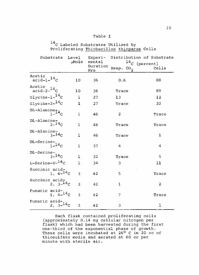
15
Table I
14C Labeled Substrates Utilized by
Proliferating Thiobacillus thioparus Cells
Substrate Level Mole
Experi- Distribution of Substrate mental 14
C (percent) Duration Hrs
Resp. CO2 Cells
Acetic acid -1 -14C 10 36 0.6 88
Acetic acid -2 -14C 10 36 Trace 89
Glycine -1 -14C 1 27 13 12
Glycine -2 -14C 1 27 Trace 22
DL- Alanine14 1- C 1 46 2 Trace
DL- Alanine- 2_14C 1 46 Trace Trace
DL- Alanine- 3 -14C 1 46 Trace 1
DL-Serine- 1-14c
1 37 4 4
DL- Serine- 3 -14C 1 32 Trace 5
L- Serine -U -14Ç 1 34 3 11
Succinic acid - 1, 4 -14C 3 42 5 Trace
Succinic acid - 2, 3 -14C 3 42 1 2
Fumaric acid - 1, 4 -14C 3 42 7 Trace
Fumaric acid- 2, 3 -14C 3 42 3 1
Each flask contained proliferating cells (approximately 0.14 mg cellular nitrogen per flask) which had been harvested during the first one -third of the exponential phase of growth. These cells were incubated at 26° C in 20 cc of thiosulfate media and aerated at 60 cc per minute with sterile air.

16
the end of the respective time courses of utilization as
indicated by the fact that the production of respiratory
14CO2 had declined to an insignificant level. It was
noted that even with a dense cell suspension, the rate
of substrate utilization was very low, ranging from
1 mole to 3 'mole of labeled substrate over a period
of 30 hours. An induction period as long as 28 hours
prior to the active utilization of the C4 dicarboxylic
acids was also observed.
Encouraged by the findings in the preliminary
experiments, the experimental conditions were modified
to increase the net amount of substrate utilization.
Labeled substrates were introduced into an incubation
medium, which had been inoculated with a very small
amount of cells. The cells were then permitted to under-
go active proliferation over an extended duration. Under
these conditions, the amount of substrate utilization
was found to be significantly increased when compared
with a similar experiment with a dense cell suspension.
In fact, the total amount of labeled substrate incorpo-
rated into the cells was respectable in magnitude when
the cells were harvested at the terminal side of the
exponential growth curve.
From the list of carbonaceous substrates found to
be utilized by T. thioparus cells, DL- alanine- 2 -14C,

17
acetate- 1 -14C, and acetate -2 -14C were selected as tracing
substrates because of their close relation with 3 -phos-
phoglycerate metabolism. Pyruvate- 2 -14C, arising from
DL- alanine -2 -14C may either be decarboxylated to yield
CO2 and acetate -1 -14C or enter into the C3 +C1 type of
carbon dioxide fixation pathway to provide C4 dicarbox-
ylic acids required for the biosynthesis of a number of
key amino acids. Using specifically 14C labeled acetate
samples to trace the role of C2 metabolism should also
give insight into the exact role played by the Krebs
Cycle reactions in this organism.
Table II
Distribution of Substrate Radioactivity in Incor- poration Experiments with Thiobacillus thioparus Cells
Substrate Level mg
Experi- mental Duration Hrs
Distribution of Sub- strate Radioactivity
(Percent) Resp. Cells Medium CO2
CH 3 14COOH 1.20 22 0.6 88 11
14CH3COOH 1.20 22 Trace 89 11
CH314CHNH2COOH 2.41 50 Trace 72 28
Each flask contained two liters of thiosulfate media into which was inoculated proliferating cells (4.6 µg of total cellular nitrogen) in the first one -third of the exponential phase of growth. These cells were incubated at 26o C in the presence of the 1 C substrates and aerated with sterile air at a rate of 260 cc per minute.

18
In Table II the inventory of radioactivity from the
respective 14C labeled substrates is given in experiments
with T. thioparus under active proliferating conditions.
It is noted that a significant amount of 14C compound
has gained entry into the cells. The fact that yields
of respiratory 14C ®2 are extremely low in magnitude
provide a clue that these substrates are not involved,
to any great extent, in energy producing processes.
In order to elucidate the nature of the biosynthetic
pathways operative in this bacteria, efforts were then
directed to investigation of whether the respective sub-
strates, upon entry into the cells, were engaged in
further metabolic processes. The 14C labeled cells were
hydrolyzed and processed for the separation of cellular
amino acids. The amino acids were separated by means of
resin column chromatography and their respective radio -
activities assayed by means of a liquid scintillation
flow counter.
It is evident, from the data present in Table III,
that the labeled substrate has entered the pool of intra-
cellular constituents and engaged in active metabolism
giving rise to carbon skeletons of several amino acids.
It is interesting to note that the variety of
cellular amino acids derived from DL- alanine -2 -14C is
in sharp contrast to that derived from 14C labeled
19
Table III
Incorporation of the Labeled Atom of DL- Alanine- 2 -14C, Acetic Acid- 1 -14C, and Acetic Acid -2 -14C into the Cellular Amino Acids of Thiobacillus thioparus
Cellular µc/ µMole Cellular Amino Acid Amino Acid DL- Alanine -2 -14C Acetic Acetic
14 14 Acid-1-
14 C Acid-2- 14
14 C
Glutamic acid .005 3.27 2.84
Proline .005 3.98 3.41
Arginine .005 3.55 3.12
Leucine .009 3.41 3.12
Valine .007 0 0
Aspartic acid .002 0 0
Threonine .002 0 0
Lysine .005 0 0
Isoleucine .005 0 0
Methionine .003 0 0
Alanine .108 0 0
Serine .001 0 0
Substrate level:
DL- Alanine- 2 -14C, 27 µMoles at 2.09 pc/ µMole; Acetic acid -1 -14C and Acetic acid- 2 -14C, 20 µMoles at 25 µc/ µMole.
20
acetates. In the alanine -2 -14C experiment, the bulk of
the radioactivity is found in cellular alanine. The
magnitude is such that it may reflect that D-alanine-
2- 14
C has undergone epimerization to L- alanine -2 -14C
prior to its incorporation into cellular proteins. The
remaining radioactivity of the alanine -2 -14C is found to
be distributed among a great number of amino acids. On
the other hand, one finds that in the experiments with
14C labeled acetates, labeling is confined to only four
amino acids, all related directly to q- ketoglutarate.
However, a close examination of the amino acids that were
preferentially labeled from alanine -2 -14C revealed that
they are related either directly to pyruvic acid (e.g.,
serine and valine), or to oxalacetic acid which is pre-
sumably derived from pyruvic acid by a CO2 fixation
process of the C3 +C1 type.
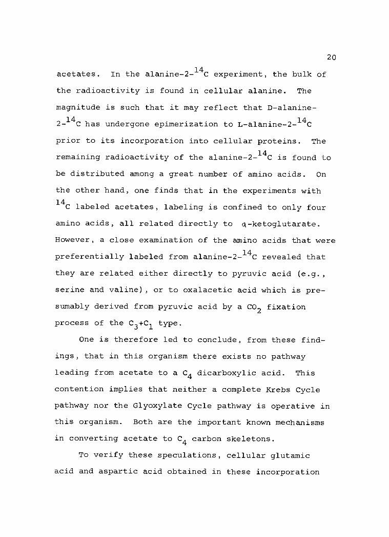
One is therefore led to conclude, from these find-
ings, that in this organism there exists no pathway
leading from acetate to a C4 dicarboxylic acid. This
contention implies that neither a complete Krebs Cycle
pathway nor the Glyoxylate Cycle pathway is operative in
this organism. Both are the important known mechanisms
in converting acetate to C4 carbon skeletons.
To verify these speculations, cellular glutamic
acid and aspartic acid obtained in these incorporation

21
experiments were subjected to degradation studies in
order to gain information on the intramolecular distribu-
tion pattern of the 14C label derived from the respective
14C labeled substrates.
The label distribution patterns of the cellular
amino acids derived from DL- alanine- 2 -14C, acetate- 1
and acetate -2 -14C are given in Table IV along with the
labeling patterns anticipated, assuming the operation of
several possible sets of reaction sequences. It can be
readily concluded that cellular glutamate is derived
from acetate by way of: citrate - cis- aconitate ->
isocitrate - a- ketoglutarate - glutamate.
Cellular aspartate is derived from oxalacetate which in
turn is derived from pyruvate by way of a CO2 fixation
process of the C3 +C1 type. The fact that acetate was
found to be converted to glutamate but not to aspartate
suggests strongly that the enzyme a- ketoglutaric acid
oxidase complex is not present in this organism and
thereby forbids the operation of the Krebs Cycle path-
way in a cyclic manner.
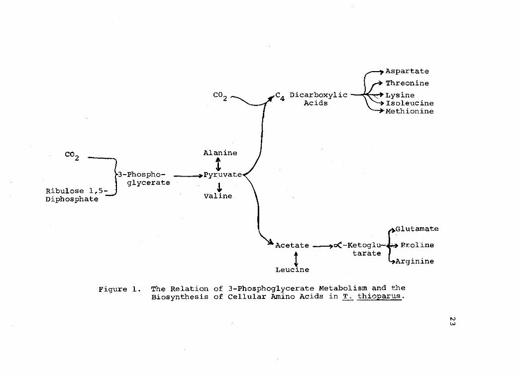
These results indicate that enzymes responsible for
several reactions of the Krebs Cycle pathway are
operative in T. thioparus, but they function only as
biosynthetic reactions. Given in Figure 1 is a graphic
summary of the understandings derived from the present
-14C,

22
Table IV
Labeling Patterns of Cellular Glutamic Acid and Aspartic Acid Derived from 14C Labeled Substrates
Pathway Followed by the Labeled Carbon Atoms
Expected and Observed Isotopic Distribution Pattern (Percent)
Glutamic Acid
Acetate -1 -14C Via Krebs Cycle at
COOH CH2 CH2 CHNH2 COOH
steady state 67 0 0 0 33 Via Krebs Cycle reactions to a- ketoglutarate 100 a___9___Jli 0
Observed 100 Acetate -2 -14C Via Krebs Cycle after two turns 0
Via Krebs Cycle at steady state 0
Via Krebs Cycle reactions to a- ketoglutarate 0
Observed 0
DL- Alanine -2 -14C Via pyr -4 acetate -4
a- ketoglutarate 100 Via pyr -4 OAA; OAA +acetate (derived from pyr)
0
50 25 25
29 29 29
100 0 0
100
0 0 0
0
0
14
0
0
0
-4 a- ketoglutarate 50 50 0_ 0
Observed 65 35 0
Aspartic Acid
COOH CH2 CHNH2 COOH DL- Alanine 14
-2 -14C Via pyr -, acetate --4
Krebs Cycle -4 OAA 50 Via C3 +C1 --4 C4 acid (unsymmetrical) -4 OAA 0
Observed 0
0 0 50
0 100 0
100 0
pyr = pyruvate, OAA = oxalacetate
CO2
Ribulose 1,5- Diphosphate
CO 2
Alanine
I 3-Phospho- =_*Pyruvate glycerate
Valine
Aspartate
Threonine
Dicarboxylic Lysine Acids Isoleucine
Methionine
Glutamate
Acetate -,c- Ketoglu Proline tarate
Arginine Leucine
Figure 1. The Relation of 3- Phosphoglycerate Metabolism and the Biosynthesis of Cellular Amino Acids in T. thioparus.
-4+-j
t
C4 .
-

24
study, relative to the fate of 3- phosphoglyceric acid in
the biosynthesis of cellular amino acids found in T.
thioparus. The energy required to implement these
reactions is derived exclusively from sulfur metabolism.
25
SUMMARY
Aseptic radiorespirometry has been used to examine
the utilization of external carbon sources by proliferat-
ing Thiobacillus thioparus cells. These studies reveal
that trace amounts of succinate, fumarate, acetate,
DL- serine, DL- alanine, and glycine can be utilized by
this chemoautotrophic organism.
Examination of the respective fates of the label in
DL- alanine- 2 -14C, acetate- 1 -14C, or acetate -2 -14C in the
cellular metabolism revealed that the Krebs Cycle path-
way is not functioning as a respiratory mechanism in
T. thioparus. However, most of the reactions of the
Krebs Cycle pathway are involved in the biosynthesis of
carbon skeletons for various amino acids. A CO2 fixation
pathway of the C3 +C1 type is instrumental in providing C4
dicarboxylic acid and those amino acids derived there-
from. Acetate can be incorporated into a- ketoglutarate
and those amino acids derived therefrom, but cannot be
incorporated into the C4 dicarboxylic acids.
It appears that the absence of the enzyme a- ketoglu-
taric acid oxidase complex accounts for the lack of oper-
ation of the Krebs Cycle pathway as the terminal respira-
tory mechanism. These findings also suggest that the
Glyoxylate Cycle pathway is inoperative in this organism.

26
BIBLIOGRAPHY
1 Bassham, J. A. et al. The path of carbon in photo- synthesis. XXI. The cyclic regeneration of carbon dioxide acceptor. Journal of the American Chemical Society 76:1760 -1770. 1954.
2 Boltjes, T. Y. K. Onderzoekingen over nitri- ficeerende bacterien. Thesis. Delft, Technische Hoogeschool. 1934.
3. Bomeke, H. Beiträge zur Physiologie nitrifizie- render Bakterien. Archiv fur Mikrobiologie 10:385 -445. 1939.
4. Cooper, Robert C. Evidence for the presence of certain tricarboxylic acid cycle enzymes in Thiobacillus thioparus. Journal of Bacteriology 88:624 -629. 1964.
5. Emoto, Y. über eine neue schwefeloxydierende Bakterie. Vorläufige Mitteilung. Botanical Magazine (Tokyo) 42 :421 -426. 1928.
6. Studien uber die physiologie der schwefeloxydierenden bakterien. Botanical Magazine 47 :405 -588. 1933.
7. Hirs, C. H. W., Stanford Moore and William H. Stein. The chromatography of amino acids on ion exchange resins. Use of volatile acids for elution. Journal of the American Chemical Society 76:6063 -6065. 1954.
8. Hutchens, Tyra et al. Techniques in the use of C14 as a tracer II Preparation of BaCO
3 by centrifugation. Nucleonics 7 :41 -44.
3 1950.
9. Johnson, E. J. and H. D. Peck, Jr. Coupling of phosphorylation and carbon dioxide fixation in a cell -free system from Thiobacillus thioparus. (Abstract) Bacteriological Proceedings of the 64th Meeting of the American Society for Microbiology, 1964, p. 91.
10. Jones, G. L. and F. C. Happold. The occurrence of polythionates as intermediates in the metabolism of thiosulfate by the Thiobacillus. Journal of General Microbiology 26:361 -366. 1961.
plates
27
11 Katz, J., S. Abraham and N. Baker. Analytical procedures using a combined combustion -diffusion vessel. Improved method for combustion of organic compounds in aqueous solution. Analytical Chemistry 26 :1503 -1504. 1954.
12 Katz, J., S. Abraham and I. C. Chaikoff. Analytical procedures using a combined combustion - diffusion vessel. An improved method for the degradation of carbon -14- labeled lactate and acetate. Analytical Chemistry 27:155 -156, 1955.
13. Lees, Howard. Energy metabolism in chemolitho- tropic bacteria. Annual Review of Microbiology 14 :83 -98. 1960.
14. Milhaud, G., J. P. Aubert and J. Millet. Metabolisme du carbone dans la chimiautotrophie. Cycle d'assimilation de l'anhydride carbonique. Comptes Rendus Hebdomandaires des Seances de l'Academie des Sciences 243:102 -105. 1956. .
15. Moore, S., D. H. Spackman and W. H. Stein, Chromatography of amino acids on sulfonated polystyrene resins. An improved system. Analytical Chemistry 30:1185 -1190. 1958.
16 Nathansohn, A. Uber eine neue Gruppe von Schwefelbakterien und ihren Stoffwechsel. Mitteilungen aus der zoologischen Station zu Neapel 15:655 -680. 1902.
17. Peck, H. D., Jr. Symposium on metabolism of inorganic compounds. V. Comparative metabolism of inorganic sulfur compounds in microorganisms. Bacteriological Reviews 26:67 -94. 1962.
18 Pigretti, M. Martha and A. O. M. Stoppani. Chemical degradation of 14C- labeled glutamic acid. Biochimica et Biophysica Acta 52:390 -392. 1961.
19. Roland, J. F., Jr. and A. M. Gross. Quantitative determination of amino acids using monodimensional paper chromatography. Analytical Chemistry 26:502 -505. 1954.
20. Santer, Melvin and Wolf Vishniac. CO incor- poration by extracts of Thiobacillus thioparus. Biochimica et Biophysica Acta 18 :157 -158. 1955.

28
21 Spackman, D. H., W. H. Stein and S. Moore. Automatic recording apparatus for use in the chromatography of amino acids. Analytical Chemistry 30:1190 -1206. 1958.
22 Starkey, R. L. Concerning the physiology of Thiobacillus thiooxidans, an autotrophic bacterium oxidizing sulfur under acid conditions. Journal of Bacteriology 10:135 -163. 1925.
23. Concerning the carbon and nitrogen nutrition of Thiobacillus thiooxidans, an autotrophic bacterium oxidizing sulfur under acid conditions. Journal of Bacteriology 10:165 -195. 1925.
24. Cultivation of organisms concerned in the oxidation of thiosulfate. Journal of Bacteriology 28:365 -386. 1934.
25. Suzuki, I. and C. H. Werkman. Chemoautotrophic carbon dioxide fixation by extracts of Thiobacillus thiooxidans. II. Formation of phosphoglyceric acid. Archives of Biochemistry and Biophysics 77:112 -123. 1958.
26. Trudinger, P. A. Phosphoglycerate formation from pentose phosphate by extracts of Thiobacillus denitrificans. Biochimica et Biophysica Acta 18:581 -582. 1955.
27. Fixation of carbon dioxide by extracts of the strict autotroph Thiobacillus denitrificans. Biochemical Journal 64:274 -286. 1956.
28. Van Slyke, Donald D., Douglas A. MacFadyen and Paul B. Hamilton. The gasometric determination of amino acids in urine by the ninhydrin- carbon dioxide method. Journal of Biological Chemistry 150:251 -258. 1943.
29. Vishniac, Wolf and Melvin Santer. The Thiobacilli. Bacteriology Review 21:195 -213. 1957.
30. Vogler, K. G., G. A. LePage and W. W. Umbreit. Studies on the metabolism of autotrophic bacteria. I. The respiration of Thiobacillus thiooxidans on sulfur. Journal of General Physiology 26:89 -102. 1942.

29
31. Wang, Chih H. and Julia K. Krackov. The catabolic fate of glucose in Bacillus subtilis. Journal of Biological Chemistry 237:3614 -3622. 1962.
32 Wang, Chih H. Metabolism studies by radio - respirometry. In: Advances in tracer methodology, ed. by Seymour Rothchild vol, 1. New York, Plenum Press. 1963. p. 274 -290.
33 White, C. G. and Samuel Helf. Suspension counting in scintillation gels. Nucleonics 14 :46 -48. 1956.
34. Yemm, E. W. and E. C. Cocking. The determination of amino acids with ninhydrin. Analyst 80:209 -213. 1955.