red blood cell disorders - università di torino
TRANSCRIPT

Adult Reference Ranges for Red Blood Cells
Men Women
Hemoglobin (g/dL) HGB 13.6-17.2 12.0-15.0
Hematocrit (%) HCT 39-49 33-43
Red cell count (106/μL) RBC 4.3-5.9 3.5-5.0
Reticulocyte count (%) 0.5-1.5
Mean cell volume (MCV; μm3) 82-96
Mean corpuscular hemoglobin (MCH; pg) 27-33
Mean corpuscular hemoglobin concentration (MCHC; g/dL) 33-37
RBC distribution width (RDW; coefficient of variation of volume) 11.5-14.5
Decreased erythrocyte number ANEMIA
Increased erythrocyte number POLICYTEMIA
RED BLOOD CELL DISORDERS

This is normal data from a complete blood count as performed on an automated
instrument, including an automated WBC differential count.

Here is data from a CBC in a person with iron deficiency anemia.
Note the low hemoglobin (HGB). Microcytosis is indicated by the low MCV
(mean corpuscular volume). Hypochromia correlates here with the low MCH
(mean corpuscular hemoglobin).

The CBC here shows a markedly increased MCV, typical for megaloblastic anemia. The
MCV can be mildly increased in persons recovering from blood loss or hemolytic anemia,
because the newly released RBC's, the reticulocytes, are increased in size over normal
RBC's, which decrease in size slightly with aging.


Induction of HIF1 target genes by hypoxia
HIF1 HIF1

Mechanism Specific Examples
Inherited genetic defects
Defects leading to stem cell depletion Fanconi anemia, telomerase defects
Defects affecting erythroblast
maturation
Thalassemia syndromes
Nutritional deficiencies
Deficiencies affecting DNA synthesis B12 and folate deficiencies
Deficiencies affecting hemoglobin
synthesis
Iron deficiency anemia
Erythropoietin deficiency
Renal failure, anemia of chronic disease
Immune-mediated injury of progenitors Aplastic anemia, pure red cell aplasia
Inflammation-mediated iron
sequestration
Anemia of chronic disease
Primary hematopoietic neoplasms Acute leukemia, myelodysplasia, myeloproliferative
disorders
Space-occupying marrow lesions Metastatic neoplasms, granulomatous disease
Infections of red cell progenitors Parvovirus B19 infection
Unknown mechanisms Endocrine disorders, hepatocellular liver disase
DECREASED RED CELL PRODUCTION

Due to reduced intake or to metabolic problems
Iron need:
•man: < 1.0 mg/day
•fertile woman: 1.5 mg/day, increases during pregnancy
SIDEROPENIC ANEMIA
Hypochromic microcytic anemia of iron deficiency
(peripheral blood smear). Note the small red cells
containing a narrow rim of peripheral hemoglobin.
Scattered fully hemoglobinized cells, present due to
recent blood transfusion, stand in contrast.

Main proteins involved in iron metabolism
- transferrin (Tr; binds 2 iron atoms) - Tr receptor (binds 2 Tr = 4 iron atoms) - ferritin (binds about 4.500 iron atoms) - hemosiderin (insoluble ferritin, in bone marrow, spleen, liver)
iron distribution in the adult (50-40 mg/kg body weight)
mg/kg
man woman
functional iron
• Hb 31 28
• myoglobin 5 4
• enzymes 2 2
transport iron
• Tr < 1 < 1
iron stores
• ferritin 8 4
• hemosiderin 4 2

HIF-1 regulates the expression of multiple genes to stimulate erythropoiesis in response to hypoxia.
HIF-1 stimulates production of the EPO in the kidney, which binds to its receptor (EPOR) on erythroid
progenitors in the bone marrow (in the adult and yolk sac in the embryo) to stimulate their survival,
proliferation, and differentiation. Erythropoiesis involves uptake by the marrow of large amounts of iron, which
are used in the synthesis of hemoglobin. In the liver, HIF-1 stimulates iron uptake by repressing the gene
encoding hepcidin, which is an inhibitor of ferroportin, the major protein responsible for intestinal iron uptake.
HIF-1 also activates hepatic synthesis of transferrin, the major plasma protein responsible for transporting
iron from the intestine to the bone marrow via the transferrin receptor. Thus, HIF-1 directly regulates the
expression of 5 gene products (EPO, EPOR, hepcidin, transferrin, and transferrin receptor) involving 5
different organs (kidney, liver, intestine, blood, and bone marrow) to control erythropoiesis (Semenza, 2009).
HIF
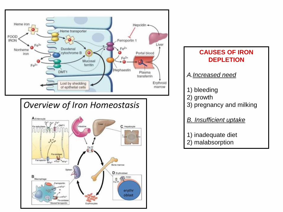
CAUSES OF IRON
DEPLETION
A.Increased need
1) bleeding
2) growth
3) pregnancy and milking
B. Insufficient uptake
1) inadequate diet
2) malabsorption
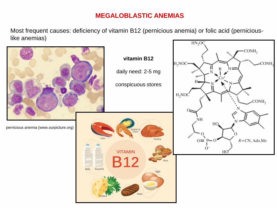
MEGALOBLASTIC ANEMIAS
Most frequent causes: deficiency of vitamin B12 (pernicious anemia) or folic acid (pernicious-
like anemias)
pernicious anemia (www.ourpicture.org)
vitamin B12
daily need: 2-5 mg
conspicuous stores
Elsevier, 2010
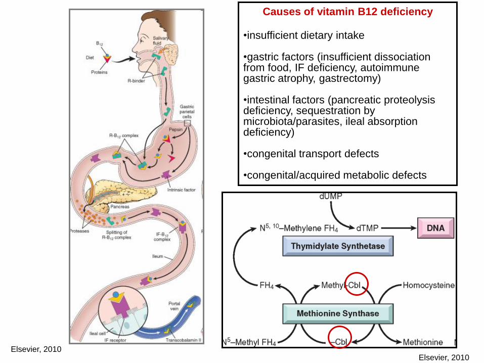
Causes of vitamin B12 deficiency
•insufficient dietary intake
•gastric factors (insufficient dissociation from food, IF deficiency, autoimmune gastric atrophy, gastrectomy)
•intestinal factors (pancreatic proteolysis deficiency, sequestration by microbiota/parasites, ileal absorption deficiency)
•congenital transport defects
•congenital/acquired metabolic defects
Elsevier, 2010

I. Structural defects hemoglobinopaties
II. Synthesis rate defects α and β thalassemias
GENETIC DISEASES OF GLOBIN CHAIN
(mendelian inheritance)
HEMOGLOBIN (Hb)
2β2 tetramer (HbA1)
each globin chain bound to one heme (Fe2+)
95% of red blood cell content

97-98% 2–3,5%
< 6%

ALTERED Hb SYNTHESIS RATES: OR β THALASSEMIAS
β-thalassemia
homozygosis β-thalassemia major (Cooley disease or mediterranean anemia)
heterozygosis β-thalassemia minor (β-thalassemic tract; compensated by HbA2 and
HbF. Need to identify carriers)
• exon nonsense mutations (recessive phenotype) β0-thalassemia (no transcript)
• intron mutations (recessive phenotype) β+-thalassemia
(uncorrect mRNA splicing reduced β chain synthesis)
- unbalanced globin chain synthesis α chains aggregate and precipitate in
erythrocytes (reduced half life) and in precursors (ineffective erythropoiesis due to
destruction in bone marrow)
- sometimes HbF produced to compensate
ANEMIA: due to both reduced production and increased destruction of erythrocytes

Thalassemia major: pathogenesis

HEMOLYTIC ANEMIAS
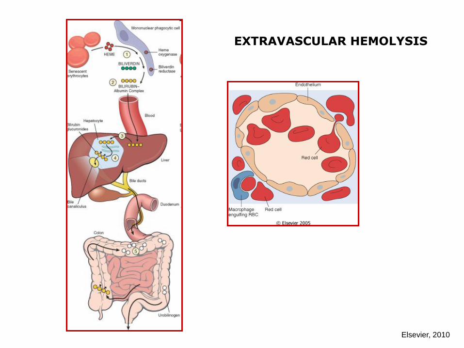
EXTRAVASCULAR HEMOLYSIS
Elsevier, 2010
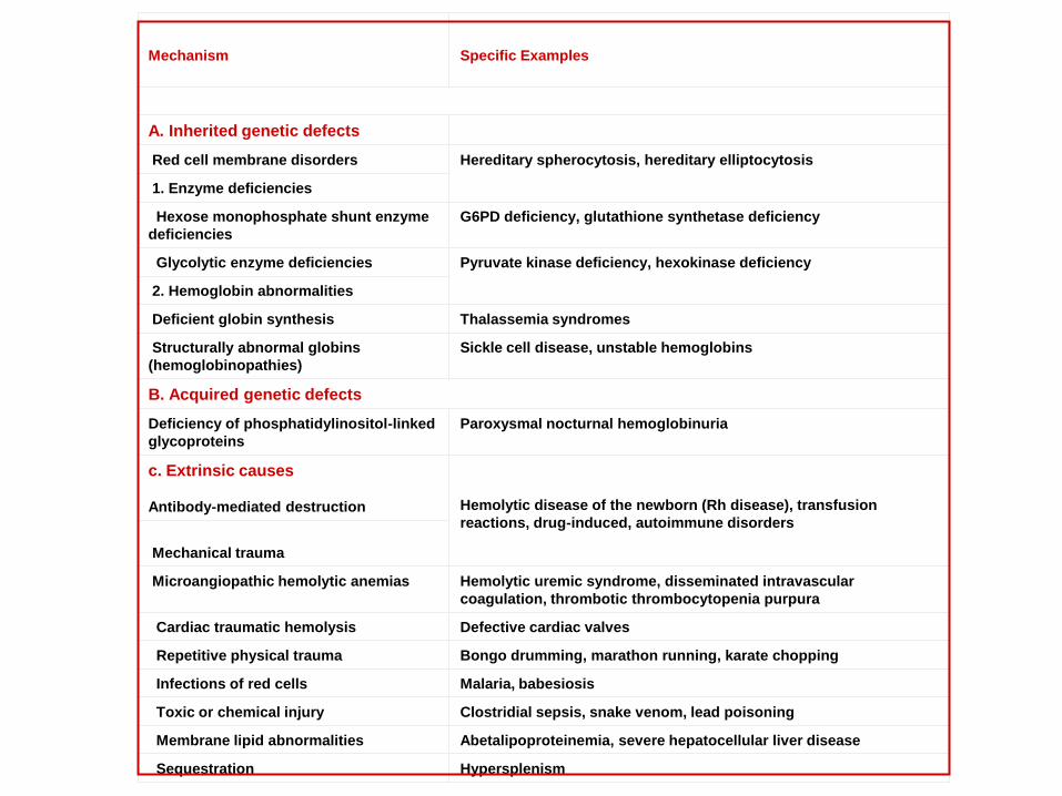
Mechanism
Specific Examples
A. Inherited genetic defects
Red cell membrane disorders Hereditary spherocytosis, hereditary elliptocytosis
1. Enzyme deficiencies
Hexose monophosphate shunt enzyme
deficiencies
G6PD deficiency, glutathione synthetase deficiency
Glycolytic enzyme deficiencies Pyruvate kinase deficiency, hexokinase deficiency
2. Hemoglobin abnormalities
Deficient globin synthesis Thalassemia syndromes
Structurally abnormal globins
(hemoglobinopathies)
Sickle cell disease, unstable hemoglobins
B. Acquired genetic defects
Deficiency of phosphatidylinositol-linked
glycoproteins
Paroxysmal nocturnal hemoglobinuria
c. Extrinsic causes
Antibody-mediated destruction
Hemolytic disease of the newborn (Rh disease), transfusion
reactions, drug-induced, autoimmune disorders
Mechanical trauma
Microangiopathic hemolytic anemias Hemolytic uremic syndrome, disseminated intravascular
coagulation, thrombotic thrombocytopenia purpura
Cardiac traumatic hemolysis Defective cardiac valves
Repetitive physical trauma Bongo drumming, marathon running, karate chopping
Infections of red cells Malaria, babesiosis
Toxic or chemical injury Clostridial sepsis, snake venom, lead poisoning
Membrane lipid abnormalities Abetalipoproteinemia, severe hepatocellular liver disease
Sequestration Hypersplenism


HEMOGLOBINOPATIES
1. Single point mutations: HbS β6glu val
2. Double point mutations in the same globin chain
3. Nonsense mutations: es. β39 CAG(gln) TAG(stop) frequent in thalassemias
4. Mutations causing elongation
5. Mutationi due to fusion genes Hb Lepore (δβ)
6. Codon deletion/insertion (in frame and frameshift)
A SICKLE CELL DISEASE
B UNSTABLE Hb
C MODIFIED O2 AFFINITY
D ALTERED O2 TRANSPORTATION

SICKLE CELL DISEASE
HbS: β6glu val; autosomal recessive inheritance
heterozygosis carriers low frequency clinical signs: HbS 35-40%, HbA1 55-60%
homozygosis SICKLE CELL DISEASE early death
Deoxygenated blood: adjacent mutated β chain polimerize (β6val)
asymptomatic until HbF can compensate (6 months)
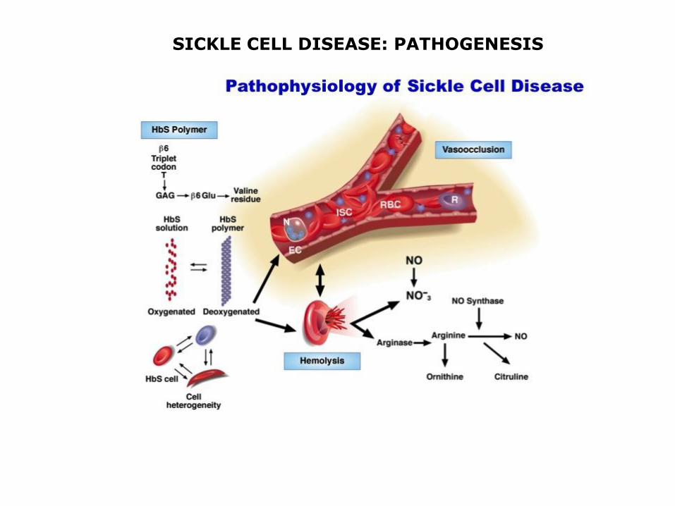
SICKLE CELL DISEASE: PATHOGENESIS

SICKLE CELL DISEASE: PATHOGENESIS


IMMUNOHEMOLYTIC ANEMIAS
ISOANTIBODIES non compatible transfusion
newborn hemolytic disease (Rh incompatibility)
AUTOANTIBODIES warm Ab caldi (IgG)
cold Ab (agglutinins, hemolysins)
erythrocyte Ag acquiring immunogenicity

Diagnosis of immunohemolytic anemia
- direct Coombs antiglobulin test, in which the patient's red cells are mixed with sera containing antibodies that are
specific for human immunoglobulin or complement.
If either immunoglobulin or complement is present on the surface of the red cells, the multivalent antibodies cause
agglutination, which is easily appreciated visually as clumping.
- indirect Coombs antiglobulin test, the patient's serum is tested for its ability to agglutinate commercially available red
cells bearing particular defined antigens.
This test is used to characterize the antigen target and temperature dependence of the responsible antibody. Quantitative
immunological tests to measure such antibodies directly are also available.
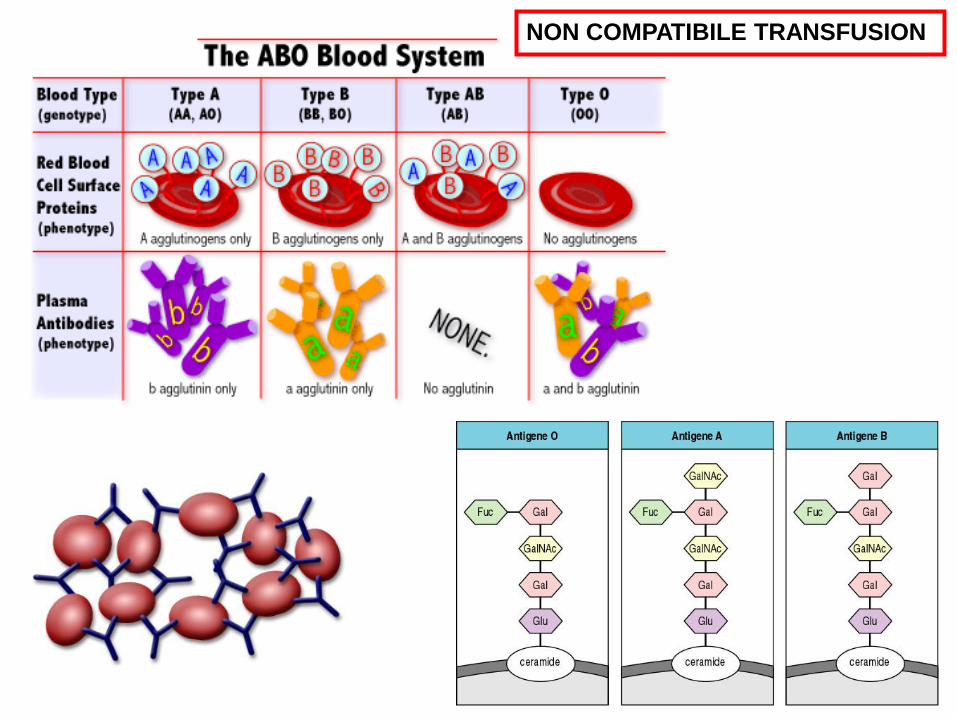
NON COMPATIBILE TRANSFUSION

Macacus rhesus; 1940
Rh Positive Rh Negative
0+ 37% 0- 6%
A+ 34% A- 6%
B+ 10% B- 2%
AB+ 4% AB- 1%
85% positive 15% negative
Van der Schoot et al., 2008
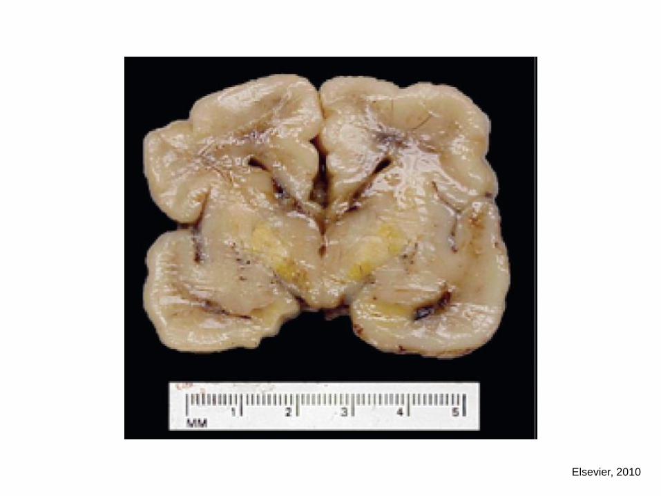
NEWBORN HEMOLYTIC DISEASE

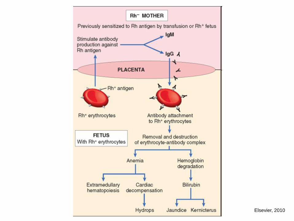
Elsevier, 2010
Elsevier, 2010

AUTOIMMUNE HEMOLYTIC ANEMIAS
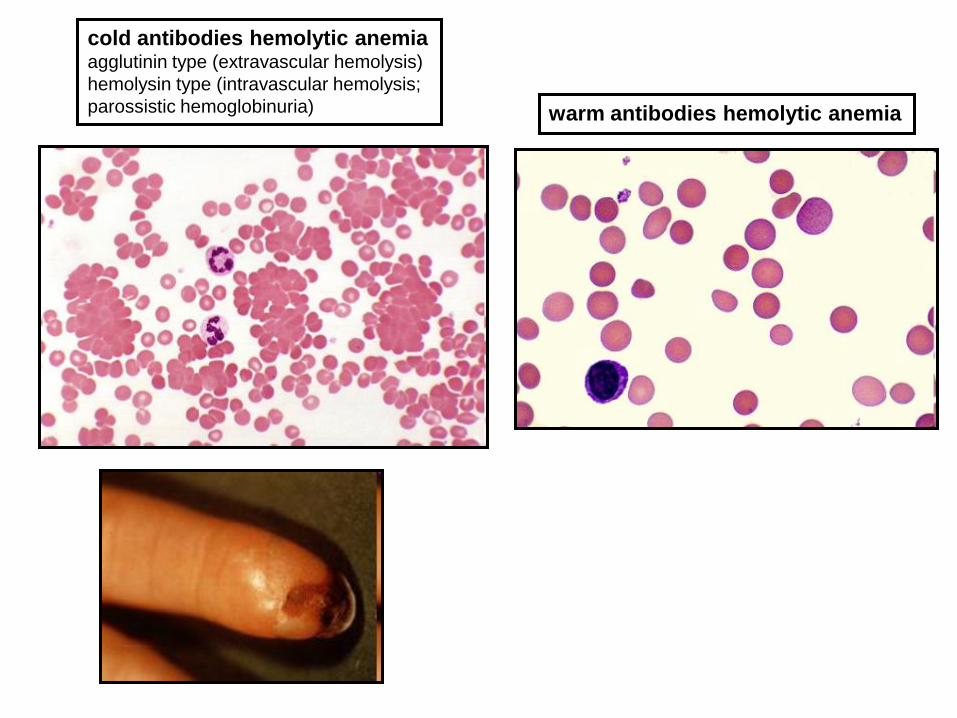
cold antibodies hemolytic anemia agglutinin type (extravascular hemolysis)
hemolysin type (intravascular hemolysis;
parossistic hemoglobinuria) warm antibodies hemolytic anemia

Cephalosporins (3rd generation)
Diclofenac
α -Methyldopa
High-dose therapy with penicillin for > 10 days
Oxaliplatin
Rifampicin
Fludarabin
Levodopa
Quinidine
Mefenamic acid
Salama, 2009
DRUG-INDUCED HEMOLYTIC ANEMIAS
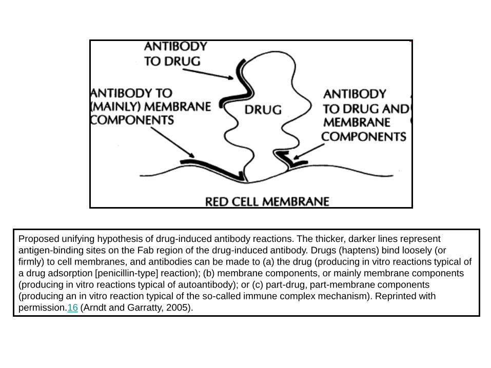
Proposed unifying hypothesis of drug-induced antibody reactions. The thicker, darker lines represent
antigen-binding sites on the Fab region of the drug-induced antibody. Drugs (haptens) bind loosely (or
firmly) to cell membranes, and antibodies can be made to (a) the drug (producing in vitro reactions typical of
a drug adsorption [penicillin-type] reaction); (b) membrane components, or mainly membrane components
(producing in vitro reactions typical of autoantibody); or (c) part-drug, part-membrane components
(producing an in vitro reaction typical of the so-called immune complex mechanism). Reprinted with
permission.16 (Arndt and Garratty, 2005).