rectal cancer: a prospective cohort study laparoscopic
TRANSCRIPT

Page 1/16
E�cacy of Anastomosis Enhancing Suture inLaparoscopic Anterior Resection of Middle-LowRectal Cancer: A Prospective Cohort StudyJin-Shui Chen
Departmetn of Surgery, No.991 Hospital of People's Liberation Army, Hubei, ChinaJia Zang
Department of Surgery, Changzheng Hospital, Shanghai, ChinaShu-Xun Wei
Shanghai Changzheng HospitalWen-Tao Yan
Department of Surgery, Changzheng Hospital, Shanghai, ChinaHai-Yang Zhou
Department of Surgery, Changzheng Hospital, SHanghai, ChinaCan-Ping Ruan
Department of Surgery, Changzheng Hospital, Shanghai, ChinaWei-Jun Wang
Department of Surgery, Changzheng Hospital, Shanghai, ChinaYi Wang
Department of Surgery, Changzheng Hospital, Shanghai, ChinaYan-Ping Sun
Department of Surgery, Changzheng Hospital, Shanghai, ChinaJian Zhang ( [email protected] )
Changzheng Hospital, Shanghai, China https://orcid.org/0000-0002-0126-6172Zhi-Qian Hu
Department of Surgery, Changzheng Hospital, Shanghai, China
Technical innovations
Keywords: Middle-low rectal cancer, Laparoscopic anastomosis enhancing suture, Preventiveenterostomy, Transanal drainage tube, Laparoscopic anterior resection, Double-stapling technique,Anastomotic leakage, Anastomotic bleeding, Anastomotic stricture
Posted Date: February 4th, 2021
DOI: https://doi.org/10.21203/rs.3.rs-165686/v1

Page 2/16
License: This work is licensed under a Creative Commons Attribution 4.0 International License. Read Full License

Page 3/16
AbstractBackground Anastomotic leakage (AL) limits the outcome after laparoscopic anterior resection (LAR) formiddle-low rectal cancer. The study investigated the e�cacy of laparoscopic anastomosis enhancingsuture (LAES), preventive ileostomy and transanal drainage tube placement in reducing anastomoticleakage after LAR for middle-low rectal cancer.
Methods From April 2016 to April 2019, a prospective cohort study was performed on consecutivepatients who underwent LAR for middle-low rectal cancer in Changzheng hospital. The patients weredivided into group A, B, C and D in which LAES, transanal drainage tube placement, protective ileostomy,and no preventive treatment were applied, respectively. Clinical characteristics, operative variables andpostoperative complication were compared between the groups.
Results Among 320 patients, 24 (7.5%) developed AL and incidence rate of AL was 1.3%, 12.5%, 1.3% and15.0% in the four groups, respectively. Left colic artery preservation and neoadjuvant chemotherapy werefound not associated with the incidence of AL. A total of 0, 2, 2 and 5 patients had anastomotic bleedingin the four groups, respectively. No patient underwent reoperation in group A and group C, while 5.0%(4/80) of the patients had reoperation in group B and group D due to grade C AL with severe symptoms.
Conclusions Compared with preventive ileostomy, LAES was effective in preventing AL after LAR formiddle-low rectal cancer and relieving the complications of AL. The transanal drainage tube placementdid not reduce the risk of AL.
The study was retrospectively registered with the Chinese Clinical Trial Registry on 28th June 2016 (code:ChiCTR-IOR-17011777).
What Does This Paper Add To The Literature?In the current prospective cohort study, we observed the e�cacy of a new suture technique, naminglaparoscopic anastomosis enhancing suture (LAES) in preventing anastomotic leakage afterlaparoscopic anterior resection (LAR) for middle-low rectal cancer, and found that LAES is effective inlowering the incidence of AL.
BackgroundLaparoscopic anterior resection (LAR) with total mesorectal excision (TME) has been the cornerstone inthe treatment of middle-low rectal cancers[1]. However, anastomotic leakage (AL) remains one of the mostdevastating complications with an incidence ranging from 1.3 to 12.9 %[2–4]. The risk factors can beclassi�ed as patient-related factors and operation-related factors. Patient-related factors mainly consistof male sex, higher body mass index (BMI), diabetes mellitus and smoking, while operation factorsincluded lower tumor location, larger tumor size, more intraoperative blood loss and longer operation time,etc[5, 6]. Many solutions, including defunctioning stoma (DS) and transanal drainage tube (TDT)
Page 4/16
placement, have been proposed to address the problem of AL and its complications. Despite its e�cacyin preventing AL, defunctioning stoma has been overused in current surgical practice, while theeffectiveness of transanal drainage tube placement needs to be con�rmed with further studies[7–10].
With the development of laparoscopic techniques, laparoscopic anastomotic enhancing suture (LAES)has come into sight to lower anastomotic tension and reduce risks of AL. The purpose of this study wasto investigate whether laparoscopic anastomosis enhancing suture can reduce AL following LAR formiddle-low rectal cancer.
Method
Study populationFrom January 2016 to April 2019, 320 cases of middle-low rectal cancer undergoing LAR with TME anddouble-stapling technique (DST) in Changzheng hospital were enrolled in this prospective cohort study.All the patients underwent standard treatment for CRC according to the Chinese guideline 2017. Theeligibility criteria were as follows: 1) age: over 18 years; 2) tumor location: 5 ~ 8cm from the anal verge; 3)pathologic stage: Tis/T1 ~ 4a, N0-2, M0, AJCC-7th TNM stage); 4) American Society of Anesthesiologist(ASA) function: class I to class III. The exclusion criteria included: 1) age: younger than 18 years; 2) tumorlocation: less than 5cm or greater than 8cm from the anal verge; 3) emergency surgery for intestinalperforation or obstruction; 4) self-expanding stent insertion for obstruction; 5) preoperative major pelvicsurgery; 6) neoadjuvant radiotherapy; 7) locally advanced(T4b) and/or distant metastasis; 8) recurrentrectal cancer; 9) Patients with severely compromised cardio-pulmonary function were also excluded.
Eligible patients were divided into A, B, C and D groups in which LAES, TDT placement, DS, and nopreventive treatment were applied, respectively. Each group included 80 cases and preventive measuresused in the four groups were the routine operative techniques of the four surgical teams, which were allled by senior surgeons with more than 20 years’ experience in TME and laparoscopic surgery.
The study was approved by the Ethics Committee of the Changzheng hospital and was registered withthe Chinese Clinical Trial Registry (code: ChiCTR-IOR-17011777) in June 2016.Written inform consent wasobtained from all the patients enrolled in this study.
Surgery procedureThe establishment of anastomosis was performed with standard practice according to the Chinese expertconsensus on diagnosis, prevention and treatment of rectal anastomotic leakage of 2019[11]. Good bloodsupply and low longitudinal tension were con�rmed by the senior surgeons.
After the construction of anastomosis, LAES was performed in group A patients as shown in Fig. 1. Theanterior half of anastomosis was reinforced with knotless absorbable sutures (Quill RA*, 3 − 0 PDOsuture) and 26mm, 3/8 circle needles in a running full-layer fashion. In the process of LAES, werecommend the �rst suture be medially placed in the front part of the anastomosis, which could make the

Page 5/16
following sutures easier, especially when the pelvis is narrow or the anastomosis is close to the anus. Wedidn’t suture the whole circumference of the anastomosis as anastomotic stenosis could occurpostoperatively.
In Group B, transanal drainage tube (Marecot catheter, 28Fr, 1.0cm diameter) was placed 3-5cm from theanastomotic site without any resistance and the tube was �xed with skin sutures and connected to adrainage bag (Fig. 2). Protective ileostomy was performed in group C and no additional preventivetreatment were used in group D.
After discharge, patients were followed in accordance with a standardized protocol, in which the patientsunderwent abdominal computed tomography scan and colonoscopy every 3 months for the �rst 6months after operation, every 3 months for the next 18 months and every 6 months afterwards. All theimaging outcomes were carefully analyzed by the experienced radiologists and surgeons. Information onrecurrence date, last follow-up, and death were recorded.
De�nition of anastomotic leakageThe endpoint in the study was occurrence of anastomotic leakage. According to the de�nition of theInternational Study Group of Rectal Cancer (ISREC)[12, 13], AL in this study was de�ned as postoperativesigns of peritonitis with high fever caused by leakage from any surgical stapler line, recto-vaginal �stula,or pelvic abscess upon clinical assessment. Diagnosis of AL was made based on feces, pus or gas fromthe abdominal drain or a peri-anastomotic abscess, and con�rmed by CT scan, ultrasonic examination,rectoscopy and/or radiological investigations. AL was classi�ed into Grade A B and C on the basis of thede�nition proposed by Rahbari et al[13]. All the patients diagnosed with clinical AL in the study wereidenti�ed within 30 days. Anastomotic Bleeding (AB) was de�ned as fresh blood or clots deriving fromthe anus or TDT and con�rmed by postoperative colonoscopy.
Clinicopathologic and operative variablesDetailed information on clinicopathologic and operative variables was carefully collected from patients.Patient-related information included demography like weight, height, gender, preoperative serumhemoglobin, albumin, and history of neoadjuvant chemotherapy. Operation-related variables includedoperation time, intraoperative blood loss, left colonic artery preservation, tumor size, tumor location,anastomosis level, postoperative pathology, AL, AB, anastomotic stricture intestinal obstruction,postoperative hospital stay, treatment cost and complications (using Clavien-Dindo classi�cationsystem). Concentration of serum hemoglobin, albumin, pre-albumin and pre-operative carcinoembryonicantigen (CEA) were obtained one week before the surgery.
Statistical analysisAll statistical analyses were performed in SPSS statistical package (version 19.0; IBM, Chicago, USA).Continuous variables were expressed as the means ± standard deviation. Differences of continuousvariables between the groups were compared with one-way analysis of variance (ANOVA), Kruskal-Wallistest and Least signi�cant difference test (LSD-test). Differences in the distribution of categorical variables

Page 6/16
were analyzed with Chi-squared test. All the statistical and P < 0.05 was considered statisticallysigni�cant.
Results
Clinical characteristicsAll the patients in four groups underwent successful laparoscopic operations without conversion tolaparotomy. The patients’ demographic and clinical data were shown in Table 1. There was no signi�cantdifference between the 4 groups in terms of age, gender, body mass index, comorbidities, serumhemoglobin, albumin, pre-albumin, pre-operative CEA concentration and ASA score (P > 0.05).

Page 7/16
Table 1Comparisons of patients’ demographical characteristics and operative variables and perioperative
outcomes after construction of rectal anastomosisVariable Anastomosis
reinforcementsuture (N = 80)
Transanaldrainage tube(N = 80)
Protectiveileostomy(N = 80)
No preventivetreatment (N = 80)
P
Age (years)* 61.6 ± 10.4 58.6 ± 10.6 64.3 ± 9.8 62.3 ± 9.1 0.013
Sex (male) 44(55.0%) 53(66.2%) 55(68.7%) 60(75.0%) 0.058
BMI (kg/m2)* 23.1 ± 2.9 22.6 ± 2.9 23.1 ± 3.2 23.6 ± 3.2 0.406
Hemoglobin(g/L)* 127.5 ± 17.5 134.2 ± 14.0 130.6 ± 15.0 134.5 ± 14.5 0.057
Albumin(g/L)* 39.9 ± 4.4 41.2 ± 4.8 39.1 ± 4.1 40.2 ± 4.7 0.043
Pre-Albumin(mg/L)*
249.0 ± 73.2 250.1 ± 73.1 238.5 ± 65.4 223.3 ± 68.6 0.175
Preoperative CEA ≥ 5.0 ug/L
31(38.8%) 32(40.0%) 32(40.0%) 31(38.8%) 0.997
ASA score > 2 33(41.3%) 36(40.0%) 34(42.5%) 29(36.3%) 0.866
Tumor Size(cm)* 3.5 ± 1.2 3.8 ± 1.0 3.5 ± 1.1 3.5 ± 1.0 0.329
Tumorlocation(cm)*
Male + Female 7.1 ± 0.8 6.4 ± 0.8 7.3 ± 0.7 7.6 ± 0.5 < 0.001
Male 7.3 ± 0.5 7.3 ± 0.7 6.6 ± 0.7 7.6 ± 0.5
Female 6.8 ± 0.9 7.2 ± 0.8 6.0 ± 0.9 7.7 ± 0.5
Operation time(min)*
150.7 ± 54.4 122.0 ± 24.9 160.6 ± 46.9 125.1 ± 34.9 < 0.001
Intraoperativeblood loss > 150ml
49(61.2%) 52(65.0%) 39(48.8%) 53(66.2%) 0.093
TNM stage
0-II 51(63.7%) 44(55.0%) 53(66.2%) 49(61.2%) 0.500
III 29(36.3%) 36(45.0%) 27(33.8%) 31(38.8%)
Tumor poordifferentiation
2(3.2%) 3(4.4%) 4(6.2%) 3(4.8%) 0.988
Number of lymphnodes harvested
13(10,17) 13(10,17) 14(11,17) 15(11,18) 0.266
* Values are mean ± standard deviation or median with range unless otherwise indicated.

Page 8/16
Variable Anastomosisreinforcementsuture (N = 80)
Transanaldrainage tube(N = 80)
Protectiveileostomy(N = 80)
No preventivetreatment (N = 80)
P
Left colonic arterypreservation
44(55.0%) 41(51.2%) 38(47.5%) 37(46.3%) 0.682
Anastomosislocation (cm)*
< 0.001
Male + Female 5.1 ± 0.7 4.4 ± 0.7 5.2 ± 0.7 5.0 ± 0.8 < 0.001
Male 5.3 ± 0.6 5.2 ± 0.7 4.4 ± 0.6 5.1 ± 0.7
Female 4.8 ± 0.7 5.2 ± 0.8 4.2 ± 0.7 7.7 ± 0.5
Anastomoticleakage
1(1.3%) 10(12.5%) 1(1.3%) 12(15.0%) < 0.001
Grade A/B 1(1.3%) 6(7.5%) 1(1.3%) 8(10.0%) 0.019
Grade C 0 4(5.0%) 0 4(5.0%) 0.042
Anastomoticbleeding
0 2(2.5%) 2(2.5%) 5(6.3%) 0.120
Anastomoticstricture
0 0 2(2.5%) 0 0.110
Intestinalobstruction
0 2(2.5%) 8(10.0%) 1(1.3%) 0.002
Reoperation 0 4(5.0%) 0 4(5.0%) 0.042
Postoperativehospital stay (d)
7.6 ± 2.7 7.5 ± 2.3 9.3 ± 4.0 12.7 ± 5.1 < 0.001
Treatment cost(×104 yuan)
51.4 ± 9.1 55.2 ± 10.0 59.4 ± 11.5 55.2 ± 10.9 < 0.001
* Values are mean ± standard deviation or median with range unless otherwise indicated.
Operative characteristicsDistance from anal margin to tumor distal edge and distance from anal margin to the anastomosis wasmeasured under the view of the rigid rectoscope after anesthesia. The operative characteristics wereshown in Table 1. Operation time, left colic artery preservation, estimated intra-operative blood loss,distance from anal margin to tumor distal edge, tumor size and distance from anal margin to theanastomosis were compared among the four groups. There was a signi�cant difference among the fourgroups in the operation time (P < 0.001). The average operation time of group A (150.7 ± 54.4min) wassigni�cantly longer than that of group B(122.0 ± 24.9min) and group D(125.1 ± 34.9min) (P < 0.001) butsimilar with group C. The rate of left colic artery preservation, intra-operative estimated blood loss and

Page 9/16
tumor size did not differ among the four groups (P > 0.05). The average distance from anal margin totumor distal edge of group A (7.1 ± 0.8cm) was signi�cantly shorter than that of group B(7.3 ± 0.7cm) andlonger than that of group C(6.4 ± 0.8cm) (P < 0.001). The average distance from anal margin toanastomosis of group A (5.1 ± 0.7cm) was similar with that of group B(5.2 ± 0.7cm) (P = 0.155) and thatof group D(5.0 ± 0.8cm) (P = 0.776), but longer than that of group C(4.4 ± 0.7cm) (P < 0.001).
Postoperative characteristicsThe postoperative characteristics were shown in Table 1, and the patients’ comorbidities and pathologicaltypes were summarized in eTable 1. There was no case of positive circumferential resection marginamong the four groups. The postoperative hospital stay, TNM tumor stage, number of lymph nodesharvested, pathological type, and differentiation level of carcinoma were compared among the fourgroups. There was a signi�cant difference between the four groups in the postoperative hospital stay(P < 0.001). The postoperative hospital stay of group A(7.6 ± 2.7d) was similar with group B(7.5 ± 2.3d) (P = 0.962), and signi�cantly shorter than group C and group D (9.3 ± 4.0d, P = 0.962; 12.7 ± 5.1d, P < 0.001,respectively). There were no signi�cant differences among the four groups in terms of TNM tumor stage,number of lymph nodes harvested, pathological type and differentiation levels of carcinoma (P > 0.05).
ComplicationsAs shown in Table 1, overall incidence rate of AL was 7.5 %. The complications and reoperation rateamong the four groups was compared. The rate of AL was 1.3%, 12.5%, 1.3%, 15.0% in group A, B, C andD, respectively. Both group A and group C had one patient of grade B AL, which was treated withconservative treatment. But four patients of grade C AL occurred in both group B and D, in which theincidence rate of AL was signi�cantly higher than that in group A and C(P < 0.05).
A total of 0, 2, 2 and 5 patients had AB in the four groups, respectively, without signi�cant difference(P > 0.05). Two cases of anastomotic stricture occurred in group C. Postoperative intestinal obstructionhappened in 8 patients of group C, 2 patients of group B and one patient of group D, while none in groupA. Compared with the other groups, intestinal obstruction rate after operation in group C was signi�cantlyhigher (P < 0.05). There was no signi�cant difference in comorbidities such as pulmonary infection, heartfailure or arrhythmia, urinary retention and infection among the 4 groups (P > 0.05). There were 4 cases ofpostoperative anastomotic stenosis in the group C, which was considered related to the disuse ofanastomotic site and the lower site of the anastomosis.
Treatment costThe average treatment cost was shown in Table 1. There were signi�cant differences among the fourgroups in the treatment cost (P < 0.001). The treatment cost in group A (51.4 ± 9.1 thousand yuan) wassigni�cantly lower than the other three groups (group B, 55.2 ± 10.0 thousand yuan; group C, 59.4 ± 11.5thousand yuan; group D, 55.2 ± 10.9 thousand yuan) (P < 0.001).
In addition, there were 2 cases of incision infection, 2 cases of postoperative incisional hernia and 1 caseof intraperitoneal bleeding in the second hospitalization for stoma reversal in group C. The second

Page 10/16
average treatment cost in group C was 21.5 ± 4.6 thousand yuan, and second average postoperativehospital stay was 6.7 ± 2.0d.
DiscussionIn the current study, we compared the incidence of AL in patients who underwent four differentpostoperative treatments after LAR for middle-low rectal cancer, and the results showed that the effect ofLAES in preventing postoperative AL is the same as defunctioning stoma. The LAES technique madepossible the laparoscopic resection of rectal cancer within 6.5cm from the anal verge for male patients,and 5cm for female patients. Tumors within 5cm from the anal margin could be resected safely bymeans of intersphincteric resection with the help of LAES. Considering the different distance of levatorani muscle to anal skin between the male and female, we recommend the anastomotic site be higher thanlevator ani muscle level for the achievement of LAES. Also, the anastomotic site was likely to becompressed and squeezed by the surrounding muscles, and LAES could provide resistance to thecompression.
We believe that the satisfying performance of LAES in preventing AL can be explained as follows. First,enhancing suture could improve the resistance to transverse tension in that the enhancing suture �xes ahalf of the anastomosis and the other part could be protected from over extension, especially when thepressure in rectum suddenly increase. Second, LAES provides reliable �xation in the triangle area of theanastomosis, where AL frequently occurs. Third, the application of spiral suture averages the tension andavoid cutting of the tissue.
In this study, LAES also show de�nite effect in prevent postoperative AB and anastomotic stenosis. LAESis a simple, easy-to-perform and cost-effective technique. Of note, edema and thickening of theanastomotic site were easy to be mistaken with local recurrence within 3–6 months after surgery, whichcan be excluded by colonoscopy and pathological biopsy. Usually, thickening of the anastomotic site wasgradually improved after 6 months postoperatively.
The importance of anastomotic tension on the incidence of AL has been fully realized by surgeons. Assuggested by the Chinese expert consensus on diagnosis, prevention and treatment of rectal anastomoticleakage, sigmoid colon should be attached to the anterior side of sacrum after rectal anastomosis,avoiding bridge-like suspending, in order to prevent longitudinal tension on the anastomosis. According toour experience, the occurrence of AL is more associated with transverse tension on the anastomosis.Colonic peristalsis usually recovers 48–120 hours postoperatively and the contents in colon (feces, gas,blood, etc.) pass through the anastomosis. The contents are stopped by the constricted anal sphincterbecause of surgical injury, pain and in�ammation, which form the transverse tension on dilatation ofanastomosis. However, 48–120 hours postoperatively, edema of the anastomosis is happening thatedema results in decreased tissue resistance to the shear force from staples and incidence of AL. Routinemeasures to lower the transverse tension on the anastomosis include reducing colon content (e.g.

Page 11/16
preventive ileostomy and TDT, etc.) and enhancing anastomotic tissue resistance to the radial sheer force(e.g. reinforcement with glue and LAES, etc.).
Recently, preventive enterostomy is popular in many medical institutions to reduce the incidence ofpostoperative AL or relieve the complications of AL. More studies suggested that preventive enterostomycould reduce the incidence of symptomatic anastomotic leakage and avoid reoperation after LAR forrectal cancer[14, 15]. Therefore, although a preventive enterostomy reduces the risk of AL, there are severalproblems associated with preventive enterostomy such as stoma prolapsed, retraction, stenosis, bleeding,incision infection, parastomal hernia, postoperative incisional hernia, stoma re-creation and permanentstoma, all leading to unsatis�ed life quality after operation[16–18]. Thus, it is important to reducepreventive enterostomy rate reasonably without increasing the risk of AL after LAR for middle-low rectalcancer and improve the patients’ life quality after operation.
It is still controversial whether TDT placement can reduce the incidence of AL after LAR for rectal cancer.Several studies have reported the effect of transanal tube placement for the prevention of AL after LAR[10,
19–21]. However, Lee SY et al. reported that transanal tube placement did not prevent AL[22]. Our resultsalso showed that TDT placement does not help to prevent the development of AL, which may be relatedto the diameter, material and depth of the tube. More clinical studies are needed to prove the e�cacy ofTDT, and we have been conducting a new clinical trial on the effect of membrane covered TDT inpreventing AL.
There were also some limitations in our study. First, the study was not a randomized controlled study andwas conducted at a single institution. Second, because of different surgeons performed the operation inthis study, there is a theoretical in�uence of surgeon factors on AL and multicenter database is needed toprove the advantages of the LAES. Third, because of low incidence rate and small sample size, the studywas underpowered to perform subgroup analyses for AB and anastomotic stenosis. Nevertheless, ourresults suggest that LAES plays different roles in preventing AL.
To our knowledge, this is the �rst study comparing the effect of LAES, transanal drainage tube andpreventive enterostomy in preventing AL. In this study, although only rectal cancers within 5–8 cm fromthe anal verge were included, we believe that LAES is also effective for the rectal cancers higher than 8cmfrom the anal verge. Further studies are still needed to con�rm our hypotheses.
Our results show that, compared with TDT and preventive ileostomy, LAES is effective in preventing ALand AB after LAR for middle-low rectal cancer. LAES is also cost-effective and can bring better life qualityfor patients and thus is worth of further clinical application.
Drs. Jin-Shui Chen, Jia Zang, Shu-Xun Wei, Wen-Tao Yan, Hai-Yang Zhou, Can-Ping Ruan, Wei-Jun Wang,Yi Wang, Yan-Ping Sun, Jian Zhang, Zhi-Qian Hu have no con�icts of interest or �nancial ties to disclose.
Declaration

Page 12/16
Ethics approval, guidelines and consent to participate
The study was approved by the Ethics Committee of the Changzheng hospital and was registered withthe Chinese Clinical Trial Registry (code: ChiCTR-IOR-17011777) in June 2017. The study was performedin accordance with the ethical standards of the Declaration of Helsinki (1964) and its subsequentamendments. Written inform consent for the publication of personal/clinical data was obtained from allthe patients enrolled in this study.
AbbreviationsLAR, laparoscopic anterior resection; TME, total mesorectal excision; DST, double-stapling technique;LAES, laparoscopic anastomosis enhancing suture; DS, defunctioning stoma; TDT, Transanal drainagetube; AL, Anastomotic leakage; AB, anastomotic bleeding; AS, anastomotic stricture; ASA-PS, AmericanSociety of Anesthesiologists Physical Status; CEA, Carcinoembryonic antigen; BMI, body mass index;ANOVA, Analysis of Variance; LSD-test, least signi�cant difference test.
DeclarationsEthics approval, guidelines and consent to participate
The study was approved by the Ethics Committee of the Changzheng hospital and was registered withthe Chinese Clinical Trial Registry (code: ChiCTR-IOR-17011777) in June 2017. The study was performedin accordance with the ethical standards of the Declaration of Helsinki (1964) and its subsequentamendments. Written inform consent for the publication of personal/clinical data was obtained from allthe patients enrolled in this study.
Consent for publication
Written inform consent for the publication of personal/clinical data was obtained from all the patientsenrolled in this study.
Availability of data and materials
The datasets used and/or analyzed during the current study are available from the corresponding authoron reasonable request.
Competing interests
No competing interests to declare.
Funding
Funding for the study was provided by the National Science and Technology Foundation for extraordinaryyoung scholars of China (No. 2019-JCJQ-ZQ-002).
Page 13/16
Authors' contributions
Drs Jin-Shui Chen, Jia Zang, Shu-Xun Wei and Wen-Tao Yan contributed equally to this work. Dr Zhanghad full access to all the data in the study and take responsibility for the integrity of the data and theaccuracy of the data analysis.
Study concept and design: Chen, Zang, Wei, Yan, Zhou, Ruan, Sun, Zhang, Hu.
Acquisition, analysis, or interpretation of data: Chen, Zang, Wei, Yan, Zhou, Ruan, Sun, W. Wang, Y. Wang,Zhang, Hu.
Drafting of the manuscript: Chen, Zang, Wei, Yan, Zhang.
Critical revision of the manuscript for important intellectual content: Chen, Yan, Zhang.
Statistical analysis: Chen, Zang, Wei, Yan, Zhang.
Obtained funding: Zhang.
Administrative, technical, or material support: Chen, Zang, Wei, Zhang, Hu. Study supervision: Zhang, Hu.
Acknowledgements
None
Study concept and design: Chen, Zang, Wei, Yan, Zhou, Zhang, Hu. Acquisition, analysis, or interpretationof data: Chen, Zang, Wei, Yan, Zhou, W. Wang, Y. Wang, Zhang, Hu. Drafting of the manuscript: Chen,Zang, Wei, Yan, Zhang. Critical revision of the manuscript for important intellectual content: Chen, Yan,Zhang. Statistical analysis: Chen, Zang, Wei, Yan, Zhang. Obtained funding: Zhang. Administrative,technical, or material support: Chen, Zang, Wei, Zhang, Hu. Study supervision: Zhang, Hu.
Con�ict of Interest Disclosures: None reported.
Role of the Funder/Sponsor: The funding sources had no role in the design and conduct of the study;collection, management, analysis, and interpretation of the data; preparation, review, or approval of themanuscript; and decision to submit the manuscript for publication.
References1. Zhang L, Xie Z, Zhang W, Lin H, Lv X. Laparoscopic low anterior resection combined with "dog-ear"
invagination anastomosis for mid- and distal rectal cancer. Tech Coloproctol. 2018;22(1):65–8.
2. Zanguie M, Abdollahi A, Salek R, Jangjoo A, Jabbari Nooghabi M, Shabahang H,Golmohammadzadeh H. Three Anastomotic Techniques Following Laparoscopic Rectal CancerResection: Our Experience in 155 Patients. Surg Innov. 2018;25(1):57–61.

Page 14/16
3. Kawada K, Hasegawa S, Hida K, Hirai K, Okoshi K, Nomura A, Kawamura J, Nagayama S, Sakai Y.Risk factors for anastomotic leakage after laparoscopic low anterior resection with DSTanastomosis. Surg Endosc. 2014;28(10):2988–95.
4. Lim SB, Yu CS, Kim CW, Yoon YS, Park IJ, Kim JC. The types of anastomotic leakage that developfollowing anterior resection for rectal cancer demonstrate distinct characteristics and oncologicoutcomes. Int J Colorectal Dis. 2015;30(11):1533–40.
5. Trencheva K, Morrissey KP, Wells M, et al. Identifying important predictors for anastomotic leak aftercolon and rectal resection: prospective study on 616 patients. Ann Surg. 2013;257(1):108–13.
�. Warschkow R, Steffen T, Thierbach J, Bruckner T, Lange J, Tarantino I. Risk factors for anastomoticleakage after rectal cancer resection and reconstruction with colorectostomy. A retrospective studywith bootstrap analysis. Ann Surg Oncol. 2011;18(10):2772–82.
7. Snijders HS, van den Broek CB, Wouters MW, et al. An increasing use of defunctioning stomas afterlow anterior resection for rectal cancer. Is this the way to go. Eur J Surg Oncol. 2013;39(7):715–20.
�. Garg PK, Goel A, Sharma S, Chishi N, Gaur MK. Protective Diversion Stoma in Low Anterior Resectionfor Rectal Cancer: A Meta-Analysis of Randomized Controlled Trials. Visc Med. 2019;35(3):156–60.
9. Waterland P, Goonetilleke K, Naumann DN, Sutcliff M, Soliman F. Defunctioning Ileostomy ReversalRates and Reasons for Delayed Reversal: Does Delay Impact on Complications of IleostomyReversal? A Study of 170 Defunctioning Ileostomies. J Clin Med Res. 2015;7(9):685–9.
10. Wang S, Zhang Z, Liu M, Li S, Jiang C. E�cacy of transanal tube placement after anterior resectionfor rectal cancer: a systematic review and meta-analysis. World J Surg Oncol. 2016;14:92.
11. Zhang ZT. Chinese expert consensus on diagnosis, prevention and treatment of rectal anastomoticleakage. Zhonghua Wei Chang Wai Ke Za Zhi. 2019;22(3):201–6.
12. Matsuda K, Hotta T, Takifuji K, Yokoyama S, Watanabe T, Mitani Y, Ieda J, Iwamoto H, Mizumoto Y,Yamaue H. Clinical characteristics of anastomotic leakage after an anterior resection for rectalcancer by assessing of the international classi�cation on anastomotic leakage. Langenbecks ArchSurg. 2015;400(2):207–12.
13. Rahbari NN, Weitz J, Hohenberger W, Heald RJ, Moran B, Ulrich A, Holm T, Wong WD, Tiret E, Moriya Y,Laurberg S, den Dulk M, van de Velde C, Büchler MW. De�nition and grading of anastomotic leakagefollowing anterior resection of the rectum: a proposal by the International Study Group of RectalCancer. Surgery. 2010;147(3):339–51.
14. Abudeeb H, Hammad A, Ugwu A, Darabnia J, Malcomson L, Maung M, Khan K, Mclaughlin C,Mukherjee A. Defunctioning stoma- a prognosticator for leaks in low rectal restorative cancerresection: A retrospective analysis of stoma database. Ann Med Surg (Lond). 2017;21:114–7.
15. Mrak K, Uranitsch S, Pedross F, Heuberger A, Klingler A, Jagoditsch M, Weihs D, Eberl T, TschmelitschJ. Diverting ileostomy versus no diversion after low anterior resection for rectal cancer: A prospective,randomized, multicenter trial. Surgery. 2016;159(4):1129–39.
1�. Song O, Kim KH, Lee SY, Kim CH, Kim YJ, Kim HR. Risk factors of stoma re-creation after closure ofdiverting ileostomy in patients with rectal cancer who underwent low anterior resection or

Page 15/16
intersphincteric resection with loop ileostomy. Ann Surg Treat Res. 2018;94(4):203–8.
17. Holmgren K, Kverneng Hultberg D, Haapamäki MM, Matthiessen P, Rutegård J, Rutegård M. Highstoma prevalence and stoma reversal complications following anterior resection for rectal cancer: apopulation-based multicentre study. Colorectal Dis. 2017;19(12):1067–75.
1�. Haksal M, Okkabaz N, Atici AE, Civil O, Ozdenkaya Y, Erdemir A, Aksakal N, Oncel M. Fortune oftemporary ileostomies in patients treated with laparoscopic low anterior resection for rectal cancer.Ann Surg Treat Res. 2017;92(1):35–41.
19. Hidaka E, Ishida F, Mukai S, Nakahara K, Takayanagi D, Maeda C, Takehara Y, Tanaka J, Kudo SE.E�cacy of transanal tube for prevention of anastomotic leakage following laparoscopic low anteriorresection for rectal cancers: a retrospective cohort study in a single institution. Surg Endosc.2015;29(4):863–7.
20. Matsuda M, Tsuruta M, Hasegawa H, Okabayashi K, Kondo T, Shimada T, Yahagi M, Yoshikawa Y,Kitagawa Y. Transanal drainage tube placement to prevent anastomotic leakage following colorectalcancer surgery with double stapling reconstruction. Surg Today. 2016;46(5):613–20.
21. Shigeta K, Okabayashi K, Baba H, Hasegawa H, Tsuruta M, Yamafuji K, Kubochi K, Kitagawa Y. Ameta-analysis of the use of a transanal drainage tube to prevent anastomotic leakage after anteriorresection by double-stapling technique for rectal cancer. Surg Endosc. 2016;30(2):543–50.
22. Lee SY, Kim CH, Kim YJ, Kim HR. Impact of anal decompression on anastomotic leakage after lowanterior resection for rectal cancer: a propensity score matching analysis. Langenbecks Arch Surg.2015;400(7):791–6.
Figures
Figure 1
Procedure and Outcomes of Laparoscopic Anastomosis Reinforcement Suture A. End-to-endanastomosis was reconstructed using circular stapler. B. The �rst suture was medially placed in the front
Page 16/16
part of the anastomosis. C. The anterior half of anastomosis was reinforced with knotless absorbablesutures in a running full-layer fashion.
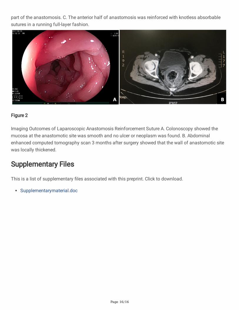
Figure 2
Imaging Outcomes of Laparoscopic Anastomosis Reinforcement Suture A. Colonoscopy showed themucosa at the anastomotic site was smooth and no ulcer or neoplasm was found. B. Abdominalenhanced computed tomography scan 3 months after surgery showed that the wall of anastomotic sitewas locally thickened.
Supplementary Files
This is a list of supplementary �les associated with this preprint. Click to download.
Supplementarymaterial.doc