recombinase-mediated gene activation and site … 251_1991.pdf · recombinase-mediated gene...
TRANSCRIPT

Recombinase-Mediated Gene Activation and Site-Specific Integration in Mammalian CellsAuthor(s): Stephen O'Gorman, Daniel T. Fox, Geoffrey M. WahlSource: Science, New Series, Vol. 251, No. 4999 (Mar. 15, 1991), pp. 1351-1355Published by: American Association for the Advancement of ScienceStable URL: http://www.jstor.org/stable/2875533Accessed: 01/07/2009 15:00
Your use of the JSTOR archive indicates your acceptance of JSTOR's Terms and Conditions of Use, available athttp://www.jstor.org/page/info/about/policies/terms.jsp. JSTOR's Terms and Conditions of Use provides, in part, that unlessyou have obtained prior permission, you may not download an entire issue of a journal or multiple copies of articles, and youmay use content in the JSTOR archive only for your personal, non-commercial use.
Please contact the publisher regarding any further use of this work. Publisher contact information may be obtained athttp://www.jstor.org/action/showPublisher?publisherCode=aaas.
Each copy of any part of a JSTOR transmission must contain the same copyright notice that appears on the screen or printedpage of such transmission.
JSTOR is a not-for-profit organization founded in 1995 to build trusted digital archives for scholarship. We work with thescholarly community to preserve their work and the materials they rely upon, and to build a common research platform thatpromotes the discovery and use of these resources. For more information about JSTOR, please contact [email protected].
American Association for the Advancement of Science is collaborating with JSTOR to digitize, preserve andextend access to Science.
http://www.jstor.org

tain two zooxanthella RFLPs: the group C RFLP plus a unique RFLP generated by one nucleotide substitution that creates a Taq I site in the larger of the two RFLP C fragments (14). These two RFLPs either represent two distinct algae or a polymor- phism within the multicopy ssRNA genes of one alga. Some other digests contain extra fragments that may identify additional ssRNA sequences. These represent small PCR amplification products that cannot be complete ssRNA genes (for example, Fig. 1C, lanes 1 and 6), or they are not interpretable from the available data; where those products occur, they occur in all individuals of that host species.
16. Replicates (Table 1) were collected from different locations so that distinct individuals, as opposed to members of one or a few clones, were sampled.
17. C. Yanisch-Perron, J. Vieira, J. Messing, Gene 33, 103 (1985).
18. In phylogenetic analyses, sequences determined from cloned PCR-amplified ssRNA genes include an uncertainty owing to the possibility of DNA repli- cation errors during PCR, and of sequence polymor- phism within multicopy ssRNA genes [M. L. Sogin,
in PCR Protocols: A Guide to Methods and Applica- tions, M. A. Innis, D. H. Gelfand, J. J. Sninsky, T. J. White, Eds. (Academic Press, New York, 1990), pp. 307-314]. Replicate sequencing suggests that this uncertainty is on the order of one or two nucleotides for the ssRNA region that we studied (13).
19. M. J. Kevin, W. T. Hall, J. J. A. McLaughlin, P. A. Zahl, J. Phycol. 5, 341 (1969).
20. M. L. Sogin, A. Ingold, M. Karlok, H. Nielsen, J. Engberg, EMBOJ. 5, 3625 (1986).
21. T. W. Vaughan and J. W. Wells, Spec. Papers Geol. Soc. Am. 44 (1943).
22. The validity of using RFLP genotypes to classify these zooxanthellae is explicitly demonstrated by their concordance with the phylogenetic groups defined by ssRNA sequences (Fig. 3).
23. A. G. Coates and J. B. C. Jackson, Paleobiology 13, 363 (1987); G. D. Stanley, Jr., Geology 9, 507 (1981).
24. I. Leizerovich, N. Kardish, M. Galun, Symbiosis 8, 75 (1990).
25. T. F. Goreau, Science 145, 383 (1964); P. W. Glynn, Environ. Conserv. 10, 149 (1983).
26. J. Maynard Smith, The Evolution of Sex (Cambridge Univ. Press, Cambridge, MA, 1978).
27. L. W. Buss, The Evolution of Individuality (Princeton Univ. Press, Princeton, NJ 1987).
28. T. H. Jukes and C. R. Cantor, in Mammalian Protein Metabolism, H. N. Munro, Ed. (Academic Press, New York, 1969), pp. 21-132.
29. J. Felsenstein, PHYLIP Version 3.2 Manual (Uni- versity of California Herbarium, Berkeley, CA, 1989).
30. R.R. thanks L. Brezinsky, C. Kanechika, and T. Humphrys for hospitality and laboratory space on Oahu, and colleagues at the West Indies Laboratory and the National Undersea Research Center on St. Croix for their help in collecting specimens. M. A. Coffroth supplied Plexaura A samples. L. Park and C. Patton got the PHYLIP programs running. Supported by an NSF Postdoctoral Fellowship in Marine Biotechnology (R.R.) and by a contract from the National Undersea Research Center, Na- tional Oceanic and Atmospheric Administration.
17 August 1990; accepted 19 December 1990
Recombinase-Mediated Gene Activation and Site-Specific Integration in Mammalian Cells
STEPHEN O'GORMAN,* DANIEL T. Fox, GEOFFREY M. WAHL
Recombinase-Mediated Gene Activation and Site-Specific Integration in Mammalian Cells
STEPHEN O'GORMAN,* DANIEL T. Fox, GEOFFREY M. WAHL
A binary system for gene activation and site-specific integration, based on the conditional recombination of transfected sequences mediated by the FLP recombinase from yeast, was implemented in mammalian cells. In several cell lines, FLP rapidly and precisely recombined copies of its specific target sequence to activate an otherwise silent p-galactosidase reporter gene. Clones of marked cells were generated by excisional recombination within a chromosomally integrated copy of the silent reporter. By the reverse reaction, integration of transfected DNA was targeted to a specific chromosomal site. The results suggest that FLP could be used to mosaically activate or inactivate transgenes for analysis of vertebrate development, and to efficiently integrate transfected DNA at predetermined chromosomal locations.
A binary system for gene activation and site-specific integration, based on the conditional recombination of transfected sequences mediated by the FLP recombinase from yeast, was implemented in mammalian cells. In several cell lines, FLP rapidly and precisely recombined copies of its specific target sequence to activate an otherwise silent p-galactosidase reporter gene. Clones of marked cells were generated by excisional recombination within a chromosomally integrated copy of the silent reporter. By the reverse reaction, integration of transfected DNA was targeted to a specific chromosomal site. The results suggest that FLP could be used to mosaically activate or inactivate transgenes for analysis of vertebrate development, and to efficiently integrate transfected DNA at predetermined chromosomal locations.
ECENT ANALYSES OF MAMMALIAN
development have made use of transfected genes to alter cell inter-
actions and trace cell lineages. This inherent- ly powerful approach could be applied to investigate a broader range of developmen- tal processes if it was possible to restrict transgene expression to specific subsets of the cells, tissues, or developmental stages in which the cis-acting sequences that typically control expression are active. Such mosaic expression is essential for many forms of lineage analyses and would additionally pro- vide a means to assess the effects of trans- genes that grossly alter development in small patches of tissue within an otherwise normal embryo. Toward this end, we have charac- terized a conditional recombination system based on the site-specific recombinase, termed FLP (1), from Saccharomyces cerevi- siae. In this system, gene activation requires prior FLP-mediated excisional recombina-
Gene Expression Laboratory, The Salk Institute for Biological Studies, La Jolla, CA 92037.
*To whom correspondence should be addressed.
tion, and expression therefore falls under the
binary control of the transgene's own cis-
acting sequences and those that direct FLP
expression. Reversal of this excisional re- combination, under different experimental conditions, provides a means for introduc-
ing DNA into specific sites in mammalian chromosomes.
A cotransfection assay was used to char- acterize FLP-mediated recombination of ex- trachromosomal DNA in a variety of cell lines. Cells were transfected with an expres- sion construct and a "reporter" plasmid that was a substrate for the recombinase. The
activity of the expression construct was as-
sayed either by recovering the transfected
reporter and looking for molecular evidence of recombination or by preparing cytoplas- mic extracts to measure p-galactosidase ac-
tivity generated by precisely recombined re-
porter molecules. The pNEOpGAL reporter plasmid used
in these assays was derived from
pFRTpGAL (Fig. 1A). pFRTPGAL con- tains the bacterial P-galactosidase coding sequence, which has been modified by inser- tion of an FLP recombination target site
(FRT) immediately 3' to the translational start (2). The FRT consisted of the two inverted 13-base pair (bp) repeats and 8-bp spacer that comprise the minimal FLP target (3, 4) plus an additional 13-bp repeat that
may augment reactivity of the minimal sub-
Table 1. p-Galactosidase activities in cotransfection assays of 293, CV-1, and F9 cells. Positive control transfections (pFRTpGAL) included 1 Vtg of pFRTpGAL and 18 Vtg of the pOG28 (6) non-FLP control plasmid. Negative control transfections (pNEO,GAL) included 1 Vtg of pNEOPGAL and 18 vtg of pOG28. Experimental transfections (pNEOpGAL + FLP) contained 1 Vtg of pNEOpGAL and 18 Vtg of the pOG44 FLP expression plasmid (Fig. 1A). The pNEOPGAL + FLP values are also shown as a percentage of the pFRTPGAL positive control values. Each value represents the mean and SEM of six plates from two experiments. Neither pOG28 nor pOG44 generated P-galactosidase activity when transfected alone (5). Transfections and assays were performed as described in the legend to Fig. 1. All transfections contained 1 Vtg of pRSVL to correct p-galactosidase activities for relative transfection efficiencies.
p-Galactosidase activity (units/mg protein) Cel line pFRTGAL ppNEO3G A L pNEOPGAL + FLP
pFRTPGAL PNEOGAL + FLP pFRTPGAL )
293 30.4 ± 1.9 0.17+ 0.02 14.2 + 2.2 47 CV-1 275 + 25 0.33 ± 0.06 22.6 + 1.2 8 F9 24.8 + 4.3 0.04 + 0.01 1.88 ± 0.02 8
15 MARCH 1991 15 MARCH 1991 REPORTS 1351 REPORTS 1351

strate (4). The insertion preserved the 3-ga- lactosidase translational reading frame and pFRT1GAL generated robust activity in mammalian cells (Table 1). pNEOpGAL was constructed by cutting pFRTIGAL within the FRT with Xba I and then insert- ing an Xba I fragment that consisted of two half-FRT sites flanking a neomycin tran- scription unit (2). This created intact FRTs on each side of the neomycin cassette and rendered the (-galactosidase transcription unit inactive (Table 1). Because the two FRT sites in pNEO1GAL are tandemly arrayed, precise FLP-mediated recombina- tion of the FRTs would be expected to excise the neomycin cassette, re-create the parental pFRT,BGAL plasmid, and restore 13-galactosidase expression.
Cotransfection of cells with a fixed amount of pNEOPGAL and increasing amounts of an FLP expression vector (pOG44) generated increasing amounts of recombined reporter plasmid and ,B-galacto- sidase activity. Molecular evidence for FLP- mediated recombination was obtained by recovering plasmids 36 hours after transfec- tion, followed by restriction endonuclease treatment and Southern (DNA) blotting (Fig. 1B). Lysates of cells from cotransfec- tions that included pOG44 showed a signal at 5.6 kb, which corresponds to the size of recombined reporter (equivalent to pFRT1GAL), and a 3.2-kb signal that was generated by unrecombined pNEO,BGAL
Fig. 2. Histochemical demonstra- tion of ,B-galactosidase activity in cell lines with a single integrated copy of pNEOI3GAL after transfec- tion with the pOG44 FLP expres- sion plasrnid. (A) CVNEO(CGAL/ E26 cells 48 hours after transfection. Cells that express 13-galactosidase con- ~ tain a blue reaction product. (B) Border between positive and nega- tive colonies of CVNEOI3GAL'E25 cells 2 weeks after transfection. (C) The B2 subclone of CVNEOt3GAL/ E25 after 8 weeks of culture. (D) A mixed colony of CVNEOP3GAIU E25 cells 1 week after transfection showingboth positive and negative cells. CVNEOI3GALIE cells growing under G418 selection were transferred to nonselective medium and transfected 24 hours later with pOG44 as described in the legend to Fig. 1. Cells were histochemically processed (26) either with (A and D) or without (B and C) prior resuspension.
receptor (Fig. 1A). The 5.6-kb band inten- sity was proportional to the amount of FLP expression plasmid included in the transfec- tion, and this band was not seen in cotrans- fections in which a non-FLP plasmid was substituted for the FLP expression vector (Fig. 1B) or in transfections that only con- tained pOG44 (5). The pOG44 vector gen- erated additional signals at 2.2 and 2.8 kb because it contained sequences homologous to the probe (6). In the same samples, ,B-galactosidase activity was also proportion- al to the amount of FLP expression plasmid included (Fig. 1C). Only background activ- ities were observed in cotransfections that included a non-FLP control plasmid (Table
Fig. 1. Cotransfection assay for A B FLP-mediated recombination of pFRTBGAL extrachromosomal DNA. (A) 0 45.6 pFRTIGAL, pNEOIGAL, and EI-IiiI1 the pOG44 FLP expression vector s E u m 4 (6). Half-arrows, positions of FRT 4 2.8 sites; E and S, Eco RI and Sca 4 422 I restriction sites; respectively; Psv, early promoter from SV40; pNEOBGAL 0 0.1 0.2 0.51 2 6 12 18 BETA-GAL, 3-galactosidase cod- pOG44 (n) ing sequence; NEO, neomycin I C expression cassette; Pcmv, cytome- E- 3.2kN- E galovirus immediate-early promot- _20
er; IN, intron; FLP, FLP coding 215 sequence; AN, SV40 adenylation 1
0 010- cassette. Thin lines represent vector pOG44 0 -,a sequcnces; thick lines in the i= 5
pOG44 diagram represent se- I O E- 2.2 -E E-2.8-S 0 6 1'2 18 quences homologous to the probe. k. kb pOG44 ()
Sizes of restriction fragments are indicatedin kilobase pairs (kb). (B) Southern blot of Hirt lysates (21) prepared from 293 (human embryonic kidney) (22) cells transfected with 1 ,ug of pNEOPGAL and varying amounts of pOG44. A non-FLP control plasmid (pOG28) (6) was included as needed to keep the total amount of plasmid and Pcmv constant. Sizes of fragments (kilobase pairs) are shown at right. Lysates were digested with Eco RI and Sca I, and Southern blots were probed with a f-galactosidase probe (Fig. lA). The identities of the hybridizing bands were confirmed with additional digests and probes. (C) ,B-Galactosidase activities in the same transfections shown in (B). Subconfluent cultures of cells growing in Dulbecco's minimum essential medium and 5% calf serum in 10-cm dishes were transfected by overnight exposure to calcium phosphate precipitates (23) and then divided into four lots. After 24 hours of incubation, one plate of each transfection was harvested by Hirt extraction (21), and a second plate was used to prepare cytoplasmic extracts (24). Approximately 5% of the DNA recovered from single plates was used for Southern analyses. ,B-Galactosidase assays were performed as described (25). Luciferase activities generated by the inclusion of 1 ,g of pRSVL (24) in all transfections were used to correct 1-galactosidase activities for relative transfection efficiencies. The experiment was repeated twice with similar results.
1) or when pOG44 alone was transfected (5). The experiment thus provides both mo- lecular and biochemical evidence for precise FLP-mediated recombination.
This paradigm was used to demonstrate FLP activity in monkey kidney (CV-1) and mouse embryonal carcinoma (F9) cells. In Table 1, the ,B-galactosidase activities in the "pFRTPGAL" transfections represent an es- timate of the expression expected if all the pNEOPGAL in a cotransfection immediate- ly underwent recombination. The highest relative expression in pNEO3GAL plus pOG44 cotransfections, 47%, occurred in 293 cells; this is a high level of activity considering that 3-galactosidase expression required prior FLP expression and recombi- nation of pNEOPiGAL. Cotransfections of CV-1 and F9 cells generated 8% of the activity seen in the pFRTiGAL transfec- tions. Even this lower relative activity clearly marked cotransfected cells in histochemical reactions for 3-galactosidase activity (5).
For this method of gene activation to be usefil in transgenic mice, FLP would need to routinely mediate recombination of FRTs integrated at a single site in the mammalian genome. Cell lines that contained single inte- grated copies of pNEOPGAL (designated CVNEOIGAIJE) were isolated by transfect- ing CV-1 cells with linearized plasmids by electroporation, selecting G418-resistant (G418R) transfectants that stably expressed the neomycin cassette, and identifying single copy lines by Southern blot analyses (5) (Fig. 3). As previously shown for other integrated constructs with similarly short direct repeats (7), the chromosomal FRTs did not sponta- neously recombine to produce a 3-galacto- sidase-positive (IGAL+) phenotype at de- tectable frequencies (Table 2).
Transient expression of FLP in the CVNEOIGAIE lines promoted a rapid conversion to a PGAL+ phenotype. When five different cell lines were transfected with pOG44, 3-galactosidase activities after 36
1352 SCIENCE, VOL. 251
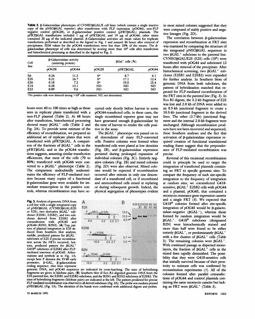
Table 2. 1-Galactosidase phenotypes of CVNEOIGAIJE cell lines (which contain a single inactive copy of the pNEO,BGAL reporter) after transfection with FLP expression (pOG44), non-FLP negative control (pOG28), or ,B-galactosidase positive control (pFRT,BGAL) plasmids. The pFRTIGAL transfections included 1 ,ug of pFRT(GAL and 19 1Lg of pOG44; other mixes contained 20 ,ug of the indicated plasmid. t-Galactosidase activities are mean values for triplicate transfections performed as described in the legend to Fig. 1 and assayed 36 hours after removal of precipitates; SEM values for the pOG44 transfections were less than 10% of the means. The 1- galactosidase phenotype of cells was determined by scoring more than 103 cells after transfection and histochemical processing as described in the legend to Fig. 2.
Cl-Galactosidase activity 3Gal+ cells (%) Cel (units/mg protein) line
pOG28 pOG44 pOG28 pFRT3GAL pOG44
E6 0.24 11.2 0* 8.7 6.1 E25 0.21 16.7 0* 17.1 12.4 E26 0.18 7.2 0* 19.5 15.4 E14 0.28 13.1 ND ND ND E22 0.09 9.6 ND ND ND
*No positive cells were detected among >106 cells examined. ND, not determined.
hours were 40 to 100 times as high as those seen in replicate plates transfected with a non-FLP plasmid (Table 2). At 48 hours after transfection, histochemical processing showed many OGAL+ cells (Table 2 and Fig. 2A). To provide some estimate of the efficiency of recombination, we prepared an additional set of replicate plates that were transfected with pFRTOGAL. A compari- son of the fractions of OGAL+ cells in the pFRT,BGAL and in the pOG44 transfec- tions suggests, assuming similar transfection efficiencies, that most of the cells (70 to 80%) transfected with pOG44 were con- verted to a IGAL+ phenotype (Table 2). The comparison undoubtedly underesti- mates the efficiency of FLP-mediated exci- sion because many copies of a functional ,B-galactosidase gene were available for im- mediate transcription in the positive con- trols, whereas recombination may have oc-
curred only shortly before harvest in some pOG44-transfected cells; in these cases, the single recombined reporter gene may not have generated enough 0-galactosidase by the time of harvest to render the cells posi- tive in the assay.
The ,BGAL+ phenotype was passed on to all descendents of many FLP-converted cells. Positive colonies were formed when transfected cells were plated at low densities (Fig. 2B), and 13-galactosidase expression persisted during prolonged expansion of individual colonies (Fig. 2C). Entirely neg- ative colonies (Fig. 2B) and mixed colonies (Fig. 2D) were also observed. Mixed colo- nies would be expected if recombination occurred after mitosis in only one descen- dant of a transfected cell, or if recombined and unrecombined cells mixed at replating or during subsequent growth. Indeed, the physical segregation of phenotypes evident
Fig. 3. Analysis of genomic DNA from A B E25 E25E25 B2 B2 a cell line with a single integrated copy E25 E2 B2 B2 of pNEOPGAL (CVNEO,GAIJE25 E25 Bi B2 Ni N2 or E25), two derivative IGAL+ sub- P O43-GAL 12.0 clones (E25B1, E25B2), and two sub- 1 clones derived from E25B2 after __ E EE 1 _ cotransfection with pOG45 and 8.5 3.2 2.0 3. 85 pOG44 (B2NI, B2N2). (A) Top, pat- tern of plasmid integration in E25 de- E25B duced from Southern blot analysis; P -GA middle, predicted pattern for 3GALF subclones of E25 if precise recombina- E E 37- tion across the FRTs occurred; bot- 10.5 2.0 3.7 3.2- tom, predicted pattern for ,BGAL- - - G418R subclones of E25B2 after FLP- B2N mediated insertion of pOG45. Abbre- viations and symbols as in Fig. IlA;- except here P denotes the SV40 early _ _E E 2.0 - promoter; ,B-GAL, P-galactosidose 12.0 3.2 2.0 3.7 coding sequence; thin lines represent genomic DNA; and pOG45 sequences are indicated by cross-hatching. The sizes of hybridizing fragments are given in kilobase pairs. (B) Southern blot of Eco RI-digested genomic DNA from the E25 parental line, the E25Bl and E25B2 subclones, and the B2Nl and B2N2 subclones of E25B2. The sizes of hybridizing fragments (kilobase pairs) are indicated at the left. The pattern predicted for precise FLP-mediated recombination was observed in all derived subclones (Fig. 3A). The probe was random-primed pNEO3GAL (Fig. IA). The identities of the bands were confirmed with additional digests and probes.
in most mixed colonies suggested that they were composed of stably positive and nega- tive lineages (Fig. 2D).
The correlation between P-galactosidase expression and recombination at FRT sites was examined by comparing the structure of the integrated pNEO,BGAL sequences in two IGAL+ subclones to the parental line. CVNEOPGA1JE25 (E25) cells (106) were transfected with pOG44 and subcloned 12 hours after removal of the precipitate. After histochemical screening, two ,BGAL+ sub- clones (E25B1 and E25B2) were expanded for further analysis. In Southem blots of genomic DNA from both subclones, the pattem of hybridization matched that ex- pected for FLP-mediated recombination of the FRT sites in the parental line (Fig. 3). In Eco RI digests, the 3.2-kb fragment of E25 was lost and 2.0 kb of DNA were added to an 8.5-kb junctional fragment to create a 10.5-kb junctional fragment in the derived lines. The other (3.7-kb) junctional frag- ment and the intemal 2.0-kb fragment were unchanged. Although recombination prod- ucts have not been recovered and sequenced, these Southem analyses and the fact that activation of P-galactosidase expression re- quired creation of functional translational reading frame suggest that the preponder- ance of FLP-mediated recombination was precise.
Reversal of this excisional recombination could in principle be used to target the integration of transfected plasmids contain- ing an FRT to specific genomic sites. To compare the frequency of such site-specific integration to the frequency of integration at random sites, we cotransfected G418- sensitive, PGAL+ E25B2 cells with pOG44 and a plasmid, pOG45, that contained a neomycin resistance gene expression cassette and a single FRT (8). We expected that G418R colonies formed after site-specific integration of pOG45 would be ,B-galacto- sidase-negative (IGAL-), whereas those formed by random integration would be ,GAL+. G418R subclones (designated B2N) were histochemically stained and more than half were found to be either entirely PiGAL-, or predominantly PGAL- with a few clusters of PGAL+ cells (Table 3). The remaining colonies were IGAL+. With continued passage as dispersed mono- layers, the fraction of PGAL+ cells in the mixed lines rapidly diminished. The possi- bility that they were G418-sensitive cells that initially survived because of their prox- imity to resistant cells was confirmed by reconstitution experiments (5). All of the colonies formed after parallel cotransfec- tions of p0OG44 and control plasmids con- taining the same neomycin cassette but lack- ing an FRT were (GAL+ (Table 3).
15 MARCH 1991 REPORTS 1353
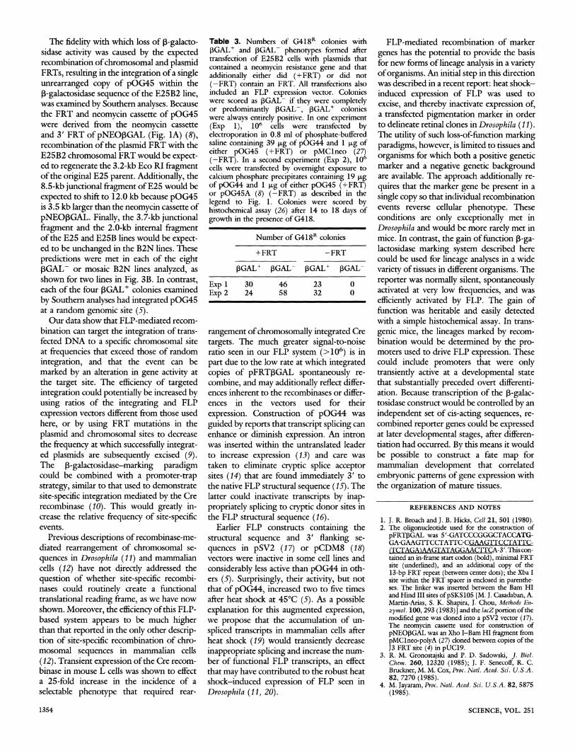
The fidelity with which loss of p-galacto- sidase activity was caused by the expected recombination of chromosomal and plasmid FRTs, resulting in the integration of a single unrearranged copy of pOG45 within the
P-galactosidase sequence of the E25B2 line, was examined by Southern analyses. Because the FRT and neomycin cassette of pOG45 were derived from the neomycin cassette and 3' FRT of pNEOpGAL (Fig. 1A) (8), recombination of the plasmid FRT with the E25B2 chromosomal FRT would be expect- ed to regenerate the 3.2-kb Eco RI fragment of the original E25 parent. Additionally, the 8.5-kb junctional fragment of E25 would be
expected to shift to 12.0 kb because pOG45 is 3.5 kb larger than the neomycin cassette of
pNEOPGAL. Finally, the 3.7-kb junctional fragment and the 2.0-kb internal fragment of the E25 and E25B lines would be expect- ed to be unchanged in the B2N lines. These
predictions were met in each of the eight pGAL- or mosaic B2N lines analyzed, as shown for two lines in Fig. 3B. In contrast, each of the four PGAL+ colonies examined
by Southern analyses had integrated pOG45 at a random genomic site (5).
Our data show that FLP-mediated recom- bination can target the integration of trans- fected DNA to a specific chromosomal site at frequencies that exceed those of random
integration, and that the event can be marked by an alteration in gene activity at the target site. The efficiency of targeted integration could potentially be increased by using ratios of the integrating and FLP
expression vectors different from those used here, or by using FRT mutations in the
plasmid and chromosomal sites to decrease the frequency at which successfully integrat- ed plasmids are subsequently excised (9). The P-galactosidase-marking paradigm could be combined with a promoter-trap strategy, similar to that used to demonstrate
site-specific integration mediated by the Cre recombinase (10). This would greatly in- crease the relative frequency of site-specific events.
Previous descriptions of recombinase-me- diated rearrangement of chromosomal se-
quences in Drosophila (11) and mammalian cells (12) have not directly addressed the
question of whether site-specific recombi- nases could routinely create a functional translational reading frame, as we have now shown. Moreover, the efficiency of this FLP- based system appears to be much higher than that reported in the only other descrip- tion of site-specific recombination of chro- mosomal sequences in mammalian cells
(12). Transient expression of the Cre recom- binase in mouse L cells was shown to effect a 25-fold increase in the incidence of a selectable phenotype that required rear-
Table 3. Numbers of G418R colonies with pGAL+ and PGAL- phenotypes formed after transfection of E25B2 cells with plasmids that contained a neomycin resistance gene and that additionally either did (+FRT) or did not (-FRT) contain an FRT. All transfections also included an FLP expression vector. Colonies were scored as PGAL- if they were completely or predominantly PGAL-, pGAL+ colonies were always entirely positive. In one experiment (Exp 1), 106 cells were transfected by electroporation in 0.8 ml of phosphate-buffered saline containing 39 jg of pOG44 and 1 ig of either pOG45 (+FRT) or pMClneo (27) (-FRT). In a second experiment (Exp 2), 106 cells were transfected by overnight exposure to calcium phosphate precipitates containing 19 ig of pOG44 and 1 jig of either pOG45 (+FRT) or pOG45A (8) (-FRT) as described in the legend to Fig. 1. Colonies were scored by histochemical assay (26) after 14 to 18 days of growth in the presence of G418.
Number of G418R colonies
+FRT -FRT
PGAL+ PGAL- pGAL+ ,GAL-
Exp 1 30 46 23 0 Exp 2 24 58 32 0
rangement of chromosomally integrated Cre
targets. The much greater signal-to-noise ratio seen in our FLP system (>106) is in
part due to the low rate at which integrated copies of pFRTPGAL spontaneously re- combine, and may additionally reflect differ- ences inherent to the recombinases or differ- ences in the vectors used for their
expression. Construction of pOG44 was
guided by reports that transcript splicing can enhance or diminish expression. An intron was inserted within the untranslated leader to increase expression (13) and care was taken to eliminate cryptic splice acceptor sites (14) that are found immediately 3' to the native FLP structural sequence (15). The latter could inactivate transcripts by inap- propriately splicing to cryptic donor sites in the FLP structural sequence (16).
Earlier FLP constructs containing the structural sequence and 3' flanking se-
quences in pSV2 (17) or pCDM8 (18) vectors were inactive in some cell lines and
considerably less active than pOG44 in oth- ers (5). Surprisingly, their activity, but not that of pOG44, increased two to five times after heat shock at 45°C (5). As a possible explanation for this augmented expression, we propose that the accumulation of un-
spliced transcripts in mammalian cells after heat shock (19) would transiently decrease
inappropriate splicing and increase the num- ber of functional FLP transcripts, an effect that may have contributed to the robust heat shock-induced expression of FLP seen in
Drosophila (11, 20).
FLP-mediated recombination of marker
genes has the potential to provide the basis for new forms of lineage analysis in a variety of organisms. An initial step in this direction was described in a recent report: heat shock- induced expression of FLP was used to excise, and thereby inactivate expression of, a transfected pigmentation marker in order to delineate retinal clones in Drosophila (11). The utility of such loss-of-function marking paradigms, however, is limited to tissues and
organisms for which both a positive genetic marker and a negative genetic background are available. The approach additionally re-
quires that the marker gene be present in a
single copy so that individual recombination events reverse cellular phenotype. These conditions are only exceptionally met in Drosophila and would be more rarely met in mice. In contrast, the gain of function P-ga- lactosidase marking system described here could be used for lineage analyses in a wide variety of tissues in different organisms. The
reporter was normally silent, spontaneously activated at very low frequencies, and was
efficiently activated by FLP. The gain of function was heritable and easily detected with a simple histochemical assay. In trans-
genic mice, the lineages marked by recom- bination would be determined by the pro- moters used to drive FLP expression. These could include promoters that were only transiently active at a developmental state that substantially preceded overt differenti- ation. Because transcription of the P-galac- tosidase construct would be controlled by an
independent set of cis-acting sequences, re- combined reporter genes could be expressed at later developmental stages, after differen- tiation had occurred. By this means it would be possible to construct a fate map for mammalian development that correlated
embryonic patterns of gene expression with the organization of mature tissues.
REFERENCES AND NOTES
1. J. R. Broach and J. B. Hicks, Cell 21, 501 (1980). 2. The oligonucleotide used for the construction of
pFRTPGAL was 5'-GATCCCGGGCTACCATG- GA-GAAGTrCCTATTC-CGAAGTrCCTATrC- (TCTAGA)AAGTATAGGAACITCA-3'. This con- tained an in-frame start codon (bold), minimal FRT site (underlined), and an additional copy of the 13-bp FRT repeat (between center dots); the Xba I site within the FRT spacer is enclosed in parenthe- ses. The linker was inserted between the Bar HI and Hind III sites ofpSKS105 [M. J. Casadaban, A. Martin-Arias, S. K. Shapira, J. Chou, Methods En- zymol. 100, 293 (1983)] and the lacZ portion of the modified gene was cloned into a pSV2 vector (17). The neomycin cassette used for construction of pNEOPGAL was an Xho I-Bam HI fragment from pMClneo-polyA (27) cloned between copies of the J3 FRT site (4) in pUC19.
3. R. M. Gronostajski and P. D. Sadowski, J. Biol. Chem. 260, 12320 (1985); J. F. Senecoff, R. C. Bruckner, M. M. Cox, Proc. Natl. Acad. Sci. U.S.A. 82, 7270 (1985).
4. M. Jayaram, Proc. Natl. Acad. Sci. U.S.A. 82, 5875 (1985).
SCIENCE, VOL. 251 1354
5. S. O'Gorman and G. M. Wahl, unpublished obser- vations.
6. pOG44 consisted of the cytomegalovirus immedi- ate-early promoter from pCDM8 (18), a 5' leader sequence and synthetic intron from pMLSIScat (13), the FLP coding sequence [nucleotides 5568 to 6318 and 1 to 626 of the 2-p.m circle (15)], and the SV40 late region polyadenylation signal from pML- SIScat (13). The following silent nucleotide substi- tutions were introduced into the structural FLP sequences with the use of the polymerase chain reaction: C for T at position 5791 (15), G for A at 5794, G for C at 5800, C for T at 55, G for A at 58, and C for T at 55, G for A at 58, and C for T at 103. These changes eliminated three canonical AATAAA polyadenylation signals and introduced a Pst I re- striction site without altering the amino acids encod- ed by the sequence. These alterations did not appear to significantly affect the expression of FLP by mammalian cells. pOG28 consisted of a murine cDNA for dihydrofolate reductase (6) cloned into pCDM8.
7. R. J. Bollag, A. S. Waldman, R. M. Liskay, Annu. Rev. Genet. 23, 199 (1989).
8. pOG45 consisted of the neomycin resistance cassette and 3' FRT from pNEO3GAL cloned into pUC19. pOG45A was derived from pOG45 by deleting a 200-bp fragment containing the FRT.
9. J. F. Senecoff, P. J. Rossmeissl, M. M. Cox, J. Mol. Biol. 201, 405 (1988).
10. B. Sauer and N. Henderson, New Biologist 2, 441 (1990).
11. K. G. Golic and S. Lindquist, Cell 59, 499 (1989). 12. B. Sauer and N. Henderson, Nucleic Acids Res. 17,
ESPITE THE POWERFUL NATURE OF
pain as a sensation, there is little consensus regarding the involve-
ment of the cerebral cortex in pain process- ing. Early this century, Head and Holmes
(1) observed individuals with war injuries and concluded that the cerebral cortex
played only a minimal role in pain percep- tion. Penfield and Boldrey (2) reached a similar conclusion when they found that
patients rarely reported a sensation of pain
J. D. Talbot, M. C. Bushnell, G. H. Duncan, Laboratoire de neurophysiologie comportementale, Faculte de mede- cine dentaire, Universite de Montr6al, Montreal, Que- bec, Canada H3C 3J7. S. Marrett, A. C. Evans, E. Meyer, Positron Imaging Laboratories, McConnell Brain Imaging Center, Mon- treal Neurological Institute, Montreal, Quebec, Canada H3A 2B4.
*To whom correspondence should be addressed.
147 (1989). 13. M. T. F. Huang and C. M. Gorman, ibid. 18, 937
(1990). 14. S. M. Mount, ibid. 10, 459 (1982). 15. J. L. Hartley and J. E. Donelson, Nature 286, 860
(1980). 16. M. T. F. Huang and C. M. Gorman, Mol. Cell. Biol.
10, 1805 (1990). 17. P. J. Southern and P. Berg, J. Mol. Appl. Genet. 1,
327 (1982). 18. A. Aruffo and B. Seed, Proc. Natl. Acad. Sci. U.S.A.
84, 8573 (1987). 19. U. Bond, EMBOJ. 7, 3509 (1988). 20. H. J. Yost and S. Lindquist, Cell 45, 185 (1986). 21. B. Hirt, J. Mol. Biol. 26, 365 (1967). 22. F. L. Graham, J. Smiley, W. C. Russel, R. Nairn,
J. Gen. Virol. 36, 59 (1977). 23. F. L. Graham and A. J. van der Eb, Virology 52, 456
(1973). 24. J. de Wet, K. V. Wood, M. DeLuca, D. R. Helenski,
S. Subramani, Mol. Cell. Biol. 7, 725 (1987). 25. C. V. Hall, P. E. Jacob, G. M. Ringold, F. Lee, J.
Mol. Appl. Genet. 2, 101 (1983). 26. J. R. Sanes, J. L. R. Rubenstein, J.-F. Nicolas,
EMBOJ. 5, 3133 (1986). 27. K. Thomas and M. Capecchi, Cell 51, 503 (1987). 28. We thank M. McKeown, R. Heyman, and R. Evans
for their insightful contributions to this work and the preparation of the manuscript. M. Jayaram pro- vided the FRT and FLP coding sequences. Support- ed by Xerox Corporation, Bayer A. G., the Weingart Foundation, and the Mathers Foundation.
28 August 1990; accepted 8 January 1991
on electrical stimulation of their exposed cerebral cortex during surgery to remove epileptic seizure foci. Thus, a commonly held view in clinical neurology is that "stim- ulation of ... any ... cortical areas in a normal, alert human being does not produce pain" (3).
Other data indicate that several areas of the cerebral cortex may process nociceptive information. Some patients with epileptic foci involving the primary or secondary so- matosensory areas of the parietal lobe (SI and SII, respectively) experience pain during seizures (4). In addition, lesions of these areas in humans can sometimes lead to re- duced pain perception (5). Single neurons in both SI and SII of the parietal cortex of awake monkey respond to nociceptive stim- uli (6); however, these findings are so rare
that the functional significance of parietal nociceptors is still in question.
Frontal cortex has also been implicated in
pain processing. In cat and in humans, nox- ious electrical stimuli induce an increase in cerebral blood flow to the frontal lobes (7). In rat there are neurons in the prefrontal cortex that respond to noxious skin stimula- tion (8). In addition, in patients resection of the anterior cingulate cortex can reduce the distress associated with chronic intractable
pain (9). Nevertheless, the unreliable nature of this surgical procedure in relieving pain (10) and the absence of precise anatomical data from humans, uncompromised by dis- ease or lesions, underscore our lack of knowledge concerning the normal function of specific cortical regions in pain process- ing.
We have now investigated the involve- ment of specific cortical areas in the percep- tion of pain in awake, healthy, human vol- unteers. To functionally isolate the
perception of pain from all other sensory and behavioral variables, we used subtrac- tive positron emission tomography (PET). This technique can identify subtle differ- ences in the activation of specific brain sites relative to sensory and evaluative processes (11, 12). In addition, we have applied meth- ods (13, 14) that combine into stereotaxic
images the functional information derived
o80
040 0
80- 0
4)
40- 0
0
o80
040 0
80- 0
4)
40- 0
0
B B
Before During After Before During After
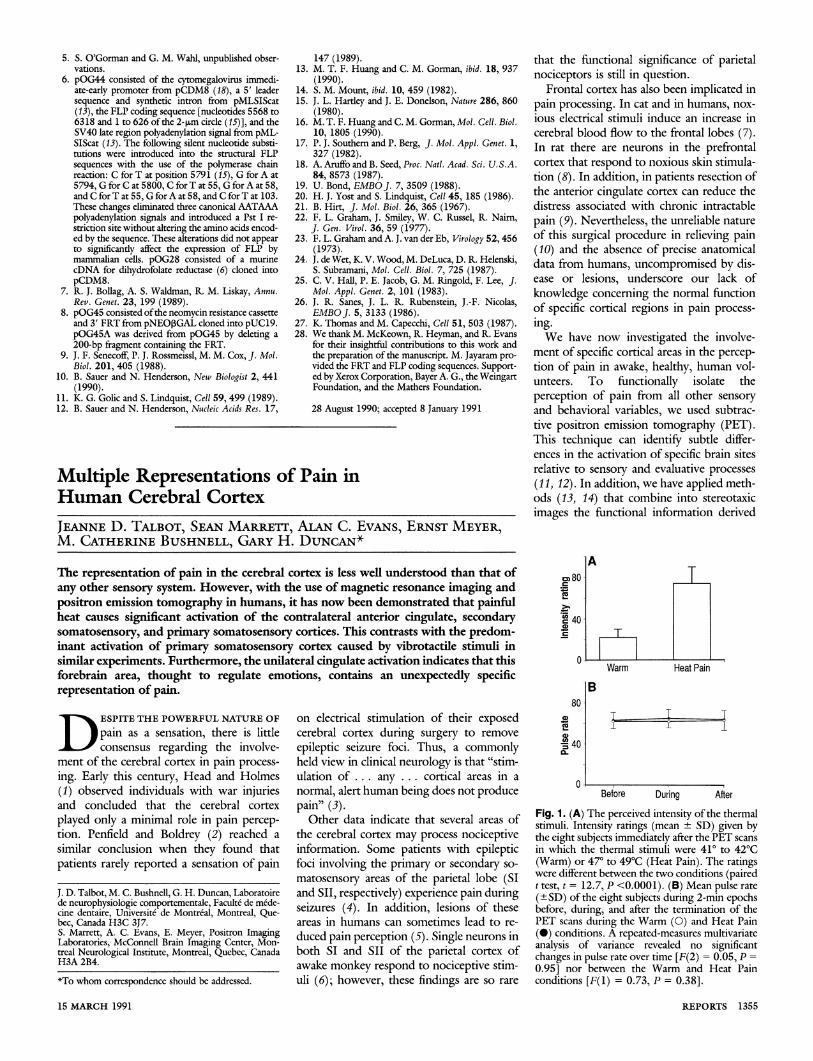
Fig. 1. (A) The perceived intensity of the thermal stimuli. Intensity ratings (mean + SD) given by the eight subjects immediately after the PET scans in which the thermal stimuli were 41° to 42°C (Warm) or 47° to 49°C (Heat Pain). The ratings were different between the two conditions (paired t test, t = 12.7, P <0.0001). (B) Mean pulse rate (-+SD) of the eight subjects during 2-min epochs before, during, and after the termination of the PET scans during the Warm (0) and Heat Pain (O) conditions. A repeated-measures multivariate analysis of variance revealed no significant changes in pulse rate over time [F(2) = 0.05, P =
0.95] nor between the Warm and Heat Pain conditions [F(1) = 0.73, P = 0.38].
15 MARCH 1991 15 MARCH 1991
Multiple Representations of Pain in Human Cerebral Cortex
JEANNE D. TALBOT, SEAN MARRETr, ALAN C. EVANS, ERNST MEYER, M. CATHERINE BUSHNELL, GARY H. DUNCAN*
Multiple Representations of Pain in Human Cerebral Cortex
JEANNE D. TALBOT, SEAN MARRETr, ALAN C. EVANS, ERNST MEYER, M. CATHERINE BUSHNELL, GARY H. DUNCAN*
The representation of pain in the cerebral cortex is less well understood than that of any other sensory system. However, with the use of magnetic resonance imaging and positron emission tomography in humans, it has now been demonstrated that painful heat causes significant activation of the contralateral anterior cingulate, secondary somatosensory, and primary somatosensory cortices. This contrasts with the predom- inant activation of primary somatosensory cortex caused by vibrotactile stimuli in similar experiments. Furthermore, the unilateral cingulate activation indicates that this forebrain area, thought to regulate emotions, contains an unexpectedly specific representation of pain.
The representation of pain in the cerebral cortex is less well understood than that of any other sensory system. However, with the use of magnetic resonance imaging and positron emission tomography in humans, it has now been demonstrated that painful heat causes significant activation of the contralateral anterior cingulate, secondary somatosensory, and primary somatosensory cortices. This contrasts with the predom- inant activation of primary somatosensory cortex caused by vibrotactile stimuli in similar experiments. Furthermore, the unilateral cingulate activation indicates that this forebrain area, thought to regulate emotions, contains an unexpectedly specific representation of pain.
REPORTS 1355 REPORTS 1355