recombinant human preproinsulin expression, purification ... · recombinant human preproinsulin at...
TRANSCRIPT
Journal of Immunological Methods, 164 (1993) 221-231 221 © 1993 Elsevier Science Publishers B.V. All rights reserved 0022-1759/93/$06.00
JIM 06789
Recombinant human preproinsulin
Expression, purification and reaction with insulin autoantibodies in sera from patients with insulin-dependent diabetes mellitus
H e i k e Berg a, Michae l W a l t e r u, Ludwig M a u c h a, J o c h e n Seissler c and Wol fgang N o r t h e m a n n a
a Department of Molecular Biology, ELIAS Entwicklungslabor, D-79114 Freiburg, Germany, b Institute of Genetics, University of Diisseldorf, D-40225 Diisseldorf,, Germany, and
c Department of Internal Medicine I, University of Ulm, D-89081 Ulm, Germany
(Received 9 December 1992, revised received 19 March 1993, accepted 10 May 1993)
A novel prokaryotic expression vector pGEX-6T was designed for high-level expression of recombi- nant fusion protein with a histidine-hexapeptide and glutathione-S-transferase at its N-terminus and the recombinant human preproinsulin at its C-terminus. Efficiency of expression was investigated in the Escherichia coli strain CAG456. The synthesized protein was sequestered in an insoluble form in inclusion bodies and was purified to homogeneity by one-step affinity chromatography based on the specific complex formation of the histidine-hexapeptide and a chelating matrix which was charged with Ni 2+ ions. The antigenic nature of the purified recombinant preproinsulin fusion protein was evaluated by ELISA screening for insulin autoantibodies in selected sera from patients with recent-onset type 1 (insulin-dependent) diabetes mellitus classified by the existence of additional autoantibodies reactive against glutamic acid decarboxylase. 14% of the tested sera (n = 43) contained insulin autoantibodies which strongly recognized the recombinant human preproinsulin. Comparable measurements with both recombinant human preproinsulin and mature insulin suggested that the observed autoantigenicity of preproinsulin was mediated by the C-peptide o r / a n d signal peptide.
Key words: Prokaryotic expression; Recombinant preproinsulin; Insulin autoantibody; Insulin-dependent diabetes mellitus; Chro- matography, affinity
Correspondence to: W. Northemann, Department of Molecular Biology, ELIAS Entwicklungslabor, Obere Hardt- strasse 18, D-79114 Freiburg, Germany. Tel.: + 49-761-47805- 58; Fax: + 49-761-47805-20.
Abbreviations: GAD65 , 65 kDa glutamic acid decarboxyl- ase, GST, glutathione-S-transferase; IAA, insulin autoanti- body; IDDM, insulin-dependent diabetes mellitus; IPTG, iso- propylthiogalactoside; PBS, phosphate-buffered saline; PPI, preproinsulin.
Introduction
Many eukaryotic proteins that are of interest for basic research or for medical applications such as diagnostic assays are often not available in the desired amount and purity from natural sources. Therefore, cloning and expression of for- eign genes in various biological systems such as bacterial, yeast and mammalian cells are common

222
procedures providing a ready source of these proteins. In the present paper we describe cDNA cloning and expression of recombinant human preproinsulin in Escherichia coli with respect to two objectives. First, we established satisfactory synthesis and purification of recombinant human preproinsulin as a fusion protein using the novel prokaryotic expression system pGEX-6T followed by a specific single-step purification based on affinity chromatography with a metal chelating matrix (Berthold et al., 1992). Second, we wished to evaluate recombinant human preproinsulin as a possible antigenic tool in ELISA measurements of insulin autoantibodies (IAA), e.g., in selected sera from patients with insulin-dependent dia- betes mellitus (IDDM) which also contained au- toantibodies directed against the 65 kDa glutamic acid decarboxylase (GAD65).
Type 1 (insulin-dependent) diabetes mellitus is one of most severe and common autoimmune diseases characterized by irreversible and selec- tive destruction of insulin-producing /3 cells in the pancreas and circulating islet specific autoan- tibodies which are present for years before clini- cal onset of the disease (for review see Castano and Eisenbarth, 1990; Boitard and Bach, 1991; Harrison, 1992). The detection of these autoanti- bodies in non-diabetic individuals indicates risk for IDDM and prediabetic insulitis. The anti- GAD65 autoantibodies are one of the earliest detectable islet-cell autoantibodies (Baekkeskov et al., 1982, 1990). While the anti-GAD65 autoan- tibody is currently the best studied of the islet- specific autoantibodies and serves as the most relevant serological marker for ongoing /3 cell destruction, the generation of insulin autoanti- bodies (IAA) alone confers relatively little risk for IDDM development.
Material and methods
Isolation of human preproinsulin cDNA Approximately 1.5 x 10⁶ clones of a human
pancreatic carcinoma cDNA library in A-gtll (Clontech) were screened with a synthetic oligonucleotide corresponding to the cDNA se- quence of human insulin (Goedel et al., 1980). 14 positive clones were obtained. Corresponding cD-
NAs were isolated from the A clones, subcloned into plasmid Bluescript SK(f-) (Stratagene) and characterized by sequence analysis. The clone pPPI.3 carried the longest insert with 541 bp in length coding for the entire human preproinsulin including extended non-coding 5' and 3' regions and with a polyA stretch of 65 adenosines.
Expression of the preproinsulin fusion protein in Escherichia coli
Various E. coli strains transformed with the plasmid pPPI.304 were cultured overnight in LB medium containing 150/zg/ml ampicillin, diluted ten-fold with fresh prewarmed LB medium and incubated for 60 min prior to induction with 1 mM IPTG for 5 h. Bacteria in aliquots of 100/zl culture medium were sedimented, resuspended in sample buffer and applied to 10% polyacryl- amide-SDS gel electrophoresis.
Immunoblotting The proteins were separated by 10% polyacryl-
amide-SDS gel electrophoresis under reducing conditions and transferred to nitrocellulose filters (Amersham) using a trans-blot semi-dry elec- trophoretic transfer cell (Bio-Rad). The unoccu- pied protein-binding sites on the filter were blocked with 5% non-fat dried milk in TBST buffer (10 mM Tris/HC1, pH 8.0, 150 mM NaCI, 0.05% Tween 20). The immobilized proteins were incubated for 2 h with a 500-fold dilution of anti-porcine insulin serum from guinea pig (Sigma) or sera from IDDM patients. The anti- porcine insulin serum was preabsorbed with 0.1 mg/ml crude extract derived from E. coli strain LE392 transformed with pGEX-6T. The bound antibodies were visualized either with anti-guinea pig immunoglobulins (Sigma) or anti-human im- munoglobulins (Promega) conjugated with alka- line phosphatase, respectively.
Purification of the recombinant preproinsulin fu- sion protein
E. coli strain CAG456 (Snyder et al., 1987) transformed with pPPI.304 was cultured in 100 ml LB medium and induced with 1 mM IPTG for 5 h. The cells were sedimented, resuspended in 20 ml PBS and treated with 1.0 mg/ml lysozyme (Sigma) and 1 mM phenylmethylsulfonyl fluoride

(Sigma) for 20 min at 0°C followed by incubation with 1% Triton X-100 (Sigma) for 10 min at 0°C. The cells were lysed by sonication with two pulses for 20 s each at about 100 W at 0°C. The soluble and insoluble cell fractions were separated by centrifugation of the cell homogenate at 8000 × g and 4°C for 5 rain. The pellet containing the insoluble recombinant preproinsulin fusion pro- tein was dissolved in 50 ml 6 M guanidinium-hy- drochloride in Tris/acetate buffer (50 mM Tris/acetate, pH 7.8, 0.5 M NaCI) and directly applied onto a 10 ml chelating Sepharose FF column (Pharmacia) which was charged with nickel ions according to the producer's standard
223
protocol (Pharmacia). The column was developed with a pH step gradient of pH 6.0, pH 5.5., pH 5.0 and pH 4.0 as described previously (Berthold et al., 1992). The recombinant protein eluted at pH 4.0 was dialysed against 6 M guanidinium-hy- drochloride in Tris/acetate buffer, pH 7.8, with- out NaCI and treated with 20 mg/ml sodium sulfite, 10 mg/ml sodium tetrathionate and 1 mM EDTA for oxidative sulfitolysis for 6 h at room temperature (Patrick and Lagu, 1992). After sub- sequent dialysis against 2 M urea in 20 mM Tris/acetate, pH 8.0, and PBS at room tempera- ture the preproinsulin fusion protein was concen- trated and stored at -20°C. The recombinant
957
V enterokinase cleavage site I multicloning site 1016
D D D D K G I
GTC GAT GAC GAT GAC AAG GGA TCC GAA GCT TCG AAT TCC CCG GGT CGA CTC GAG GTA CCA i i i J i N i L /
l B I m H I H i n d I I I E c o R I S m a I X h o I 1
,j Xma I , S i l l I K p n
224
GST was expressed by pGEX-6T and purified with the same protocol.
ELISA screening for anti-insulin autoantibodies The antigens were diluted in phosphate-
buffered saline (PBS) and applied to microtiter plates at concentrations of 1 ~zg/ml. The coating was performed with 120/zl/well for 16 h at 4°C. The plates were blocked with PBS containing 0.5% bovine serum albumin (BSA), 1% lactose and 0.02% sodium azide, dried and stored in sealed bags until use at 4°C. The ELISA proce- dure was performed with 100/xl aliquots of serum from each patient which were diluted 100-fold with PBS supplemented with 0.5% BSA and 0.05% Tween 20. Samples were incubated in the plate for 30 min at room temperature. To prevent nonspecific immunoreaction the diluted sera were pretreated with a 0.1 mg/ml extract from E. coli strain LE392 transformed with pGEX-6T. After washing the plates, bound antibodies were visual- ized with rabbit anti-human IgG conjugated with horseradish peroxidase for 15 min at room tem- perature followed by a color reaction with o- phenylenediamine. The extinction was measured bichromatically at 492 nm with a 640 nm refer- ence. The concentrations of IAA expressed in arbitrary units (U/ml) were calculated from a standard curve. The standard used in this im- munoassay was taken from a commercially avail-
able insulin antibody ELISA kit (SYNELISA, ELIAS Medizintechnik) and comprised an iso- lated and defined immunoglobulin fraction specifically directed against highly purified hu- man mature insulin. The interassay variance was less than 10% at all analyte levels. All measure- ments were undertaken with an automatic ELISA reader (SLT Labinstruments) using commercially available SYNELISA software (ELIAS Medizin- technik).
Autoantibodies All sera from autoimmune patients with re-
cent-onset IDDM tested in this study were posi- tively assayed for anti-GAD65 autoantibodies by immunoprecipitation as described previously (Seissler et al., 1992). The antigen sample for screening the sera of patients was metabolically labeled recombinant full-length human GAD65 which was expressed in infected Spodoptera frugiperda (Sf9) insect cells mediated by recombi- nant baculovirus Autographa californica (L. Mauch, unpublished results).
Results
Construction of vector pGEX-6T The initial expression vector pGEX-3T (Fro-
rath et al., 1992) was modified in order to insert a
I<-- GST MSPIHHHHHH LLGYWKIKGL VQPTRLLLEY LEEKYEEHLY ERDEGDKWRK 50
KKFELGLEFP NLPYYIDGDV KLTQSMAIIR YIADKHNMLG GCPKERAEIS I00
MLEGAVLDIR YGVSRIAYSK DFETLKVDFL SKLPEMLKMF EDRLCHKTYL 150
NGDHVTHPDF MLYDALDWL YMDPMCLDAF PKLVCFKKRI EAIPQIDKYL 200
GST -->I I<-- preproinsulin KSSKYIAWPL QGWQATFGGG DHPPKSVDDD DKGALWMRLL PLLALLALWG 250
PDPAAAFVNQ HLCGSHLVEA LYLVCGERGF FYTPKTRREA EDLQVGQVEL 300
-->I GGGPGAGSLQ PLALEGSLQK RGIVEQCCTS ICSLYQLENY CN 342
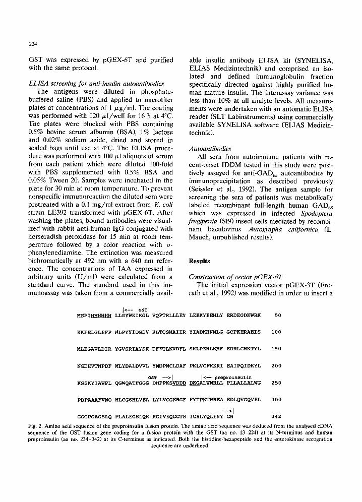
Fig. 2. Amino acid sequence of the preproinsulin fusion protein. The amino acid sequence was deduced from the analysed cDNA sequence of the GST fusion gene coding for a fusion protein with the GST (aa no. 13-224) at its N-terminus and human preproinsulin (aa no. 234-342) at its C-terminus as indicated. Both the histidine-hexapeptide and the enterokinase recognition
sequence are underlined.
coding sequence for his t idine-hexapept ide (HI_ 6) and to replace the th rombin cleavage site by a cor responding recogni t ion sequence ( V D D D - D K G ) for the site-specific enterokinase by corre- sponding ol igonucleot ide linker cloning (data not shown). The enterokinase cleavage site provided the opt ion of cleaving the GST moiety of the purified fusion protein, if necessary (Hopp et al.,
225
1988; Su et al., 1992). The new expression vector, p G E X - 6 T (Fig. 1), is 5023 bp long and directs the expression of recombinant fusion prote in with glutathione-S-transferase (GST) combined with a hist idine-hexapeptide as an affinity tail at its N- terminus. The highly efficient synthesis of recom- binant prote in is control led by a t a c - p r o m o t e r
( A m m a n et al., 1983) which is normally repressed
A kDa
- 2 0 0 . 0
9 7 . 4
6 8 . 0
B kDa
- 200 .0
- 9 7 . 4
68.0
43 .0 4 3 . 0
2 9 . 0
29 .0
~ 18.4
1 2 3 4 5 6 M
1 8 . 4
1 2 3 4 5 6
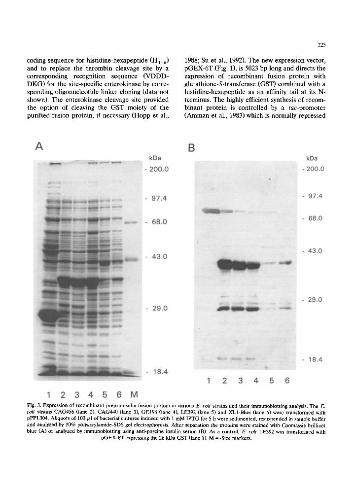
Fig. 3. Expression of recombinant preproinsulin fusion protein in various E. coli strains and their immunoblotting analysis. The E. coli strains CAG456 (lane 2), CAG440 (lane 3), GEl96 (lane 4), LE392 (lane 5) and XL1-Blue (lane 6) were transformed with pPPI.304. Aliquots of 100 ~1 of bacterial cultures induced with 1 mM IPTG for 5 h were sedimented, resuspended in sample buffer and analyzed by 10% polyacrylamide-SDS gel electrophoresis. After separation the proteins were stained with Coomassie brilliant blue (A) or analyzed by immunoblotting using anti-porcine insulin serum (B). As a control, E. coli LE392 was transformed with
pGEX-6T expressing the 26 kDa GST (lane 1). M = -Size markers.

226
by the lac repressor mediated by the lacI q gene located on the plasmid until induction with IPTG.
Construction and expression of preproinsulin cDNA clone
In order to express the recombinant human preproinsulin as a GST fusion protein the full- length cDNA with 541 bp was isolated from the clone pPPI.3 in Bluescript, trimmed to remove the 5' non-coding region including the initial codon for methionine by oligonucleotide linker replacement and inserted into the multicloning site of pGEX-6T resulting in the cDNA clone pPPI.304. The correct protein reading frame of the resulting GST fusion gene was established by DNA sequence analysis (Fig. 2).
Five E. coli strains, CAG456, CAG440, GEl96, LE392, and XL1-Blue, were transformed with pPPI.304 and analysed for yield of the recombi- nant preproinsulin fusion protein. As a control the E. coil strain LE392 was transformed with pGEX-6T expressing the 26 kDa GST. After induction with 1 mM IPTG for 5 h, aliquots of the bacterial culture were analysed by polyacryl- amide-SDS gel electrophoresis prior to staining of the proteins (Fig. 3A). Correct expression of the preproinsulin fusion protein with the ex- pected molecular weight of 38 kDa was con- firmed by immuno blotting analysis using com- mercially available anti-porcine insulin (Fig. 3B). The highest yield of recombinant protein was obtained with E. coil strains CAG456 and CAG440 (Fig. 3, lanes 2 and 3). The synthesised recombinant protein was gradually accumulated in CAG456 cells over a time period of 24 h (Fig. 4). Under optimal conditions the preproinsulin fusion protein accounted for up to 20% of total cellular proteins.
Purification of the recombinant preproinsulin fu- sion protein by affinity chromatography
The recombinant preproinsulin fusion protein was designed with an N-terminal histidine hexapeptide that permitted the simplified purifi- cation of recombinant proteins from bacterial lysates by affinity chromatography using a metal chelating matrix as previously described (Berthold et al., 1992). To examine whether the recombi- nant preproinsulin fusion protein directed by the
k D a
- 2 0 0 . 0
- 9 7 . 4
- 6 8 . 0
- 4 3 . 0
2 9 . 0
1 8 . 4
1 2 3 4 5 6 7 8 M Fig. 4. Expression kinetics of recombinant human preproin- sulin fusion protein. Cultures of E. coli CAG456 transformed with pPPI.304 were induced with 1 mM IPTG for 0 h (lane 1), 1 h (lane 2), 2 h (lane 3), 4 h (lane 4), 6 h (lane 5), 8 h (lane 6), 12 h (lane 7) and 24 h (lane 8). Bacterial proteins in 100 ~1 aliquots of culture fluid were separated by 10% poly- acrylamide-SDS gel electrophoresis and stained. (M)= Size
markers.
clone pPP.304 was synthesized as a soluble or insoluble protein, the transformed E. coli strain CAG456 was induced with IPTG and lysed by sonication. The cell homogenate was separated into soluble and insoluble cell fractions by cen- trifugation and analysed by gel electrophoresis (Fig. 5, lanes 1, 2 and 3). The synthesized pre- proinsulin fusion protein was mainly sequestered as an insoluble cell fraction in inclusion bodies (Fig. 5, lane 3). In order to purify the recombi- nant protein the inclusion bodies were solubilized in 6 M guanidinium-hydrochloride and applied to a chelating Sepharose column charged with Ni 2+

227
ions (Berthold et al., 1992). The recombinant protein was eluted gradually at p H 5.5 and 4.0 (Fig. 5, lanes 5 and 6). The eluate at pH 4.0 contained recombinant preproinsulin fusion pro- tein in almost pure form. Following dialysis the low solubility of the recombinant preproinsulin fusion protein could induce precipitation and in order to prevent this the intermolecular and in- t ramolecular disulfide bridges were permanent ly disrupted by reaction with sulfite and tetrathion- ate a technique known as oxidative sulfitolysis (Patrick and Lagu, 1992). With this technique the purification of preproinsul in fusion protein yielded about 1.2 mg soluble recombinant protein
k D a
- 2 0 0 . 0
9 7 . 4
- 8.o
- 4 3 . 0
- - - 2 9 . 0
- 1 8 . 4
1 2 3 4 5 6 M Fig. 5. Purification of recombinant human preproinsulin fu- sion protein. E. coli strain CAG456 transformed with pPPI.304 was cultured, induced with 1 mM IPTG for 5 h and fraction- ated to purify the recombinant fusion protein. 100/~l aliquots of culture fluid were removed from different cell fractions and from several steps of the purification by affinity chromatogra- phy on a metal chelating Sepharose FF column. E. coli lysate (lane 1), soluble cell fraction (lane 2), insoluble cell fraction (lane 3), fraction eluted at pH 5.5 (lane 4), at pH 5.0 (lane 5)
and at pH 4.0 (lane 6). (M) = Size markers.
per 100 ml bacterial culture of t ransformed E.
coli.
ELISA screening of IAA in sera of patients with IDDM using recombinant preproinsulin fusion pro- tein
For the determination of IAA in sera from patients with I D D M a quantitative immunoassay (ELISA) was established with recombinant hu- man preproinsulin fusion protein as an immobi- lized antigen target. In total 43 sera derived from patients with recent-onset I D D M and 39 sera from blood donors were compared (Fig. 6). The sera of the patients were positively selected for ant i -GAD autoantibodies as a specific serum marker for I D D M measured by immunoprecipi- tation of metabolically labeled recombinant hu- man GAD65 which was expressed by baculovirus in infected Sf9 cells (data not shown). With refer- ence to the standard a cut-off was set at 10 U / m l between positive and negative sera. Six sera from patients with I D D M (14.0%) and one serum among blood donors (2.6%) reacted significantly with the recombinant preproinsulin fusion pro- tein and exhibited I A A concentrations above the cut-off value (Fig. 6). The sera from the blood donors were randomly selected from a large pool. All sera used for the ELISA screening of IAA were pret reated with lysates of E. coli trans- formed with pGEX-6T which provided sufficient amounts of recombinant GST to prevent nonspe- cific reactions of the sera with the GST moiety of the preproinsulin fusion protein as well as with contaminating E. coli proteins (data not shown). No positive reactions were observed in ELISA measurements of the same pool of sera from I D D M patients based on recombinant mature human insulin as antigen target (data not shown).
Immunoblot t ing analysis with either purified recombinant preproinsulin fusion protein or GST as control were performed in order to confirm the positive ELISA measurements of the six sera from I D D M patients (Figs. 7 B - 7 G ) and the one serum from blood donors (Fig. 7H). As shown in Fig. 7 only five of the positive I D D M sera (11.6%) and the positive serum from the blood donors (2.6%) maintained their strong reactivity with the recombinant preproinsulin fusion protein. These data clearly indicated the existence of linear anti-

228
5O
~0
<~ (/)
30
, , 20 E
10
.!, i group I group 2
Fig. 6. ELISA screening of insulin autoantibodies in sera from patients with IDDM and blood donors using the recombinant preproinsulin fusion protein. Concentration of IAA illustrated in arbitrary units (U/ml) were assayed in 43 sera from patients with
IDDM (group 1) and in 39 sera from blood donors (group 2) by an ELISA procedure using the preproinsulin fusion protein.
A B C D E F G H I J
=
w g
1 2 1 2 1 2 1 2 1 2 1 2 1 2 1 2 1 2 1 2 Fig. 7. Immunoblotting analysis of recombinant preproinsulin fusion protein using IAA positive IDDM sera. 2 /zg of purified recombinant GST (lane 1) and preproinsulin fusion protein (lane 2) were separated under denaturing and reducing conditions by 10% polyacrylamide-SDS gel eleetrophoresis and blotted onto nitrocellulose filter. The separated proteins were stained with Coomassie brilliant blue (A) or analyzed by immunoblotting with sera from IDDM patients (B-G) and blood donors (H) which were positively assayed for IAA in an ELISA procedure as shown in Fig. 6. As a control the recombinant proteins were visualized
by immunoblotting using a negative serum from blood donors (I) and an anti-porcine insulin serum from a guinea pig (J).

genic epitopes within the preproinsulin. One serum from the blood donors contained antibod- ies directed against recombinant GST. Therefore, some reactivity of the recombinant preproinsulin fusion protein may also be mediated non-specifi- cally by the GST moiety (Fig. 7H). A negative serum from blood donors (Fig. 71) and anti- porcine insulin serum (Fig. 7J) were included as negative and positive controls, respectively.
Discussion
The use of modern recombinant technology permits the synthesis and purification of human proteins in heterologous cell systems which are potenially useful for basic research or for many medical applications such as diagnostic assay sys- tems. Many proteins such as human preproinsulin cannot be obtained from their natural sources in suitable quantities because of their low abun- dance or difficulty of purification by conventional methods from human tissue samples, organs or cell lines. One solution is the production of re- combinant human proteins in heterologous prokaryotic cell systems such as Escherichia coli mediated by suitable and powerful expression vectors. In the present paper we describe the construction of a novel highly efficient prokary- otic expression vector as well as its use for the cloning, expression and purification of recombi- nant human preproinsulin. An additional aim of the present study was to investigate the use of recombinant human preproinsulin in ELISA screening for insulin autoantibodies (IAA).
In addition to autoantibodies directed against islet cell proteins (Bottazzo et al., 1980) and glutamic acid decarboxylase (Baekkeskov et al., 1982, 1990; Kaufman et al., 1992) the IAA are useful in the investigation diagnosis of IDDM (Palmer et al., 1983; Atkinson et al., 1986; Dean et al., 1986; Wilkin, 1990). In newly diagnosed IDDM patients the frequency of IAA is about 40% in children, but only 4% in adults (Karja- lainen et al., 1989). The IAA may arise secondary to injury of the/3 cells with subsequent release of stored precursors of insulin having antigenic po- tential. Therefore, the use of preproinsulin repre- senting the entire precursor molecule of insulin
229
may be an appropriate technique for the mea- surement of IAA in the sera of patients with IDDM because it to also permits the detection of antibodies directed against the C-peptide or sig- nal peptide.
A cDNA coding for the full-length human preproinsulin was isolated from a cDNA library derived from a pancreatic carcinoma cell line by screening with corresponding synthetic oligonu- cleotides based on the published cDNA sequence of human insulin (Goedel et al., 1980). For the production of recombinant human preproinsulin the cDNA was inserted into the newly designed prokaryotic vector pGEX-6T which provides high-level expression of a fusion protein with a histidine hexapeptide and glutathione-S-trans- ferase (GST) at its N-terminus under the control of the tac promoter (Amman et al., 1983).
The pGEX-6T was designed with several ob- jectives in mind. Firstly, we wished to express the human preproinsulin as a high molecular weight fusion protein with the carboxyl terminus of the GST from Schistosoma japonicum (Smith et al., 1988). The GST moiety is able to mediate protec- tion of recombinant proteins against the prote- olytic defense system of the host (Smith and Johnson, 1988) which is particularly common in the bacterial expression of small recombinant proteins such as the 12.5 kDa preproinsulin (data not shown). As shown in Figs. 3 and 4 the recom- binant preproinsulin fusion protein was over-pro- duced in the E. coli after induction with 1 mM IPTG and was intracellularly stable for up to 24 h. Secondly, we introduced a recognition se- quence for the site-specific enterokinase (Sue t al., 1992; Hopp et al., 1988) providing the oppor- tunity, if necessary to cleave the GST moiety from the recombinant protein. This might be advantageous in certain situations, although it was not essential in the case of the preproinsulin used as antigen in the ELISA measurements. Thirdly, we fused a histidine hexapeptide to the N-terminus of the recombinant preproinsulin fu- sion protein to permit single-step purification by affinity chromatography using a metal chelating Sepharose charged with Ni 2+ ions (Berthold et al., 1992). In general, high-level expression often leads to the intracellular accumulation of recom- binant proteins in the form of insoluble inclusion

230
bodies. The inclusion bodies are only soluble in detergents, strong chaotropic reagents such as guanidinium hydrochloride, and in urea. These agents normally destroy the natural conformation of the recombinant proteins, and therefore pre- vent purification by conventional chromatography based on interactions with biologically active lig- ands. The fusion to a histidine hexapeptide per- mitted the ready purification of recombinant pre- proinsulin to near homogeneity after solubilizing the inclusion bodies with guanidinium-hydrochlo- ride followed by affinity chromatography based on retention on a Ni2+-Sepharose column and elution by a pH step gradient. Subsequent sulfi- tolysis (Patrick and Lagu, 1992) is suitable for the purification of recombinant proteins with either low solubility or high hydrophobicity by prevent- ing precipitation after dialysis. Fourthly, we eval- uated the most efficient host/vector system for pGEX-6T. The recombinant human preproin- sulin fusion protein was preferentially produced at high levels in the E. coli strains CAG440 and CAG456 (Snyder et al., 1987). Because the pGEX-6T carries the gene coding for the E. coli lac repressor regulation of the expression of re- combinant protein is independent on lac repres- sor synthesis by the host.
In order to examine the autoantigenic property of the bacterially expressed preproinsulin we analysed 43 selected sera from patients with re- cently diagnosed IDDM aged between 18 and 30 years. All sera were positively assayed for autoan- tibodies directed against GAD65 (Seissler et al., 1992). Such autoantibodies are commonly be- lieved to be one of the major predictive serum markers for IDDM (Baekkeskov et al., 1982, 1990; Kaufman et al., 1992). The ELISA procedure identified six patients (14%) with autoantibodies able to react strongly with recombinant preproin- sulin fusion protein. The frequency of IAA in sera from IDDM patients measured with the recombinant ELISA correlated with previously described results (Karjalainen et al., 1989; Wilkin, 1990; Greenbaum et al., 1992). Only five of the IAA positive sera (11.6%) maintained their reac- tion with recombinant preproinsulin in im- munoblotting analysis confirmes the existence of linear antigenic epitopes within the preproin- sulin. Bacterially expressed proteins normally lack
any secondary structure or post-translational modifications. Interestingly, comparable studies of the same pool of IDDM sera were uniformly negative when recombinant mature human in- sulin was used as the antigen (data not shown).
In conclusion, the pGEX-6T construct repre- sents a suitable cloning vector for high-level pro- duction of recombinant proteins as illustrated here for human preproinsulin. The comparable measurements of IAA with either recombinant preproinsulin or mature insulin suggest that IAA in IDDM sera assayed by this recombinant ELISA in IDDM could be directed against C-peptide or /and signal peptide. Future investigations should focus on the mapping of autoantigenic epitope(s) within human preproinsulin with a view to verifying the autoantigenicity of the C-peptide or signal peptide and the generation of IAA in the pathogenesis of IDDM.
Acknowledgements
The authors like to thank Drs. U. Sonnen- schein and G. Stahnke (ELIAS Entwicklungsla- bor) for their helpful discussions as well as advice in performing and interpretation of ELISA mea- surements. Mr. M. Scanarini and T. Massell (ELIAS Entwicklungslabor) are gratefully ac- knowledged for synthesis of the synthetic oligonu- cleotides and preparation of the art work, respec- tively.
References
Atkinson, M.A., Maclaren, N.K., Riley, W.J., Winter, W.E., Fisk, D.D. and Spillar, R.P. (1986) Are insulin autoanti- bodies markers for insulin-dependent diabetes mellitus?. Diabetes 35, 894-898.
Amman, E., Brosius, J. and Ptashne, M. (1983) Vectors bear- ing a hybrid try-lac promoter useful for regulated expres- sion of cloned genes in Escherichia coli. Gene 25, 167-178.
Baekkeskov, S., Nielsen, J.H., Marner, B., Bilde, T., Ludvigs- son, J. and Lernmark, A. (1982) Autoantibodies in newly diagnosed diabetic children immunoprecipitate human pancreatic islet cell proteins. Nature 298, 167-169.
Baekkeskov, S., Anstoot, H.J., Christgau, S., Reetz, A., Soli- mena, M., Cascalho, M., Foili, F., Richter-Olesen, H. and De Camilli, P. (1990) Identification of the 64 K autoanti-

231
gen in insulin-dependent diabetes as the GABA-synthesiz- ing enzyme glutamic acid decarboxylase. Nature 347, 151- 156.
Berthold, H., Scanarini, M., Abney, C.C., Frorath, B. and Northemann, W. (1992) Purification of recombinant anti- genic epitopes of the human 68-kDa (U1) ribonucleopro- tein antigen using the expression system pH6EX3 followed by metal chelating affinity chromatography. Protein Expr. Purif. 3, 50-56.
Boitard, C. and Bach, J.F. (1991) Insulin-dependent diabetes mellitus: an autoimmune disease. In: N. Talal (Ed.), Molecular Autoimmunity. Academic Press, London, pp. 273-318.
Bottazzo, G.F., Dean, B.M., Gorsvoh, A.N., Cudworth, A.G. and Doniach, D. (1980) Complement fixing islet-cell anti- bodies in type I diabetes; possible monitors of active beta cell damage. Lancet 1,668-672.
Castano, L. and Eisenbarth, G.S. (1990) Type 1 diabetes: a chronic autoimmune disease of human, mouse and rat. Ann. Rev. Immunol. 8, 647-679.
Dean, B.M., Gale, E.A.M. and Bottazzo, G.F. (1986) Insulin autoantibodies in the prediabetic period: correlation with islet cell antibodies and development of diabetes. Dia- betologia 29, 339-342.
Frorath, B., Abney, C.C., Berthold, H., Scanarini, M. and Northemann, W. (1992) Production of recombinant rat interleukin-6 in Escherichia coli using a novel highly effi- cient expression vector pGEX-3T. Biotechnigues 12, 558- 563.
Goedel, D.V., Gray, A. and Ullrich, A. (1980). Nucleotide sequence of human preproinsulin complementary DNA. Nature 208, 57-59.
Greenbaum, C.J., Palmer, J.P., Kuglin, B., Koib, H. and participating laboratories (1992). Insulin autoantibodies measured by radioimmunoassay methodology are more related to insulin-dependent diabetes mellitus than those measured by enzyme-linked immunosorbent assay: results of the fourth international workshop on the standardiza- tion of insulin autoantibody measurement. J. Clin. En- docrinoi. Metab. 74, 1040-1044.
Harrison, L.C. (1992) Islet cell antigens in insulin-dependent diabetes: Pandora's box revisited. Immunol. Today 13, 348-352.
Hopp, T.P., Prickett, K.S., Price, V.L., Libby, R.T., March, C.J., Cerretti, D.P., Urdal, D.L. and Conlon, P.J. (1988). A short polypeptide marker sequence useful for recombinant
protein identification and purification. Biotechnology 6, 1204-1210.
Karjalainen, J., Salmela, P., Ilonen, J., Surcel, H.M. and Knip, M. (1989) A comparison of childhood and adult type 1 diabetes. New Engl. J. Med. 320, 881-886.
Kaufman, D.L., Erlander, M.G., Clare-Salzler, M., Atkinson, M.A., Maclaren, N.K. and Tobin, A.J. (1992) Autoimmu- nity to two forms of glutamate decarboxylase in insulin-de- pendent diabetes mellitus. J. Clin. Invest. 89, 283-292.
Kuglin, B., Gries, F.A. and Kolb, H. (1988) Evidence of IgG autoantibodies against human proinsulin in patients with IDDM before insulin treatment. Diabetes 37, 130-136.
Kuglin, B., Rjasanovski, I., Bertrams, J., Gries, F.A., Kolb, H. and Michaelis, D. (1990) Antibodies to proinsulin and insulin as predictive markers of type 1 diabetes. Diabet. Med. 7, 310-314.
Palmer, J., Asplin, C., Clemons, P., Lyen, K., Tatpati, O., Raghu, P. and Paquette, I. (1983) Insulin antibodies in insulin dependent diabetes before insulin treatment. Sci- ence 222, 1337-1339.
Patrick, J.S. and Lagu, A.L. (1992) Determination of recombi- nant human proinsulin fusion protein produced in Es- cher~hia coli using oxidative sulfitolyis and two-dimen- sional HPLC. Anal. Chem. 64, 507-511.
Seissler, J., Hering, B., Richter, W., Gliick, M., Yassin, N., Bretzel, R.G., Boehm, B.O., Federlin, K. and Scherbaum, W,A. (1992) Antibodies to the M r 64,000 (64 K) protein in islet cell antibody positive non-diabetic individuals indi- cate high risk for imparired beta-cell function. Diabetolo- gia 35, 550-554.
Smith, D.B. and Johnson, K.S. (1988) Single-step purification of polypeptides expressed in Escherichia coli as fusions with glutathione S-transferase. Gene 67, 31-40.
Smith, D.B., Rubira, M.R., Simpson, K.M., Davern, W.U., Tiu, W.U., Board, P.G. and Mitchell, G.F. (1988) Expres- sion of an enzymatically parasite molecule in Eseherichia coli: Schistosoma japonicum glutathione S-transferase. Mol. Biochem. Parasitol. 27, 249-256.
Snyder, M., Elledge, S., Sweetser, D., Young, R.A. and Davis, R.W. (1987) Lambda-gtll: Gene isolation with antibody probes and other applications. Methods Enzymol. 154, 107-128.
Su, X., Prestwood, A.K. and McGraw, R.A. (1992) Production of recombinant porcine tumor necrosis factor alpha in a novel E. coli expression system. Biotechniques 13, 756- 762.