receptor-mediated endocytosis: concepts emerging from … · goldstein 1984). during the last...
TRANSCRIPT

Ann. Rev. Cell Biol. 1985.1 : 1-39Copyright © 1985 by Annual Reviews Inc. All rights reserved
RECEPTOR-MEDIATEDENDOCYTOSIS: ConceptsEmerging from the LDLReceptor System
Joseph L. Goldstein, Michael S. Brown,Richard G. W. Anderson, David W, Russell, andWolfgan9 J. Schneider
Departments of Molecular Genetics, Cell Biology, and Internal Medicine,University of Texas Health Science Center at Dallas, Dallas, Texas 75235
CONTENTS
INTRODUCTION ........................................................................................................................................ 1
PATHWAYS OF RECEPTOR-MEDIATED ENDOCYTOSIS ....................................................................... 3Entry Into Coated Pits ................................................................................................................... 3Intracellular Routes ......................................................................................................................... 4
THE LDL RECEPTOR: STRUCTURE-I~XJNCTION RELATIONSHIPS ..................................................... 9Protein Purification and cDNA CI, oning ................................................................................... 10A Single Polypeptide Chain with Five Domains ..................................................................... 10Comparison With Five Other Coated Pit Receptors ............................................................. 18
THE LDL RECEPTOR AT A GENETIC LEVEL ........................................................................................ 20mRN A Structure ............................................................................................................................... 20Gene Structure ................................................................................................................................... 21
BIOSYNTHESIS OF THE HUMAN LDL RECEPTOR ................................................................................ 22
NATURALLY OCCURRING MUTATIONS IN THE LDL RECEPTOR .................................................... 24Class 1 Mutations : No Detectable Precursor ......................................................................... 25Class 2 Mutations: Precursor Not Processed ........................................................................ 25Class 3 Mutations: Precursor Processed, Abnormal Binding of LDL by Receptor ...... 28Class 4 Mutations : Precursor Processed, LDL Bound, Not Clustered in Coated Pits 29
EXPERIMENTALLY INDUCED MUTATIONS IN TIlE LDL RECEPTOR ................................................ 32
INTRODUCTION
The concept of receptor-mediated endocytosis was formulated in 1974 toexplain the observation that regulation of cellular cholesterol metabolism
10743-4634/85/1115-0001 $02.00
Annual Reviewswww.annualreviews.org/aronline

2 GOLDSTEIN ET AL
depended on the sequential cell surface binding, internalization, andintracellular degradation of plasma low density lipoprotein (LDL)(Goldstein & Brown 1974, Goldstein et al 1976). This uptake mechanismwas postulated on the basis of biochemical studies; it was soon verifiedmorphologically when the receptors for LDL were observed to be clusteredin coated pits that pinched off from the surface to form coated vesicles thatcarried the LDL into the cell (Anderson et al 1976, 1977a).
Coated pits and coated vesicles had been recognized by electronmicroscopy in the mid-1960s (Roth & Porter 1964, Fawcett 1965, Friend Farquhar 1967). Their role in receptor-mediated endocytosis was ap-preciated a decade later as a result of the convergence of twin events: 1) thedemonstration that coated vesicles were the sites at which LDL receptorswere concentrated, and 2) the demonstration by Pearse (1975) that a singleprotein, clathrin, formed the cytoplasmic coat, an observation thatprovided a biochemical definition of coated vesicles. The biologicalimplications of receptor-mediated endocytosis were vividly underscored bythe finding that genetic defects in the LDL receptor preclude cellular uptakeof LDL, producing hypercholesterolemia and’ heart attacks (Brown Goldstein 1984).
During the last decade, receptor-mediated endocytosis was recognized asa mechanism by which animal ceils internalize many macromolecules inaddition to LDL (Goldstein et al 1979a, Pastan & Willingham 1981,Bretscher & Pearse 1984). The process is initiated when receptors on the cellsurface bind macromolecules and slide laterally into clathrin-coated pits.Within minutes the coated pits invaginate into the cell and pinch off to formcoated endocytic vesicles. After shedding their clathrin coats the vesiclesfuse with one another to form endosomes whose contents are acidified byATP-driven proton pumps (Tycko & Maxfield 1982, Helenius et al 1983,Pastan & Willingham 1983). Within the endosome the ligand and receptorpart company. Often, but not always, the ligand is carried to lysosomes fordegradation, while the receptor cycles back to the cell surface to bind newligand (Brown et al 1983).
More than 25 specific receptors have been observed to participate inreceptor-mediated endocytosis. These include receptors for transportproteins that deliver nutrients to cells, such as the cholesterol-carryinglipoprotein LDL, the iron transport protein transferrin, and the vitaminBla transport protein transcobalamin II. Receptor-mediated endocytosisalso applies to many nontransport plasma proteins, including asialoglyco-proteins, c~-2-macroglobulin, and immune complexes. Moreover, the pro-cess mediates the cellular uptake of lysosomal enzymes, which occurs whenthese enzymes bind to receptors that recognize mannose-6-phosphateresidues uniquely attached to this class of proteins. Certain viruses and
Annual Reviewswww.annualreviews.org/aronline

RECEPTOR-MEDIATED ENDOCYTOSIS 3
toxins use receptor-mediated endocytosis to enter cells, apparently bybinding opportunistically to receptors that normally function in the uptakeof other substances.
Protein growth factors, such as epidermal growth factor (EGF) andplatelet-derived growth factor (PDGF), as well as classic polypeptidehormones, such as insulin and luteinizing hormone, also enter cells byreceptor-mediated endocytosis. The same receptors that mediate en-docytosis of these proteins mediate their physiologic actions. However,frequently cellular entry of the ligand does not seem to be required for theaction of the growth factor or the hormone. Rather, the entry mechanismfunctions in the rapid control of receptor number and in the removal of thegrowth factor or hormone from the circulation (Carpenter & Cohen 1979,Terris et al 1979).
Progress in this field has been rapid. Within a single year--1984--complementary DNAs (cDNAs) for five different coated pit receptors wereisolated, and their nucleotide and corresponding amino acid sequenceswere determined (Mostov et al 1984, Ullrich et al 1984, Russell et al 1984,Yamamoto et al 1984, McClelland et al 1984, Schneider et al 1984, Hollandet al 1984). The cDNA cloning and structure of a sixth coated pit receptor,the insulin receptor, was reported early in 1985 (Ullrich et al 1985, Ebina etal 1985). In this review we summarize the information that is emerging fromstudy of the amino acid sequences of the receptor proteins, with emphasison the LDL receptor.
PATHWAYS OF RECEPTOR-MEDIATED
ENDOCYTOSIS
Entry Into Coated Pits
The various pathways of receptor-mediated endocytosis share onecommon feature: in each case the receptors move to coated pits and coatedvesicles. However, there are differences in the mechanisms that triggermovement to coated pits as well as differences in the routes the ligands andreceptors follow after entering the cell. We can divide the process ofreceptor-mediated endocytosis into subc-ategories according to thesedifferences, as described below.
The first distinction is whether the receptors spontaneously move tocoated pits and enter cells continuously (even in the absence of ligand), whether the receptors wait on the surface until a ligand is bound,whereupon they are captured by coated pits. The receptors in the firstcategory include those for LDL (Anderson et al 1982, Basu et al 1981),transferrin (Hopkins & Trowbridge 1983, Hopkins 1985), ~-2-macroglobulin (Hopkins 1982, Via et al 1982), asialoglycoproteins (Wall
Annual Reviewswww.annualreviews.org/aronline

4 GOLDSTEIN ET AL
al 1980, Berg et al 1983), and insulin (Krupp & Lane 1982). Conversely, receptor for EGF is diffusely distributed on the cell surface, and is nottrapped in coated pits unless it is occupied with ligand (Schlessinger 1980,Dunn & Hubbard 1984).
The propulsive force for movement of receptors to coated pits may bcsimple diffusion, or it may involve a more directed type of propulsion(Bretscher 1984). The rate of diffusion of receptors on cell surfaces sufficiently fast in itself to explain movement into coated pits (Goldstein etal 1981, Barak & Webb 1982). However~ considerable evidence suggeststhat membrane lipids are continuously flowing toward coated pits(Bretscher 1984). This lipid flow may carry membrane proteins alongpassively (Bretscher & Pearse 1984, Hopkins 1985), but why are onlycertain cell surface proteins trapped in coated pits? One possibility is thatreceptors are marked for such entry by the attachment of prosthetic groups.Many receptors (such as those for transferrin, asialoglycoproteins, EGF,PDGF, and insulin) have phosphate groups attached to serine, threonine,or tyrosine residues in their cytoplasmic domains (see Table 1 in Brown et al1983).
Recent attention has focused on phosphorylation or dephosphorylationas a potential mechanism for signaling entry, perhaps through induction ofreceptor binding to clathrin, the protein that covers the cytoplasmic surfaceof coated structures. Phosphorylation of the receptors for EGF (Hunter1984), transferrin (Klausner et al 1984), and insulin (Jacobs ¢t al 1983) be enhanced by treatment of cells with phorbol esters, which activateprotein kinase C. Phorbol esters cause transferrin receptors in K562 cells tobecome trapped within the cell, which suggests that phosphorylation eitherincreases the rate of their cellular entry or slows their return to the cellsurface, or both (Klausner et al 1984).
A few receptors undergo acylation of cysteine residues with fatty acids,but this modification does not apply to all receptors that participate inendocytosis: Moreover, in the one case that has been studied in detail, thatof the transferrin receptor, the turnover of the fatty acid moiety is muchslower than the internalization rate (Omary & Trowbridge 1981), whichimplies that acylation-deacylation does not occur during each recyclingevent.
Intracellular Routes
A second variation in the systems of receptor-mediated endocytosis is thefate of the ligand and receptor. It appears that all endocytotic receptorsenter cells in the same coated pits, and are delivered to the same acidifiedendosomes (Pastan & Willingham 1981, Via et al 1982, Carpentier et al1982). Thereafter, the pathways diverge. The receptor-ligand complex may
Annual Reviewswww.annualreviews.org/aronline
RECEPTOR-MEDIATED ENDOCYTOSIS 5
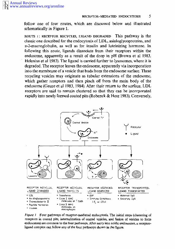
follow one of four routes, which are discussed below and illustratedschematically in Figure 1.
ROUTE i: RECEPTOR RECYCLES, LIGAND DEGRADED This pathway is theclassic one described for the endocytosis of LDL, asialoglycoproteins, andct-2-macroglobulin, as well as for insulin and luteinizing hormone. Infollowing this route, ligands dissociate from their receptors within theendosome, apparently as a result of the drop in pH (Brown et al 1983,Helenius et al 1983). The ligand is carried further to lysosomes, where it isdegraded. The receptor leaves the endosome, apparently via incorporationinto the membrane of a vesicle that buds from the endosome surface. Theserecycling vesicles may originate as tubular extensions of the endosom¢,which gather receptors and then pinch off from the main body of theendosome (Geuze et al 1983, 1984). After their return to the surface, LDLreceptors are said to remain clustered so that they can be incorporatedrapidly into newly formed coated pits (Robenek & Hesz 1983). Conversely,
Receptor
Ligand
RECEPTOR TRANSPORTED,LIGAND TRANSPORTED
RECEPTOR RECYCLES, RECEPTOR RECYCLES, RECEPTOR DEGRADED,LIGAND DEGRADED LIGAND RECYCLES LIGAND DEGRADED
¯ LDL * Transferrin ¯ EGF ¯ Malerna’l¯ Asialoglycoproteins ¯ Class ] MHC ¯ Tmrnune Complexes ¯ Secretory IgA¯ Transcobalamin 1T molecules on T Cells ( c or D3b)¯ Peptide Hormones ¯ ClassY1" MHC
molecules on¯ Viruses rnacrophages
Figure 1 Four pathways of receptor-mediated endocytosis. The initial steps (clustering ofreceptors in coated pits, internalization of coated vesicles, and fusion of vesicles to formendosomes) are common to the four pathways. After entry into acidic endosomes, a receptor-ligand complex can follow any of the four pathways shown in the figure.
Annual Reviewswww.annualreviews.org/aronline

6 GOLDSTEIN ET AL
recycling transferrin receptors seem to go through a transient phase ofmonomolecular dispersion on the cell surface before clustering andinternalizing again (Hopkins 1985).
Route 1 seems ideally adapted for use by receptors that transport ligandsinto cells at a high rate : It allows reuse of receptors once every 10-20 min.Thus, one receptor can mediate the uptake of hundreds ofligands during itsusual lifespan of 10-30 hr. Recycling requires that the receptors have astable structure that will permit them to pass repeatedly through the acidicenvironment of the endosome without denaturation. In the acidic endo-some the receptors must undergo sufficient conformational change torelease their ligands (DiPaola & Maxfield 1984), but they must not becomeirreversibly denatured. The LDL receptor, for example, can make up to 150trips through the endosome without losing its function (Goldstein et al1979a, Brown et al 1982). Maintenance of stability may require unusualprotein structures, some of which are detailed below.
ROUTE 2: RECEPTOR RECYCLES, LIGAND RECYCLES This pathway was
originally described for the transferrin receptor (Octave et al 1983). Whenthe transferrin/receptor complex reaches the endosome the two proteins donot dissociate. In vitro binding studies show that the transferrin receptor, incontrast to the receptors for LDL, asialoglycoproteins, and EGF, fails todissociate from its ligand at pH 5 (Klausner et al 1983, Dautry-Varsat et al1983). However, iron does dissociate from transferrin at acidic pH. Thus, inthe endosome the iron is stripped from transferrin while the apo-transferrinremains attached to the receptor. The apo-transferrin/receptor complexthen returns to the cell surface. The recycling transferrin receptor seems toleave the endosome by a network of membrane tubules and vesicles thateventually leads it back to the cell surface (Geuze et al 1984). Once on thesurface and again at neutral pFl, the apo-transferrin dissociates from thereceptor. (Iron-containing transferrin binds to the receptor at neutral pH,but apo-transferrin dissociates from the receptor at this pH). The trans-ferrin receptor is now free to bind another molecule of iron-containingtransferrin and to repeat the cycle. Like the LDL receptor, the transferrinreceptor is degraded very slowly with a half life of > 30 hr (Omary Trowbridge 1981), even though it enters the cell every 15-20 min (Bleil Bretscher 1982, Ciechanover et al 1983).
Recent studies suggest a new role for internalization and recycling ofligands by this route. Such recycling may provide the mechanism by whichcells of the immune system process antigen and "present" it to effector cells(for reviews see Unanue 1984, Pernis 1985, Pernis & Tse 1985).Macrophages and certain B lymphocytes internalize foreign antigens byreceptor-mediated endocytosis. The receptors responsible for this uptake
Annual Reviewswww.annualreviews.org/aronline

RECEPTOR-MEDIATED ENDOCYTOSIS 7
are poorly characterized. Their function is to deliver the antigen to an acidicintracellular compartment where the antigen undergoes partial proteolysis.The proteolytic fragments are transported back to the cell surface wherethey are presented to the well-characterized antigen receptors on neighbor-ing T lymphocytes. Presentation requires that the surface of the antigen-presenting cell express Class II major histocompatibility (MHC) proteins the same genotype as the T lymphocyte. The Class II MHC proteins aredimers of two nonidentical transmembrane glycoproteins, which arecontinuously internalized and recycled without degradation. One reasonfor such recycling may be that after proteolysis the antigen and the Class IImolecules must pass through the same acidic compartment so they canform a complex that presents the fragmented antigen to the T-cell antigenreceptor (Brodsky 1984).
Once a responding T cell is stimulated by exposure to antigen, it begins tointernalize and recycle its own MHC molecules, which are of the Class Itype (Pernis 1985). These Class I MHC molecules are a complex of transmembrane glycoprotein and another protein (~O2-microglobulin) thatis adherent to the outer surface of the cell membrane. The Class I MHCmolecules are not internalized by the T cells unless the cells are activated(i.e. stimulated by antigen). After activation the internalized Class molecules are delivered to acidic endosomes and cycled back to the cellsurface every 15 rain (Pernis 1985). Each Class I molecule makes many tripsin and out of the activated T cell during its half-life of 14 hr (Tse & Pernis1984, Tse et al 1985). Monoclonal antibodies directed against the Class molecules do not affect this recycling. In fact, the bound antibody enters thecell and cycles back to the cell surface with the Class I molecule stillattached, in a manner analogous to the co-recycling of apo-transferrin andthe transferrin receptor (Tse et al 1985).
The specificity of regulation of internalization of Class I MHC moleculesis striking. Internalization and recycling occur only on activated Tlymphocytes and not on resting T lymphocytes, B lymphocytes (resting oractivated), or on any other known cell type. It is not possible to induce rapidinternalization of these molecules in B cells, even when the Class I moleculeshave been cross-linked by exposure to a monoclonal anti-Class I antibodyfollowed by a second layer of polyclonal antibodies (Pernis & Tse 1985).Moreover, nonlymphoid cells, such as fibroblasts and mouse L cells, do notrapidly internalize their Class I MHC molecules even though they rapidlyinternalize other membrane molecules, such as the receptors for LDL andtransferrin (Pernis & Tse 1985). Internalization of Class I molecules mouse L cells or fibroblasts can be observed when the molecules have beencross-linked by a double layer of antibodies, but the rate is considerablyslower than receptor-mediated endocytosis--it takes hours rather than
Annual Reviewswww.annualreviews.org/aronline

8 GOLDSTEIN ET AL
minutes. Moreover, this internalization leads to lysosomal degradation ofthe Class I molecules rather than to recycling (Pernis & Tse 1985, Huet et al
1980).Do the internalized antigens and MHC molecules follow the classic
coated pit to coated vesicle to endosome to lysosome pathway described forLDL? There are no electron microscopic data that address this question;however, there is circumstantial evidence that coated pits and vesicles areinvolved. For instance, recycling of MHC molecules is strictly regulated,has rapid kinetics, and is inhibited by drugs that raise the pH of endosomes
(Tse & Pernis 1984, Tse et al 1985, Unanue 1984), all of which are features endocytosis via coated pits and vesicles (Brown et al 1983).
ROUTE 3 : RECEPTOR DEGRADED, LIGAND DEGRADED This pathway has beendescribed in greatest detail for EGF. After the EGF/receptor complexreaches the endosome both components are degraded, probably as a resultof subsequent cotransport to the lysosome (Carpenter & Cohen 1979). Themechanism for this cotransport is unclear. Since EGF dissociates from itsreceptor at acidic pH (DiPaola & Maxfield 1984), it would presumablydissociate in the endosome. Somehow this dissociation does not allow theEGF receptor to return to the surface, but rather it is carried further intolysosomes. If the EGF receptor is delivered to lysosomes by vesicular fusionthen the cytoplasmic domain of the receptor would remain outside of thelysosome, facing the cytoplasm. This domain of the EGF receptor consistsof 542 amino adds and contains tyrosine kinase activity (Hunter 1984,Ullrich et al 1984). It is tempting to speculate that this tyrosine kinasedomain of the receptor might be liberated from its hydrophobic anchorthrough proteolytic cleavage and then migrate elsewhere in the cell, where itcould phosphorylate proteins that trigger cell division. The selective releaseof such a cytoplasmic fragment has not yet been demonstrated.
In certain cells, one population of EGF receptors may escape degrada-tion and recycle. In cultured fibroblasts (Carpenter & Cohen 1979) and the perfused rat liver (Dunn & Hubbard 1984) the addition of EGF causes decrease of up to 80~ in EGF receptors, apparently due to ligand-inducedinternalization and degradation. However, the remaining 20~o of EGFreceptors continue to bind, internalize, and degrade EGF with kinetics thatsuggest recycling.
ROUTE 4." RECEPTOR TRANSPORTED, LIGAND TRANSPORTED This pathwayhas been described most clearly for the receptor that carries polymericimmunoglobulin A (!gA) and immunoglobulin M (IgM) across epithelialsurfaces, such as across liver cells for excretion into the bile, and acrossmammary epithelia for excretion into milk (Solari & Kraehenbuhl 1984,Mostov et al 1984). In the liver the newly synthesized receptor appears on
Annual Reviewswww.annualreviews.org/aronline

RECEPTOR-MEDIATED ENDOCYTOSIS 9
the sinusoidal surface of the hepatocyte, where it binds dimeric IgA. Thereceptor/immunoglobulin complex is incorporated into vesicles and car-ried into the cell (Renston et al 1980). Coated vesicles have not beenimplicated formally, but such involvement seems likely. At some point afterinternalization, the receptor is clipped proteolytically so that part of thereceptor with the immunoglobulin still bound to it is released from themembrane. This released receptor fragment is the so-called secretorycomponent. The IgA-containing vesicle eventually migrates to the bilecanalicular face of the hepatocyte, where it discharges the IgA/secretorycomponent adduct into the bile.
In neonatal animals, transepithelial transport of maternal IgG from thelumen of the intestine to the interstitial space is mediated by a receptor thatbinds to the Fc domain of the IgG. This transport probably does notinvolve cleavage of the receptor, since a secretory component has not beenidentified (Rodewald & Abrahamson 1982).
The delineation of four routes for disposal of receptors and ligands(Figure 1) implies that cells have multiple mechanisms for sorting receptorsafter they enter the cell. These mechanisms must be regulated so as to allowdifferent cells to process the same receptor by different routes or a single cellto process the same receptor by~ different routes at different times. In somecases sorting involves passage of the receptors through vesicles that arelocated near the Golgi complex. However, the receptors do not seem totransit through classic Golgi stacks, which are the sites of sorting in theexocytotic pathway. Rather, they pass through nearby vesicles that may ormay not contain Golgi-associated enzymes (Dunn & Hubbard 1984,Hanover et al 1984).
To assure accuracy of the multiple sorting and targeting events, eachreceptor must contain multiple functional domains. It must contain abinding domain that is specific for a given set of ligands, and regions thatallow it to interact with other macromolecules so it can be transported tovarious sites within the cell. Often a receptor will proceed successively fromone compartment to another, at each stage being sorted from othermembrane molecules that are stationary or are moving to different sites.Therefore, each receptor must contain multiple sorting signals that actsequentially. These signals will be revealed only when the completestructures of the receptors are known.
THE LDL RECEPTOR: STRUCTURE-FUNCTIONRELATIONSHIPS
We recently carried out detailed studies of the structure and biosynthesis ofthe LDL receptor. This receptor performs a simple function: it carries
Annual Reviewswww.annualreviews.org/aronline

10 GOLDSTEIN ET AL
cholesterol-bearing lipoproteins into ceils. To accomplish this task, the
receptor must move from its site of synthesis in the membranes of theendoplasmic reticulum (ER) through the Golgi complex to the cell surface,where it is targeted to coated pits. It must then recycle from the endosome tothe cell surface. Naturally occurring mutations in the gene for the LDLreceptor disrupt several of these transport steps and produce a clinicalcondition of receptor deficiency known as familial hypercholesterolemia(FH).
Protein Purification and cDNA Clonin9
The LDL receptor was purified from the bovine adrenal cortex, an organthat contains a relative abundance of LDL receptors (~ 105 molecules percell), which it uses to supply cholesterol for conversion to steroid hormones(Schneider et al 1982). Biochemical tools were developed that permittedcloning of cDNAs for the receptor. Thus, polyclonal antibodies raisedagainst the purified bovine protein were used to enrich for the rare LDL-receptor mRNA by polysome immune purification. A cDNA library wasconstructed from the purified mRNA of bovine adrenal cortex, and wasscreened with two families of oligonucleotides derived from the amino acidsequence of a cyanogen bromide fragment of the bovine protein. Thesemethods led to the isolation of a partial cDNA for the bovine receptor(Russell et al 1983).
The bovine cDNA was used as a probe to isolate a fragment of the humanLDL-receptor gene. In turn, an exon probe from this genomic fragment wasemployed to isolate a eDNA clone representing the complete 5.3-kilobase(kb) human LDL-receptor mRNA. Transfection studies indicated that thiscDNA could direct the expression of functional human LDL receptors insimian COS cells (Yamamoto et al 1984).
The nucleotide sequence of this eDNA was used to derive the completeamino acid sequence of the human LDL receptor (Yamamoto et al 1984).This sequence, together with biochemical experiments (Russell et al 1984),revealed that the mature receptor is divided into five distinguishabledomains. A model of the domain structure is shown in Figure 2.
A Single Polypeptide Chain With Five Domains
FIRST DOMAIN : LIGAND BINDING The extreme NH2-terminus of the LDLreceptor consists of a hydrophobic sequence of 21 amino acids that iscleaved from the receptor and is not present in the mature protein. Thissegment presumably functions as a classic signal sequence to direct thereceptor-synthesizing ribosomes to the ER membrane. Because it does notappear in the mature receptor, the signal sequence is omitted from thenumbering system that is described below.
Annual Reviewswww.annualreviews.org/aronline

RECEPTOR-MEDIATED ENDOCYTOSLS 11
The mature receptor (without the signal sequence) consists of 839 aminoacids. The first domain of the mature LDL receptor consists of the NH2-terminal 292 amino acids, which are extremely rich in cysteines (42 out of292 amino acids). Studies with anti-peptide antibodies revealed that thisdomain is located on the external surface of the plasma membrane(Schneider et al 1983b). The cysteines are spaced at intervals of 4-7 aminoacids (Figure 3). An initial computer analysis suggested that the firstdomain was made up of eight repeat sequences (Yamamoto et al 1984).More recent analysis of the sequence, considered together with exon/intronmapping data (see below and Figure 2), suggests that the number of repeatsis only seven, as shown in Figure 3.
Each of the seven repeats consists of ~ 40 amino acids and contains 6cysteine residues, which are essentially in register for all of the repeats. Thereceptor cannot be labeled with [3H]iodoacetamide without prior reduc-tion, suggesting that all of these cysteines are involved in disulfide bonds.This region of the receptor must therefore exist in a tightly cross-linked,convoluted state.
A striking feature of the COOH-terminus of each repeat sequence is acluster of negatively-charged amino acids (Figure 3). These sequences arecomplementary to positively-charged sequences in the best-characterizedligand for the LDL receptor, apolipoprotein E (apo E), a 33-kilodaltonprotein component of the plasma lipid transport system. Apo E contains acluster of positively charged residues that are believed to face one side of asingle a-helix (Innerarity et al 1984). Studies with mutant and proteolyzedforms of apo E, and with monoclonal antibodies against different regions ofapo E showed that the positively charged region contains the site by which
Domains~ Ligond- -inding )
JEGF Precur~r Homology
C9 Complement Homology
O- LinkedSignal Sugars
Sequence
DCOOH
Cytoplasm ic
Transmembrane
Fioure 2 Exon organization and protein domains in the human LDL receptor. The domainsof the protein are delimited by thick black lines and are labeled in the lower portion of thefigure. The 7 cysteine-rich, 40-amino acid repeats in the LDL binding domain (see also Figure3) are assigned roman numerals I-VII. Repeats IV and V are separated by 8 amino acids. The cysteine-rich repeats in the EGF precursor homology domain are lettered A~. The positionsat which introns interrupt the coding region are indicated by arrow heads. Exon numbers areshown between the arrow heads. (Reprinted from SiJdhof et al 1985a with permission.)
Annual Reviewswww.annualreviews.org/aronline

12 GOLDSTEIN ET AL
Annual Reviewswww.annualreviews.org/aronline

RECEPTOR-MEDIATED ENDOCYTOSIS 13
this protein binds to the LDL receptor (Innerarity et al 1984). It is thereforetempting to speculate that the negatively charged clusters of amino acidswithin the cysteine-rich repeat sequence of the LDL receptor constitutemultiple binding sites, each of which binds a single apo E molecule byattaching to its positively-charged s-helix (Table 1).
This speculation is supported by several observations. First, Innerarity &Mahley (1978) and Pitas et al (1980) showed through kinetic analysis multiple apo E molecules (4-8) bind to a single LDL receptor. Second,receptor binding of apo E and of apo B (the other ligand for the LDLreceptor) is blocked by modification oflysine or arginine residues of the twoligands, a reaction that is achieved with acetylation or cyclohexanedionetreatment, respectively (Basu et al 1976, Mahley & Innerarity 1983). Third,although both apo E and apo B have a net negative charge, theynevertheless bind tightly to polyanions, such as heparin, which suggeststhat both have exposed clusters of basic residues. Fourth, the binding of125I-LDL to the LDL receptor is inhibited by negatively and positivelycharged molecules such as heparin, suramin, protamine, and plateletfactor 4 (Goldstein et al 1976, Brown et al 1978, Schneider et al 1982). Fifth,the binding activity of the LDL receptor is destroyed by reduction of thedisulfide bonds. Sixth, proteolytic treatment of the purified bovine LDLreceptor with thrombin yields a 60~kilodalton fragment (isolated on SDSpolyacrylamide gels) that is recognized by an anti-peptide antibodydirected against the NH2-terminus of the receptor, and that also specificallybinds LDL (Schneider et al, manuscript in preparation). Thus, the LDLbinding site is located within the NH2-terminal 60 kilodaltons of protein,which includes the cysteine-rich acidic region.
The disulfide bonds confer great stability upon the binding site of the
Table I Complementarity between amino acid sequences in the LDL receptor and in one ofits ligands
LDL Receptor
Apo E
(-Cys-Asp-X-X-X-Asp-Cys-X-Asp-Gly-Ser-Asp-Glu-)7
-His-Leu-Ar#-Lys-Leu-Aro-Lys-Aro-Leu-Leu-Ara-
140 150
The LDL receptor sequence corresponds to the consensus from the most conserved part of theseven repeat units shown in Figure 3. The apo E sequence (amino acids 140-150) has beenidentified as being responsible for binding to the LDL receptor (McLean et al 1984, Innerarityet al 1984). Negatively charged amino acids in the LDL receptor sequence and positivelycharged amino acids in the apo E sequence are italicized. No amino acid sequence data areavailable for apo B, the other ligand for the LDL receptor.
Annual Reviewswww.annualreviews.org/aronline

14 GOLDSTEIN ET AL
receptor. The receptor can be boiled in SDS or guanidine and still retainbinding activity as long as the disulfide bonds are not reduced (Daniel et al1983). The disulfide bonds in this region may preserve the stability of thereceptor when it delivers LDL to endosomes. In this acidic environment,the negatively-charged residues of the receptor become protonated and losetheir charges, allowing the LDL to be released. (LDL is known to dissociatefrom the receptor in vitro when the pH falls below 6.5) (Basu et al 1978).Despite the titration of its carboxyl groups, the receptor is not irreversiblydenatured by this acid exposure, apparently because of the structuralstability afforded by the multiple disulfide bonds.
Each of the seven 40-amino acid repeats in the LDL receptor is stronglyhomologous to a single 40-residue sequence that occurs within the cysteine-rich region of human complement component C9, a plasma protein of 537amino acids (Stanley et al 1985, DiScipio et al 1984). Of the 19 conservedamino acids in the LDL receptor repeats, 14 are found in the C9 sequence,including the highly conserved negatively charged cluster:
LDL Receptor Consensus (Figure 3):
x x T x x x x x x xxW x D xxx x G x
E D D - G N Q T R M R n N G D N~D C~G[_~IF D
Complement factor C9 (residues 77-113) (above).
This finding raises the possibility that C9 might have measurable bindingaffinity for lipoproteins containing apo B or E, the two ligands for theLDL receptor.
SECOND DOMAIN ; HOMOLOGY WITH THE EGF PRECURSOR Epidermal growthfactor (EGF) is a peptide of 53 amino acids that is synthesized as a largeprecursor of 1217 amino acids (Scott et a11983, Gray et a11983). Analysis the amino acid sequence, as revealed from the sequence of the clonedeDNA, suggests that the EGF precursor is synthesized as a membrane-bound molecule (Doolittle et al 1984). During synthesis, the first 1038amino acids of the precursor penetrate into the lumen of the ER, whereupona stretch of 22 hydrophobic amino acids is encountered. According to thecurrent view of protein synthesis in the ER, such a hydrophobic stretchshould become anchored in the membrane and stop further transfer(Sabatini et al 1982). Upon completion of translation a short tail of 158amino acids would face the cytoplasm, constituting the cytoplasmicdomain of the precursor. The 53-amino acid EGF sequence lies just outsidethe membrane-spanning region in the external domain of 1038 amino acids.This external sequence also contains multiple repeats of the EGF sequencethat have diverged during evolution, as well as spacer sequences that are
Annual Reviewswww.annualreviews.org/aronline

RECEPTOR-MEDIATED ENDOCYTOSIS 15
not related to EGF. EGF is presumably liberated from this putativemembrane-bound precursor by proteolysis, and the peptide is then
released, and eventually gains access to receptors on epithelial cells, which itstimulates to divide.
The second domain of the LDL receptor, consisting of ~ 400 amino acids(Figure 2), is homologous to a portion of the extracellular domain of theEGF precursor (Russell et al 1984, Yamamoto et al 1984, Siidhof et al1985b). Within this region approximately 35~ of the amino acids areidentical, with a few short gaps (Russell et al 1984, Yamamoto et al 1984).This overall domain is flanked by several short repetitive sequences of ~ 40amino acids each that are designated A, B, and C in Figure 2. Each of theserepeats contains six cysteine residues spaced at similar intervals. The A, B,and C sequences are homologous to four repeat sequences in the EGFprecursor (Scott et a11983, Doolittle et al 1984, Siidhofet al 1985a). RepeatsA, B, and C in the LDL receptor are also homologous to certain proteins ofthe blood clotting system, including Factor IX, Factor X, and protein C(Doolittle et al 1984, SiJdhof et al 1985a).
The existence of these homologies implies that all of these proteinscontain regions derived from a common ancestral protein. Does thisobservation bear any further significance? Does the homology imply thatthe functions of these regions are conserved among the proteins? It isdifficult to imagine functions that would be conserved among a bloodclotting factor, the EGF precursor, and the LDL receptor. All of theseproteins are made in the ER and reach the cell surface, but it is unlikely thatthe homology relates to this shared characteristic since other proteins thatfollow similar routes do not have homologous regions.
Another possibility is that the EGF precursor and the LDL receptorevolved from the duplication of a single ancestral gene that played roles ingrowth control as well as nutrient delivery (Russell et al 1984). Comparisonof the structures of the genes for the human LDL receptor and the humanEGF precursor reveals that the region of homology is encoded by eightcontiguous exons in each respective gene (Siidhof et at 1985b). Of the nineintrons that separate these exons, five are located in identical positions inthe two protein sequences. This finding strongly suggests that thehomologous region arose by a duplication of an ancestral gene. Each copyof the duplicated gene would have further evolved by recruitment of exonsfrom other genes, which provided specialized functions, i.e. the provision ofa nutrient (cholesterol) or the signaling of cell growth via the secretion of peptide (EGF). It is even possible that at some phase of its life cycle the EGFprecursor exists in the intact form on the cell surface, where it functions as areceptor, thus increasing the analogy with the LDL receptor. In this regard,Rail et al (1985) have recently shown that the EGF precursor is synthesized
Annual Reviewswww.annualreviews.org/aronline

16 GOLDSTEIN ET AL
in the distal tubules of the mouse kidney where it accumulates in the intactform and is not detectably processed to EGF.
THIRD DOMAIN : O-LINKED SUGARS Immediately external to the membrane-spanning domain of the human LDL receptor is a sequence of 58 amino
acids that contains 18 serine or threonine residues (Yamamoto et al 1984).This domain is encoded within a single exon (see below). Proteolysis studiesreveal that this region contains carbohydrate chains attached by O-linkage(Cummings et al 1983, Russell et al 1984). Each O-linked sugar chainconsists of a core N-acetylgalactosamine, plus a single galactose and one ortwo sialic acids. In this respect, the LDL receptor resembles glycophorin, ared-cell membrane protein that contains short O-linked sugar chainsattached to clusters of serines and threonines (Marchesi et a11976). Anothercell surface receptor, that for interleukin-2 (IL-2) on T lymphocytes,contains O-linked sugars and a cluster of serine and threonine residuesimmediately external to the membrane-spanning region (Leonard et al1984, Nikaido et al 1984).
What is the function of the clustered O-linked sugars? Their similarlocation in two plasma membrane receptors suggests that these sugars mayfunction as struts to keep the receptors extended from the membranesurface so they can bind their ligands. Why only certain receptors requiresuch struts is not known.
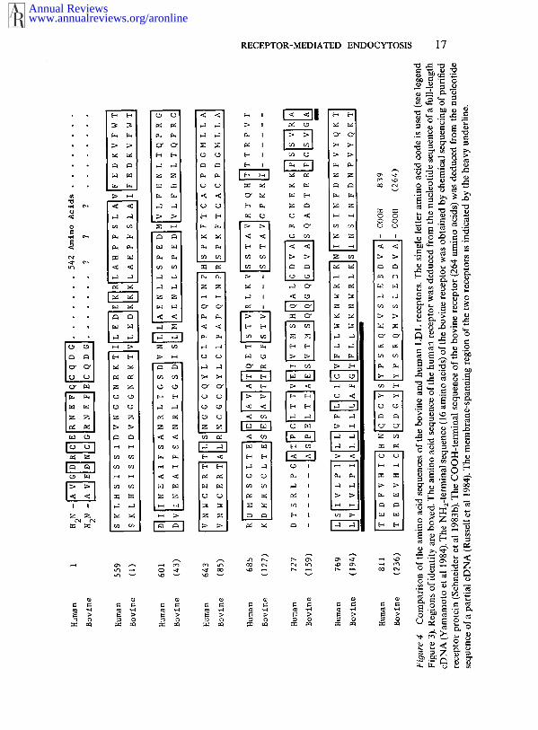
FOURTH DOMAIN : MEMBRANE-SPANNING REGION This domain consists of astretch of 22 hydrophobic amino acids. Proteolysis experiments (see below)confirmed that this domain spans the membrane (Russell et al 1984).Comparison of the amino acid sequences of the bovine and human LDLreceptors reveals that the membrane-spanning region is relatively poorlyconserved (Figure 4). Of the 22 amino acids in this region, 7 differ betweenhuman and cow, but all of the substitutions are also hydrophobic. Thehuman receptor contains a single cysteine in the membrane-slSanningregion. In the bovine receptor this cysteine is replaced by an alanine (Figure4). Since the bovine and the human receptors function similarly, it seemslikely that this intramembraneous cysteine exists in a reduced state in thehuman LDL receptor.
FIFTH DOMAIN : CYTOPLASMIC TAIL The human and bovine LDL receptorsboth contain a COOH-terminal segment of 50 amino acids that projectsinto the cytoplasm. This sequence is strongly conserved; only four of the 50amino acids differ between the two species, and each of these substitutions isconserv.a.tive with respect to the charge of the amino acid (Figure 4).Localization of this domain to the cytoplasmic side of the membrane wasdetermined through use of an anti-peptide antibody directed against the
Annual Reviewswww.annualreviews.org/aronline
RECEPTOR-MEDIATED ENDOCYTOSIS 17
Annual Reviewswww.annualreviews.org/aronline

18 GOLDSTEIN ET AL
COOH-terminal sequence (Russell et al 1984). When inside-out membranevesicles containing receptor were digested with pronase, the antibody-reactive material was removed, and the molecular weight of the receptorwas reduced by approximately 5000.
Immediately internal to the membrane the cytoplasmic tail contains acluster of positively charged amino acids (3 of the first 6 residues are lysinesor arginines). This is a frequent feature of plasma membrane proteins(Sabatini et al 1982). Near the COOH-terminal end of the receptor lies cluster of negatively charged residues (glutamic-aspartic-aspartic) (Figure4). The cytoplasmic segment also contains several serine and threonineresidues and three tyrosines, which may be sites for phosphorylation. Thisdomain also contains a single cysteine, which may be a site for disulfidebond formation or for fatty acylation. None of these modifications havebeen detected as yet.
The cytoplasmic domain of the LDL receptor plays an important role inclustering in coated pits, either through interaction with clathrin itself orwith some protein associated with clathrin on the cytoplasmic side of themembrane (see below). For this reason, it is important to compare thesequences of the cytoplasmic domain of the LDL receptor with those ofother receptors known to enter coated pits.
Comparison With Five Other Coated Pit Receptors
In 1984 and early 1985 complete eDNA sequences for 6 coated pit receptorswere reported. When the predicted amino acid sequences are compared, noobvious conserved feature is apparent (Figure 5). In particular, thecytoplasmic domains show tremendous differences, varying in size from 38amino acids (asialoglycoprotein receptor) to 542 amino acids with tyrosinekinase activity (EGF receptor). In addition, the orientations of the receptorsare different. Four receptors, those for LDL, EGF, insulin, and polymericIgA/IgM, are oriented with their NH2-termini outside the cell and theirCOOH-termini in the cytoplasm. Two receptors, for transferrin andasialoglycoprotein, exhibit an inverted orientation with their NH/-terminiin the cytoplasm and their COOH-termini outside the cell. The cytoplasmicdomains of the 4 receptors that lack tyrosine kinase domains containclusters of negatively charged amino acids (glutamic and aspartic),generally in regions predicted to have an a-helical conformation. Theseacidic residues may play some role in interaction with coated pits.
The transferrin receptor exists as a homodimer, linked by a disulfidebond between two cysteine residues that are immediately external to theplasma membrane (McClelland et a11984, Schneider et a11984). The insulinreceptor is composed of two ~-subunits (ligand binding domain) and two/~-subunits (tyrosine kinase domain) linked by disulfide bonds. The ~ and
Annual Reviewswww.annualreviews.org/aronline

RECEPTOR-MEDIATED ENDOCYTOSIS 19
0
Annual Reviewswww.annualreviews.org/aronline

20 GOLDSTEIN ET AL
chains are derived from a single precursor molecule that undergoesproteolytic cleavage to assume the configuration shown in Figure 5 (Ullrichet al 1985, Ebina et al 1985). There is no published evidence that the other coated pit receptors form disulfide-linked dimers. Four coated pit receptors(those for LDL, IgA/IgM, transferrin, and asiaioglycoproteins) have single cysteine residue in their cytoplasmic regions; the EGF and insulinreceptors have several such residues. This observation suggests that a novelcytoplasmic interchain disulfide bond may play a role in clustering incoated pits.
As mentioned above, the amino acid sequences of the cytoplasmicdomains of the bovine and human LDL receptors are highly conserved. Incontrast, no significant conservation is observed between the cytoplasmicdomains of the asialoglycoprotein receptor in the rat and the analogousreceptor in the chicken, even though the extracellular domains of these tworeceptors are conserved (Drickamer et al 1984).
THE LDL RECEPTOR AT A GENETIC LEVEL
mRNA StructureIn the human tissues studied so far (cultured diploid fibroblasts, SV40-transformed fibroblasts, A-431 epidermal carcinoma cells, fetal and adultadrenal glands, and fetal and adult liver) the LDL-receptor mRNA appearson Northern blots as a single species of approximately 5.3 kb (Yamamoto etal 1984, unpublished observations). (Minor heterogeneity in size cannot excluded by this technique.) About half of the mRNA consists of anunusually long 3’ untranslated region of 2.5 kb. It terminates with a poly(A)÷ tract that is about 15 nucleotides downstream from a likelypolyadenylation signal (AAUUAAA).
An unusual feature of the 3’ untranslated region is the presence of 2½RNA copies of a middle repetitive sequence present in mammaliangenomes. This sequence, designated Alu, occurs on average once in every5000 base pairs (bp) in the human genome, for a total of ~ 300,000 copies(Schmid & Jelinek 1982). Each Alu repeat is about 300 bp long, and consistsof a tandem repeat of two monomeric units--a left monomer of 130 bp anda right monomer 160 bp long, owing to a 30 bp insertion. The human LDL-receptor mRNA has two complete Alu sequences and an extra rightmonomeric unit, all clustered together within a region of about 750nucleotides (Yamamoto et al 1984).
The bovine mRNA does not contain these Al’u sequences or any otherrepetitive sequences (Hobbs et al, manuscript in preparation). Thesequences on either side of the Alu repeats are conserved in the human andthe cow, suggesting that the Alu sequences were inserted after the human
Annual Reviewswww.annualreviews.org/aronline
RECEPTOR-MEDIATED ENDOCYTOSIS21
and bovine evolutionary lines diverged. Restriction maps of genomic DNAsuggest that the Alu repeats are present at the same location in the 3’untranslated regions of the LDL-receptor genes of the gorilla andchimpanzee, but not the baboon (unpublished observations). If this findingis confirmed by direct cloning studies, it would suggest that the Alusequences have inserted into this location late in the evolution of theprimates. Whether this insertion has any functional consequence for theprocessing, translation, or stability of the receptor mRNA is unknown.
Gene Structure
Southern blotting of genomic DNA demonstrated that the haploid humangenome contains a single copy of the LDL-receptor gene (Lehrman et al1985). This gene resides on chromosome 19, as determined by somatic cellgenetic techniques (Francke et al 1984). The gene spans more than 45 kb.Sequences representing almost the entire gene have been isolated frombacteriophage lambda and cosmid libraries (Siidhof et al 1985a,b). Theposition of each intron within the gene has been mapped, and the sequenceof each exon-intron junction has been determined.
These studies reveal that the receptor gene is made up of 18 exons. Thesites of the introns in relation to the protein sequence are indicated inFigure 2 (Siidhof et al 1985a,b). Most of the introns separate regions of theprotein that correspond to domains that were identified through theprotein chemistry studies described above. The first intron is located just atthe end of the DNA encoding the cleaved signal sequcnc~ of the protein.Within the binding domain of the receptor (which contains the sevencysteine-rich repeats), introns occur precisely between repeats I and II; IIand III; V and VI; and VI and VII (Figures 2, 3). Repeats III, IV, and V areincluded in one exon. The binding domain is terminated by an intron atamino acid 292, the last residue in the seventh repeat.
The next domain, the region of homology with the EGF precursor, isencoded in 8 contiguous exons. Within this 400-amino acid region ofhomology are located 3 copies of a repeated sequence (repeats A, B, and C inFigure 2), each of which is encoded by a single exon (Siidhof et al 1985a,b).(The striking similarity in the exon-intron organization of this region of theLDL receptor gene and the EGF precursor gene is discussed in thepreceding section).
The O-linked sugar domain is also demarcated neatly by two introns(Figure 2). However, not all domains of the protein are encoded by singleexons. Thus, the membrane-spanning region is interrupted by an intron.Another intron interrupts the coding region for the cytoplasmic tail 11amino acids from the COOH-tcrminus.
The placement of the introns is consistent with the notion that the human
Annual Reviewswww.annualreviews.org/aronline

22 GOLDSTEIN ET AL
LDL receptor gene was constructed by the stepwise assembly of exons thatencode useful protein sequences. Thirteen of the 18 exons comprising theLDL receptor gene encode protein sequences that are homologous tosequences in other proteins: 5 of these exons encode a sequence similar toone in the C9 component of complement; 3 exons encode a sequencesimilar to a repeat sequence in the EGF precursor and in 3 proteins of theblood clotting system; and 5 other exons encode nonrepeated sequencesthat are shared only with the EGF precursor. The LDL receptor thusappears to be a mosaic protein built up of exons shared with differentproteins (Siidhof it al 1985a,b).
The 5’ untranslated region of the receptor gene is less than 100 base pairs,and it is not interrupted by an intron (Siidhof et al 1985a). Two TATA-likeboxes occur 20-30 base pairs to the 5’ side of the two major sites oftranscription initiation located between nucleotides - 79 to - 93. Analysisof the upstream promoter region will be of interest because transcription ofthe gene into mRNA appears to be regulated by a feedback mechanism.When cholesterol accumulates in cells, the level of cytoplasmic mRNA ~forthe receptor declines dramatically (Russell et al 1983, Yamamoto et al1984), and this leads to a decrease in the rate of synthesis of the receptorprotein (Goldstein & Brown 1977). It is likely, but not yet proven, that thedecrease in cytoplasmic mRNA is due to a cholesterol-mediated suppres-sion of transcription of the receptor gene.
BIOSYNTHESIS OF THE HUMAN LDLRECEPTOR
The presence of a cleaved NH2-terminal hydrophobic signal sequencesuggests that the LDL receptor is synthesized on membrane-boundribosomes. At the earliest time point that can be studied (15 min after theaddition of [35S]methionine to cultured human fibroblasts) the receptorappears in immunoprecipitates as a protein with an apparent molecularweight of 120,000, as estimated on SDS polyacrylamide gels (Tolleshaug etal 1982). This precursor contains asparagine-linked (N-linked), high-mannose oligosaccharide chains, which are sensitive to endoglycosidase-H(endo-H) (Tolleshaug et al 1983). According to the best estimates available,there are two N-linked sugar chains on the purified bovine LDL receptor(Cummings et al 1983). Although the protein sequence for the humanreceptor shows five potential N-linked giyeosylation sites (Figure 5), it possible that the three N-linked sites in the cysteineorich, disulfide-linkedregion of the receptor are not glycosylated (Yamamoto et al 1984).
The earliest detectable receptor precursor also contains N-acetylgalactosamine (GalNAc) residues attached to serines and threonines
Annual Reviewswww.annualreviews.org/aronline

RECEPTOR-MEDIATED ENDOCYTOSIS23
by O linkage. This finding emerged from experiments in which theEZH]glucosamine-labeled 120-kilodalton precursor of human A-431epi-dermal carcinoma cells was isolated by SDS gel electrophoresis anddigested with pronase. Multiple GalNAc residues were found on a singlepronas¢-resistant fragment (Cummings et al 1983).
The presence of O-linked GalNAc residues at a time when the N-linkedsugar chains are still in the high-mannose (endo-H sensitive) configurationimplies that the GalNAc transferase that initiates synthesis of O-linkedsugar chains is proximal to the cis-Golgi stacks. This follows from theobservation that once a protein reaches the cis-Golgi the mannose residuesare trimmed from the N-linked sugars and the chains become endo-Hresistant (Hubbard & Ivatt 1981). Whether the O-linked GalNAc residuesare added in the ER, or whether they are added in some transitional zonebetween the ER and the cis-Golg~ is not known.
Between 30 and 60 min after synthesis, the LDL receptor precursorundergoes a sudden shift in apparent molecular weight from 120,000 to160,000 (Tolleshaug et al 1982, 1983 ; Schneider et al 1983a). The timing this shift coincides with the maturation of the N-linked and O-linkedchains. The shift is not the result of the alteration in N-linked sugars,because a change of nearly equal magnitude occurs in cells that are treatedwith tunicamycin, which blocks the addition of N-linked chains.Conversely, the increase in apparent molecular weight is minimized in amutant strain of Chinese hamster ovary (CHO) cells that is unable to addgalactose to the core GalNAc residues of the O-linked chains (Cummings etal 1983). These findings suggest that the 40,000 change in apparentmolecular weight is attributable to the elongation of the O-linked chains.This elongation consists of the addition of a single galactose and one or twosialic acids to the GalNAc core sugar of each O-linked chain.
The apparent molecular weight of the mature receptor is reduced by onlyabout 10,000 when the sialic acids are completely removed with neura-minidase (Schneider et al 1982, Cummings et al 1983). Thus, most of thechange from 120,000 to 160,000 daltons is contributed not by the sialicacids, but by the simple addition of galactose residues to the GalNAc coresugars. This change selectively retards the mobility of the receptor on SDSgels, so that the mature receptor migrates more slowly than would beappropriate for its true molecular weight. The calculated molecular weightof the protein component of the receptor is 93,102. When the maturecarbohydrates are included, the molecular weight will be ~ 115,000, not160,000 as observed on SDS gels. Aberrant migration of other membraneglycoproteins that contain clustered O-linked sugars has been previouslydocumented (Marchesi et al 1976).
The increase in apparent molecular weight of the LDL receptor is
Annual Reviewswww.annualreviews.org/aronline

24 GOLDSTEIN ET AL
partially blocked when cells are incubated with monensin, an ionophorethat blocks vesicular transport in the Golgi complex (unpublished obser-vations). Under these conditions there is no longer a discrete jump fromapparent molecular weight of 120,000 to 160,000. Rather, the receptorappears as a smear between these two extremes.
The LDL receptor in human fibroblasts can be labeled with 35S-sulfate,which attaches to N-linked sugars; incorporation is blocked by tunica-mycin (Cummings et al 1983). The receptor synthesized in tunicamycin-treated fibroblasts appears to undergo normal internalization andrecycling, but subtle changes in receptor half-life have not been ruled out(unpublished observations). Since these tunicamycin-treated receptors not contain sulfate, it is unlikely that sulfate performs a crucial function inthe LDL receptor. Incorporation of a 5S.sulfate into the receptor in humanA-431 carcinoma cells could not be demonstrated (Cummings et al 1983).
NATURALLY OCCURRING MUTATIONS INTHE LDL RECEPTOR
The power of the LDL receptor as a system for the study of receptor-mediated endocytosis derives from the existence of many naturallyoccurring mutations in the LDL receptor gene that disrupt receptorfunction in revealing ways. The mutations occur in individuals with familialhypercholesterolemia (FH) (Goldstein & Brown 1983). Those who inheritone mutant LDL-receptor gene produce half the usual number of normalreceptors. In tissue culture their cells degrade LDL at about half the normalrate. In the body the receptor deficiency causes LDL to build up in plasmato levels about twofold greater than normal. Eventually, the high plasmaLDL levels lead to atherosclerosis and heart attacks as early as 40 years ofage (Brown & Goldstein 1984):
Individuals with two mutant LDL-receptor genes are termed FHhomozygotes. Their cells produce few or no functional LDL receptors. As aresult, plasma LDL accumulates to levels eight to ten times greater thannormal, and they develop atherosclerosis and heart attacks in childhood.
At least ten different mutant alleles at the LDL-receptor locus have beendescribed (Goldstein & Brown 1983, Tolleshaug 1982, 1983, Schneider et al1983a, Lehrman et al 1985). Many of the phenotypic FH homozygotesactually represent compound heterozygotes who inherit different mutantalleles of the receptor gene from each parent. Study of cultured skinfibroblasts from 104 FH homozygotes revealed that the mutations could bedivided into four broad classes based upon their effects on receptorstructure and function. These mutations are summarized in Table 2 anddiscussed below.
Annual Reviewswww.annualreviews.org/aronline

RECEPTOR-MEDIATED ENDOCYTOSIS 25
Class 1 Mutations: No Detectable Precursor
These alleles, designated R-O for "receptor-zero," are the most frequent ofthe mutant alleles. It is difficult to determine their frequency directly (sincethey cannot be identified unequivocally in the heterozygous state), but theyprobably account for about one-third to one-half of all mutant alleles at theLDL-receptor locus. Class 1 alleles fail to express receptor proteins asmeasured by-functional assays (binding of 125I-LDL) or immunologicalassays (immunoblotting or precipitation by a variety of monoclonal andpolyclonal antibodies directed against the LDL receptor). It is likely thatthis class includes nonsense mutations, which introduce terminationcodons early in the protein coding region. It may also include: pointmutations in the promoter that block transcription of mRNA; pointmutations in intron-exon junctions that alter the splicing of mRNA; andlarge deletions.
Class 2 Mutations: Precursor Not ProcessedThese alleles encode receptor precursors that are synthesized in normal orreduced amounts, but that do not undergo any apparent increase inmolecular weight after synthesis. These receptors remain in the endo-Hsensitive form, and they do not receive sialic acid, as indicated by their lackof susceptibility to neuraminidase. Thus, we believe these receptors are nottransported to the Golgi complex. Receptors specified by these alleles neverreach the cell surface, and hence they are protected when the surface ofintact cells is treated with pronase (Tolleshaug et al 1983).
Most of the alleles in this class encode receptors with apparent molecularweights on SDS gels of 120,000, which is similar to the apparent molecularweight of the normal precursor. These alleles are designated R-120(Tolleshaug et al 1982, 1983). Detailed structural analysis of the oligosac-charide chains of one mutant receptor encoded by the R-120 allele showedit to contain N-linked high mannose chains and O-linked core GalNAcresidues indistinguishable from the normal 120-kilodalton receptor pre-cursor (Cummings et al 1983). We have also observed precursor proteinswith abnormal apparent molecular weights of 100,000 and 135,000 that fallinto this class (designated R-IO0 and R-135 alleles, respectively). These.molecular weight abnormalities may result from alterations in the length ofthe protein chain, rather than from alterations in carbohydrate, since themolecular weight remains abnormal after endo-H treatment (Tolleshaug etal 1983).
Variants of the Class 2 mutation were observed in a consanguineousblack American family, in several Afrikaners, and in a strain of rabbits thathas a syndrome similar to FH, i.e. Watanabe heritable hyperlipidemic
Annual Reviewswww.annualreviews.org/aronline

26 GOLDSTEIN ET AL
0 0 0 ¢~
+ ++ +
Annual Reviewswww.annualreviews.org/aronline

RECEPTOR-MEDIATED ENDOCYTOSIS 27
~ ~ ~ z z
~" + +
+ + +"
Annual Reviewswww.annualreviews.org/aronline
28 GOLDSTEIN ET AL
(WHHL) rabbits (Schneider et al 1983a, unpublished observations). these variants the receptor is produced as a 120-kilodalton precursor that isprocessed to the mature form at a slow but finite rate. Eventually about 10~oof the receptors appear on the cell surface as 160-kilodalton matureproteins. Even after they reach the surface, these receptors have a reducedability to bind LDL. Normal receptors bind equimolar amounts of IgCi-C7(a monoclonal antibody against the external domain of the LDL receptor)and LDL protein (Beisiegel et al 1981). In the Class 2 variants the ratio LDL binding to monoclonal antibody binding is reduced, suggesting thatthese receptors have an abnormality in the LDL binding site as well as aslower rate of transport to the surface (Schneider et al 1983a).
The molecular basis of the defect in the Class 2 mutations is not known.These receptors are all recognized by monoclonal and polyclonal anti-bodies against the receptor, so their structures are not drastically differentfrom the normal receptor. It seems likely that the failure of transport arisesfrom some subtle alteration in structure. Elucidation of this change shouldlead to new insights into the signals that govern transport of proteins fromthe ER to the cis-Golgi.
Scheckman and co-workers have described a mutation in yeast invertase,a secreted enzyme, that is analogous to the Class 2 mutations (Schauer et al1985). The defect in invertase results from the alteration of a single aminoacid at the site at which the hydrophobic NHE-terminal signal sequence iscleaved from the protein. In the absence of cleavage, invertase is transportedto the Golgi at 2~o of the normal rate. A similar transport defect has beencreated in yeast by in vitro mutagenesis of the gene for acid phosphatase(Haguenauer-Tsapis & Hinnen 1984). It seems likely that some of the Class2 mutations in FH may result from the failure to cleave the signal sequencefrom the protein.
Class 3 Mutations: Precursor Processed, Abnormal
Binding of LDL by Receptor
Receptors specified by Class 3 mutant alleles reach the surface at a normalrate and are recognized on the surface by monoclonal anti-receptorantibody (IgG-C7). However, these receptors bind less than 15~ of the
normal amount of 125I.LDL (Goldstein & Brown 1983, Beisiegel et al 1981,Tolleshaug et al 1983).
Most commonly, the receptors produced by the Class 3 alleles have anormal molecular weight on SDS gel electrophoresis. This allele is desig-nated R-160 b-. Receptors with molecular weights of 140,000 (R-140 b-)
(Tolleshaug et al 1983) and 210,000 (R-210 b-)(Tolleshaug et al 1982)have also been described. Both of these proteins originate as precursors
Annual Reviewswww.annualreviews.org/aronline

RECEPTOR-MEDIATED ENDOCYTOSIS 29
with apparent molecular weights that are 40,000 less than their maturespecies, i.e. 100,000 and 170,000, respectively. The correct increase inapparent molecular weight suggests that the carbohydrate processingreactions occur normally. Structural analysis of the carbohydrates of thereceptor specified by the Ro210 b- allele showed no abnormality(Cummings et al 1983). We believe, therefore, that the abnormal molecularweight is due to alterations in the amino acid sequence, and not to changesin carbohydrate content.
An explanation for the abnormally sized receptors is suggested by thestructure of the binding domain for LDL. As discussed above, this bindingdomain is made up of seven repeats of a 40 amino acid sequence. Because ofthis internal homology, the DNA encoding such a repeat structure wouldbe susceptible to deletion or duplication following "slipped mispairing" andrecombination during meiosis. Such duplications or deletions wouldchange the size of the receptor and might reduce LDL binding withoutaffecting the binding of monoclonal antibody IgG-C7 (which does notrecognize the LDL binding site). This hypothesis is presently being testedwith the available cDNA and genomic probes.
Class 4 Mutations: Precursor Processed, Receptor
Binds LDL But Does Not Cluster In Coated Pits
These are the so-called internalization-defective mutations. The originalexample was patient JD (Brown & Goldstein 1976). Biochemical studiesshowed that patient JD is a compound heterozygote (Goldstein et al1977). From his mother he inherited a gene that produces a nonfunctionalreceptor (R-0 allele). From his father he inherited a gene that produces receptor of normal size that reaches the surface and binds LDL normally,but is not able to carry the bound LDL into the cell (R-160 i- allele).Electron microscopic studies revealed that the internalization-defectivereceptors in JD and his father are present on the surface in small clusters,but they are not sequestered in coated pits, even though coated pits arepresent in these cells (Anderson et al 1977b) and the coated pits functionnormally in the receptor-mediated endocytosis of other ligands such asEGF (Goldstein et al 1978).
Subsequently, four other FH patients with internalization defects havebeen identified. One is a young man from Minnesota (designated FH 274 orBH) who is also a compound heterozygote with an internalization-defectiveallele inherited from his mother and a nonfunctional allele from his father(Goldstein et al 1982, Lehrman et al 1985). Another is a patient from ~apan,born of consanguineous parents, who appears to be homozygous for aninternalization-defective allele (Miyake et al 1981).. The third and fourth
Annual Reviewswww.annualreviews.org/aronline
30 GOLDSTEIN ET AL
patients are Arab siblings from a consanguineous marriage, who appear tobe homozygous for an internalization-defective allele (Lehrman et al1985a).
We have recently elucidated the molecular defect in the internalization-defective allele in patient FH 274 (Lehrman et al 1985b). Protein chemistrystudies demonstrated that his mutant receptor has two abnormal pro-perties. First, it is about 10,000 daltons smaller in apparent molecularweight than the normal receptor. Second, about 80~ of the receptors aresecreted into the culture medium, and only about 20~ remain associatedwith the cell. (In normal fibroblasts no detectable amounts of receptor aresecreted.) The allele giving rise to this abnormal receptor was inherited fromthe mother. Since the mother’s cells had the internalization-defectivephenotype, the shortened receptor must be responsible for theinternalization-defective state.
Through restriction endonuclease mapping of genomic DNA andsubsequent cloning of the relevant genomic fragments into bacteriophagelambda, Lehrman et al (1985b) demonstrated that the mutantinternalization-defective allele in FH 274 cells (designated R-150 i-, sec)had undergone a large deletion. The deletion resulted from a recombinationbetween one of the Alu sequences in the 3’-untranslated region of themRNA and an Alu sequence that is located 5 kb upstream in the interveningsequence that separates the exon encoding the O-linked sugar region fromthe exon encoding the membrane-spanning domain. This deletion elimin-ated the DNA sequences that encode the membrane-spanning region andthe cytoplasmic domain of the receptor.
According to the above results the protein produced by the deleted geneis expected to have a normal sequence from the NH2-terminus through theO-linked sugar region. Thereafter, the protein should terminate becausethe deletion joint should produce a random sequence of nucleotides in themRNA. By chance, a protein termination codon is expected to be reachedwithin 20 codons. These predictions derived from the genomic cloning wereconfirmed by studies of the FH 274 protein. The truncated receptorprecursor (110 kilodaltons) showed the normal 40,000 increase in molecularweight after synthesis, indicating that the O-linked sugars were present(Lehrman et al 1985b). Moreover, the FH 274 protein reacted with anti-peptide antibodies directed against the NH2-terminal domain of thereceptor, but failed to react with anti-peptide antibodies directed againstthe COOH-terminal cytoplasmic tail, which confirms that the cytoplasmicdomain was eliminated (Lehrman et al 1985b).
The secretion of the truncated receptor from the FH 274 cells is anexpected result based on earlier studies with truncated mutants for viral
Annual Reviewswww.annualreviews.org/aronline

RECEPTOR-MEDIATED ENDOCYTOSIS 31
envelope proteins, such as that of the influenza hemagglutinin (Gething Sambrook 1982) and the G protein of vesicular stomatitis virus(Florkiewicz et al 1983). When such truncated proteins are synthesized membrane-bound ribosomes, the lack of a hydrophobic membrane-spanning region allows the entire protein to translocate across the ERmembrane and to appear in the lumen of the ER, from which it is eventuallyincorporated into secretory vesicles.
Why are some of the mutant receptors produced by the FH 274 cellsfound attached to the cell surface? These molecules appear to be firmlyattached since they cannot be washed off easily by high salt, EDTA, highlycharged polymers, or reducing agents (Lehrman et al 1985b, unpublishedobservations). The membrane-adherent receptors are found in noncoatedregions of the cell surface where they bind LDL (Anderson et al,unpublished observations) and give rise to the internalization-defectivephenotype by which the FH 274 cells were originally identified (Goldsteinet al 1982). It is possible that these truncated receptors bind with highaffinity to some other protein that keeps them anchored to the membrane.Alternatively, perhaps some of the mutant receptors have acquired ahydrophobic sequence by chance, as a result of alternative splicing of themRNA within the intervening sequence that contains the deletion joint.Such splicing might lead to a random sequence of nucleotides that happensto encode a stretch of hydrophobic amino acids prior to the terminationcodon. Thus, some of the receptors might have sticky hydrophobic COOH-terminal tails that make them adhere to the membrane.
We have recently identified defects in two other internalization-defectiveLDL receptor mutations (Lehrman et al 1985a and manuscript inpreparation). Both of these mutations involve single base substitutions inthe exon encoding the majority of the cytoplasmic domain of the receptor(exon 17 in Figure 2), and both were identified through cloning andsequencing of genomic DNA fragments that contain this exon. One of thesepoint mutations, found in the Arab family mentioned above, results from aguanosine to adenosine transition, which changes a tryptophan codon(UGG) to a termination codon (UGA). The receptor produced by this terminates at a position corresponding to the tryptophan that is just at thebeginning of the cytoplasmic domain of the receptor (Figure 4). Theresulting protein has a truncated cytoplasmic domain of only 2 (rather than50) amino acids. This protein moves to the cell surface, but the lack of cytoplasmic domain renders it incapable of clustering in coated pits.
A different point mutation occurs in JD, the first internalization-defectiveFH patient to be described (Brown & Goldstein 1976, Goldstein et a11977).In this case, an adenosine to guanosine transition converts a codon for
Annual Reviewswww.annualreviews.org/aronline

32 GOLDSTEIN ET AL
tyrosine (UAU) into a codon for cysteine (UGU). This mutation occurs the cytoplasmic domain 33 residues from the COOH-terminus (Lehrman etal, manuscript in preparation). The mutant receptor has 2 cyste~ne residuesin its cytoplasmic domain : the normal one at position 818 and a new one atposition 807. We do not yet know whether the failure to cluster in coatedpits and the abnormal internalization are attributable to the acquisition ofan extra cysteine or to the loss of a crucial tyrosine.
The finding of defects in the cytoplasmic domain in three internalization-defective mutations (one deletion, one nonsense mutation, and onemissense mutation) supports our earlier proposal that this domain is crucialin directing the LDL receptor to coated pits (Anderson et a11977, Goldsteinet al 1979).
EXPERIMENTALLY INDUCED MUTATIONS INTHE LDL RECEPTOR
To increase the repertoire of available mutations, Krieger et al (1981, 1983)developed two methods for creating LDL-receptor mutations in tissueculture cells. In the first procedure the cholesteryl esters of LDL areextracted and replaced with hydrophobic molecules that convert the LDLinto either toxic or fluorescent particles (Krieger et al 1981). Mutagen-treated Chinese hamster ovary (CHO) cells are incubated with re-constituted LDL-containing toxic 25-hydroxycholesteryl oleate. Wild-typecells take up this lipoprotein via the LDL receptor, liberate the 25-hydroxycholesterol in lysosomes, and die. The few surviving clones areincubated with LDL reconstituted with fluorescent cholesteryl ester, andthe colonies that fail to accumulate fluorescence are picked. This two-stepisolation procedure yielded LDL-receptor-deficient cells at a frequency of1 x 10-s (Krieger et al 1981).
The second selection procedure takes advantage of two fungal meta-bolites: compactin, a potent inhibitor of cholesterol biosynthesis, andamphotericin B, a polyene antibiotic that forms toxic complexes withsterols in membranes (Krieger et al 1983). Mutagen-treated CHO cells arepreincubated in a medium containing compactin, LDL, and small amountsof mevalonate, a combination that makes CHO cells dependent on theLDL receptor for obtaining cholesterol (Goldstein et al 1979b). After thepreincubation, mutant cells that cannot utilize the cholesterol of LDLbecome cholesterol-deficient. Subsequent incubation with amphotericin Bkills the wild-type cells through formation of complexes with membranecholesterol. The receptor-deficient clones, which are depleted in cholesterol,do not bind amphotericin B and therefore they survive. With this procedure
Annual Reviewswww.annualreviews.org/aronline

RECEPTOR-MEDIATED ENDOCYTOSIS 33
mutant cells are isolated at a frequency of approximately 2.6 x 10⁻⁵
(Krieger et al 1983).All of the mutants obtained to date, by either of the two procedures,
express an LDL-receptor-deficient or negative phenotype, i.e. there is aproportional reduction in the binding, internalization, and degradation ofLDL. Complementation studies suggest that the mutants fall into fourgroups, designated IdIA, ldlB, ldIC, and ldlD (Kingsley & Krieger 1984). TheldlA locus appears to be the structural gene for the LDL receptor on thebasis of two observations: 1) fusion of these cells with normal humanfibroblasts, but not FH homozygote fibroblasts, leads to complementation;and 2) mutants in the ldlA group show abnormal LDL receptors byimmunoprecipitation and SDS gel electrophoresis (Kozarsky & Krieger,manuscript in preparation). The ldlA locus appears to be diploid in CHOcells, as defined by the isolation of a heterozygous revertant of ahomozygous mutant (Kingsley & Krieger 1984).
The biochemical basis of the LDL-receptor deficiency in the ldlB, ldlC,and ldID mutants is unclear at the present time; it may involve defects in theposttranslational processing of the LDL receptor. Most of these mutantsexhibit altered sensitivity to plant lectins, which suggests they harbora pleiotropic abnormality in glycoprotein processing (Krieger et al,unpublished observations). The ldlD mutants are unique in that the re-ceptor-negative phenotype can be corrected by co-cultivation with othermammalian cells (Krieger 1983). LDL-receptor activity can also be in-duced in the ldlD cells by addition of a factor found in human Or bovineserum (Krieger 1983).
Sege et al (1984) recently used the technique of calcium phosphate-mediated gene transfer to introduce human genomic DNA into the IdlAcells. After transfection the ldlA cells were incubated in the presence ofcompactin, LDL, and mevalonate, a combination that selects for functionalLDL receptors. One of the clones that survived this selection was shown toexpress the human LDL receptor on its cell surface, as determined byimmunoprecipitation with monoclonal antibody IgG-C7, which reactswith the receptor of human but not of hamster origin (Beisiegel et al 1981).The transfected human receptor gene was functional in the binding, uptake,and degradation of LDL, and its expression was suppressed by cholesterol(Sege et al 1984).
The ability to introduce a functional human LDL-receptor gene intomutant CHO cells illustrates the power of the LDL-receptor system for usein studies of experimentally induced mutations. Not only are efficientselection systems available for the production of LDL-receptor-deficientmutants, but efficient systems are also now available for selecting revertants
Annual Reviewswww.annualreviews.org/aronline

34 GOLDSTEIN ET AL
of these mutants, and for introducing transfected genes into these cells.Finally, the ability to express functional LDL receptors from clonedcDNAs following transfection of cultured cells (Yamamoto et al 1984),together with the techniques of in vitro mutagenesis, should provide muchexciting information regarding the LDL receptor in particular, and theprocess of receptor-mediated endocytosis in general.
ACKNOWLEDGMENT
The original research described in this article was supported by grants fromthe National Institutes of Health (HL 20948 and HL 31346). D.W.R. is therecipient of an NIH Research Career Development Award (HL 01287).W.J.S. is an Established Investigator of the American Heart Association.
Literature Cited
Anderson, R. G. W., Brown, M. S., Beisiegel,U., Goldstein, J. L. 1982. Surface distri-bution and recycling of the LDL receptoras visualized by anti-receptor antibodies.J. Cell. Biol. 93:523-31
Anderson, R. G. W., Brown, M. S., Goldstein,J. L. 1977a. Role of the coated endocyticvesicle in the uptake of receptor-boundlow density lipoprotein in human fibr0b-lasts. Cell 10:351-64
Anderson, R. G. W., Goldstein, J. L., Brown,M. S. 1977b. A mutation that impairs theability of lipoprotein receptors to localisein coated pits on the cell surface of humanfibroblasts. Nature 270: 695-99
Anderson, R. G. W., Goldstein, J. L., Brown,M. S. 1976. Localization of low densitylipoprotein receptors on plasma mem-brane of normal human fibroblasts andtheir absence in cells from a familial hyper-cholesterolemia homozygote. Proc. Natl.Acad. Sci. USA 73 : 2434-38
Barak, L. S., Webb, W. W. 1982. Diffusion oflow density lipoprotein-receptor complexon human fibroblasts. J. Cell Biol. 95 : 846-52
Basu, S. K., Goldstein, J. L., Anderson, R. G.W., Brown, M. S. 1976. Degradation ofcationized low density lipoprotein andregulation of cholesterol metabolism inhomozygous familial hypercholestero-lemia fibroblasts. Proc. Natl. Acad. Sci.USA 73 : 3178-82
Basu, S. K., Goldstein, J. L., Anderson, R. G.W., Brown, M. S. 1981. Monensin inter-rupts the recycling of low density lipo-prQtein receptors in human fibroblasts.
Cell 24: 493-502Basu, S. K., Goldstein, J. L., Brown, M. S.
1978. Characterization of the low densitylipoprotein receptor in membranes pre-pared from human fibroblasts. J. Biol.Chem. 253 : 3852-56
Beisiegel, U., Schneider, W. J., Brown, M. S.,Goldstein, J. L. 1982. Immunoblot analysisof low density lipoprotein receptors infibroblasts from subjects with familialhypercholesterolemia. J. Biol. Chem. 257:13150-56
Beisiegel, U., Schneider, W. J., Goldstein, J.L., Anderson, R. G. W., Brown, M. S. 1981,Monoclonal antibodies to the low densitylipoprotein receptor as probes for study ofreceptor-mediated endocytosis and thegenetics of familial hypercholesterolemia.J. Biol. Chem. 256:11923-31
Berg, T., Blomhoff, R., Naess, L., Tolleshaug,H., Drevon, C. A. 1983. Monensin inhibitsreceptor-mediated endocytosis of asialog-lycoproteins in rat hepatoeytes. Exp. CellRes. 148:319-30
Bleil, J. D., Bretscher, M. S. 1982. Transferrinreceptor and its recycling in HeLa cells.EMBO J. 1:351-55
Bretscher, M.S. 1984. Endocytosis : Relationto capping and cell locomotion. Science244: 681-86
Bretscher, M. S., Pearse, B. M. F. 1984.Coated pits in action. Cell 38 : 3-4
Brodsky, F. M. 1984. The intracellular trafficof immunologically active molecules.Immunol. Today 5 : 350-57
Brown, M. S., Anderson, R. G. W., Basu, S.K., Goldstein, J. L. 1982. Recycling of cell
Annual Reviewswww.annualreviews.org/aronline

RECEPTOR-IVIEDIATED ENDOCYTOSIS 3~
surface receptors: Observations from theLDL receptor system. Cold Spring HarborSymp. Quant. Biol. 46:713-21
Brown, M. S., Anderson, R. G. W., Goldstein,J. L. 1983. Recycling receptors :The round-trip itinerary of migrant membrane pro-teins. Cell 32 : 663-67
Brown, M. S., Deuel, T. F., Basu, S. K.,Goldstein, J. L. 1978. Inhibition of thebinding of low-density lipoprotein to itscell surface receptor in human fibroblastsby positively charged proteins. J.SupramoL Struct. 8 : 223-34
Brown, M. S., GoldsteiI1, J. L. 1976. Analysisof a mutant strain of human fibroblastswith a defect in the internalization ofreceptor-bound low density lipoprotein.Cell 9 : 663-74
Brown, M. S., Goldstein, J. L. 1984. HowLDL receptors influence cholesterol andatherosclerosis. Sci. Am. 251:58-66
Carpenter, G., Cohen, S. 1979. Epidermalgrowth factor. Ann. Rev. Biochem. 48 : 193-216
Carpentier, J.-L., Gorden, P., Anderson, R.G. W., Goldstein, J. L., Brown, M. S., et al.1982. Co-localization of 125I-epiden:nalgrowth factor and ferritin-low densitylipoprotein in coated pits: A quantita-tive electron microscopic study in normaland mutant fibroblasts. J. Cell Biol. 95:73-77
Chiacchia, K. B., Drickamer, K. 1984. Directevidence for the transmembrane orien-tation of the hepatic glycoprotein recep-tors. J. Biol. Chem. 259:15440-46
Ciechanover, A., Schwartz, A. L., Dautry-Varsat, A., Lodish, H. F. 1983. Kinetics ofinternalization and recycling of transferrinand the transferrin receptor in a humanhepatoma cell line. Effect of lysosomo-tropic agents. J. Biol. Chem. 258:9681-89
Cummings, R. D., Kornfeld, S., Schneider, W.J., Hobgood, K. K., Tolleshaug, H., et al.1983. Biosynthesis of the N- and O-linkedoligosaccharides of the low density lipo-protein receptor. J. Biol. Chem. 258:15261-73
Daniel, T. O., Schneider, W. J., Goldstein, J.L., Brown, M. S. 1983. Visualization oflipoprotein receptors by ligand blotting. J.Biol. Chem. 258 : 4606-11
Dautry-Varsat, A., Ciechanover, A., Lodish,H. F. 1983. pH and the recycling of trans-ferrin during receptor-mediated endocy-tosis. Proc. Natl. Acad. Sci. USA 80:2258-62
DiPaola, M., Maxfield, F. R. 1984. Con-formational changes in the receptors forepidermal growth factor and asialoglyco-proteins induced by the mildly acidic pH
found in endocytic vesicles. J. Biol. Chem.259 : 9163-71
DiScipio, R. G., Gehring, M. R,, Podack, E.R., Kan, C. C., Hugli, T. E., et al. 1984.Nudeotide sequence of cDNA and derivedamino acid sequence of human comple-ment component C9. Proc. Natl. Acad. Sci.USA 81 : 7298-7302
Doolittle, R. F., Feng, D.-F., Johnson, M. S.1984. Computer-based characterization ofepidermal growth factor precursor. Nature307 : 558-66
Drickamer, K., Mamon, J. F., Binns, G.,Leung, J. O. 1984. Primary structure of therat liver asialoglycoproteiri receptor:Structural evidence for multiple polypep-tide species. J. Biol. Chem. 259 : 770-78
Dunn, W. A., Hubbard, A. L. 1984. Receptor-mediated endocytosis of epidermal growthfactor by hepatOcytes in the perfused ratliver: Ligand and receptor dynamics. J.Cell Biol, 98 : 2148-59
Ebina, Y., Ellis, L., Jarnagin, K., Edery, M.,Graf, L., et al. 1985. The human insulinreceptor cDNA: The structural basis forhormone-activated transmernbrane sig-nalling. Cell 40: 747-58
Fawcett, D. W. 1965. Surface specializationsof absorbing cells. J. H istochem. C ytochem.13 : 75-91
Florkiewicz, R. Z., Smith, A., Bergmann, J. E.,Rose, J. K. 1983. Isolation of stable mousecell lines that express cell surface andsecreted forms of the vesicular stomatitisvirus glycoprotein. J. Cell. Biol. 97:1381-88
Francke, U., Brown, M. S., Goldstein, J. L.1984. Assignment of the human gene forthe low density lipoprotein receptor tochromosome 19: Synteny of a receptor, aligand, and a genetic disease, Proc. Natl.Acad. Sci. USA 81:2826-30 -
Friend, D, S., Farquhar, M. G. 1967.Functions of coated vesicles during pro-tein absorption in the rat vas deferens. J.Cell Biol. 35 : 357-76
Gething, M.-J., Sambrook, J. 1982. Con-struction of influenza haemagglutinin genesthat code for intraeellular and secretedforms of the protein. Nature 300:598-603
Geuze, H. J., Slot, J. W., Strous, G. J. A. M.,Lodish, H. F., Schwartz, A. L. 1983.Intracellular site of asialoglycoproteinreceptor-ligand uncoupling. Double-labelimmunoelectronmieroscopy during recep-tor mediated endocytosis. Cell 32 : 277-87
Geuze, H. J., Slot, J. W., Strous, G. J. A. M.,Peppard, J., von Figura, K., et al. 1984.Intracellular receptor sorting during en-docytosis: Comparative immunoelectron
Annual Reviewswww.annualreviews.org/aronline

36 GOLDSTEIN ET AL
microscopy of multiple receptors in ratliver. Cell 37 : 195-204
Goldstein, B., Wofsy, C., Bell, G. 1981.Interactions of low density lipoproteinreceptors with coated pits on human fib-roblasts: Estimate of the forward rateconstant and comparison with the diffu-sion limit. Proc. Natl. Acad. Sci. USA78 : 5695-98
Goldstein, J. L., Anderson, R. G. W., Brown,M. S. 1979a. Coated pits, coated vesicles,and receptor-mediated endocytosis.Nature 279 : 679-85
Goldstein, J. L., Basu, S. K., Brunschede, G.Y., Brown, M. S. 1976. Release of lowdensity lipoprotein from its cell surfacereceptor by sulfated glycosaminoglycans.Cell 7 : 85-95
Goldstein, J. L., Brown, M. S. 1974. Bindingand degradation of low density lipo-proteins by cultured human fibroblasts:Comparison of cells from a normal subjectand from a patient with homozygousfamilial hypercholesterolemia. J. Biol.Chem. 249:5153-62
Goldstein, J. L., Brown, M. S. 1977. The low-density lipoprotein pathway and its re-lation to atherosclerosis. Ann. Rev. Bio-chem. 46 : 897-930
Goldstein, J. L., Brown, M. S. 1979. The LDLreceptor locus and the genetics of familialhypercholesterolemia. Ann. Rev. Genet. 13 :259-89
Goldstein, J. L., Brown, M. S. 1983. Familialhypercholesterolemia. In The MetabolicBasis of Inhfrited Disease, ed. J. B. Stan-bury, J. B. Wyngaarden, D. S. F~redricl~-son, J. L. Goldstein, M. S. Brown, Chapter33, pp. 672-712. New York: McGraw-Hill. 2032 pp. 5th ed.
Goldstein, J, L., Brown, M. S., Stone, N. J.1977. Genetics of the LDL receptor:Evidence that the mutations affectingbinding and internalization are allelic.Cell 12 : 629-41
Goldstein, J. L., Buja, L. M., Anderson, R. G.W., Brown, M. S. 1978. Receptor-mediateduptake of macromolecules and theirlivery to lysosomes in human fibroblasts.In Protein Turnover and L~sosomeFunction, ed. H. L. Segal, D. F. Doyle, pp.455-77. New York : Academic. 790 pp.
Goldstein, J. L., Helgeson, J. A. S., Brown, M.S. 1979b. Inhibition of cholesterol syn-thesis with compactin renders growth ofcultured cells dependent on the low densitylipoprotein receptor. J. Biol. Chem. 254:5403-9
Goldstein, J. L., Kottke, B. A., Brown, M. S.1982. Biochemical genetics of LDL recep-tor mutations in familial hypercholestero-
lemia. In Human Genetics, Part B: MedicalAspects, ed. B. Bonne-Tamir, T. Cohen, R.M. Goodman, pp. 161-76. New York:Alan R. Liss. 619 pp.
Gray, A., Dull, T. J., Ullrich, A. 1983. Nucleo-tide sequence of epidermal growth factorcDNA predicts a 128,000-molecular weightprotein precursor. Nature 303 : 722-25
Haguenauer-Tsapis, R., Hinnen, A. 1984. Adeletion that includes the signal peptidasecleavage site impairs processing, glycosy-lation, and secretion of cell surface yeastacid phosphatase. Mol. Cell. Biol. 4: 2668-75
Hanover, J. A., Willingham, M. C., Pastan, I.1984. Kinetics of transit of transferrin andepidermal growth factor through clathrin-coated membranes. Cell 39 : 283-93
Helenius, A., Mellman, I., Wall, D., Hubbard,A. 1983. Endosomes. Trends Biochem. Sci.8 : 245-50
Holland, E. C., Leung, J. O., Drickamer, K.1984. Rat liver asialoglycoprotein receptorlacks a cleavable NH2-terminal signalsequence. Proc. Natl. Acad. Sci. USA 81 :7338-42
Hopkins, C. R. 1982. Membrane recycling.Ciba Foundation Symposium 92: 239-42
Hopkins, C. R. 1985. The appearance andinternalization of transferrin receptors atthe margins of spreading human tumorcells. Cell 40: 199-208
Hopkins, C. R., Trowbridge, I. S. 1983.Internalization and processing of trans-ferrin and the transferrin receptor inhuman carcinoma A431 cells. J. Cell Biol.97 : 508-21
Hubbard, S. C., Ivatt, R. J. 1981. Synthesisand processing of asparagine-linked oligo-saccharides. Ann. Reo. Biochem. 50:555-83
Huet, C., Ash, J. F., Singer, S. J. 1980, Theantibody-induced clustering and endocy-tosis of HLA antigens on cultured humanfibroblasts. Cell 21:429-38
Hunter, T. 1984. The epiddrmal growthfactor receptor gene and its product.Nature 311:414-16
lnnerarity, T. L., Mahley, R. W. 1978.Enhanced binding by cultured human fib-roblasts of apo-E-containing lipoproteinsas compared with low density lipoproteins.Biochemistry 17 : 1440~7
Innerarity, T..L., Weisgraber, K. H., Arnold,K. S., Rail, S. C. Jr., Mahley, R. W. 1984.Normalization of receptor binding ofapolipoprotein E2: Evidence for modula-tion of the binding site conformation. J.Biol. Chem. 259 : 7261-67
Jacobs, S., Sahyoun, N. E., Saltiel, A. R.,Cuatrecasas, P. 1983. Phorbol esters
Annual Reviewswww.annualreviews.org/aronline

RECEPTOR-MEDIATED ENDOCYTOSIS
stimulate the phosphorylation of receptorsfor insulin and somatomedin C. Proc. Natl.Acad. $ci. USA 80 : 6211-13
Kingsley, D. M., Krieger, M. 1984. Receptor-mediated endocytosis of low density lipo-protein: Somatic cell mutants definemultiple genes required for expression ofsurface-receptor activity. Proc. Natl. Acad.Sci. USA 81:5454-58
Klausner, R. D., Ashwell, G., Van Rens-woude, J., Harford, J. B., Bridges, K. R.1983. Binding of apotransferrin to K562cells : Explanation of the transferrin cycle.Proc. Natl. Acad. Sci. USA 80:2263~56
Klausner, R. D., Harford, J., van Renswoude,J. 1984. Rapid internalization of the trans-ferrin receptor in K562 cells is triggered byligand binding or treatment with a phorbolester. Proc. Natl. Acad. Sci. USA 81 : 3005-9
Krieger, M. 1983. Complementation of mu-tations in the LDL pathway of receptor-mediated endocytosis by cocultivationof LDL receptor-defective hamster cellmutants. Cell 33 : 413-22
Krieger, M., Brown, M. S., Goldstein, J. L.1981. Isolation of Chinese hamster cellmutants defective in the receptor-mediatedendocytosis of low density lipoprotein. J.Mol. Biol. 150: 167-84
Krieger, M., Martin, J., Segal, M., Kingsley,D. 1983. Amphotericin B selection ofmutant Chinese hamster cells with defectsin the receptor-mediated endocytosis oflow density lipoprotein and cholesterolbiosynthesis. Proc. Natl. Acad. Sci. USA80: 5607-11
Krupp, M. N., Lane, M. D. 1982. Evidencefor different pathways for the degradationof insulin and insulin receptor in the chickliver cell. J. Biol. Chem. 257:1372-77
Lehrman, M. A., Goldstein, J. L, Brown, M.S., Russell, D. W., Schneider, W. J. 1985a,Internalization-defective LDL receptorsproduced by genes with nonsense andframeshift mutations that truncate thecytoplasmic domain. Cell. In press
Lehrman, M. A., Schneider, W. J., S/idhof, T.C., Brown, M. S., Goldstein, J. L., et al.1985b. Mutation in LDL receptor: Alu-Alurecombination deletes exons encodingtransmembrane and cytoplasmic domains.Science 227 : 140-46
Leonard, W. J., Depper, J. M., Crabtree, G.R., Rudikoff, S., Pumphrey, J., et al. 1984.Molecular cloning and expression ofeDNAs for the human interleukin-2 recep-tor. Nature 311:626-31
McClelland, A., Kuhn, L. C., Ruddle, F. H.1984. The human transferrin receptorgene: Genomic organization, and the corn-
37plete primary structure of the receptordeduced from a cDNA sequence. Cell 39 :267-74
McLean, J. W., Elshourbagy, N. A., Chang,D. J., Mahley, R. W., Taylor, J. M. 1984.Human apolipoprotein E mRNA :cDNAcloning and nucleotide sequencing of anew variant. J. Biol. Chem. 259 : 6498-6504
Mahley, R. W., lnnerarity, T. L. 1983. Lipo-protein receptors and cholesterol homeo-statis. Biochim. Biophys. Acta 737:197-222
Marchesi, V. T., Furthmayr, H., Tomita, M.1976. The red cell membrane. Ann. Reo.Biochem. 45 : 667-98
Miyake, Y., Tajima, S., Yamamura, T.,Yamamoto, A. 1981. Homozygous familialhypercholesterolemia mutant with a defectin internalization of low density lipop-rotein. Proc. Natl. Aead. Sci. USA 78:5151-55
Mostov, K. E., Friedlander, M., Blobel, G.1984. The receptor for transepithelialtransport of IgA and lgM contains mul-tiple immunoglobulin-like domains.Nature 308 : 37-43
Nikaido, T., Shimizu, A., Ishida, N., Sabe, H.,Teshigawara, K., et al. 1984. Molecularcloning of eDNA encoding humaninterleukin-2 receptor. Nature 311 : 631-35
Octave, J.-N., Schneider, Y,-J., Trouet, A.,Crichton, R. R. 1983. Iron uptake andutilization by mammalian cells. I : Cellularuptake of transferrin and iron. TrendsBiochem. Sci. 8 : 217-20
Omary, M. B., Trowbridge, I. S. 1981.Biosynthesis of the human transferrinreceptor in cultured cells. J. Biol. Chem.256 : 12888-92
Pastan, I. H., Willingham, M. C. 1981.Journey to the center of the cell : Role ofthe receptosome. Science 214:504-9
Pastan, I., Willingham, M. C. 1983. Re-ceptor-mediated endocytosis: Coated pits,receptosc.mes and the Golgi. Trends Bio-chem. Sci. 8 : 250-54
Pearse, B. M, F. 1975. Coated vesicles frompig brain: Purification and biochemicalcharacterization. J. Mol. Biol. 97 : 93-98
Pernis, B. 1985. Internalization of lym-phocyte membrane components. Immunol.Today. 6: 45-49
Pernis, B., Tse, D. B. 1985. Dynamics ofMHC molecules in lymphoid cells: Factsand speculations. In The Cell Biolooy ofthe Major Histocompatibility Complex, ed.B. Pernis, H. J, Vogel. New York: Aca-demic. P & S Biomedical Sciences Sym-posium, pp. 15~64
Pitas, R. E., Innerarity, T. L., Mahley, R. W.1980. Cell surface receptor binding of
Annual Reviewswww.annualreviews.org/aronline

38 GOLDSTEIN ET AL
phospholipid : protein complexes contain-ing different ratios of receptor-acti~;e and-inactive E apoprotein. J. Biol. Chem. 255:5454-60
Rail,L. B., Scott, J., Bell, G. I., Crawford, R.J., Penschow, J. D., et al. 1985. Mouseprepro-epidermal growth factor synthesisby the kidney and mouse tissues. Nature313:228-31
Renston, R. H., Jones, A. L., Christiansen, W.D., Hradek, G. T., Underdown, B. J. 1980.Evidence for a vesicular transport mechan-ism in hepatocytes for biliary secretion ofimmunoglobulin A. Science 208 : 1276-78
Robenek, H., Hesz, A. 1983. Dynamics oflow-density lipoprotein receptors in theplasma membrane of cultured human skinfibroblasts as visualized by colloidal goldin conjunction with surface replicas. Eur. J.Cell Biol. 31:275-82
Rodewald, R., Abrahamson, D. R. 1982. Re-ceptor-mediated transport of IgG acrossthe intestinal epithelium of the neonatalrat. Ciba Foundation Symp. 92 : 209-32
Roth, T. F., Porter, K. R. 1964. Yolk proteinuptake in the oocyte of the mosquito Aedesaegypti. L. J. Cell. Biol. 20:313-32
Russell, D. W., Schneider, W. J., Yamamoto,T., Luskey, K. L., Brown, M. S., et al. 1984.Domain map of the LDL receptor:Sequence homology with the epidermalgrowth factor precursor. Cell 37 : 577-85
Russell, D. W., Yamamoto, T., Schneider, W.J., Slaughter, C. J., Brown, M. S., et al.1983. cDNA cloning of the bovine lowdensity lipoprotein receptor: Feedbackregulation of a receptor mRNA. Proc.Natl. Acad. Sci. USA 80: 7501-5
Sabatini, D. D., Kriebich, G., Morimoto, T.,Adesnik, M. 1982. Mechanisms for theincorporation of protein in membranesand organelles. J. Cell Biol. 92:1-21
Schauer,I., Emr, S., Gross, C., Schekman, R.1985.Invertase signal and mature se-quence substitutions that delay transportof active enzyme from the endoplasmicreticulum. J. Cell Biol. 100:1664-75
Schlessinger, J. 1980. The mechanism androle of hormone-induced clustering ofmembrane receptors. Trends Biochem. Sci.5 : 210-14
Schmid, C. W., Jelinek, W. R. 1982. The Alufamily of dispersed repetitive sequences.Science 216:1065-70
Schneider, C., Owen, M. J., Banville, D.,Williams, J. G. 1984. Primary structure ofhuman transferrin receptor deduced fromthe mRNA sequence. Nature 311:675-78
Schneider, W. J., Beisiegel, U., Goldstein, J.L., Brown, M. S. 1982. Purification of thelow density lipoprotein receptor, an acidicglycoprotein of 164,000 molecular weight.
J. Biol. Chem. 257 : 2664-73Schneider, W. J., Brown, M. S., Goldstein, J.
L. 1983a. Kinetic defects in the processingof the LDL receptor in fibroblasts fromWHHL rabbits and a family with familialhypercholesterolemia. Mol. Biol. Med.1 : 353-67
Schneider, W. J., Slaughter, C. J., Goldstein,J. L., Anderson, R. G. W., Capra, D. J., etal. 1983b. Use of anti-peptide antibodies todemonstrate external orientation of NH2-terminus of the LDL receptor in theplasma membrane of fibroblasts. J. CellBiol. 97 : 1635-40
Scott, J., Urdea, M., Quiroga, M., Sanchez-Pescador, R., Fong, N., et al. 1983. Struc-ture of a mouse submaxillary messengerRNA encoding epidermal growth factor andseven related proteins. Science 221:236-40
Sege, R. D., Kozarsky, K., Nelson, D. L.,Krieger, M. 1984. Expression and regu-lation of human low-density lipoproteinreceptors in Chinese hamster ovary cells.Nature 307 : 742-45
Solari, R., Kraehenbuhl, J.-P. 1984. Bio-synthesis of the IgA antibody receptor: Amodel for the transepithelial sorting of amembrane glycoprotein. Cell 36: 61-71
Stanley, K. K., Kocher, H.-P., Luzio, J. P.,Jackson, P., Tschopp, J. 1985. Thesequence and topology of human comple-ment component C9. EMBO J. 4 : 375-82
Siidhof, T. C., Goldstein, J. L., Brown, M. S.,Russell, D. W. 1985a. The LDL receptorgene: A mosaic of exons shared withdifferent proteins. Science 228:815-22
Stidhof, T. C., Russell, D. W., Goldstein, J. L.,Brown, M. S., Sanchez-Pescador, R., et al.1985b. Cassette of eight exons shared bygenes for LDL receptor and EGF pre-cursor. Science 228 : 815-22
Terris, S., Hofmann, C., Steiner, D. F. 1979.Mode of uptake and degradation of 12~I-labelled insulin by isolated hepatocytesand H4 hepatoma cells. Can. J. Biochem.57 : 459-68
Tolleshaug, H., Goldstein, J. L., Schneider,W. J., Brown, M. S. 1982. Posttrans-lational processing of the LDL receptorand its genetic disruption in familial hyper-cholesterolemia. Cell 30: 715-24
Tolleshaug, H., Hobgood, K. K., Brown, M.S., Goldstein, J. L. 1983. The LDL receptorlocus in familial hypercholesterolemia:Multiple mutations disrupting the trans-port and processing of a membrane recep-tor. Cell 32 : 941-51
Tse, D. B., Cantor, C. R., McDowdl, J.,Pernis, B. 1985. Recycling Class I MHCantigens: Dynamics of internalization,acidification and ligand-degradation in
Annual Reviewswww.annualreviews.org/aronline

RECEPTOR-MEDIATED ENDOCYTOSIS
murine T lymphoblasts. Submitted forpublication
Tse, D, B., Pernis, B. 1984. Spontaneousinternalization of Class I major histocom-patibility complex molecules in T lym-phoid cells. J. Exp. Med. 159:193-207
Tycko, B., Maxfield, F. R. 1982. Rapidacidification of endocytic vesicles contain-ing ~q-macroglobulin. Cell 28 : 643-51
Ullrich, A., Bell, J. R., Chen, E. Y., Herrera,R., Petruzzelli, L. M., et al. 1985. Humaninsulin receptor and its relationship to thetyrosine kinase family of oncogenes.Nature 313 : 756--61
Ullrich, A., Coussens, L., Hayflick, J. S., Dull,T. J., Gray, A., et al. 1984. Human epi-dermal growth factor receptor eDNAsequence and aberrant expression of theamplified gene in A431 epidermoid car-cinoma cells. Nature 309:418-25
39Unanue, E, R. 1984. Antigen-presenting
function of the macrophage. Ann. Rev.Immunol. 2 : 395-428
Via, D, P., Willingham, M. C., Pastan, I.,Gotto, A. M. Jr., Smith, L. C. 1982. Co-clustering and internalization of low-density lipoproteins and ~t2-macroglobulinin human skin fibroblasts. Exp. Cell Res.141 : 15-22
Wall, D. A., Wilson, G., Hubbard, A. L. 1980.The galactose-specific recognition systemof mammalian liver: The route of ligandinternalization in rat hepatocytes. Cell21 : 79-93
Yamamoto, T., Davis, C. G., Brown, M. S.,Schneider, W. J., Casey, M. L., et al. 1984.The human LDL receptor: A cysteine-richprotein with multiple Alu sequences in itsmRNA. Cell 39 : 27-38
Annual Reviewswww.annualreviews.org/aronline