recent advances of in vitro embryogenesis of monocotyledon and
TRANSCRIPT

12
Recent Advances of In Vitro Embryogenesis of
Monocotyledon and Dicotyledon
Sun Yan-Lin1,2 and Hong Soon-Kwan2,3
1School of Life Sciences, Ludong University, Yantai, Shandong 2Department of Bio-Health Technology, College of Biomedical Science,
Kangwon National University, Chuncheon, Kangwon-Do, 3Institute of Bioscience and Biotechnology,
Kangwon National University, Chuncheon, Kangwon-Do, 1China 2,3Korea
1. Introduction
Plant tissue and cell culture is a rapid way of achieving plant breeding through the protoplast fusion and regeneration novel hybrid, the production of large numbers of identical individuals and disease and/or pest resistant varieties, thus indirectly increasing the crop yield. Particularly for some plant species, they cannot be improved by conventional breeding because of poor seed germination, frequency of seedling death, or/and environmental challenges such as habitat destruction and illegal and indiscriminate collection. Based on the plasticity and totipotency of plants, plant tissue and cell culture techniques offer a viable tool for mass multiplication and germplasm conservation of some plants, especially those rare and endangered medicinal plants while at the same time facilitating pharmaceutical and other commercial needs (Sahoo & Chand, 1998; Anis & Faisal, 2005). Owing to these useful applications, plant tissue culture technology has now become a remarkably important, useful tool in experimental studies.
The concept of in vitro plant cell culture was firstly developed by Gottlieb Haberlandt, a German scientist in 1902. He isolated single fully differentiated individual plant cells from different plant species and cultured them in a nutrient medium containing glucose, peptone, and Knop’s salt solution. However, Haberlandt did not succeed to induce plant cells to divide. Later, Hanning (1904) initiated a new line of investigation involving the culture of embryogenic tissue. He excised embryogenic tissues like mature embryos from Raphanus sativus, R. landra, R. caudatus, and Cochlearia donica to culture them to maturity on mineral salts and sugar solution. Until in 1934, Gautheret (1934) found successful results on in vitro culture of plants. In the following few years, single somatic cells of some green plants have been induced to develop into entire individuals and eventually produce flowers and fruits (Vasil & Hilderbrandt, 1965). In addition, studies of plant tissue culture in monocotyledons were a bit later than that in dicotyledons: Loo (1945) firstly performed stem cultures in vitro from apical meristems of monocotyledonous Asparagus officinalis; until in 1951, Morel &
www.intechopen.com

Embryogenesis
270
Wetmore (1951) successfully obtained the proliferation in vitro from tuber of monocotydelonous Amorphophallus rivieri. Based on one hundred years’ investigation, plant tissue culture technologies have achieved a great progress in many aspects including the effects of plant growth regulators, auxins, and cytokinins, genotype-dependence, callus type-dependence and so on. However, plant tissue and cell cultures in medicinal plants and recalcitrant crops, especially monocotyledonous species and grass species are still deficient. In this chapter, recent advances of in vitro embryogenesis of monocotyledon, the halophyte Leymus chinensis (Trin.) Tzvel (=Aneurolepidium chinensis Trin. Kitag, Poaceae, LC, thereafter) and dicotyledon, the medicinal plant, Eleutherococcus senticosus (Rupr. et Maxim.) Harms (=Acanthopanax senticosus, Araliaceae, ES, thereafter) will be presented.
LC, a perennial rhizomatous grass belonging to the tribe Poaceae (Czerepanov, 2007), is widely distributed through Northern China, Mongolia and Siberia (Liu et al., 2002a). Due to its intrinsic adaptation to highly alkaline-sodic soil conditions (Jin et al., 2006), this plant species has been used to protect soil and water from loss in arid areas of Northwest of China. Combined with its fine agronomic properties such as rich productivity, high protein content, and palatable to cattle, this plant species has become a major candidate in artificial grassland construction and grassland ecological environment improvement (Jia, 1987). Despite the LC population is common in distinctive regions of China, especially in Songnen Steppe, LC grasslands are being seriously ruined owing to deteriorating environmental conditions, animal destroy, and human destructive activities (Wang et al., 2005). Moreover, the protandry in LC, which limits pollination within flowering shoots, results in self-incompatible and then causes the propagation problem in low seed-set and fecundity (Huang et al., 2004; Wang et al., 2005). Plant breeding or trait improvement in this plant species becomes important and urgently needed.
For in vitro embryogenesis of LC, the first report was performed by Gao (1982), using rhizome as explants resulting in about 20% callus induction frequency and 24.2% plant regeneration frequency. Later in 1990, Cui et al. (1990) investigated young rhizome and mature seeds as explants to induce callus induction, and referred to the relationship with callus status and plant regeneration in LC for the first time. However, their callus induction and plant regeneration frequencies were still not very high. In the following few years, many scientists continued to attempt the optimal tissue culture conditions and explants for in vitro tissue culture of LC (Liu et al., 2002b; Liu et al., 2004; Sun & Hong 2009, 2010a, 2010b). Induction of embryogenic calli, considered as the most critical step for the success in plant regeneration, is influenced by genotype, explants type, and medium composition as well as by their interaction (Rachmawati & Anzai, 2006). In this chapter, we will summarize the factors influencing LC callus induction, embryogenesis, and plant regeneration efficiency, and focus their interaction.
ES, called Siberian ginseng, Ciwujia in Chinese and Gasiogalpi in Korean, is a woody medicinal plant, distributed in southeast Russia, northeast China, Korea, and Japan (Lee, 1979; Hahn et al., 1985). The cortical root and stem tissues of this species have long been used for medicinal properties (Umeyama et al., 1992; Davydov & Krikorian, 2000). Main active compounds such as triterpene saponins isolated from ES possess important pharmacological activities, including inhibiting histamine release, improving immune system, fighting cancer and aging, and improving adrenal function (Umeyama et al., 1992; Gaffney et al., 2001). However, the poor and/or even failed seed setting, seed dormancy and
www.intechopen.com
Recent Advances of In Vitro Embryogenesis of Monocotyledon and Dicotyledon
271
over-exploitation always puzzle this species (Yu et al., 2003). Thus, improving its propagation efficiency on enhancing yield and quality to achieve efficient farm cultivation and considerable economic benefits has become an important issue. To achieve this goal, many investigations have been reported, including conventional propagations, habitat conditions, molecular classification, and mass production through in vitro tissue cultures.
Conventional propagations of ES have two means: seed propagation and stem cutting propagation. However, until now, two propagations are still considered difficult because of long-term stratification prior to the maturation of the zygotic embryos in mature seeds or difficultly rooting induction from stem cuts (Isoda & Shoji, 1994). Based on this situation, plant cell culture techniques have been applied as a new means for propagation of this species (Choi et al., 1999a, b). Compared with the rise and development of tissue culture in LC, the tissue culture studies in ES initiate relatively late. The first callus induction attempt was done in 1991, and this work reported plant regeneration could be successfully achieved through direct secondary somatic embryogenesis from immature zygotic embryos (Gui et al., 1991). Later, somatic embryos were produced directly from the surface of zygotic embryos of this species without forming an intervening callus (Choi & Soh, 1993). In this report, two kinds of somatic embryos were induced from various explants, including hypocotyls, cotyledon, radicle: one was single embryos with closed radicle mainly formed on cotyledon and radicle, the other was polyembryos mainly formed on hypocotyls. To improve the in vitro tissue culture conditions, Yu et al. (1997a, b) attempted to induce embryogenic callus from immature embryos, and obtained high callus formation of 83% on modified SH medium and 100% on B5 medium with 2,4-D addition. Plant regeneration capability of embryogenic callus was different depending on the mature degree of the explants, immature embryos. Choi et al. (1999a) established a high frequency of plant production via somatic embryogenesis from callus with cultured on MS medium with 1.0 mg/l 2,4-D for somatic embryo induction and then MS medium lacking 2,4-D before plant regeneration. In the following report by Choi et al. (1999b), various explants such as cotyledon, hypocotyl and root were investigated in plant regeneration via direct somatic embryogenesis, of which hypocotyls segments showed the highest somatic embryo formation frequency (75%). This report obtained the highest germination rate of 93% from somatic embryos, and thus established an efficient means for mass propagation though somatic embryogenesis of ES. As known that the somatic embryogenesis and plant regeneration in plants were genotype-specific and explants-specific (Liu et al., 2004; Sun & Hong, 2010), Li & Yu (2002) investigated somatic embryogenesis from various explants including young leaf, stem, node, petiole, peduncle, flower and root using three different genotypes of ES accession Korea, Russia, and Japan. In this report, the highest callus formation frequency was obtained from flower explants, and normal plantlets were produced from somatic embryos when transferred to 1/4 MS medium.
To achieve in vitro mass propagation of ES, cell suspension cultures using hypocotyls-derived callus have been firstly conducted by Choi et al. (1999a). However, the somatic embryo formation capacity of suspension cultured cells was significantly lower compared to that from callus cultures. Later, improved cell suspension cultures were observed that 35 g dotyledonary embryos (about 12,000) were converted to 567 g fresh mass of plantlets with initially culture in 500-ml flask, followed by culture in 10-l plastic tank, and then low-
www.intechopen.com

Embryogenesis
272
strength MS medium (Choi et al., 2002). This report established an efficient protocol for the mass production of ES plantlet from tank culture of somatic embryos. In the year 2003, the in
vitro mass propagation conditions were further improved by shortening the maturation time from immature zygotic embryos to somatic embryos within one month (Han & Choi, 2003). Based on the above results, it indicated that in vitro mass propagation could be practically applicable for systematic procedure of plant production of ES, and the in vitro plantlets could be satisfied as a source of medicinal raw materials, just like Panax ginseng (Furuya et al., 1983). Due to no comprehensive review of in vitro embryogenesis and plant regeneration on ES to date, we here, summarize the currently available scientific information on ES, aiming to provide the basis of further understanding this species.
2. In vitro embryogenesis of monocotyledon
The halophyte forage grass, LC was used as the model monocotyledonous plant for understanding embryogenic callus induction and plant regeneration. The factors affecting embryogenic callus induction efficiency and plant regeneration potential would be summarized as follow:
2.1 Explants type
Plant tissue culture of LC has been investigated using nearly all readily available explants such as mature embryos (Liu et al., 2002b; Kim et al., 2005), mature seeds (Cui et al., 1990; Qu et al., 2004; Kim et al., 2005; Wei et al., 2005; Kong et al., 2008; Sun & Hong, 2009, 2010a), leaf base segments (Liu et al., 2002b; Kim et al., 2005; Sun & Hong, 2009, 2010a), rhizoma (Gao, 1982; Lu et al., 2009), immature inflorescence (Liu et al., 2004), immature spikes (Liu et al., 2002b; Zhang et al., 2007), and root segments (Sun & Hong, 2009), shown in Table 1. In our previous studies (Sun & Hong, 2009; 2010a), mature seed is considered as the optimal explants to induce embryogenic callus, with 56.4 ~ 88.3% of callus induction frequencies. Similar results have been observed in reports of Cui et al. (1990) and Kim et al. (2005) that found mature seeds could produced the highest callus induction frequencies among young rhizome, embryos and leaves as explants, respectively. Using mature seeds as explants to induce callus, it is not only due to the highest callus induction efficiency, but also several advantages such as convenient acquisition and easy conservation in bulk quantities. Except using mature seeds as explants, Liu et al. (2002) suggested that immature stacys were the optimal explants for callus induction with compared to mature embryos and leaf sections, and only calli from immature stacys could regenerate plants. Lu et al. (2009) investigated roots, rhizoma and leaves as explants to induce callus, and found rhizoma are the optimal explants among these three explants. Sun & Hong (2009) have further attempted root segments as explants for callus induction, and increased callus induction frequencies to 71.0 ~ 75.0 %, respectively. However, because the status of calli derived from root segments was less efficient to regenerate shoots or plantlets than that from mature seeds followed by that from leaf base segments, root segments did not use as the optimal explants in further experiments. And in later studies, Sun & Hong (2010a) continued to use mature seeds as the optimal explants and obtained high callus induction frequencies, and authors have also successfully transformed some genes into this grass using this system (data not published).
www.intechopen.com

Recent Advances of In Vitro Embryogenesis of Monocotyledon and Dicotyledon
273
Plant species Isolate Explants Reference Aneurolepidium
chinensis --- Rhizoma Gao 1982
Aneurolepidium chinensis (Trin.)
Kitag
Wild-type collected from Jilin, China Young rhizoma Cui et al. (1990) Wild-type collected from Nei
Mongolia, China Mature seeds
Leymus chinensis (Trin.) Tzvel.
NM-1 Immature stacys
Liu et al. (2002) Mature embryos Leaf sections
Leymus chinensis (Trin.)
Wild-type collected from Jilin, China in 2001
Mature seeds Qu et al. (2004)
Leymus chinensis
Nongmu 1
Immature inflorescence Liu et al. (2004)
Jisheng 1 C-5 C-4 C-3 W4 C-2 C-6
Leymus chinensis Wild-type collected from Jilin, China
in 2002 Mature seeds Qu et al. (2005)
Leymus chinensis (Trin.)
Wild-type collected from Anda, Heilongjiang, China in 2003
Embryos Kim et al. (2005) Seeds
Leaves
Aneurolepidium chinensis (Trin.)
Kitag
A (grey-green leaf) collected from Daqing, Heilongjiang, China
Mature seeds Wei et al. (2005) B ( yellow-green leaf) collected from
Daqing, Heilongjiang, China C (grey leaf) collected from Daqing,
Heilongjiang, China Leymus chinensis Zaipei-3 Young spikes Zhang et al. (2007)
Leymus chinensis Wild-type collected from Daan, China
in July, 2004 Mature seeds Kong et al. (2008)
Leymus chinensis --- Roots
Lu et al. (2009) Rhizoma Leaves
Leymus chinensis (Trin.) Tzvel.
WT, wild-type collected from Siping, Jilin, China
Mature seeds
Sun & Hong (2009)
Leaf base segments Root segments
JS, a new variety collected from Jisheng Wildrye Excellent Seed Station, Changchun, Jilin, China
Mature seeds Leaf base segments
Root segments
Leymus chinensis (Trin.)
WT, wild-type collected from Siping, Jilin, China
Mature seeds
Sun & Hong (2010a) Leaf base segments
JS, a new variety collected from Jisheng Wildrye Excellent Seed Station, Changchun, Jilin, China
Mature seeds
Leaf base segments
Table 1. Summary of different isolates and explants of Leymus chinensis (Trin.) Tzvel. or Aneurolepidium chinensis (Trin.) Kitag., used in different tissue culture systems. --- means undefined in the relevant reference
www.intechopen.com
Embryogenesis
274
2.2 Genotypes
Tissue culture capacities are estimated by callus induction and plant regeneration efficiency. For LC, the tissue culture capacities according to different genotypes are shown in Table 2. Cui et al. (1990) only could induce 29.05% of explants into calli and 23.68% of calli into shoots or plantlets using wild-type collected from Inner Mongolia of China, while in the following few years, Liu et al. (2002) have obtained nearly 3 times of callus induction frequency (88%) using NM-1 collected from Inner Mongolia of China compared to that in the study of Cui et al. (1990). In the report of Liu et al. (2002), they suggested that only embryogenic calli derived from immature stacys could be used for plant regeneration, but not other explants; NM-1 had the highest plant regeneration frequency (38%) among all ten genotypes such as WZMQY, SL, and JIS-1. Of them, WZMQY could only induce 3% of embryogenic calli into shoots or plantlets, YHT-w had obtained just 20% of plant regeneration frequency, and JIS-1 as a new variety from Jisheng Chinese Wildrye Excellent Seed Station, Jilin, China, had only resulted in 5% of plant regeneration frequency. Liu et al. (2004) optimized further the tissue culture systems of this grass, suggested that all eight genotypes including Nongmu 1 [the same as NM-1 in Liu et al. (2002)], C-2 ~ 6 (populations derived from Nongmu 1), Jisheng 1 [the same as JIS-1 in Liu et al. (2002)] and W4, had relatively high callus induction frequencies and plant regeneration frequencies, especially in Nongmu 1 and Jisheng 1. Nongmu 1 showed 90.29% of callus induction frequency, and C-6, one of its populations showed 93.21% of callus induction frequency, while the plant regeneration frequencies of Nongmu 1 and C-6 reached 43.66% and 9.46%, respectively. Qu et al. (2004) investigated mature seeds of wild-type collected in Jilin of China as explants and obtained 24% of callus induction frequency and 26.67% of plant regeneration frequency. Kim et al. (2005) used various explants of wild-type collected from Heilongjiang of China and found seeds as the optimal explants for callus induction had 68% of callus induction frequency and 36% of plant regeneration frequency; Wei et al. (2005) used mature seeds of wild-type plants collected from Heilongjiang of China and obtained relatively low callus induction and plant regeneration frequencies, of which A with grey-green leaves had the highest callus induction frequency (20%), but relatively low plant regeneration frequency (2%), B with yellow-green leaves had the lowest callus induction frequency (6%), but the highest plant regeneration frequency (4%). Kong et al. (2008) optimized the tissue culture conditions using mature seeds of wild-type collected from Heilongjiang of China as explants, and obtained 48.3% of callus induction frequency. In the study of Sun & Hong (2009), they used both genotypes, WT (wild-type) and JS [a new variety, same as JIS-1 in the report of Liu et al. (2002) and Jisheng 1 in the report of Liu et al. (2004)], collected from Jilin of China, suggesting that WT had slightly higher callus induction and plant frequency frequencies than JS which had been improved to 88.3% and 70.8 %, respectively. In another study of Sun & Hong (2010a), they also used WT and JS as explants and had 75.6% and 71.0% of callus induction and plant regeneration frequencies, respectively. In this report, WT also showed higher callus induction and plant regeneration potential than JS.
Isolate Collection origin and
year Explants
Callus induction frequency
(%)
Plant regeneration
frequency (%) Reference
w Inner Mongolia, China Mature seeds 29.05 23.68 Cui et al. (1990) w Jilin, China Young rhizoma 22.50 14.80
www.intechopen.com
Recent Advances of In Vitro Embryogenesis of Monocotyledon and Dicotyledon
275
Isolate Collection origin and
year Explants
Callus induction frequency
(%)
Plant regeneration
frequency (%) Reference
NM-1 Ximeng, Inner
Mongolia, China Immature stacys 88 38
Liu et al. (2002)
NM-1 Ximeng, Inner
Mongolia, China Leaf sections 60 0
NM-1 Ximeng, Inner
Mongolia, China Mature embryos 45 0
WZMQY Hailaer, Inner
Mongolia, China Immature stacys --- 3
SL Shuangliao, Jilin,
China Immature stacys --- 11
GLT Gaolintun, Inner Mongolia, China
Immature stacys --- 21
YHT-w Yihuta, Inner
Mongolia, China Immature stacys --- 20
CL-w Changling, Jilin, China Immature stacys --- 8
HUIG Changlin, Jilin, China Immature stacys --- 8
CHC-01 Changchun, Jilin,
China Immature stacys --- 10
JIS-1 Changchun, Jilin,
China Immature stacys --- 6
JIS-4 Changchun, Jilin,
China Immature stacys --- 12
Nongmu 1 Inner Mongolia, China Immature
inflorescence 90.29 43.66
Liu et al. (2004)
C-2 Inner Mongolia, China Immature
inflorescence 54.23 7.69
C-3 Inner Mongolia, China Immature
inflorescence 90.72 6.67
C-4 Inner Mongolia, China Immature
inflorescence 87.12 5.71
C-5 Inner Mongolia, China Immature
inflorescence 87.79 12.82
C-6 Inner Mongolia, China Immature
inflorescence 93.21 9.46
Jisheng 1 Jilin, China Immature
inflorescence 33.35 10.34
W4 Inner Mongolia, China Immature
inflorescence 64.95 4.71
w Jilin, China in 2001 Mature seeds 24 26.67 Qu et al. (2004)
w Anda, Heilongjiang,
China in 2003 Seeds 68 36
Kim et al. (2005) w
Anda, Heilongjiang, China in 2003
Leaves 51 36
w Anda, Heilongjiang, Embryos 39 36
www.intechopen.com

Embryogenesis
276
Isolate Collection origin and
year Explants
Callus induction frequency
(%)
Plant regeneration
frequency (%) Reference
China in 2003
A (grey-green leaf)
Daqing, Heilongjiang, China
Mature seeds 20 2
Wei et al. (2005)
B (yellow-green leaf)
Daqing, Heilongjiang, China
Mature seeds 6 4
C (grey leaf)
Daqing, Heilongjiang, China
Mature seeds 12 2
w Daqing, Heilongjiang,
China Mature seeds 48.3 ---
Kong et al. (2008)
WT Siping, Jilin, China Mature seeds 88.3 70.8
Sun & Hong (2009)
WT Siping, Jilin, China Root segments 71.0 70.8
WT Siping, Jilin, China Leaf base segments
66.7 70.8
JS Changchun, Jilin,
China Mature seeds 83.3 68.1
JS Changchun, Jilin,
China Root segments 75.0 68.1
JS Changchun, Jilin,
China Leaf base segments
74.7 68.1
WT Siping, Jilin, China Mature seeds 75.6 71.0
Sun & Hong (2010a)
WT Siping, Jilin, China Leaf base segments
30.0 71.0
JS Changchun, Jilin,
China Mature seeds 56.4 69.2
JS Changchun, Jilin,
China Leaf base segments
28.9 69.2
Table 2. Summary of callus induction and plant regeneration frequencies in tissue culture systems using different genotypes. --- means undefined or unverified in the relevant reference. w means wild-type plants in its collection origin
2.3 Medium compositions
2.3.1 Culture media for callus induction
Except effects of explants type and genotypes, the effect of medium compositions in each stage of LC tissue culture is also important and never neglected (Table 3). Gao (1982) has conducted three culture media to induce callus using rhizoma, but could only induce 20% of explants into callus, which also had not high potential of plant regeneration. To improve callus induction conditions, Cui et al. (1990) attempted Murashige and Skoog (MS, Murashige & Skoog, 1962), B5 and 8114 containing 1 ~ 4 mg/l 2,4-dichlorophenoxyacetic acid (2,4-D) to induce callus, suggesting that B5 or MS with 4 mg/l 2,4-D is the most appropriate for callus induction of this grass. Despite there is no a great increase in callus induction frequencies, with about 22%, the study of Cui et al. (1990) is the first report talking about the importance of callus type for regeneration and optimization method of callus
www.intechopen.com

Recent Advances of In Vitro Embryogenesis of Monocotyledon and Dicotyledon
277
types with cultured on B5 or MS with 1 mg/l 2,4-D before shoot organogenesis. Later, Liu et al. (2002) detected effect of various 2,4-D concentrations (0.5 ~ 2.5 mg/l) on callus induction frequency, suggesting that 2.0 mg/l 2,4-D could induce the highest callus induction frequency, but the callus induction frequencies depend on different plant genotypes. Qu et al. (2004) talked effects of various culture medium types (MS, B5, N6 and MSB) and 2,4-D concentrations (0 ~ 4.0 mg/l) on callus induction frequency, and showed that 2.5 mg/l 2,4-D when added into MS culture medium, induce relatively higher callus compared to other 2,4-D concentrations and there was no significant change on callus induction frequencies among MS, B5, N6 and MSB media all supplemented with 2.0 mg/l 2,4-D. Liu et al. (2004) firstly attempted N6 medium in LC, that is more appropriate for tissue culture of gramineous plants due to lower concentrations of inorganic salts, and some components such as glutamine, proline, and casein hydrolytes that might act as nitrogen supplier also helped enhancement of callus induction. Kim et al. (2005) increased the concentrations of thiamine·HCl (VB1), glycine, and inositol in MS basic salts with additional application of 1.0 mg/l, 2.0 mg/l, and 100 mg/l, respectively. With the addition of 1.5 mg/l 2,4-D, it could cause the highest callus induction frequency. Lower and higher 2,4-D concentrations did not satisfy the demands of high callus induction frequency. However, Wei et al. (2005) suggested that effect of 1.0 mg/l 2,4-D on callus induction is remarkable, and the effect on callus induction is inversely proportionate to the 2,4-D concentration. In this study, compact embryogenic callus could be translated from soft and watery callus with 1 ~ 2 times of subculture on the same medium used for callus induction. Zhang et al. (2007) continued to use 2.0 mg/l 2,4-D as the optimal 2,4-D concentration for callus induction of LC according to the report of Liu et al. (2004). The difference was that there was a process of callus status regulation with transferring callus onto MS medium supplemented with 1.0 mg/l abscisic acid (ABA), 100 mg/l casein hydrolytes, 300 ~ 500 mg/l glutamine, 500 mg/l proline and 2.0 mg/l 2,4-D from MS medium only containing 2.0 mg/l 2,4-D. Newly formed callus appeared white, translucent, watery, and nearly ropy with slow growth, that could not be used for plant regeneration (Zhang et al., 2007; Sun & Hong, 2010a). In Zhang et al. (2007) study, they also investigated N6/MS medium alternation to stimulate the formation of embryogenic callus and the proliferation, and make embryogenic callus compact. Compared to MS medium that is in favor of the embryogenesis of callus and the proliferation of embryogenic callus, N6 medium contains higher nitrate-nitrogen concentrations that results in the formation of compact structure of embryogenic callus, the maintenance of the embryogenesis. In the studies of Kong et al. (2008) and Lu et al. (2009), they all applied only 2,4-D to induce callus production, however, the former suggested 2.0 mg/l 2.4-D is the optimal concentration, while the later suggested moderate low 2,4-D concentration (0.5 mg/l) is more suitable in callus induction from rhizoma than high concentration (1.0 mg/l) and low concentration (0.1 mg/l). To optimize the tissue culture conditions further, Sun & Hong (2009) investigated effects of plant hormone 2,4-D, nitrogen supplier and high osmosis maker, glycine and proline, nitrate-nitrogen enhancer, KNO3 on callus induction by L9(34) orthogonal test, suggesting that using mature seeds as explants demands higher 2,4-D concentration, the optimal medium compositions varies in different explants and genotypes. In our following study, Sun & Hong (2010a) added freshly 5.0 mg/l L-glutamic acid combined with 2.0 mg/l 2,4-D in MS medium, suggesting that the inclusion could significantly promote primary callus induction. Culturing on the same
www.intechopen.com
Embryogenesis
278
medium for 1 ~ 2 months was essential for the embryogenic callus maturation and the optimization of callus status.
Isolate Optimal components in media plant regeneration Reference
w B5 or MS + 2,4-D 4 mg/l MS + 0.5 mg/l BA Cui et al.
(1990)
NM-1
MS + 2 mg/l 2,4-D
MS + 1.0 mg/l KT, 0.5 mg/l NAA
Liu et al. (2002)
WZMQY MS + 1.0 mg/l KT, 0.5 ~ 1.0 mg/l NAA
SL MS + 1.0 mg/l KT, 0.5 mg/l NAA
GLT MS + 1.0 mg/l KT, 0.5 mg/l NAA
YHT-w MS + 1.0 mg/l KT, 0.5 mg/l NAA
CL-w MS + 1.0 mg/l KT, 1.5 mg/l NAA
HUIG MS + 1.0 mg/l KT, 0.5 ~ 1.0 mg/l NAA
CHC-01 MS + 1.0 mg/l KT, 0.5 mg/l NAA
JIS-1 MS + 1.0 mg/l KT, 1.0 mg/l NAA
JIS-4 MS + 1.0 mg/l KT, 0.6 mg/l NAA
w MS + 2.5 mg/l 2,4-D MS + 0.5 mg/l BA Qu et al. (2004)
Nongmu 1
N6 + 5.0 mg/l Glutamine, 500 mg/l Proline, 500 mg/l Casein
hydrolytes, 2.0 mg/l 2,4-D N6 + 1.0 mg/l KT, 1.0 mg/l BA
Liu et al. (2004)
Jisheng 1
C-5
C-4
C-3
W4
C-2
C-6
w MS + 1.5 mg/l 2,4-D, 1.0 mg/l
Thiamine·HCl, 2.0 mg/l Glycine, 100 mg/l Myo-inositol
MS + 2.0 mg/l KT, 0.5 mg/l NAA Kim et al.
(2005)
A (grey-green leaf)
MS + 1.0 mg/l 2,4-D
1/2 MS + 0.5 ~ 1.5 mg/l NAA Wei et al.
(2005) B (yellow-green leaf)
MS + 2.0 mg/l 2,4-D
C (grey leaf)
MS + 1.0 mg/l 2,4-D
Zaipei-3 MS + 2.0 mg/l 2,4-D
MS/N6 + 2.0 mg/l 2,4-D, 1.0 mg/l ABA, 100 mg/l Casein hydrolytes, 300
~ 500 mg/l Glutamine, 500 mg/l Proline
Zhang et al. (2007)
w MS + 2.0 mg/l 2,4-D --- Kong et al.
(2008)
--- MS + 0.5 mg/l 2,4-D --- Lu et al. (2009)
www.intechopen.com

Recent Advances of In Vitro Embryogenesis of Monocotyledon and Dicotyledon
279
Isolate Optimal components in media plant regeneration Reference
WT
MS + 1.0 mg/l 2,4-D, 4.0 mg/l Glycine, 0.3 g/l Proline, 1.0 g/l
KNO3
MS + 0.2 mg/l NAA, 2.0 mg/l KT, 2.0 g/l casamino acid
Sun & Hong (2009)
MS + 1.0 mg/l 2,4-D, 4.0 mg/l Glycine, 0.3 g/l Proline, 1.0 g/l
KNO3 MS + 0.5 mg/l 2,4-D, 2.0 mg/l Glycine, 0.5 g/l Proline, 1.0 g/l
KNO3
JS
MS + 2.0 mg/l 2,4-D, 1.0 mg/l Glycine, 1.0 g/l Proline, 1.0 g/l
KNO3 MS + 0.5 mg/l 2,4-D, 2.0 mg/l Glycine, 0.5 g/l Proline, 1.0 g/l
KNO3 MS + 1.0 mg/l 2,4-D, 1.0 mg/l Glycine, 0.5 g/l Proline, 2.0 g/l
KNO3
WT MS + 2.0 mg/l 2,4-D, 5.0 mg/l L-
glutamic acid
MS + 0.2 mg/l NAA, 2.0 mg/l KT, 2.0 g/l casamino acid Sun & Hong
(2010a) JS
MS + 0.5 mg/l NAA, 2.0 mg/l KT, 2.0 g/l casamino acid
Table 3. Summary of the optimal medium compositions in callus induction and plant regeneration stages. --- means undefined or unverified in the relevant reference. w means wild-type plants in its collection origin
2.3.2 Culture media for plant regeneration
The final aim of plant tissue culture is still plant regeneration, of which the appropriate concentration combination of medium compositions in plant regeneration media plays an important role (Table 3). From 1982, Gao (1982) has been able to regenerate whole plants from rhizoma, just with low plant regeneration frequency (24.2%). Later in 1990, Cui et al. (1990) performed plant regeneration on MS medium containing 0.5 mg/l 6-benzyladenine (BA), but this still did not largely enhance plant regeneration frequency (14.8 ~ 23.68%). Qu et al. (2004) also investigated plant regeneration on MS medium containing 0.5 mg/l BA, and similar results were obtained, with 26.67% of the plant regeneration frequency. Liu et al. (2002) attempted kinetin (KT) and α-naphthalene acetic acid (NAA) to induce shoot organogenesis in plant regeneration stage, and obtained 38% of the highest plant regeneration frequency using NM-1 genotype. In 2004, Liu et al. (2004) further optimized plant regeneration medium compositions using N6 medium supplemented with 1.0 mg/l KT and 1.0 mg/l BA, and increased plant regeneration frequency (43.66%) once again using Nongmu 1 genotype. In this study, plant regeneration efficiencies varying according to different genotypes are obvious that C-4 and W4 showed only 5.71 ~ 4.71% of plant regeneration frequency. In following study of Liu et al. (2006), it was reported that N6 medium supplemented with 0.3 ~ 2.5 mg/l BA and 0.3 ~ 2.5 mg/l KT could efficiently regulate callus status and thus induce high plant regeneration frequency. Kim et al. (2005) also detected effects of NAA and KT on plant regeneration frequency and found 0.5 mg/l
www.intechopen.com

Embryogenesis
280
NAA the most suitable for plant regeneration of wild-type plants collected from Heilongjiang, China when combined with 2.0 mg/l KT in MS medium. Wei et al. (2005) reported that embryogenic callus induction and shoot organogenesis could be accomplished one-step on consistent culture media. Zhang et al. (2007) found ABA, casein hydrolytes, glutamine and proline combined with alternately culture on MS/N6 medium could efficiently improve callus status, and stimulate plant regeneration. Summarized previous studies, Sun & Hong (2009, 2010a) freshly added 2.0 g/l casamino acid combined with 2.0 mg/l KT and low concentrations of NAA (0.2 ~ 0.5 mg/l) in MS medium to increase plant regeneration efficiency, and resulted in relatively high frequencies (54.0 ~ 71.0%).
2.4 Other effects on callus induction
2.4.1 Temperature
Optimal temperature is mainly considered as the requirement of plant growth, however, temperature as one influence factor in plant tissue culture is rarely reported. For LC, most tissue culture systems have been performed under 22 ~ 28℃ without special explanation (Table 4). Until in 2008, Kong et al. (2008) firstly brought forward that variable temperature results in high callus induction and proliferation frequencies through improving the callus status. Callus induction frequency under variable 16℃/26℃ was twice higher compared to that under invariable 26℃, that was explained that alternating temperature could break seed dormancy and thus induce callus induction.
Isolate Explants Temperature
(襖) Objection and Remarks Reference
--- Rhizoma --- Plant regeneration Gao 1982 Wild-type collected from
Jilin, China Young rhizoma ---
Regulation of callus status and plant regeneration
Cui et al. (1990)
Wild-type collected from Nei Mongolia, China
Mature seeds
NM-1 Immature stacys 25 Plant regeneration
Liu et al. (2002)
Mature embryos Non regenerated plants Leaf sections Non regenerated plants
Wild-type collected from Jilin, China in 2001
Mature seeds 26 Plant regeneration Qu et al.
(2004) Nongmu 1
Immature inflorescence
25 Plant regeneration Liu et al.
(2004)
Jisheng 1 C-5 C-4 C-3 W4 C-2 C-6
Wild-type collected from Jilin, China in 2002
Mature seeds --- Research on salt-tolerance of
callus Qu et al.
(2005) Wild-type collected from
Anda, Heilongjiang, China in 2003
Embryos 24 ± 2 Plant regeneration
Kim et al. (2005)
Seeds Leaves
www.intechopen.com

Recent Advances of In Vitro Embryogenesis of Monocotyledon and Dicotyledon
281
Isolate Explants Temperature
(襖) Objection and Remarks Reference
A (grey-green leaf) collected from Daqing,
Heilongjiang, China
Mature seeds 25 ± 2 In vitro 12 plants regenerated
from this system Wei et al.
(2005)
B ( yellow-green leaf) collected from Daqing,
Heilongjiang, China C (grey leaf) collected
from Daqing, Heilongjiang, China
Zaipei-3 Young spikes 22-26 Regulation of callus status Zhang et al.
(2007)
Wild-type collected from Daan, China in July,
2004 Mature seeds 16/26
Research on the relationship between variable cultivating
temperature and seed dormancy and callus induction frequency
Kong et al. (2008)
--- Roots
--- Optimation of callus
induction Lu et al. (2009)
Rhizoma Leaves
WT, wild-type collected from Siping, Jilin, China
Mature seeds
28 ± 2
Plant regeneration and optimization of callus
induction medium by four-factor-thee-level [L9(34)]
orthogonal test
Sun & Hong (2009)
Leaf base segments
Root segments JS, a new variety
collected from Jisheng Wildrye Excellent Seed
Station, Changchun, Jilin, China
Mature seeds Leaf base segments
Root segments
WT, wild-type collected from Siping, Jilin, China
Mature seeds
28 ± 2
Optimization of callus induction and plant
regeneration media by the addition of growth regulators
and plant regeneration
Sun & Hong (2010a)
Leaf base segments
JS, a new variety collected from Jisheng Wildrye Excellent Seed
Station, Changchun, Jilin, China
Mature seeds
Leaf base segments
Table 4. Summary of temperature used for callus induction and plant regeneration of L. chinensis. --- means undefined or unverified in the relevant reference
2.4.2 Seed dormancy
Seed dormancy of LC is the key factor in the inhibition of germination rate that is considered as the main connection with callus induction frequency (Cui et al., 1990), thus, breaking dormancy becomes the sticking point of increasing germination rate and subsequent enhancing callus induction frequency. Ma et al. (2005) has reported that dormancy style of LC belongs to inhibitor-induced physiological dormancy, and one of the key inhibitors is ABA. However, Zhang et al. (2007) reported that low concentration of ABA helps the callus embryogenesis and maintenance of callus compact structure. Zhang et al.
www.intechopen.com

Embryogenesis
282
(2007) also suggested that low concentration of ABA creates high-osmotic and dry conditions to stimulate cell growth, while high concentration inhibits the callus embryogenesis regulation and even cause callus browning. Except the variable temperature applied by Kong et al. (2008), many methods including polyethylene glycol (PEG) treatment, exogenous hormone addition and saturation in flowing cold water have also been investigated by scientists (Ma et al., 2005).
2.4.3 Nitrogen source
High ammonium-nitrogen MS medium is reported to be able to stimulate the callus embryogenesis, while low ammonium-nitrogen B5 medium is more suitable for tissue and suspension cell culture of some plant species than MS medium (Table 5). Early in 1990, Cui et al. (1990) have investigated callus induction of LC on B5 medium, but have not obtained significant results compared to that on MS medium. Later, the results of Qu et al. (2004) suggested further B5 did not cause significantly high callus induction frequency in tissue culture of LC. Except B5 medium, high nitrate-nitrogen N6 medium is reported to favor in the formation of callus compact structure and maintenance of callus embryogenesis (Table 5). From the year of 2004, Qu et al. (2004) and Liu et al. (2004) have chosen N6 as basic salts in optimal tissue culture media. However, Qu et al. (2004) found that using N6 and even MSB medium for callus induction did not have remarkable changes compared to using MS medium. Until in 2007, Zhang et al. (2007) further investigated MS/N6 alternating medium to meet needs of callus induction, embryogenesis and maintenance. To improve the callus status and maintain the embryogenesis, many scientists also added some organic nitrogen
Component MS N6 B5 MSB
Concentration (mg/l) Concentration (mg/l) Concentration (mg/l) Concentration (mg/l) KNO3 1900 2830 2500 1900
NH4NO3 1650 463 1650 KH2PO4 170 400 170
MgSO4·7H2O 370 185 250 370 CaCl2·2H2O 440 165 150 440
KI 0.83 0.80 0.75 0.83 H3BO3 6.2 1.6 3.0 6.2
MnSO4·4H2O 22.3 4.4 10 22.3 (NH4)2SO4 134
ZnSO4·7H2O 8.6 1.5 2.0 8.6 Na2MoO4·2H2O 0.25 0.025 0.25
CuSO4·5H2O 0.025 0.025 0.025 CoCl2·6H2O 0.025 0.025 0.025 Na2-EDTA 37.3 37.3 37.3 37.3
FeSO4·7H2O 27.8 27.8 27.8 27.8 Inositol 100 100 100 Glycine 2.0 2.0 2.0
Nicotinic acid 0.5 0.5 1.0 1.0 VB1 0.1 1.0 10 10 VB6 0.5 0.5 1.0 1.0
Table 5. Components of common culture media including MS, N6, B5 and MSB
www.intechopen.com
Recent Advances of In Vitro Embryogenesis of Monocotyledon and Dicotyledon
283
sources, such as glutamine, proline, casein hydrolytes, glycine and casamino acids (Liu et al., 2004; Zhang et al., 2007; Sun & Hong 2009, 2010a), and many evidence suggested that these accessions of organic nitrogen sources, or even additional nitrate-nitrogen sources like KNO3 (Sun & Hong 2009), have greatly improved the callus status and enhanced the callus embryogenesis and maintenance.
2.4.4 Others
Pre-culture on medium with lower 2,4-D concentrations than medium used for primary callus induction before transferring onto plant regeneration medium results in efficient and rapid plant regeneration (Liu et al., 2002b). Cui et al. (1990) firstly attempted this method in improvement of callus status, suggesting that removing 2,4-D and adding inositol and casein hydrolytes could regulate callus status to increase plant regeneration frequency. Later, this method continued to be used in many studies (Liu et al., 2002b; Qu et al., 2004; Kim et al., 2005; Wang et al., 2006; Kong et al., 2008), as well as suggesting that lower 2,4-D concentration could help the following plant regeneration. In addition, to quicken the production of embryogenic callus and in vitro regenerated plants of LC, a novel plant regeneration system from suspension-derived callus has been established by Sun & Hong (2010). This cell suspension culture system makes significantly greater increment of callus biomass and more stable culture conditions than the conventional tissue culture system.
3. In vitro embryogenesis of dicotyledon
The medicinal plant, ES was used as the model dicotyledonous plant for understanding the embryogenic callus induction and plant regeneration. The factors affecting embryogenic callus induction efficiency and plant regeneration potential would be presented here.
3.1 General introduction of ES
ES is a woody medicinal plant that grows only in cold regions of Asia (Lee, 1979). Due to the over-exploitation of ES, combined with poor seed setting and/or failure to set seed (Yu et al., 2003), this plant has become an endangered species in several countries, and even classified as rare, protected plants by the Environmental Ministry in Korea (Jung et al., 2004). To develop the farm cultivation of ES, many investigations are involved in the natural growth conditions of habitats. As known that the region, Hokkaida in Japan is the location adapting to the natural growth and seed production of ES, Park et al. (1995) therefore compared the natural condition factors such as local temperature and sunshine duration of Hokkaida in Japan with several locations in Korea to select a proper seed production site in Korea. This investigation suggested that Daegwanryeong in Korea is the most suitable for ES cultivation from seed propagation, because its climate characteristics are mostly similar to those of Hokkaido. In the further investigation, Park et al. (1996) mentioned Mountain Deokyu situated at 127º45’E, 35º52’N, is one of main habitats of ES in Korea. To understand more habitat information to instruct farm cultivation of ES in Korea, Park et al. (1996) surveyed the local climate, soil components, and symbiotic plant species as information inferences. To optimize the cultivation conditions of ES, Han et al. (2001) investigated the effect of shading treatments on the growth of ES, and suggested that 50% shading net treatment was most effective for yield. Kim et al. (2003) deemed that shading treatments could increase not only apparent quantum yield, but carboxylation efficiency and re-phosphorylation.
www.intechopen.com

Embryogenesis
284
Since long-term stratification during afterripening period is required to induce maturation of the zygotic embryos in mature seeds (Isoda & Shoji, 1994), Park et al. (1997) studied the characteristics of embryo elongation after stratification and the dehiscence rate during afterripening period, which would help improve seed propagation of this species.
In addition, seed dormancy also entangles germination and propagation of this species. ES is known to have double dormancy: morphological rudimentary dormancy influenced by surrounding endosperm, and physiological dormancy after post-maturation of zygotic embryos (You et al., 2005). To date, several studies have been attempted to break seed dormancy in order to promote the seed germination, but most studies only focus on its physiological dormancy. For example, Li et al. (2003a) investigated a method for breaking the physiological dormancy of dehisced ES seeds, and suggested that storage at 5℃ for 85 d could most effectively increase germination rates up to more than 90%. In the report by Li et al. (2003b), they performed cold stratification before sowing, combined with gibberellic acid (GA3) soak. This result showed 10 d-cold stratification at 4℃ following afterripening, and soaking in 500 ppm GA3 for 3 d could also effectively promote germination. As the effective influence of GA3 soak on germination, Lim et al. (2008) also applied this method as pretreatment of ES seeds, however, due to the different experimental materials and specific sensitivity to GA3 soak, they elucidated the optimal GA3 concentration was 300 mg/l for promoting the seed afterripening. And Toros sterilization was synchronously performed in ES seeds, showing positive effect on reducing dehiscent rates and suppressing fungi actions. To break another dormancy of ES, You et al. (2005) applied endosperm removal during in
vitro culture of excised seeds and plant regeneration, and the removal of endosperm tissue not only broke the morphological rudimentary dormancy but markedly stimulated the growth of rudimentary zygotic embryos. To improve the efficiency of dormancy breaking, GA3 treatment in 2.0 mg/l was together used in the in vitro culture of excised seeds.
Except of seed propagation, stem cutting propagation is widely used for propagation of ES, however, difficult rooting is a major problem to resolve. Park et al. (1994) suggested that rooting could be successfully induced from cut of stems after 3 ~ 12 d-culture, and the season for cutting propagation is also important, the late September being the best cutting season in Korea. Han et al. (2001) indicated that up-ground 30 cm-length cutting was the most effective for branching stem length, plant height and yield.
Despite a great process has been achieved on conventional propagation, this propagation pattern is known limited by some disadvantages, such as requiring enormous time and labor, and particularly long-term stratification for ES (Choi & Jeong, 2002). Thus, the establishment of more efficient propagation methods is urgently needed. Decently, in vitro callus induction and plant regeneration through embryogenesis has become rapid, efficient propagation means.
3.2 In vitro plant regeneration
Based on the plasticity and totipotency of plants, tissue culture technology has now become a remarkably useful tool in experimental studies, such as rapidly achievement of plant breeding and mass propagation. Based on one hundred years’ investigation, plant tissue culture technologies have achieved a great progress in many aspects including the effects of plant growth regulators, auxins, and cytokinins, genotype-dependence, and callus type-dependence.
www.intechopen.com

Recent Advances of In Vitro Embryogenesis of Monocotyledon and Dicotyledon
285
3.2.1 Effect of 2,4-D
In general, 2,4-D is an important inducer for somatic embryogenesis, and this inducer has also been used for the induction of somatic embryos of ES in many investigations (Gui et al., 1991; Han & Choi, 2003). And the importance of 2,4-D has been early affirmed by Gui et al. (1991) that 0.5 mg/l 2,4-D could produce mature embryos developed somatic embryos directly from swollen cotyledon and embryo axes, but most embryos only germinated on the above medium without 2,4-D. In the following investigations, the most optimal callus induction media were mainly composed with 2,4-D alone or combined with the addition of another growth inducer, thidiazuron (TDZ). For instance, Choi & Soh (1993) suggested that 1.0 mg/l 2,4-D could induce more calli from various explants of ES, and successfully achieve the transference from callus induction to plant regeneration on the same medium. Yu et al. (1997b) suggested that treatment with 2,4-D had better efficiency in callus induction than treatment of TDZ, however the plant regeneration was reversed. Yu et al. (1997a) have investigated the effects of 2,4-D and TDZ on callus formation and plant regeneration, suggesting that treatment of 2,4-D induced more calli than treatment of TDZ alone, and treatment of 2,4-D combined with TDZ had higher callus formation than treatment of 2,4-D alone. In addition, Yu et al. (1997a) also attempted various basic salts as main medium, such as WPM and SH, among which SH medium containing 1.0 mg/l 2,4-D showed 83% of callus induction frequency. Yu et al. (1997a) suggested that plant regeneration differed depending on the mature degree of immature embryo. Choi et al. (1999a) induced directly embryogenic callus without intervening callus formation on MS medium containing 1.0 mg/l 2,4-D, but the embryogenic callus formation frequency was not very high. However, embryogenic calli were transferred to MS medium lacking 2,4-D to induce somatic embryo development, and amounts of somatic embryos were produced. Li & Yu (2002) attempted to induce callus from various explants, different genotypes, and both 2,4-D concentrations (2.0 mg/l and 4.0 mg/l), and suggested that MS medium containing 2.0 mg/l 2,4-D combined with 2.0 mg/l TDZ, or 4.0 mg/l 2,4-D and 1.0 mg/l TDZ had the highest efficiency in callus formation. According to callus induction of ES, the most optimal 2,4-D concentrations were reported to be arranged between 0.5 mg/l and 4.0 mg/l (Table 6). Generally, the concentrations of alone 2,4-D were relatively low, arranging between 0.5 mg/l and 1.0 mg/l, while the concentrations of 2,4-D combined with TDZ showed higher than those with alone 2,4-D addition, arranging between 1.0 mg/l and 4.0 mg/l.
From our summary, it was obvious that 2,4-D is critical for callus initiation and embryogenic callus formation in ES, particularly when combined with the supplement of TDZ.
3.2.2 Effects of other callus inducers
Except the important growth inducer, 2,4-D, other many growth inducers also played important roles in callus induction, somatic embryo maturity, and even plant regeneration (Table 7). In the early report of Gui et al. (1991), BA and NAA were also used for callus induction and embryogenesis. Medium supplemented BA combined with NAA or 2,4-D only caused embryos enlarge, swell, callus, but did not produce somatic embryos or adventitious buds or shoots. This suggested that BA or NAA was not more efficient than 2,4-D in embryogenesis of ES. However, 0.5 mg/l 2,4-D or 1.0 ~ 3.0 mg/l indole-3-acetic acid (IAA) or 0.5 mg/l zeatin or 0.2 mg/l NAA was suggested to be favored in somatic embryo development and maturity by Gui et al. (1991). Later, TDZ that has both auxin- and cytokinin-
www.intechopen.com

Embryogenesis
286
like activity and can be substituted for auxins or combinations of auxins and cytokinins (Shen et al., 2007; Singh et al., 2003), was used to improve the callus induction conditions of ES (Yu et al., 1997a). It was suggested that alone TDZ, IAA, NAA, or BA with different concentrations was investigated to induce callus induction (Yu et al., 1997a), only alone BA could induce callus successfully. The callus induction frequency caused by alone BA was very low, reaching only 20%. However, if optimal concentration of TDZ (0.7 mg/l) mixed with the addition of 2,4-D, the callus induction frequency was largely increased, having 4-5-fold increase (Yu et al., 1997a). For plant regeneration, Yu et al. (1997a) suggested that the combination of growth inducers did not had better regeneration efficiency than single addition of growth inducer, and lower concentration of TDZ showed the highest plant regeneration frequency in MS, MSB5, and B5 medium. In the following study by Yu et al. (1997b), the optimal concentration of TDZ showed closed relationships with the basic salts in callus induction medium, suggesting that alone 2,4-D without the addition of TDZ produced high callus induction frequency using SH medium as the basic salts, while higher concentration of TDZ was required to be combined with 2,4-D when using WPM medium as the basic salts (Table 6). In addition, Li & Yu (2002) firstly attempted to use indole-3-butyric acid (IBA), combined with TDZ to induce callus, however, the callus induction frequencies of different ES genotypes were generally lower than those with the treatment of 2,4-D combined with TDZ.
Growth inducers Concentration (mg/l) Remarks References
2.4-D
0.5
Gui et al. (1991)
1
Choi & Soh (1993)
2 Combined with the addition of 0.7 mg/l
TDZ Yu et al. (1997a)
1 Using SH medium as the basic salts Yu et al. (1997b)
1 Combined with the addition of 3.0 mg/l
TDZ, using WPM medium as the basic salts
1
Choi et al. (1999a)
1
Choi et al. (1999b)
2 Combined with the addition of 2.0 mg/l
TDZ Li & Yu (2002)
4 Combined with the addition of 1.0 mg/l
TDZ
Table 6. The most optimal concentrations of 2,4-D in callus induction
Growth inducers Concentration (mg/l) Treatment for somatic embryo development References BA 0.5 ~ 2.0 2,4-D 0.5 mg/l, or IAA 1.0 ~ 3.0 mg/l, or
zeatin 0.5 mg/l, or NAA 0.2 mg/l Gui et al. (1991)
NAA 0.5
TDZ 0.7 2,4-D 1.0 ~ 4.0 mg/l, or TDZ 0.02 ~ 2.2 mg/l,
or IAA 1.0 ~ 2.0 mg/l, or NAA 1.0 ~ 2.0 mg/l, or BA 1.0 mg/l
Yu et al. (1997a)
TDZ 3.0 Combination of 2,4-D 0.1 ~ 2.0 mg/l and
TDZ 0.1~3.0 mg/l Yu et al. (1997b)
TDZ 0.07 ~ 10 Combination of the addition of TDZ with 2,4-D or IBA, and culture in MS free liquid
medium Li & Yu (2002)
IBA 10
Table 7. Other growth inducers during callus induction stage and treatment for somatic embryo development
www.intechopen.com

Recent Advances of In Vitro Embryogenesis of Monocotyledon and Dicotyledon
287
3.2.3 Explant type
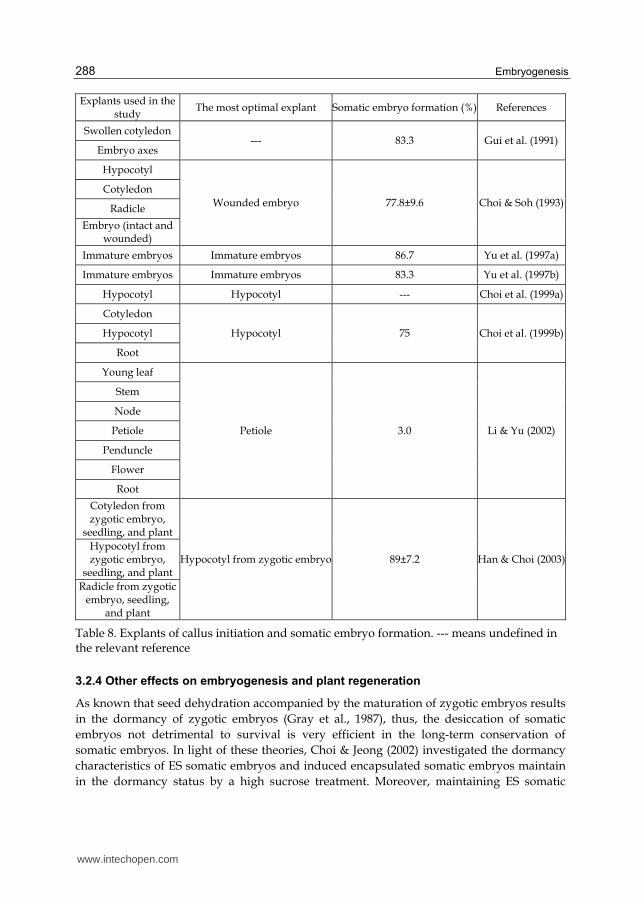
Explant type is another important factor affecting callus initiation efficiency. Generally, younger, more rapidly growing tissue is most effective. There have been many evidences indicating that plant regeneration potentials in many plant species have a direct correlation with the developmental stage of the explants tissue, such as in rice (Sahrawat & Chand, 2001), wheat (Wernicke & Milkovits, 1984), oat (Chen et al., 1995). For the monocotyledonon LC, mature seed has been reported to be the optimal explant for callus initiation according to the description of Part 2.1. For the dicotyledon ES, the investigations and attempts about appropriate explants have also been much studied (Table 8).
First, swollen cotyledon and embryo axes were used to induce callus, and they develop somatic embryos from the epidermal or subepidermal layer of the cotyledons or embryonic axes (Gui et al., 1991). The precedence of both explants, though, was not discussed, the somatic embryos rapidly developed into globular or heart-shaped structures, and germinated normally. Choi & Soh (1993) attempted various explants to initiate callus induction, including cotyledon, hypocotyl, radicle, and intact embryo and wounded embryo, of which wounded embryo produced the highest somatic embryo formation (77.8%). With 6 week-culture, wounded embryo-derived callus appeared torpedo shape and even cotyledonary embryo, while other calli derived from intact embryo, cotyledon, hypocotyl, or radicle appeared globular, heart shape, few torpedo shape, but no cotyledonary embryo. Later, immature embryo was firstly used as explant by Yu et al. (1997a, b), and produced high somatic embryo formation.
Embryogenic cells treated as artificial seeds for obtaining plants directly have been reported for several crops of agricultural interest (Kitto & Janick, 1985; Redenbaugh et al., 1986; Choi & Jeong, 2002). To achieve ES embryogenic cells as artificial seeds, Choi et al. (1999a, b) investigated different explants to attempt to initiate callus induction in a simple and efficient way. Among cotyledon, hypocotyl, and root of zygotic embryos, hypocotyl was considered as the most optimal explant of ES, and had the highest frequency of somatic embryo formation. Using hypocotyl-derived embryogenic cells from this system, mass production through large-scale tank culture were successfully obtained, with approximately 27-fold increment of fresh weight of somatic embryo after 4 week-culture (Choi et al., 2002). In addition, mass production through cell suspension culture was also done by comparing the somatic embryo formation from cotyledon, hypocotyl, and radicle-derived embryogenic cells (Han & Choi, 2003). In this study, embryogenic cells derived from hypocotyl of zygotic embryos showed the highest growth rate and somatic embryo formation of 89%. Based on the above suggestions, it is indicated that even mass production of plant cell through large-scale suspension culture has been successfully obtained, ES plantlets produced from this system could be more convenient to be used as a source of medicinal raw materials. However, due to direct sowing of artificial seeds in the field for practical use, low soil survival becomes a major problem (Redenbaugh et al., 1986). Herein, Choi & Jeong (2002) overcame the problem of low soil survival, and reported firstly encapsulated somatic embryos as ES artificial seeds to achieve all development status from artificial seeds to whole plants. Later, Jung et al. (2004) further improved this system with the addition of carbon sources to the encapsulation matric, and obtained that 96% of the encapsulated embryos converted to plantlets with well-elongated epicotyls.
www.intechopen.com
Embryogenesis
288
Explants used in the study
The most optimal explant Somatic embryo formation (%) References
Swollen cotyledon --- 83.3 Gui et al. (1991)
Embryo axes
Hypocotyl
Wounded embryo 77.8±9.6 Choi & Soh (1993) Cotyledon
Radicle
Embryo (intact and wounded)
Immature embryos Immature embryos 86.7 Yu et al. (1997a)
Immature embryos Immature embryos 83.3 Yu et al. (1997b)
Hypocotyl Hypocotyl --- Choi et al. (1999a)
Cotyledon
Hypocotyl 75 Choi et al. (1999b) Hypocotyl
Root
Young leaf
Petiole 3.0 Li & Yu (2002)
Stem
Node
Petiole
Penduncle
Flower
Root
Cotyledon from zygotic embryo,
seedling, and plant
Hypocotyl from zygotic embryo 89±7.2 Han & Choi (2003)Hypocotyl from zygotic embryo,
seedling, and plant Radicle from zygotic
embryo, seedling, and plant
Table 8. Explants of callus initiation and somatic embryo formation. --- means undefined in the relevant reference
3.2.4 Other effects on embryogenesis and plant regeneration
As known that seed dehydration accompanied by the maturation of zygotic embryos results in the dormancy of zygotic embryos (Gray et al., 1987), thus, the desiccation of somatic embryos not detrimental to survival is very efficient in the long-term conservation of somatic embryos. In light of these theories, Choi & Jeong (2002) investigated the dormancy characteristics of ES somatic embryos and induced encapsulated somatic embryos maintain in the dormancy status by a high sucrose treatment. Moreover, maintaining ES somatic
www.intechopen.com

Recent Advances of In Vitro Embryogenesis of Monocotyledon and Dicotyledon
289
embryos from cell suspension cultures under low temperature (4襖) was also considered to be able to achieve long-term animatingly conservation (Li et al., 2004). These treatments help a long-term conservation of artificial seeds and an enhanced resistance to dehydration of somatic embryos. You et al. (2005) carried out that removal of endosperm from seeds could markedly stimulate the growth of rudimentary zygotic embryos to induce more rapid germination of rudimentary zygotic embryos by in vitro culture of excised seeds. And in their later investigation (You et al., 2006), the roles of plasmolyzing pretreatment for zygotic embryos were evaluated on the induction of somatic embryos, suggesting that this pretreatment could result in sharply increased callose concentration in ES zygotic embryos, and callose accumulation could then stimulate the reprogramming of epidermal cells into embryogenically competent cells and finally induce somatic-embryo development from single cells. The further and detailed mechanism of enhanced somatic embryo formation through plasmolysis treatment was revealed that the expression level of callose synthase gene increased with response to 2,4-D, sucrose, and mannitol, and the callose played an important role in separating cell in epidermis from neighboring cells and consequently developing into embryogenic potential cells (Xilin et al., 2010).
3.3 Application for biochemical and biological events
Somatic embryogenesis has been studied as a model system for understanding the physiological, biochemical, and molecular biological events occurring during plant embryo development (Zimmerman, 1993). Among them, production of secondary metabolites through cell culture, particularly in medical plant, has long been used for commercial purposes (Roberts, 2007). To improve the culture conditions and then increase the production efficiency, many scientists have been investigated many factors affecting growth of culture materials and in vivo accumulation of active compounds. Ahn et al. (2003) investigated the effect of inorganic nitrogen sources such as KNO3 and NH4NO3 on cell growth and production of chlorogenic acid and eleutheroside E derivative. In another investigation by Ahn et al. (2007), the effect of NO3- and NH4+ on the adventitious root growth of ES and production of eleutheroside derivatives were investigated, and eleutheroside B (249 μg/g), E (788 μg/g), and E1 (43 μg/g) were increased at the highest levels by 40, 120, and 40 mM total nitrogen source, respectively. These results suggested that production of secondary metabolites through in vitro cultured cells could be manipulated by controlling the total concentration of nitrogen sources and the concentration ratio of NO3- and NH4+ in the culture medium.
Except of nitrogen sources, light is another important factor affecting growth and organogenesis, but a factor stressing plants to consequently regulate the secretion mechanism of secondary products (Shohael et al., 2006a). Higher H2O2 content, malondialdehyde content and lipoxygenase activities were observed in cultured embryos under red light compared dark grown embryos, as well as activities of some antioxidant enzymes such as catalase, superoxide dismutase, glutathione S transferase, and ascorbate peroxidase were also stimulated in red light irradiated embryos. Of course, the contents of eleutheroside E and E1 were synchronously accumulated 51% and 21% higher than control under red light irradiation. Jeong et al. (2009) compared the effects of red, blue, and far-red light by irradiation of light emitting diodes (LEDs) with white fluorescent lamp, on growth,
www.intechopen.com

Embryogenesis
290
morphogenesis and eleutheroside contents of in vitro cultured ES. The results indicated that in vitro cultured plantlets under the red/blue LEDs were taller than control, and those under blue LED showed greater leaf area, root length, and fresh weight than other light sources. Contents of eleuthroside B and E in plantlets were higher under blue LED, while content of eleuthroside E1 was the highest under fluorescent lamps.
Ahn et al. (2007, 2010) investigated the impacts of jasmonic acid (JA) on adventitious root culture of ES and eleutherosides accumulation, suggesting that JA inhibited the root growth but increased eleutherosides accumulation, as well as total phenolic contents and antioxidant activity. The highest levels of accumulation of eleutheroside B (359.9 μg/g), E (798.1 μg/g), and E1 (197 μg/g) were found under 40, 10, and 10 μM of methyl jasmonate addition, respectively.
Effects of temperature on secondary metabolite production such as eleutheroside B, E, E1, total phenolics, flavonoids, and chlorogenic acid and antioxidant enzyme activities were investigated by Shohael et al. (2006b), suggesting that culture at 24襖 caused the highest production efficiency of secondary metabolites, and either lower or higher temperature could cause severe oxidative stress to form a cellular damage. Based on above results, the production of secondary metabolites, one side, was considered as the consequent result of cultured cell metabolism, the other side, as the outcome stimulated by some stress treatments. Therefore, to control the balance between reactive oxygen species (ROS) formation derived by stress treatments and consumption correlated with an array of antioxidant enzymes and redox metabolites becomes required and important. Shohael et al. (2007) further examined the ascorbate-glutathione cycle enzymes and other enzymes metabolism during somatic embryogenesis of ES, and suggested that the alterations of the glutathione redox systems play a significant role in somatic embryo development.
Genetic improvement of another application of plant tissue culture, and a good approach to improve plant physiological traits and augment the drug-yielding capacity of medicinal plants (Tejavathi & Shailaja, 1999). To authors’ knowledge, only two transformation events through Agrobacterium-mediated transformation occurred in ES. Jo et al. (2005) transformed the human lactoferrin (hLf) gene into ES cells, and these transgenic ES cultured cells could produce hLf protein as cell growth increasing proportionally. As lactoferrin is an iron-binding glycoprotein with many biological roles, including the protection against microbial and virus infection and stimulation of the immune system, hLf transgenic ES plants could be used as a medicinal raw material for production of secondary metabolites. Another successful transformation event of ES was obtained in the report by Seo et al. (2005) that a squalene synthass-encoding gene derived from Panax ginseng (PgSS1) was successfully introduced into ES plants through Agrobacterium-mediated transformation. The transgenic plants showed up to 3-fold of squalene synthase enzyme activity higher than that of wild-type plants. Moreover, the introduced PgSS1 gene in transgenic plants enhanced the metabolisms of phytosterol and triterpenoides, with 2.0 ~ 2.5-fold increments of their levels. These results indicated that transgenic ES cultured cells would be biotechnologically useful for the commercial production of medicinal plant cell cultures.
4. Conclusions
All biotechnological approaches like genetic engineering, haploid induction, or somaclonal variation to improve traits strongly depend on an efficient in vitro plant
www.intechopen.com

Recent Advances of In Vitro Embryogenesis of Monocotyledon and Dicotyledon
291
regeneration system. Since LC as a monocotyledonous grass species and also a halophyte and ES as a dicotyledonous medicinal plant species, have increasingly great ecological and economic significant, this review would help efficiently improve traits through genetically modification.
5. Acknowledgment
This work was supported by Nutraceutical Bio Brain Korea 21 Project Group.
6. References
Ahn, J.K.; Lee, W.Y. & Park, E.J. (2010). Effect of methyl jasmonate on the root growth and the eleutheroside accumulation in the adventitious root culture of Eleutherococcus senticosus. Journal of Korean Forest Society, Vol.99, No.3, (June 2010), pp. 331-336, ISSN 0445-4650
Ahn, J.K.; Lee, W.Y. & Park, S.Y. (2003). Effect of nitrogen source on the cell growth and production of secondary metabolites in bioreactor cultures of Eleutherococcus senticosus. Korean Journal of Plant Biotechnology, Vol.30, No.3, pp. 301-305, ISSN 1598-6365
Ahn, J.K.; Park, Y.K.; Lee, W.Y. & Park, S.Y. (2007). Increment of eleutherosides and antioxidant activity in Eleutherococcus senticosus adventitious root by jasmonic acid. Journal of Korean Forest Society, Vol.96, No.5, pp. 539-542, ISSN 0445-4650
Anis, M. & Faisal, M. (2005). In Vitro regeneration and mass multiplication of Psoralea corylifolia–An endangered medicinal plant. Indian Journal of Biotechnology, Vol.4, No.2, (April 2005), pp. 261-264, ISSN 0972-5849
Chen, H.C.; Xu, G.J.; Loschke, D.C.; Tomaska, L. & Rolfe, B.G. (1995). Efficient callus formation and plant regeneration from leaves of oats (Avena sativa L.). Plant Cell Reports, Vol.14, No.6, pp. 393-397, doi:10.1007/BF00238604, ISSN 0721-7714
Choi, Y.E. & Jeong, J.H. (2002). Dormancy induction of somatic embryos of Siberian ginseng by high sucrose concentrations enhances the conservation of hydrated artificial seeds and dehydration resistance. Plant Cell Reports, Vol.20, No., pp. 1112-1116, ISSN 0721-7714. doi:10.1007/s00299-002-0455-y
Choi, Y.E. & Soh, W.Y. (1993). Structural aspects of somatic embryos derived from cultured zygotic embryos in Acanthopanax senticosus L. Korean Journal of Plant Tissue Culture, Vol.20, No.5, pp. 261-266, ISSN 1015-5880
Choi, Y.E.; Kim, J.W. & Yoon, E.S. (1999a). High frequency of plant production via somatic embryogenesis from callus or cell suspension cultures in Eleutherococcus senticosus. Annals of Botany, Vol.83, No.3, pp. 309-314, ISSN 0305-7364
Choi, Y.E.; Lee, K.S.; Kim, E.Y.; Kim, Y.S.; Han, J.Y.; Kim, H.S.; Jeong, J.H. & Ko, S.K. (2002). Mass production of Siberian ginseng plantlets through large-scale tank culture of somatic embryos. Plant Cell Reports, Vol.21, No.1, pp. 24-28, ISSN 0721-7714. doi:10.1007/s00299-002-0470-z
Choi, Y.E.; Yang, D.C. & Yoon, E.S. (1999b). Rapid propagation of Eleutherococcus senticosus via direct somatic embryogenesis from explants of seedlings. Plant Cell, Tissue and Organ Culture, Vol.58, No.2, pp. 93-97, ISSN 0167-6857
Cui, Q.H.; Zhang, Y.Z.; Piao, T.F.; Gu, D.F.; Zhang, W.Q.; Xu, Y.K.; Sun, Z.L. & Liu, H.X. (1990). Embryogenic callus and plant regeneration of Aneurolepidium chinensis (Trin.) Kitag. Journal of Jilin Agricultural University, Vol.12, pp. 1-5 ISSN 1000-5684
www.intechopen.com

Embryogenesis
292
Czerepanov, S.K. (2007). Vascular plants of Russia and adjacent states (the former USSR), Cambridge University Press, pp. 377-378, ISBN 0-521-45006-3, Cambridge, UK
Davydov, M. & Krikorian, A.D. (2000). Eleutherococcus senticosus (Rupr. and Maxim.) Maxim. (Araliaceae) as an adaptogen: a closer look. Journal of Ethnopharmacology, Vol.72, No.2000, pp. 345-393, ISSN 0378-8741
Fowler, M.W. (1983). Commercial applications and economic aspects of mass plant cell culture, In: Plant biotechnology, S.H. Mantell & H. Smith, (Ed.), 3-37, Cambridge University Press, Cambridge, UK
Furuya, T.; Yoshikawa, T.; Orihara, Y. & Oda, H. (1983). Saponin production in cell suspension cultures of Panax ginseng. Planta Medica, Vol.48, No.2, (June 1983), pp. 83-87, ISSN 0032-0943
Gaffney, B.T.; Hügel, H.M. & Rich, P.A. (2001). The effects of Eleutherococcus senticosus and Panax ginseng on steroidal hormone indices of stress and lymphocyte subset numbers in endurance athletes. Life Sciences, Vol.70, No.4, (December 2001), pp. 431-442, ISSN 0024-3205
Gao, T.S. (1982). Induction of callus and regeneration of plantlets from the rhizome explants of Aneurolepidium chinensis. Acta Botanica Sinica, Vol.24, pp. 182-185, ISSN 1672-6650
Gautheret, R.J. (1934). Culture du tissus cambial. Comptes Rendus Hebdomadaires des Séances de l’Académie des Sciences, Vol.198, pp. 2195-2196, ISSN 0001-4036
Gray, D.J.; Conger, B.V. & Songstad, D.D. (1987). Desiccated quiescent somatic embryos of orchardgrass for use as synthetic seeds. In Vitro Cellular & Developmental Biology, Vol.23, No.1, (January 1987), pp. 29-33, ISSN 0883-8364
Gui, Y.; Guo, Z.; Ke, S. & Skirvin, R.H. (1991). Somatic embryogenesis and plant regeneration in Acanthopanax senticosus. Plant Cell Reports, Vol.9, No.9, pp. 514-516, ISSN 0721-7714
Hahn, D.R.; Kim, C.J. & Kim, J.H. (1985). A study on chemical constituents of Acanthopanax koreanum Nakai and its pharmaco-biological activities. Yakhak Hoeji, Vol.29, pp. 357-361, ISSN 0377-9556
Han, J.S.; Kim, S.K.; Kim, S.W. & Kim, Y.J. (2001). Effects of shading treatments and harvesting methods on the growth of Eleutherococcus senticosus Maxim. Korean Journal of Medicinal Crop Science, Vol.9, No.1, pp. 1-7, ISSN 1225-9306
Han, J.Y. & Choi, Y.E. (2003). Mass production of Eleutherococcus senticosus plants through in vitro cell culture. Korean Journal of Plant Biotechnology, Vol.30, No.2, pp. 167-172, ISSN 1598-6365
Hanning, E. (1904). Zur Physiologie pflanzlicher Embryonen. I. Über die Kultur von cruciferen Embryonen ausserhalb des Embryosacks. Botanische Zeitung, Vol.62, pp. 45-80
Huang, Z.; Zhu, J.; Mu, X. & Lin, J. (2004). Pollen dispersion, pollen viability and pistil receptivity in Leymus chinensis. Annals of Botany, Vol.93, No.3, (January 2004), pp. 295-301, ISSN 0305-7364
Isoda, S. & Shoji, J. (1994). Studies on the cultivation of Eleutherococcus senticosus Maxim. II. On the germination and raising of seedling. Nature Medicine, Vol.48, pp. 75-81, ISSN 1078-8956
Jeong, J.H.; Kim, Y.S.; Moon, H.K.; Hwang, S.J. & Choi, Y.E. (2009). Effects of LED on growth, morphogenesis and eleutheroside contents of in vitro cultured plantlets of Eleutherococcus senticosus Maxim. Korean Journal of Medicinal Crop Science, Vol.17, No.1, pp. 39-45, ISSN 1225-9306
www.intechopen.com

Recent Advances of In Vitro Embryogenesis of Monocotyledon and Dicotyledon
293
Jia, S.X. (1987). Forage floras of China, Agricultural Press, pp. 19-35, ISBN 978-1-930723-59-7, Beijing, China
Jin, H.; Plaha, P.; Park, J.Y.; Hong, C.P.; Lee, I.S.; Yang, Z.H.; Jiang, G.B.; Kwak, S.S.; Liu, S.K.; Lee, J.S.; Kim, Y.A. & Lim, Y.P. (2006). Comparative EST profiles of leaf and root of Leymus chinensis, a xerophilous grass adapted to high pH sodic soil. Plant Science, Vol.170, No.6, (June 2006), pp. 1081-1086, ISSN 0168-9452
Jo, S.H.; Kwon, S.Y.; Kim, J.W.; Lee, K.T.; Kwak, S.S. & Lee, H.S. (2005). Transgenic Siberian ginseng cultured cells that produce high levels of human lactoferrin. Korean Journal of Plant Biotechnology, Vol.32, No.3, pp. 209-215, ISSN 1598-6365
Jung, S.J.; Yoon, E.S.; Jeong, J.H. & Choi, Y.E. (2004). Enhanced post-germinative growth of encapsulated somatic embryos of Siberian ginseng by carbohydrate addition to the encapsulation matrix. Plant Cell Reports, Vol.23, No.6, (November 2004), pp. 365-370, doi:10.1007/s00299-004-0821-z, ISSN 0721-7714
Kim, M.D.; Jin, H.; Park, E.J.; Kwon, S.Y.; Lee, H.S. & Kwak, S.S. (2005). Plant regeneration through somatic embryogenesis of Leymus chinensis Trin. Korean Journal Plant Biotechnology, Vol.32, No.1, pp. 51-55, ISSN 1598-6365 (in Korean with English abstract)
Kim, P.G.; Lee, K.Y.; Hur, S.D.; Kim, S.H. & Lee, E.J. (2003). Effects of shading treatment on photosynthetic activity of Acanthopanax senticosus. Korean Journal of Ecology, Vol.26, No.6, pp. 321-326, ISSN 1225-0317
Kitto, S. & Janick, J. (1985). Production of synthetic seeds by encapsulating asexual embryos of carrot. Journal of the American Society for Horticultural Science, Vol.110, pp. 277-282, ISSN 0003-1062
Kong, X.J.; Liang, Z.W.; Ma, H.Y. & Liu, M. (2008). Effect of variable cultivating temperature on frequency of embryogenic callus inducting from Leymus chinensis. Biotechnology, Vol.18, No.5, pp. 60-62, ISSN 1004-311X (in Chinese with English abstract)
Lee, W.T. (1979). Distribution of Acanthopanax plants in Korea. Korean Journal of Pharmacognosy, Vol.10, No.3, pp. 103-107, ISSN 0253-3073
Li, C.H. & Yu, C.Y. (2002). Effect of genotype and explant on somatic embryogenesis and acclimatization of Acanthopanax senticosus. Korean Journal of Medicinal Crop Science, Vol.10, No.3, pp. 217-221, ISSN 1225-9306
Li, C.H.; Lim, J.D.; Heo, K.; Kim, M.J.; Lee, C.O.; Lee, J.G.; Cui, X.S. & Yu, C.Y. (2004). Long-term cold storage and plant regeneration of suspension cultured somatic embryos of Eleutherococcus senticosus Maxim. Korean Journal of Medicinal Crop Science, Vol.12, No.6, (December 2004), pp. 494-499, ISSN 1225-9306
Li, C.H.; Lim, J.D.; Kim, M.J. & Yu, C.Y. (2003b). Effects of GA3 on seed germination and seedling survival rate of Acanthopanax senticosus Maxim. Korean Journal of Medicinal Crop Science, Vol.11, No.3, pp. 207-211, ISSN 1225-9306
Li, C.H.; Lim, J.D.; Kim, M.J.; Heo, K. & Yu, C.Y. (2003a). Dehisced seed germination and seedling growth affected by chilling period in Eleutherococcus senticosus Maxim. Korean Journal of Medicinal Crop Science, Vol.11, No.5, pp. 347-351, ISSN 1225-9306
Lim, S.H.; Jeong, H.N.; Kang, A.S. & Jeon, M.S. (2008). Influence of GA3 soak and seed dressing with Toros (Tolclofos methyl) wp. on the dehiscence of Eleutherococcus senticosus Maxim seeds. Korean Journal of Medicinal Crop Science, Vol.16, No.2, pp. 106-111, ISSN 1225-9306
Liu, G.S.; Li, X.F.; Su, M. & Qi, D.M. (2006). A protocol for enhancing plant regeneration frequency of Leymus chinensis and the appropriative tissue culture media, State
www.intechopen.com

Embryogenesis
294
Intellectual Property Office of the People’s Republic of China, Institute of Botany of the Chinese Academy of Science, No.200610078404
Liu, G.S.; Liu, J.S.; Qi, D.M.; Chu, C.C. & Li, H.J. (2004). Factors affecting plant regeneration from tissue cultures of Chinese leymus (Leymus chinensis). Plant Cell Tissue & Organ Culture, Vol.76, No.2, (February 2004) pp. 175-178,
doi:10.1023/B:TICU.0000007251.96785.18, ISSN 0167-6857 Liu, G.S.; Wang, E.H.; Liu, J.; Qi, D.M. & Li, F.F. (2002b). Plant regeneration of Leymus
chinensis via in vitro culture. Acta Agrestia Sinica, Vol.10, No.3, pp. 198-202, ISSN 1007-0435
Liu, J.; Zhu, Z.Q.; Liu, G.S.; Qi, D.M. & Li, F.F. (2002a). AFLP variation analysis on the germplasm resources of Leymus chiensis. Acta Botanica Sinica, Vol.44, No.7, pp. 845-851, ISSN 1672-9072
Loo, S.W. (1945). Cultivation of excised stem tips of Asparagus in vitro. American Journal of Botany, Vol.32, pp. 13-17, ISSN 0002-9122
Lu, X.Y.; Li, X.Z.; Zhou, H.X.; Xia, H.Z.; Li, Z.Y. & Wang, Y.M. (2009). Callus induction of Leymus chinensis. Beijing Agriculture, Vol.15, No., pp. 18-20, ISSN 1000-6966 (in Chinese with English abstract)
Ma, H.Y.; Liang, Z.W. & Chen, Y. (2005). Research progress on improving germination rate of Leymus chinensis. Chinese Journal of Grassland, Vol.27, No.4, pp. 64-68, ISSN 1673-5021 (in Chinese with English abstract)
Morel, G. & Wetmore, R.H. (1951). Tissue culture of monocotyledons. American Journal Botany, Vol.38, pp. 18-140, ISSN 0002-9122
Murashige, T. & Skoog, F. (1962). A revised medium for rapid growth and bio assays with tobacco tissue cultures. Physiologia Plantarum, Vol.15, No.3, pp. 473-497, ISSN 0031-9317, doi:10.1111/j.1399-3.54.1962.tb08052.x
Park, H.K.; Park, M.S.; Kim, T.S.; Choi, I.L.; Jang, Y.S. & Kim, G.S. (1994). Cutting propagation of Eleutherococcus senticosus MAXIM. Korean Journal of Medicinal Crop Science, Vol.2, No.2, pp. 133-139, ISSN 1225-9306
Park, H.K.; Park, M.S.; Kim, T.S.; Kim, S.; Choi, K.G. & Park, K.H. (1997). Characteristics of embryo growth and dehiscence during the after-ripening period in Eleutherococcus senticosus. Korean Journal of Crop Science, Vol.42, No.6, pp. 673-677, ISSN 0252-9777
Park, M.S.; Kim, Y.J.; Park, H.K.; Chang, Y.S. & Lee, J.H. (1995). Using air temperature and sunshine duration data to select seed production site for Eleutherococcus senticosus Max. Korean Journal of Crop Science, Vol.40, No.4, pp. 444-450, ISSN 0252-9777
Park, M.S.; Kim, Y.J.; Park, H.K.; Kim, S.; Kim, G.S. & Chang, Y.S. (1996). Habitat environment of Eleutherococcus senticosus Max. at Mt. Deokyu. Korean Journal of Crop Science, Vol.41, No.6, pp. 710-717, ISSN 0252-9777
Qu, T.B.; Wang, P.W.; Guan, S.Y. & Liu, L.Z. (2004). Establishment of regeneration system of Leymus chinensis culture. Acta Pratacultural Science, Vol.13, No.5, pp. 91-94, ISSN 1004-5759 (in Chinese with English abstract)
Rachmawati, D. & Anzai, H. (2006). Studies on callus induction, plant regeneration and transformation of Javanica rice cultivars. Plant Biotechnology, Vol.23, No.5, pp. 521-524, ISSN 1342-4580
Redenbaugh, K.; Paasch, B.D.; Nichol, J.W.; Kossler, M.E.; Viss, P.R. & Walker, K.A. (1986). Somatic seeds: encapsulation of asexual plant embryos. Nature Biotechnology, Vol.4, pp. 797-801, ISSN 1087-0156. doi:10.1038/nbt0986-797
www.intechopen.com

Recent Advances of In Vitro Embryogenesis of Monocotyledon and Dicotyledon
295
Roberts, S.C. (2007). Production and engineering of terpenoids in plant cell culture. Nature Chemical Biology, Vol.3, (June 2007), pp. 387-395, ISSN 1552-4469.
doi:10.1038/nchembio.2007.8 Sahoo, Y.& Chand, P.K. (1998). Micropropagation of Vitex negundo L., a woody aromatic
medicinal shrub, through high-frequency axillary shoot proliferation. Plant Cell Reports, Vol.18, No.3, (July 1998), pp. 301-307, doi:10.1007/s002990050576, ISSN 0721-7714
Sahrawat, A.K. & Chand S. (2001). High-frequency plant regeneration from coleoptiles tissue of indica rice (Oryza sativa L.). In Vitro Cellular & Developmental Biology. Plant, Vol.37, No.1, (January 2001), pp. 55-61, doi:10.1079/IVP2000114, ISSN 1054-5476
Seo, J.W.; Jeong, J.H.; Shin, C.G.; Lo, S.C.; Han, S.S.; Yu, K.W.; Harada, E.; Han, J.Y. & Choi, Y.E. (2005). Overexpression of squalene synthase in Eleutherococcus senticosus increases phytosterol and triterpene accumulation. Phytochemistry, Vol.66, No.8, (February 2005), pp. 869-877, ISSN 0031-9422. doi:10.1016/j.phytochem.2005.02.016
Shen, X.L.; Chen, J.J. & Kane, M.E. (2007). Indirect shoot organogenesis from leaves of Dieffenbachia cv. Camouflage. Plant Cell Tissue & Organ Culture, Vol.89, No.2-3, (February 2007), pp. 83-90, doi:10.1007/s11240-007-9214-7, ISSN 0167-6857
Shohael, A.M.; Ali, M.B.; Hahn, E.J. & Paek, K.Y. (2007). Glutathione metabolism and antioxidant responses during Eleutherococcus senticosus somatic embryo development in a bioreactor. Plant Cell Tissue & Organ Culture, Vol.89, No.2-3, (May 2007), pp. 121-129, doi:10.1007/s11240-9220-9, ISSN 0167-6857
Shohael, A.M.; Ali, M.B.; Yu, K.W.; Hahn, E.J. & Paek, K.Y. (2006b). Effect of temperature on secondary metabolites production and antioxidant enzyme activities in Eleutherococcus senticosus somatic embryos. Plant Cell Tissue & Organ Culture, Vol.85, No.2, pp. 219-228, ISSN 0167-6857. doi:10.1007/s11240-005-9075-x
Shohael, A.M.; Ali, M.B.; Yu, K.W.; Hahn, E.J.; Islam, R. & Park, K.Y. (2006a). Effect of light on oxidative stress, secondary metabolites and induction of antioxidant enzymes in Eleutherococcus senticosus somatic embryos in bioreactor. Process Biochemistry, Vol.41, pp. 1179-1185, ISSN 1359-5113. doi:10.1016/j.procbio.2005.12.015
Singh, N.D.; Sahoo, L.; Sarin, N.B. & Jaiwal, P.K. (2003). The effect of TDZ on organogenesis and somatic embryogenesis in pigeonpea (Cajanus cajan L. Millsp). Plant Science Vol.164, No.3, (March 2003), pp. 341-347, doi:10.1016/S0168-9452(02)00418-1, ISSN 0168-9452
Sun, Y.L. & Hong, S.K. (2009). Somatic embryogenesis and in vitro plant regeneration from various explants of the halophyte Leymus chinensis (Trin.). Journal of Plant Biotechnology, 36, No.3, pp. 236-243, ISSN 1598-6365
Sun, Y.L. & Hong, S.K. (2010a). Effects of plant growth regulators and L-glutamic acid on shoot organogenesis in the halophyte Leymus chinensis (Trin.). Plant Cell Tissue & Organ Culture, Vol.100, No.3, (December 2009), pp. 317-328, doi:10.1007/s11240-009-9653-4, ISSN 0167-6857
Sun, Y.L. & Hong, S.K. (2010b). Establishment of a novel plant regeneration system from suspension-derived callus in the halophytic Leymus chinensis (Trin.). Journal of Plant Biotechnology, Vol.27, No.2, (June 2010), pp. 228-235, ISSN 1598-6365
Tejavathi, D.H. & Shailaja, K.S. (1999). Regeneration of plants from the cultures of Bacopa monnieri (L.) Pennell. Phytomorphology, Vol.49, No.4, pp. 447-452, ISSN 0031-9449
Umeyama, A.; Shoji, N.; Takei, M.; Endo, K. & Arihara, S. (1992). Ciwujianosides D1 and C1: powerful inhibitors of histamine release induced by anti-immunoglobulin E from
www.intechopen.com

Embryogenesis
296
rat peritoneal mast cells. Journal of Pharmaceutical Sciences, Vol.81, No.7, (July 1992), pp. 661-662, ISSN 0022-3549
Vasil, V. & Hildebrandt, A.C. (1965). Differentiation of tobacco plants from single, isolated cells in microcultures. Science, Vol.150, No.3698, (November 1965), pp. 889-892, doi:10.1126/science.150.3698.889, ISSN 0036-8075
Wang, P.W.; Qu, T.B.; Guan, S.Y. & Qu, J. (2006). Selection of salt-tolerant mutants of Leymus chinensis. State Intellectual Property Office of the People’s Republic of China, Jilin Agriculture University, No.200610163263
Wang, Y.S.; Zhao, L.M.; Wang, H.; Wang, J.; Huang, D.M.; Hong, R.M.; Teng, X.H. & Miki, N. (2005). Molecular genetic variation in a clonal plant population of Leymus chinensis (Trin.) Tzvel. Journal of Integrative Plant Biology, Vol.47, No.9, pp. 1055-1064, ISSN 1672-9072
Wei, Q.; Hu, G.F.; Li, F.L. & Hu, B.Z. (2005). Study on callus inducement and plant regeneration in seeds of Aneurolepidium chinensis (Trin.) Kitag.. Journal of Northeast Agricultural University, Vol.36, No.1, pp. 41-44, ISSN 1005-9369 (in Chinese with English abstract)
Wernicke, W. & Milkovits, L. (1984). Developmental gradients in wheat leaves – Response of leaf segments in different genotypes cultured in vitro. Journal of Plant Physiology, Vol.115, pp. 49-58, ISSN 0176-1617
Xilin, H.; An, Y.; Xia, D.A. & You, X.L. (2010). Plasmolysis treatment enhances the expression of callose synthase gene in zygotic embryos of Eleutherococcus senticosus. Journal of Forestry Research, Vol.21, No.2, pp. 189-192, ISSN 1993-0607. doi:10.1007/s11676-010-0030-2
You, X.L.; Choi, Y.E. & Yi, J.S. (2005). Rapid in vitro germination of zygotic embryos via endosperm removal in Eleutherococcus senticosus. Journal of Plant Biotechnology, Vol.7, No.1, pp. 75-80, ISSN 1598-6365
You, X.L.; Yi, J.S. & Choi, Y.E. (2006). Cellular change and callose accumulation in zygotic embryos of Eleutherococcus senticosus caused by plasmolyzing pretreatment result in high frequency of single-cell-derived somatic embryogenesis. Protoplasma, Vol.227, pp. 105-112, doi:10.1007/s00709-006-0419-3, ISSN 1615-6102
Yu, C.Y.; Kim, J.K. & Ahn, S.D. (1997a). Callus formation and plant regeneration from immature embryos of Eleutherococcus senticosus. Korean Journal of Medicinal Crop Science, Vol.5, No.1, pp. 49-55, ISSN 1225-9306
Yu, C.Y.; Kim, S.H.; Lim, J.D.; Kim, M.J. & Chung, I.M. (2003). Intraspecific relationship analysis by DNA markers and in vitro cytotoxic and antioxidant activity in Eleutherococcus senticosus. Toxicology in Vitro, Vol.17, No.2, (April 2003), pp. 229-236, doi:10.1016/S0887-2333(03)00008-0, ISSN 0887-2333
Yu, C.Y.; Lim, J.D.; Seong, E.S. & Kim, J.K. (1997b). Effect of embryo maturity and medium on callus formation regeneration from immature embryos of Eleutherococcus senticosus. Korean Journal of Plant Resources, Vol.10, No.2, pp. 122-127, ISSN 1226-3591
Zhang, Y.; Li, X.F.; Liu, G.S. & Chen, Y.F. (2007). Regulation of callus status of Leymus chinensis. Journal of Northwest Sci-Tech University of Agriculture and Forestry (Natural Science Edition), Vol.35, No.5, pp. 111-114, ISSN 1671-9387 (in Chinese with English abstract)
Zimmerman, J.L. (1993). Somatic embryogenesis: a model for early development in higher plants. The Plant Cell, Vol.5, No.10, (October 1993), pp. 1411-1423, ISSN 1040-4651
www.intechopen.com
EmbryogenesisEdited by Dr. Ken-Ichi Sato
ISBN 978-953-51-0466-7Hard cover, 652 pagesPublisher InTechPublished online 20, April, 2012Published in print edition April, 2012
InTech EuropeUniversity Campus STeP Ri Slavka Krautzeka 83/A 51000 Rijeka, Croatia Phone: +385 (51) 770 447 Fax: +385 (51) 686 166www.intechopen.com
InTech ChinaUnit 405, Office Block, Hotel Equatorial Shanghai No.65, Yan An Road (West), Shanghai, 200040, China
Phone: +86-21-62489820 Fax: +86-21-62489821
The book "Embryogenesis" is a compilation of cutting edge views of current trends in modern developmentalbiology, focusing on gametogenesis, fertilization, early and/or late embryogenesis in animals, plants, and someother small organisms. Each of 27 chapters contributed from the authorships of world-wide 20 countriesprovides an introduction as well as an in-depth review to classical as well as contemporary problems thatchallenge to understand how living organisms are born, grow, and reproduce at the levels from molecule andcell to individual.
How to referenceIn order to correctly reference this scholarly work, feel free to copy and paste the following:
Sun Yan-Lin and Hong Soon-Kwan (2012). Recent Advances of In Vitro Embryogenesis of Monocotyledon andDicotyledon, Embryogenesis, Dr. Ken-Ichi Sato (Ed.), ISBN: 978-953-51-0466-7, InTech, Available from:http://www.intechopen.com/books/embryogenesis/recent-advances-of-in-vitro-embryogenesis-of-monocotyledon-and-dicotyledon

© 2012 The Author(s). Licensee IntechOpen. This is an open access articledistributed under the terms of the Creative Commons Attribution 3.0License, which permits unrestricted use, distribution, and reproduction inany medium, provided the original work is properly cited.