recent advances of curcumin in the prevention and...
TRANSCRIPT

Review ArticleRecent Advances of Curcumin in the Prevention andTreatment of Renal Fibrosis
Xuejiao Sun,1 Yi Liu,1 Cheng Li,1 XitingWang,1 Ruyuan Zhu,1 Chenyue Liu,2
Haixia Liu,1 Lili Wang,1 Rufeng Ma,1 Min Fu,3 Dongwei Zhang,4 and Yu Li1
1Preclinical Medicine School, Beijing University of Chinese Medicine, Beijing 100029, China2Chinese Material Medical School, Beijing University of Chinese Medicine, Beijing 100029, China3The Research Institute of McGill University Health Center, Montreal, QC, Canada H4A 3J14Diabetes Research Center, Beijing University of Chinese Medicine, Beijing 100029, China
Correspondence should be addressed to Dongwei Zhang; [email protected] and Yu Li; [email protected]
Received 7 January 2017; Accepted 1 February 2017; Published 4 May 2017
Academic Editor: Ekaterina A. Ivanova
Copyright © 2017 Xuejiao Sun et al. This is an open access article distributed under the Creative Commons Attribution License,which permits unrestricted use, distribution, and reproduction in any medium, provided the original work is properly cited.
Curcumin, a polyphenol derived from the turmeric, has received attention as a potential treatment for renal fibrosis primarilybecause it is a relatively safe and inexpensive compound that contributes to kidney health. Here, we review the literatures on theapplications of curcumin in resolving renal fibrosis in animal models and summarize the mechanisms of curcumin and its analogs(C66 and (1E,4E)-1,5-bis(2-bromophenyl) penta-1,4-dien-3-one(B06)) in preventing inflammatory molecules release and reducingthe deposition of extracellular matrix at the priming and activation stage of renal fibrosis in animal models by consulting PubMedand Cnki databases over the past 15 years. Curcumin exerts antifibrotic effect through reducing inflammation related factors (MCP-1, NF-𝜅B, TNF-𝛼, IL-1𝛽, COX-2, and cav-1) and inducing the expression of anti-inflammation factors (HO-1, M6PRBP1, andNEDD4) as well as targeting TGF-𝛽/Smads, MAPK/ERK, and PPAR-𝛾 pathways in animal models. As a food derived compound,curcumin is becoming a promising drug candidate for improving renal health.
1. Introduction
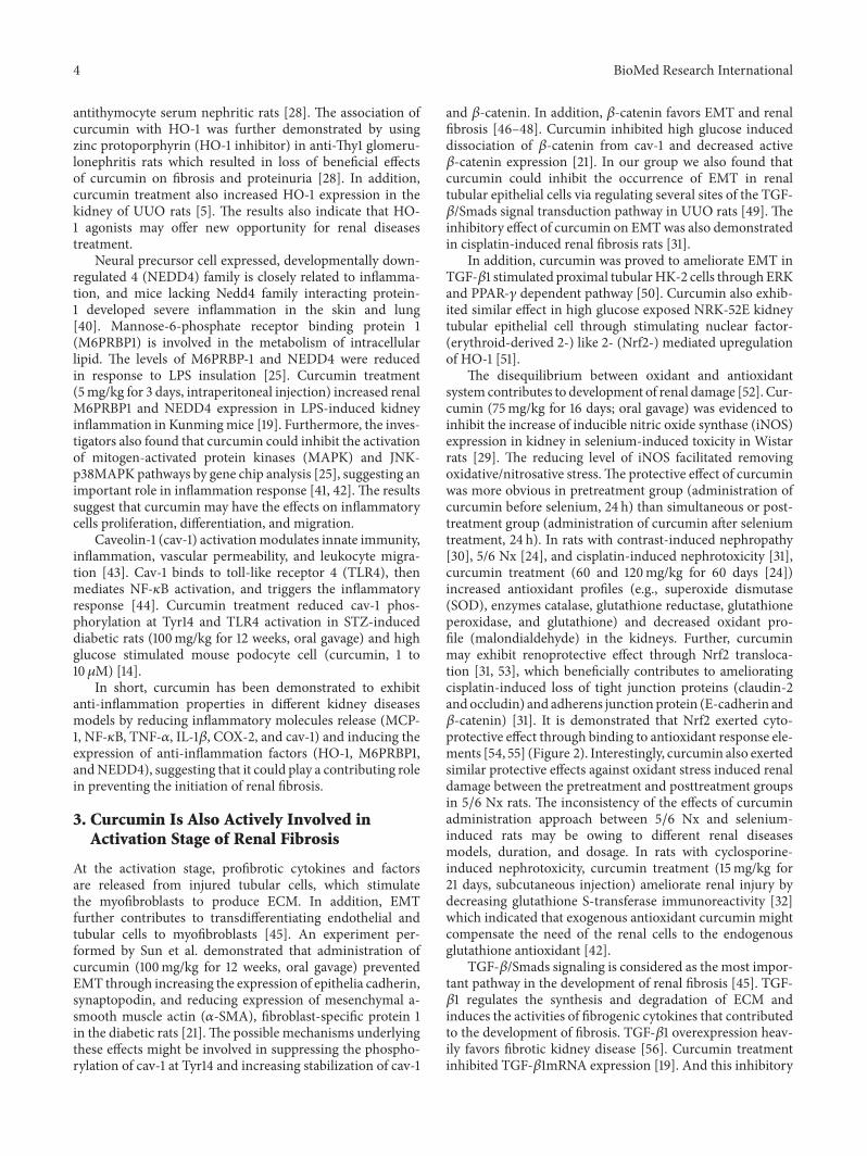
Curcumin, a polyphenol isolated from the Curcuma longaplant, is commonly known as turmeric in Asia (Figure 1)[1, 2]. As a traditional used herbal medicine and also afood spice in global cuisines, turmeric was reported to haveextensively clinical applications in various kinds of diseases,such as asthma, fibrosis, diabetes, and abdominal pain [2–4]. Curcumin is one of active ingredients in turmeric andhas been reported to attenuate the expression of apoptoticand chemokine genes in rat model of unilateral ureteralobstruction (UUO) in 2000 [5]. After that, a lot of researchersconducted various experiments to study the effects andmech-anisms regarding curcumin as a potential source in the pre-vention and treatment of renal fibrosis. In addition, there is notoxicity concern rising when curcumin is taken at the recom-mended doses, which increased the potential of therapeuticagent of this compound. A number of reviews concerningthe use of Chinese medicine for fibrosis have been recently
published [4, 6–8]. This review intends to summarize therecent studies on curcumin in delaying advance of renal fibro-sis through searching PubMed (https://www.pubmed.com/)and Cnki (https://www.cnki.com/) databases, which willprovide additional evidence and also highlight the futureresearch regarding curcumin in the management of kidneydiseases.
Renal fibrosis is the principal process underlying theprogression of chronic kidney disease (CKD) to end stagerenal disease (ESRD). With a high prevalence of morbidityand mortality, CKD brings great pressure to patients andincreases the burden on the society. In addition, currentlythere are no effective drugs to prevent the development ofthe ESRD. Characterized as glomerulosclerosis and tubularinterstitial fibrosis, renal fibrosis is considered as a dynamicand converging process that consists of four overlappingphases: priming, activation, execution, and progression [9].In the first stage, lasting inflammatory stimulation triggers
HindawiBioMed Research InternationalVolume 2017, Article ID 2418671, 9 pageshttps://doi.org/10.1155/2017/2418671

2 BioMed Research International
O O
HO OH
OCH3H3CO
(a)
O CF3CF3
(b)
Br Br
O
(c)
Figure 1: The chemical structure of curcumin (a), C66 (b), and B06 (c).
the activation of renal tubular epithelial cells and the infil-tration of inflammatory cells, including lymphocytes, mono-cytes/macrophages, dendritic cells, and mast cells [10]. Dur-ing the activation and execution stages, profibrotic cytokinesare released from injured tubular cells accompanying theactivation of matrix-producing cells. It is accepted that themyofibroblasts are the main source of extracellular matrix(ECM), which derived from renal interstitial fibroblasts, bonemarrow-derived fibrocytes, vascular pericytes, and endothe-lial and tubular cells by epithelial-to-mesenchymal transd-ifferentiation (EMT) [11]. The excessive deposition of ECMsuch as fibronectin and types I and III collagen contributes tothe development of renal fibrosis. In the final stage, the renalstructure and function gradually disappear with sustainingECMdeposition, which leads to the undesirable consequenceof fibrosis.
2. Curcumin Is Involved inthe Priming Stage of Renal Fibrosis
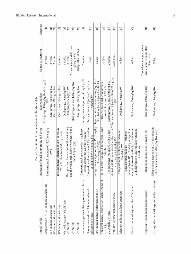
At the priming stage of renal fibrosis, inflammation initiatesa fibrotic process [12]. Sustaining inflammatory stimulustriggers the activation of renal tubular epithelial cells andthe infiltration of inflammatory cells, including lymphocytes,monocytes/macrophages, dendritic cells, andmast cells. Cur-cuminhas been demonstrated to regulatemultiple proinflam-matory molecules and reduce recruitment of inflammatorymacrophages [13–16] in various animal renal fibrosis models(Table 1).
Monocyte chemotactic protein-1 (MCP-1) is an importantmedium for monocyte/macrophage infiltration and a princi-ple cytokine that may induce tubulointerstitial fibrosis (TIF)[17]. Macrophages are attracted to the site of injury byMCP-1and its receptor CCR2. Blocking MCP-1/CCR2 pathway wasshown to prevent kidney fibrosis through reducing recruit-ment of M1 inflammatory macrophages [18]. In the UUOrats models [5], curcumin treatment (0.5mL of 30mg/mLfor 10 days, subcutaneous injection) significantly attenuatedMCP-1mRNA overexpression in the obstructed kidney com-pared with that of control [5]. Further, curcumin treatmentalso decreased MCP-1 level in the factor-H-deficient mice
(30mg/kg for 5 weeks, intraperitoneal injection) [19] andlipopolysaccharide (LPS) stimulated mice (5mg/kg for 3days, intraperitoneal injection) [20].
Under the stimulation of inflammatory factors (inter-leukin (IL), tumor necrosis factor 𝛼 (TNF-𝛼)), nuclearfactor-kappa B (NF-𝜅B) is activated and this activationfurther promotes the expression of transforming growthfactor 𝛽 (TGF-𝛽) 1, intercellular adhesion molecule-1, andother fibrogenesis factors [33–35]. It has been demonstratedthat curcumin treatment (100mg/kg/day for 8 weeks, oralgavage) suppressed NF-𝜅B activation, prevented inhibitorof NF-𝜅B (I𝜅Ba) degradation, and decreased intercellularadhesion molecule-1 protein expression in streptozotocin-(STZ-) induced diabetic nephropathy rats [15], which wasalso reflected in LPS-induced kidney inflammationmice [20].
Proinflammatory cytokines, including TNF-𝛼 and IL-1,are involved in the development of chronic kidney disor-ders, including glomerulonephritis [36]. In 5/6 nephrectomy(5/6 Nx) rats, the high levels of TNF-𝛼 and IL-1𝛽 furthertriggered the production of cytosolic phospholipase A2(cPLA2), calcium-independent intracellular PLA2 (iPLA2),and cyclooxygenase (COX) isoforms, whichmight contributeto inflammation [9]. Curcumin treatment (75mg/kg, oralgavage) for 10 weeks significantly reduced the levels of theabove-mentioned factors in 5/6 Nx rats [23]. In addition,administration of turmeric-based diet (5% w/w for 30 days)significantly decreased TNF-𝛼 mRNA expression in UUOrats [26]. C66 (0.2mg/kg for 6 weeks, oral gavage), a novelcurcumin derivative, has also been reported to reduce theproduction of TNF-𝛼, IL-1𝛽, COX-2, and NF-𝜅B in highglucose stimulated diabetic rats [37]. The above-mentionedresults suggest that curcumin and its analogs may havestrong ability of anti-inflammation in different renal rodent’sdiseases models.
Heme oxygenase-1 (HO-1) is the inducible isoform ofthe rate-limiting enzyme involved in the degradation ofheme. It is a cytoprotective molecule that could restorerenal function via resolving fibrosis factors [38, 39]. Inanti-Thy1 glomerulonephritis rats, curcumin treatment (10to 200mg/kg, intraperitoneal injection) dose-dependentlyinduced the expression of HO-1 in glomerular cells and
BioMed Research International 3
Table1:Th
eeffectso
fcurcumin
inrenalfi
brosismod
els.
Animalmod
elIndu
ctionof
renalinjury(rou
teanddo
se)
Routea
nddo
seCou
rseo
ftreatment
Reference
Streptozotocin-(ST
Z-)ind
uced
diabeticrats
Intraperito
nealinjectionof
STZ(60m
g/kg
BW)
Oralgavage,100m
g/kg
(bod
yweight)
BW12
weeks
[14]
STZ-indu
ceddiabeticrats
Oralgavage,100m
g/kg
BW12
weeks
[21]
STZ-indu
ceddiabeticrats
Oralgavage,150m
g/kg
BW12
weeks
[22]
STZ-indu
ceddiabeticrats.
Intraperito
nealinjectionof
STZ(55m
g/kg
BWin
20mM
citrateb
uffer,pH4.5)
Oralgavage,100m
g/kg
BW8weeks
[15]
5/6neph
rectom
y(5/6
Nx)
rats
Theu
pper
andlower
third
softhe
leftkidn
eyandthew
holerig
htkidn
eywereligated
and
excisedby
surgery
Oralgavage,75
mg/kg
BW8weeks
[16]
5/6Nxrats
Oralgavage,75
mg/kg
BW10
weeks
[23]
5/6Nxrats
Oralgavage,60
and120m
g/kg
BW7days
before
and60
days
after
5/6NX
[24]
5/6Nxrats
Oralgavage,120m
g/kg
BW30
days
after
5/6NX
[24]
Immun
e-complex-m
ediatedglom
erulon
ephritis
Intraperito
nealinjectionwith
4mgho
rse
spleen
apoferritin
daily
for5
weeks
Intraperito
nealinjection,
30mg/kg
BW5weeks
[19]
Lipo
polysaccharid
e(LP
S)indu
cedrenal
inflammationmice
Intraperito
nealinjectionof
LPSatthed
ose
of1m
g/kg
BWor
5mg/kg
BWIntraperito
nealinjection,
1mg/kg
or5m
g/kg
BW3days
[20]
LPSindu
cedkidn
eyinflammationmice
Intraperito
nealinjectionwith
LPS(5mg/kg
body
weight(BW
))Injectionwith
curcum
in(5mg/kg)for
3days
before
beinginjected
with
LPS
3days
[25]
Unilateralu
reteralobstructio
n(U
UO)m
odelof
renalinjury
Theleft
ureter
was
ligated
attwopo
intswith
3-0silk
Turm
eric-based
dietwith
turm
eric
powderinad
oseo
f5%w/w
30days
[26]
UUOmaleC
57mice
Theleft
ureter
was
ligated
with
4-0silk
Gastro
gavage,50and100m
g/kg
BW2weeks
[27]
Anti-Th
y1g
lomerulon
ephritism
odel
Injected
with
them
onoclonalantibod
yOX-
7atad
oseo
f2.2mg/kg
BWIntraperito
nealinjection,
10to
200m
g/kg
BWDays3
to5
[28]
Selenium
-indu
cedoxidatives
tressrats
Intraperito
nealinjectionwith
selenium
15𝜇M/kgBW
Oralgavage,75
mg/kg
BW16
days
[29]
Con
trast-ind
uced
neph
ropathy(C
IN)rats
10mg/kg
furosemideIM
+10mg/kg
indo
methacinIP
+10mL/kg
iomeprolIV
was
administered
onthe5
thdayfollo
wing
24hdehydrationun
derm
ildsevoflu
rane
anesthesia
Oralgavage,200m
g/kg
BW10
days
[30]
Cisplatin
(CIS)ind
uced
neph
rotoxicity
Intraperito
nealinjection,
5mg/kg
CIS
Oralgavage,200m
g/kg
BWTh
reed
oses
(30m
inbefore
and24
and48
hoursa
fter
CISinjection)
[31]
Cyclo
sporine-indu
cedneph
rotoxicitymaler
ats
Subcutaneous
injectionof
CsAdissolvedin
oliveo
ilin
adoseo
f20m
g/kg
BWdaily
Oralgavage,15mg/kg
BW21
days
[32]
4 BioMed Research International
antithymocyte serum nephritic rats [28]. The association ofcurcumin with HO-1 was further demonstrated by usingzinc protoporphyrin (HO-1 inhibitor) in anti-Thy1 glomeru-lonephritis rats which resulted in loss of beneficial effectsof curcumin on fibrosis and proteinuria [28]. In addition,curcumin treatment also increased HO-1 expression in thekidney of UUO rats [5]. The results also indicate that HO-1 agonists may offer new opportunity for renal diseasestreatment.
Neural precursor cell expressed, developmentally down-regulated 4 (NEDD4) family is closely related to inflamma-tion, and mice lacking Nedd4 family interacting protein-1 developed severe inflammation in the skin and lung[40]. Mannose-6-phosphate receptor binding protein 1(M6PRBP1) is involved in the metabolism of intracellularlipid. The levels of M6PRBP-1 and NEDD4 were reducedin response to LPS insulation [25]. Curcumin treatment(5mg/kg for 3 days, intraperitoneal injection) increased renalM6PRBP1 and NEDD4 expression in LPS-induced kidneyinflammation in Kunmingmice [19]. Furthermore, the inves-tigators also found that curcumin could inhibit the activationof mitogen-activated protein kinases (MAPK) and JNK-p38MAPK pathways by gene chip analysis [25], suggesting animportant role in inflammation response [41, 42]. The resultssuggest that curcumin may have the effects on inflammatorycells proliferation, differentiation, and migration.
Caveolin-1 (cav-1) activationmodulates innate immunity,inflammation, vascular permeability, and leukocyte migra-tion [43]. Cav-1 binds to toll-like receptor 4 (TLR4), thenmediates NF-𝜅B activation, and triggers the inflammatoryresponse [44]. Curcumin treatment reduced cav-1 phos-phorylation at Tyr14 and TLR4 activation in STZ-induceddiabetic rats (100mg/kg for 12 weeks, oral gavage) and highglucose stimulated mouse podocyte cell (curcumin, 1 to10 𝜇M) [14].
In short, curcumin has been demonstrated to exhibitanti-inflammation properties in different kidney diseasesmodels by reducing inflammatory molecules release (MCP-1, NF-𝜅B, TNF-𝛼, IL-1𝛽, COX-2, and cav-1) and inducing theexpression of anti-inflammation factors (HO-1, M6PRBP1,andNEDD4), suggesting that it could play a contributing rolein preventing the initiation of renal fibrosis.
3. Curcumin Is Also Actively Involved inActivation Stage of Renal Fibrosis
At the activation stage, profibrotic cytokines and factorsare released from injured tubular cells, which stimulatethe myofibroblasts to produce ECM. In addition, EMTfurther contributes to transdifferentiating endothelial andtubular cells to myofibroblasts [45]. An experiment per-formed by Sun et al. demonstrated that administration ofcurcumin (100mg/kg for 12 weeks, oral gavage) preventedEMT through increasing the expression of epithelia cadherin,synaptopodin, and reducing expression of mesenchymal a-smooth muscle actin (𝛼-SMA), fibroblast-specific protein 1in the diabetic rats [21].The possible mechanisms underlyingthese effects might be involved in suppressing the phospho-rylation of cav-1 at Tyr14 and increasing stabilization of cav-1
and 𝛽-catenin. In addition, 𝛽-catenin favors EMT and renalfibrosis [46–48]. Curcumin inhibited high glucose induceddissociation of 𝛽-catenin from cav-1 and decreased active𝛽-catenin expression [21]. In our group we also found thatcurcumin could inhibit the occurrence of EMT in renaltubular epithelial cells via regulating several sites of the TGF-𝛽/Smads signal transduction pathway in UUO rats [49]. Theinhibitory effect of curcumin on EMTwas also demonstratedin cisplatin-induced renal fibrosis rats [31].
In addition, curcumin was proved to ameliorate EMT inTGF-𝛽1 stimulated proximal tubularHK-2 cells through ERKand PPAR-𝛾 dependent pathway [50]. Curcumin also exhib-ited similar effect in high glucose exposed NRK-52E kidneytubular epithelial cell through stimulating nuclear factor-(erythroid-derived 2-) like 2- (Nrf2-) mediated upregulationof HO-1 [51].
The disequilibrium between oxidant and antioxidantsystem contributes to development of renal damage [52]. Cur-cumin (75mg/kg for 16 days; oral gavage) was evidenced toinhibit the increase of inducible nitric oxide synthase (iNOS)expression in kidney in selenium-induced toxicity in Wistarrats [29]. The reducing level of iNOS facilitated removingoxidative/nitrosative stress.The protective effect of curcuminwas more obvious in pretreatment group (administration ofcurcumin before selenium, 24 h) than simultaneous or post-treatment group (administration of curcumin after seleniumtreatment, 24 h). In rats with contrast-induced nephropathy[30], 5/6 Nx [24], and cisplatin-induced nephrotoxicity [31],curcumin treatment (60 and 120mg/kg for 60 days [24])increased antioxidant profiles (e.g., superoxide dismutase(SOD), enzymes catalase, glutathione reductase, glutathioneperoxidase, and glutathione) and decreased oxidant pro-file (malondialdehyde) in the kidneys. Further, curcuminmay exhibit renoprotective effect through Nrf2 transloca-tion [31, 53], which beneficially contributes to amelioratingcisplatin-induced loss of tight junction proteins (claudin-2andoccludin) and adherens junction protein (E-cadherin and𝛽-catenin) [31]. It is demonstrated that Nrf2 exerted cyto-protective effect through binding to antioxidant response ele-ments [54, 55] (Figure 2). Interestingly, curcumin also exertedsimilar protective effects against oxidant stress induced renaldamage between the pretreatment and posttreatment groupsin 5/6 Nx rats. The inconsistency of the effects of curcuminadministration approach between 5/6 Nx and selenium-induced rats may be owing to different renal diseasesmodels, duration, and dosage. In rats with cyclosporine-induced nephrotoxicity, curcumin treatment (15mg/kg for21 days, subcutaneous injection) ameliorate renal injury bydecreasing glutathione S-transferase immunoreactivity [32]which indicated that exogenous antioxidant curcumin mightcompensate the need of the renal cells to the endogenousglutathione antioxidant [42].
TGF-𝛽/Smads signaling is considered as the most impor-tant pathway in the development of renal fibrosis [45]. TGF-𝛽1 regulates the synthesis and degradation of ECM andinduces the activities of fibrogenic cytokines that contributedto the development of fibrosis. TGF-𝛽1 overexpression heav-ily favors fibrotic kidney disease [56]. Curcumin treatmentinhibited TGF-𝛽1mRNA expression [19]. And this inhibitory

BioMed Research International 5
Nrf2 Keap1
Nrf2 Keap1
Nrf2HO-1,SOD,etc.
Oxidativestress
Nucleus
Cytoplasm
Figure 2: Nrf2 signaling pathway. Nrf2, as a transcription factor,resides within cytoplasm binding to the actin-associated Keap1protein and is normally degraded. Upon oxidation stress, theassociation will be disrupted, resulting in the translocation of Nrf2to nuclei and then increased expression of cytoprotective enzymes(HO-1, SOD, etc.).
effect was mediated through reducing the phosphorylationof Smad2 and Smad3 [27]. In addition, pretreatment ofcurcumin resisted renal fibrosis by downregulating TGF-𝛽1 receptor II in TGF-𝛽1 stimulated NRK49F rat renalfibroblasts [57]. B06, one of the curcumin analogs, hasalso been proved to reduce the expression of collagen IVand fibronectin which further favored attenuating the accu-mulation of extracellular matrix and glomerular mesangialproliferation [58].
Furthermore, sphingosine 1-phosphate (S1P) activatesTGF-𝛽 and contributed the renal fibrosis process [59]. How-ever, the formation of S1P is catalyzed by sphingosine kinase1 (SphK1) [60]. Huang et al. found that curcumin treatment(150mg/kg for 12 weeks, oral gavage) significantly inhibitedexpression and activity of SphK1 and the production of S1Pin STZ-induced diabetic rats [22].
In addition, our group found that curcumin treatment(20𝜇M for 72 h) significantly decreased the expression ofcollagen I, 𝛼-SMA, and chemokine receptor 7 (CCR7), aswell as TGF-𝛽l secretion in human circulating fibrocytes [61].The inhibitory effect of curcumin on the differentiation andmigration of human circulating fibrocytes is likely throughregulating the CCR7/CCL21 signaling pathway, in particularby reducing CCR7 expression.
TheMAPK/ERK signaling pathway is also involved in thedevelopment of renal fibrosis [62, 63]. Pretreatment with cur-cumin blocked angiotensin II- (Ang-II-) induced profibroticresponses in renal tubular epithelial cells [64]. Ang-II exertedits fibrotic response and hypertension effect through TGF𝛽1-MAPK/ERKpathway [65] and renin-angiotensin system. Panet al. [37] further demonstrated that C66 prevented STZ-induced diabetic nephropathy through inhibition of MAPKmediated angiotensin converting enzyme (ACE) expression.Wang et al. [66] also found that the antifibrotic effect of C66was exhibited through inhibition of JNK phosphorylation
and p300/CBP-mediated histone acetylation. It is demon-strated that inhibition of histone deacetylase prevented renalinterstitial fibroblasts activation and renal tubular cell apop-tosis in a rat renal interstitial fibroblast line (NRK-49F) andin UUOmice models [67].
Emerging evidence suggests that peroxisomeproliferator-activated receptor-𝛾 (PPAR-𝛾) is implicated in cell cycle [68]and its agonists exert protective effect on glucose control,alleviating proteinuria and inhibiting tissue fibrosis [69]. InUUO mice, curcumin treatment (50mg/kg and 100mg/kgfor 14 days, oral gavage) increased PPAR-𝛾 expression anddecreased phosphorylated Smad 2/3 [27]. This was alsoreflected in TGF-𝛽1 stimulated proximal tubular epithelialcell HK-2 cells [50] and 5/6 Nx rats [16]. Since PPAR-𝛾is also associated with ACE [70, 71], it is also useful toprobe the effect of curcumin on the ACE expression in renaldiseases.
In summary, at activation stage of renal fibrosis, cur-cumin treatment inhibits EMT and rebuilds the oxidative-antioxidant balance. In addition, curcumin shows antifi-brogenic properties by regulating TGF-𝛽 expression andblocking MAPK/ERK and PPAR-𝛾 pathways.
4. Outlook and Conclusions
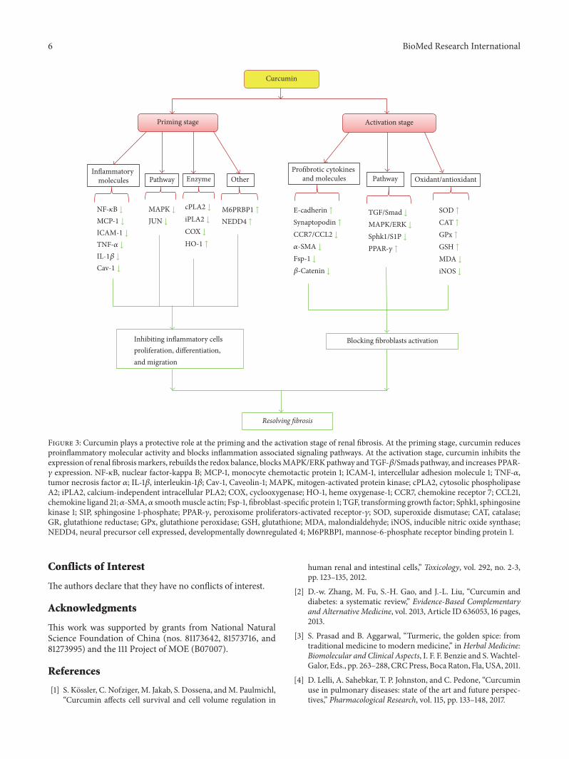
Curcumin has been demonstrated to be beneficially involvedin resolving renal fibrosis at priming and activation stagesthroughpreventing inflammation initiation, rebuilding redoxbalance, inhibiting EMT, and resolving ECM excess deposi-tion. These actions are mediated by reducing inflammationrelated factors (MCP-1, NF-𝜅B, TNF-𝛼, IL-1𝛽, COX-2, andcav-1) and inducing the expression of anti-inflammationfactors (HO-1, M6PRBP1, and NEDD4) as well as targetingTGF-𝛽/smads,MAPK/ERK, and PPAR-𝛾 pathways in animalmodels (Figure 3). In addition, no data supports the notionthat curcumin could restore renal injury during ESRD sofar. Meanwhile, cautions must be excised that pretreatmentand posttreatment may affect the effects of curcumin onrenal fibrosis. Prospective studies are also needed to fur-ther elucidate the effects of curcumin in the developmentof renal fibrosis with in-depth understanding of this dis-ease.
However, concerns are rising regarding the efficacy ofcurcumin in the management of renal fibrosis owing to itsinherent low bioavailability. In most of the studies, curcuminwas administrated by oral gavage. Further investigations areneeded to explore the real active ingredients of curcumin afteradministration. Fortunately, some of curcumin derivativeswith good bioavailability (such as C66 and B06) and newformulations of curcumin have been developed in recentlyyears. However, the efficacy and safety of these new analogsand formulations remain largely unexplored. Taken together,as a food derived compound with golden-yellow fluores-cence, curcumin may offer a new option in the treatmentof renal fibrosis and also provide a new druggable chemicalstructure for chemists in designing new antifibrosis drugcandidates.
6 BioMed Research International
Profibrotic cytokinesand molecules Pathway Oxidant/antioxidant
Blocking fibroblasts activationInhibiting inflammatory cellsproliferation, differentiation,and migration
Curcumin
Priming stage Activation stage
Inflammatorymolecules Pathway Enzyme Other
Resolving �brosis
NF-𝜅B ↓
MCP-1 ↓
ICAM-1 ↓
TNF-𝛼 ↓
IL-1𝛽 ↓
Cav-1 ↓
MAPK ↓
JUN ↓
cPLA2 ↓
iPLA2 ↓
COX ↓
HO-1 ↑
M6PRBP1 ↑
NEDD4 ↑
E-cadherin ↑
Synaptopodin ↑
CCR7/CCL2 ↓
𝛼-SMA ↓
Fsp-1 ↓
𝛽-Catenin ↓
TGF/Smad ↓
MAPK/ERK ↓
Sphk1/S1P ↓
PPAR-𝛾 ↑
SOD ↑
CAT ↑
GPx ↑
GSH ↑
MDA ↓
iNOS ↓
Figure 3: Curcumin plays a protective role at the priming and the activation stage of renal fibrosis. At the priming stage, curcumin reducesproinflammatory molecular activity and blocks inflammation associated signaling pathways. At the activation stage, curcumin inhibits theexpression of renal fibrosismarkers, rebuilds the redox balance, blocksMAPK/ERKpathway andTGF-𝛽/Smads pathway, and increases PPAR-𝛾 expression. NF-𝜅B, nuclear factor-kappa B; MCP-1, monocyte chemotactic protein 1; ICAM-1, intercellular adhesion molecule 1; TNF-𝛼,tumor necrosis factor 𝛼; IL-1𝛽, interleukin-1𝛽; Cav-1, Caveolin-1; MAPK, mitogen-activated protein kinase; cPLA2, cytosolic phospholipaseA2; iPLA2, calcium-independent intracellular PLA2; COX, cyclooxygenase; HO-1, heme oxygenase-1; CCR7, chemokine receptor 7; CCL21,chemokine ligand 21;𝛼-SMA,𝛼 smoothmuscle actin; Fsp-1, fibroblast-specific protein 1; TGF, transforming growth factor; Sphk1, sphingosinekinase 1; S1P, sphingosine 1-phosphate; PPAR-𝛾, peroxisome proliferators-activated receptor-𝛾; SOD, superoxide dismutase; CAT, catalase;GR, glutathione reductase; GPx, glutathione peroxidase; GSH, glutathione; MDA, malondialdehyde; iNOS, inducible nitric oxide synthase;NEDD4, neural precursor cell expressed, developmentally downregulated 4; M6PRBP1, mannose-6-phosphate receptor binding protein 1.
Conflicts of Interest
The authors declare that they have no conflicts of interest.
Acknowledgments
This work was supported by grants from National NaturalScience Foundation of China (nos. 81173642, 81573716, and81273995) and the 111 Project of MOE (B07007).
References
[1] S. Kossler, C. Nofziger, M. Jakab, S. Dossena, andM. Paulmichl,“Curcumin affects cell survival and cell volume regulation in
human renal and intestinal cells,” Toxicology, vol. 292, no. 2-3,pp. 123–135, 2012.
[2] D.-w. Zhang, M. Fu, S.-H. Gao, and J.-L. Liu, “Curcumin anddiabetes: a systematic review,” Evidence-Based Complementaryand Alternative Medicine, vol. 2013, Article ID 636053, 16 pages,2013.
[3] S. Prasad and B. Aggarwal, “Turmeric, the golden spice: fromtraditional medicine to modern medicine,” in Herbal Medicine:Biomolecular and Clinical Aspects, I. F. F. Benzie and S.Wachtel-Galor, Eds., pp. 263–288,CRCPress, BocaRaton, Fla,USA, 2011.
[4] D. Lelli, A. Sahebkar, T. P. Johnston, and C. Pedone, “Curcuminuse in pulmonary diseases: state of the art and future perspec-tives,” Pharmacological Research, vol. 115, pp. 133–148, 2017.

BioMed Research International 7
[5] E. A. Jones, A. Shahed, andD. A. Shoskes, “Modulation of apop-totic and inflammatory genes by bioflavonoids and angiotensinII inhibition in ureteral obstruction,” Urology, vol. 56, no. 2, pp.346–351, 2000.
[6] J. Trujillo, Y. I. Chirino, E. Molina-Jijon, A. C. Anderica-Romero, E. Tapia, and J. Pedraza-Chaverrı, “Renoprotectiveeffect of the antioxidant curcumin: recent findings,” RedoxBiology, vol. 1, no. 1, pp. 448–456, 2013.
[7] L.-C. Li and L.-D. Kan, “Traditional Chinese medicine forpulmonary fibrosis therapy: progress and future prospects,”Journal of Ethnopharmacology, vol. 198, pp. 45–63, 2017.
[8] J. Xia, L.-Q. He, and X. Su, “Interventional mechanisms ofherbs or herbal extracts on renal interstitial fibrosis,” Journal ofIntegrative Medicine, vol. 14, no. 3, pp. 165–173, 2016.
[9] Y. Liu, “Cellular and molecular mechanisms of renal fibrosis,”Nature Reviews Nephrology, vol. 7, no. 12, pp. 684–696, 2011.
[10] X.-M. Meng, D. J. Nikolic-Paterson, and H. Y. Lan, “Inflamma-tory processes in renal fibrosis,”Nature Reviews Nephrology, vol.10, no. 9, pp. 493–503, 2014.
[11] B. Conway and J. Hughes, “Cellular orchestrators of renalfibrosis,” QJM, vol. 105, no. 7, pp. 611–615, 2012.
[12] D. J. Nikolic-Paterson, S. Wang, and H. Y. Lan, “Macrophagespromote renal fibrosis through direct and indirect mecha-nisms,” Kidney International Supplements, vol. 4, no. 1, pp. 34–38, 2014.
[13] N. Kuwabara, S. Tamada, T. Iwai et al., “Attenuation of renalfibrosis by curcumin in rat obstructive nephropathy,” Urology,vol. 67, no. 2, pp. 440–446, 2006.
[14] L.-N. Sun, Z.-Y. Yang, S.-S. Lv, X.-C. Liu, G.-J. Guan, and G.Liu, “Curcumin prevents diabetic nephropathy against inflam-matory response via reversing caveolin-1 Tyr14 phosphorylationinfluenced TLR4 activation,” International Immunopharmacol-ogy, vol. 23, no. 1, pp. 236–246, 2014.
[15] V. Soetikno, F. R. Sari, P. T. Veeraveedu et al., “Curcumin ame-liorates macrophage infiltration by inhibiting NF-𝜅B activationand proinflammatory cytokines in streptozotocin induced-diabetic nephropathy,” Nutrition and Metabolism, vol. 8, no. 1,article 35, 2011.
[16] S. S. Ghosh,H.D.Massey, R. Krieg et al., “Curcumin amelioratesrenal failure in 5/6 nephrectomized rats: role of inflammation,”American Journal of Physiology—Renal Physiology, vol. 296, no.5, pp. F1146–F1157, 2009.
[17] T. Wada, K. Furuichi, N. Sakai et al., “Gene therapy via block-ade of monocyte chemoattractant protein-1 for renal fibrosis,”Journal of the American Society of Nephrology, vol. 15, no. 4, pp.940–948, 2004.
[18] K. Kitagawa, T. Wada, K. Furuichi et al., “Blockade of CCR2ameliorates progressive fibrosis in kidney,” The American Jour-nal of Pathology, vol. 165, no. 1, pp. 237–246, 2004.
[19] A. Jacob, L. Chaves, M. T. Eadon, A. Chang, R. J. Quigg, and J.J. Alexander, “Curcumin alleviates immune-complex-mediatedglomerulonephritis in factor-H-deficient mice,” Immunology,vol. 139, no. 3, pp. 328–337, 2013.
[20] F. Zhong, H. Chen, L. Han, Y. Jin, and W. Wang, “Cur-cumin attenuates lipopolysaccharide-induced renal inflamma-tion,” Biological and Pharmaceutical Bulletin, vol. 34, no. 2, pp.226–232, 2011.
[21] L.-N. Sun, Z.-X. Chen, X.-C. Liu, H.-Y. Liu, G.-J. Guan,and G. Liu, “Curcumin ameliorates epithelial-to-mesenchymaltransition of podocytes in vivo and in vitro via regulatingcaveolin-1,” Biomedicine and Pharmacotherapy, vol. 68, no. 8,pp. 1079–1088, 2014.
[22] J. Huang, K. Huang, T. Lan et al., “Curcumin ameliorates dia-betic nephropathy by inhibiting the activation of the SphK1-S1Psignaling pathway,” Molecular and Cellular Endocrinology, vol.365, no. 2, pp. 231–240, 2013.
[23] S. S. Ghosh, R. Krieg, H. D. Massey et al., “Curcumin andenalapril ameliorate renal failure by antagonizing inflammationin 5/6 nephrectomized rats: role of phospholipase and cyclooxy-genase,” American Journal of Physiology—Renal Physiology, vol.302, no. 4, pp. F439–F454, 2012.
[24] E. Tapia, Z. L. Zatarain-Barron, R. Hernandez-Pando et al.,“Curcumin reverses glomerular hemodynamic alterations andoxidant stress in 5/6 nephrectomized rats,” Phytomedicine, vol.20, no. 3-4, pp. 359–366, 2013.
[25] F. Zhong, H. Chen, Y. Jin, S. Guo,W.Wang, andN. Chen, “Anal-ysis of the gene expression profile of curcumin-treated kidneyon endotoxin-induced renal inflammation,” Inflammation, vol.36, no. 1, pp. 80–93, 2013.
[26] R. M. Hashem, H. M. Soliman, and S. F. Shaapan, “Turmeric-based diet can delay apoptosis without modulating NF-𝜅B inunilateral ureteral obstruction in rats,” Journal of Pharmacy andPharmacology, vol. 60, no. 1, pp. 83–89, 2008.
[27] X. Zhou, J. Zhang, C. Xu, andW.Wang, “Curcumin amelioratesrenal fibrosis by inhibiting local fibroblast proliferation andextracellular matrix deposition,” Journal of PharmacologicalSciences, vol. 126, no. 4, pp. 344–350, 2014.
[28] J. Gaedeke, N. A. Noble, and W. A. Border, “Curcumin blocksfibrosis in anti-Thy 1 glomerulonephritis through up-regulationof heme oxygenase 1,” Kidney International, vol. 68, no. 5, pp.2042–2049, 2005.
[29] R.Manikandan, R.Thiagarajan, S. Beulaja, G. Sudhandiran, andM. Arumugam, “Curcumin protects against hepatic and renalinjuries mediated by inducible nitric oxide synthase duringselenium-induced toxicity in Wistar rats,” Microscopy Researchand Technique, vol. 73, no. 6, pp. 631–637, 2010.
[30] M. Buyuklu, F. Mehmet Kandemir, M. Ozkaraca, T. Set, E.Murat Bakirci, and E. Topal, “Protective effect of curcuminagainst contrast induced nephropathy in rat kidney: what ishappening to oxidative stress, inflammation, autophagy andapoptosis?” European Review for Medical and PharmacologicalSciences, vol. 18, no. 4, pp. 461–470, 2014.
[31] J. Trujillo, E. Molina-Jijon, O. N. Medina-Campos et al.,“Curcumin prevents cisplatin-induced decrease in the tightand adherens junctions: relation to oxidative stress,” Food andFunction, vol. 7, no. 1, pp. 279–293, 2016.
[32] E. A. Abdel Fattah, H. E. Hashem, F. A. Ahmed, M. A. Ghallab,I. Varga, and S. Polak, “Prophylactic role of curcumin againstcyclosporine-induced nephrotoxicity: histological and immun-ohistological study,” General Physiology and Biophysics, vol. 29,no. 1, pp. 85–94, 2010.
[33] H.-Y. Xue, L. Yuan, Y.-J. Cao, Y.-P. Fan, X.-L. Chen, and X.-Z.Huang, “Resveratrol ameliorates renal injury in spontaneouslyhypertensive rats by inhibiting renal micro-inflammation,”Bioscience Reports, vol. 36, no. 3, Article ID e00339, 2016.
[34] T. D. Gilmore, “Introduction to NF-𝜅B: players, pathways, per-spectives,” Oncogene, vol. 25, no. 51, pp. 6680–6684, 2006.
[35] R. P. Nagarajan, F. Chen, W. Li et al., “Repression of trans-forming-growth-factor-𝛽-mediated transcription by nuclearfactor 𝜅B,” Biochemical Journal, vol. 348, no. 3, pp. 591–596,2000.
[36] F. Kayama, T. Yoshida, Y. Kodama, T. Matsui, J. M. Matheson,and M. I. Luster, “Pro-inflammatory cytokines and interleukin

8 BioMed Research International
6 in the renal response to bacterial endotoxin,” Cytokine, vol. 9,no. 9, pp. 688–695, 1997.
[37] Y. Pan, Y. Wang, L. Cai et al., “Inhibition of high glucose-induced inflammatory response andmacrophage infiltration bya novel curcumin derivative prevents renal injury in diabeticrats,” British Journal of Pharmacology, vol. 166, no. 3, pp. 1169–1182, 2012.
[38] M. Correa-Costa, P. Semedo, A. P. F. S. Monteiro et al.,“Induction of heme oxygenase-1 can halt and even reverse renaltubule-interstitial fibrosis,” PLoS ONE, vol. 5, no. 12, Article IDe14298, 2010.
[39] X. Chen, S.-Y. Wei, J.-S. Li et al., “Overexpression of hemeoxygenase-1 prevents renal interstitial inflammation and fibro-sis induced by unilateral ureter obstruction,” PLoS ONE, vol. 11,no. 1, Article ID e0147084, 2016.
[40] P. M. Oliver, X. Cao, G. S. Worthen et al., “Ndfip1 proteinpromotes the function of itch ubiquitin ligase to prevent Tcell activation and T helper 2 cell-mediated inflammation,”Immunity, vol. 25, no. 6, pp. 929–940, 2006.
[41] X. Che, Q. Wang, Y. Xie et al., “Astragaloside IV suppressestransforming growth factor-𝛽1 induced fibrosis of culturedmouse renal fibroblasts via inhibition of the MAPK and NF-𝜅B signaling pathways,” Biochemical and Biophysical ResearchCommunications, vol. 464, no. 4, pp. 1260–1266, 2015.
[42] W. Wang, P.-H. Zhou, C.-G. Xu, X.-J. Zhou, W. Hu, and J.Zhang, “Baicalein attenuates renal fibrosis by inhibiting inflam-mation via down-regulating NF-𝜅B and MAPK signal path-ways,” Journal ofMolecularHistology, vol. 46, no. 3, pp. 283–290,2015.
[43] S. Akira, K. Takeda, and T. Kaisho, “Toll-like receptors: crit-ical proteins linking innate and acquired immunity,” NatureImmunology, vol. 2, no. 8, pp. 675–680, 2001.
[44] G.H.Tesch, “Macrophages anddiabetic nephropathy,” Seminarsin Nephrology, vol. 30, no. 3, pp. 290–301, 2010.
[45] Y. B. Y. Sun, X. Qu, G. Caruana, and J. Li, “The origin of renalfibroblasts/myofibroblasts and the signals that trigger fibrosis,”Differentiation, vol. 92, no. 3, pp. 102–107, 2016.
[46] S. Hao,W.He, Y. Li et al., “Targeted inhibition of𝛽-catenin/CBPsignaling ameliorates renal interstitial fibrosis,” Journal of theAmerican Society of Nephrology, vol. 22, no. 9, pp. 1642–1653,2011.
[47] J. Heuberger and W. Birchmeier, “Interplay of cadherin-mediated cell adhesion and canonical Wnt signaling,” ColdSpring Harbor Perspectives in Biology, vol. 2, no. 2, Article IDa002915, 2010.
[48] F. Galbiati, D. Volonte, A. M. C. Brown et al., “Caveolin-1expression inhibits Wnt/𝛽-catenin/Lef-1 signaling by recruting𝛽-catenin to caveolae membrane domains,” Journal of BiologicalChemistry, vol. 275, no. 30, pp. 23368–23377, 2000.
[49] Y. Li, Z.-Q. Chen, and Y.-D. Li, “Effects of curcumin on theepithelial mesenchymal transition and TGF-beta/Smads signal-ing pathway in unilateral ureteral obstruction rats,” ZhongguoZhong Xi Yi Jie He Za Zhi, vol. 31, no. 9, pp. 1224–1228, 2011.
[50] R. Li, Y. Wang, Y. Liu et al., “Curcumin inhibits transforminggrowth factor-𝛽1-induced EMT via PPAR𝛾 pathway, not smadpathway in renal tubular epithelial cells,” PLoS ONE, vol. 8, no.3, Article ID e58848, 2013.
[51] X. Zhang, D. Liang, L. Guo et al., “Curcumin protects renaltubular epithelial cells from high glucose-induced epithelial-to-mesenchymal transition through Nrf2-mediated upregulationof heme oxygenase-1,”Molecular Medicine Reports, vol. 12, no. 1,pp. 1347–1355, 2015.
[52] Z. L. Li, L. Mo, G. Le, and Y. Shi, “Oxidized casein impairsantioxidant defense system and induces hepatic and renal injuryinmice,” Food and Chemical Toxicology, vol. 64, pp. 86–93, 2014.
[53] E. Tapia, V. Soto, K. M. Ortiz-Vega et al., “Curcumin inducesNrf2 nuclear translocation and prevents glomerular hyper-tension, hyperfiltration, oxidant stress, and the decrease inantioxidant enzymes in 5/6 nephrectomized rats,” OxidativeMedicine and Cellular Longevity, vol. 2012, Article ID 269039,2012.
[54] S. Boddupalli, J. R. Mein, S. Lakkanna, and D. R. James, “Induc-tion of phase 2 antioxidant enzymes by broccoli sulforaphane:perspectives in maintaining the antioxidant activity of vitaminsA, C, and E,” Frontiers in Genetics, vol. 3, article 7, 2012.
[55] T. Nguyen, P. Nioi, and C. B. Pickett, “The Nrf2-antioxidantresponse element signaling pathway and its activation byoxidative stress,” Journal of Biological Chemistry, vol. 284, no.20, pp. 13291–13295, 2009.
[56] S.-Y. Lee, S. I. Kim, and M. E. Choi, “Therapeutic targets fortreating fibrotic kidney diseases,” Translational Research, vol.165, no. 4, pp. 512–530, 2015.
[57] J. Gaedeke, N. A. Noble, and W. A. Border, “Curcumin blocksmultiple sites of the TGF-𝛽 signaling cascade in renal cells,”Kidney International, vol. 66, no. 1, pp. 112–120, 2004.
[58] C.-C. Zeng, X. Liu, W.-W. Liu et al., “Protective effect ofcurcumin derivative B06 on kidney of type 2 diabetic rats,”Chinese Journal of Applied Physiology, vol. 31, no. 1, pp. 38–42,2015.
[59] C. Xin, S. Ren, B. Kleuser et al., “Sphingosine 1-phosphate cross-activates the Smad signaling cascade and mimics transforminggrowth factor-𝛽-induced cell responses,”The Journal of Biologi-cal Chemistry, vol. 279, no. 34, pp. 35255–35262, 2004.
[60] B. Ogretmen and Y. A. Hannun, “Biologically active sphin-golipids in cancer pathogenesis and treatment,” Nature ReviewsCancer, vol. 4, no. 8, pp. 604–616, 2004.
[61] X.-Y. Fu, D.-W. Zhang, Y.-D. Li et al., “Curcumin treatmentsuppresses CCR7 expression and the differentiation and migra-tion of human circulating fibrocytes,” Cellular Physiology andBiochemistry, vol. 35, no. 2, pp. 489–498, 2015.
[62] T. Xu, N.-S. Wang, L.-L. Fu, C.-Y. Ye, S.-Q. Yu, and C.-L.Mei, “Celecoxib inhibits growth of human autosomal domi-nant polycystic kidney cyst-lining epithelial cells through theVEGF/Raf/MAPK/ERK signaling pathway,” Molecular BiologyReports, vol. 39, no. 7, pp. 7743–7753, 2012.
[63] S. Sun, X. Ning, Y. Zhai et al., “Egr-1 mediates chronic hypoxia-induced renal interstitial fibrosis via the PKC/ERK pathway,”American Journal of Nephrology, vol. 39, no. 5, pp. 436–448,2014.
[64] J. Ni, Y. Shen, Z. Wang et al., “P300-dependent STAT3 acety-lation is necessary for angiotensin II-induced pro-fibroticresponses in renal tubular epithelial cells,” Acta PharmacologicaSinica, vol. 35, no. 9, pp. 1157–1166, 2014.
[65] Q. Li, X. Liu, and J. Wei, “Ageing related periostin expressionincrease from cardiac fibroblasts promotes cardiomyocytessenescent,” Biochemical and Biophysical Research Communica-tions, vol. 452, no. 3, pp. 497–502, 2014.
[66] Y. Wang, Y. Wang, M. Luo et al., “Novel curcumin analogC66 prevents diabetic nephropathy via JNK pathway withthe involvement of p300/CBP-mediated histone acetylation,”Biochimica et Biophysica Acta—Molecular Basis of Disease, vol.1852, no. 1, pp. 34–46, 2015.
[67] M. Pang, J. Kothapally, H. Mao et al., “Inhibition of histonedeacetylase activity attenuates renal fibroblast activation and
BioMed Research International 9
interstitial fibrosis in obstructive nephropathy,” American Jour-nal of Physiology—Renal Physiology, vol. 297, no. 4, pp. F996–F1005, 2009.
[68] Y. Okunuki, Y. Usui, H. Nakagawa et al., “Peroxisome prolif-erator-activated receptor-𝛾 agonist pioglitazone suppressesexperimental autoimmune uveitis,” Experimental Eye Research,vol. 116, pp. 291–297, 2013.
[69] H. Kusunoki, Y. Taniyama, H. Rakugi, and R. Morishita, “Car-diac and renal protective effects of irbesartan via peroxisomeproliferator-activated receptor𝛾-hepatocyte growth factor path-way independent of angiotensin II Type 1a receptor blockadein mouse model of salt-sensitive hypertension,” Journal of theAmerican Heart Association, vol. 2, no. 2, Article ID e000103,2013.
[70] E. L. Santos, K. de Picoli Souza, E. D. da Silva et al., “Long termtreatment with ACE inhibitor enalapril decreases body weightgain and increases life span in rats,” Biochemical Pharmacology,vol. 78, no. 8, pp. 951–958, 2009.
[71] S. Zhao, Y. Mugabo, J. Iglesias et al., “𝛼/𝛽-Hydrolase domain-6-accessible monoacylglycerol controls glucose-stimulatedinsulin secretion,” Cell Metabolism, vol. 19, no. 6, pp. 993–1007,2014.

Submit your manuscripts athttps://www.hindawi.com
PainResearch and TreatmentHindawi Publishing Corporationhttp://www.hindawi.com Volume 2014
The Scientific World JournalHindawi Publishing Corporation http://www.hindawi.com Volume 2014
Hindawi Publishing Corporationhttp://www.hindawi.com
Volume 2014
ToxinsJournal of
VaccinesJournal of
Hindawi Publishing Corporation http://www.hindawi.com Volume 2014
Hindawi Publishing Corporationhttp://www.hindawi.com Volume 2014
AntibioticsInternational Journal of
ToxicologyJournal of
Hindawi Publishing Corporationhttp://www.hindawi.com Volume 2014
StrokeResearch and TreatmentHindawi Publishing Corporationhttp://www.hindawi.com Volume 2014
Drug DeliveryJournal of
Hindawi Publishing Corporationhttp://www.hindawi.com Volume 2014
Hindawi Publishing Corporationhttp://www.hindawi.com Volume 2014
Advances in Pharmacological Sciences
Tropical MedicineJournal of
Hindawi Publishing Corporationhttp://www.hindawi.com Volume 2014
Medicinal ChemistryInternational Journal of
Hindawi Publishing Corporationhttp://www.hindawi.com Volume 2014
AddictionJournal of
Hindawi Publishing Corporationhttp://www.hindawi.com Volume 2014
Hindawi Publishing Corporationhttp://www.hindawi.com Volume 2014
BioMed Research International
Emergency Medicine InternationalHindawi Publishing Corporationhttp://www.hindawi.com Volume 2014
Hindawi Publishing Corporationhttp://www.hindawi.com Volume 2014
Autoimmune Diseases
Hindawi Publishing Corporationhttp://www.hindawi.com Volume 2014
Anesthesiology Research and Practice
ScientificaHindawi Publishing Corporationhttp://www.hindawi.com Volume 2014
Journal of
Hindawi Publishing Corporationhttp://www.hindawi.com Volume 2014
Pharmaceutics
Hindawi Publishing Corporationhttp://www.hindawi.com Volume 2014
MEDIATORSINFLAMMATION
of