recent advances in biosynthesis of bioactive compounds in
TRANSCRIPT
Review Life & Medical Sciences
Recent advances in biosynthesis of bioactive compoundsin traditional Chinese medicinal plants
Lei Yang • Changqing Yang • Chenyi Li •
Qing Zhao • Ling Liu • Xin Fang • Xiao-Ya Chen
Received: 24 August 2015 / Accepted: 15 October 2015 / Published online: 2 November 2015
� Science China Press and Springer-Verlag Berlin Heidelberg 2015. This article is published with open access at Springerlink.com
Abstract Plants synthesize and accumulate large amount
of specialized (or secondary) metabolites also known as
natural products, which provide a rich source for modern
pharmacy. In China, plants have been used in traditional
medicine for thousands of years. Recent development of
molecular biology, genomics and functional genomics as
well as high-throughput analytical chemical technologies
has greatly promoted the research on medicinal plants. In
this article, we review recent advances in the elucidation of
biosynthesis of specialized metabolites in medicinal plants,
including phenylpropanoids, terpenoids and alkaloids. Th-
ese natural products may share a common upstream path-
way to form a limited numbers of common precursors, but
are characteristic in distinct modifications leading to highly
variable structures. Although this review is focused on
traditional Chinese medicine, other plants with a great
medicinal interest or potential are also discussed. Under-
standing of their biosynthesis processes is critical for pro-
ducing these highly value molecules at large scale and low
cost in microbes and will benefit to not only human health
but also plant resource conservation.
Keywords Medicinal plant � Biosynthesis �Phenylpropanoid � Terpenoid � Alkaloid
1 Introduction
China is rich in plant resources. Of the *300,000 species
of higher plants on the earth, around 10 % can be found in
China. As in many other countries, people in China have
used plants for treatment of diseases for thousands of years.
Compendium of Materia Medica has been held in high
esteem since it was first published in 1593, and this ancient
encyclopedia of traditional Chinese medicine (TCM)
described more than 1,000 species of plants. Plants produce
a wealth of specialized (or secondary) metabolites also
known as natural products, which are small molecular
weight compounds with enormous structural diversity and
show various biological activities. It is estimated that there
are approximately 200,000 secondary metabolites in plant
kingdom [1], which, based on biosynthetic origins, can be
classified into three major categories: phenylpropanoids,
terpenoids and alkaloids, plus a few other less abundant
groups. The usage records of China’s ancient medical
books, such as Sheng Nong’s Herbal Classic, Huang Di’s
Canon of Medicine and Compendium of Materia Medica,
already recognized that plant extracts contain active prin-
ciples in treating illness and classified them into assump-
tive, intuitive or largely philosophic categories, such as
cold, neutral or hot, toxic or nourishing. Over the past
century, hunting the active ingredients has led to important
findings, such as artemisinin for malaria, huperzine A for
Alzheimer’s disease, ephedrine for cold and camptothecin
SPECIAL TOPIC: Advances in Artemisinin Study
L. Yang � L. Liu � X.-Y. Chen (&)
Plant Science Research Center, Shanghai Chenshan Botanical
Garden, Shanghai Key Laboratory of Plant Functional Genomics
and Resources, Shanghai 201602, China
e-mail: [email protected]
C. Yang � C. Li � Q. Zhao � X. Fang � X.-Y. ChenNational Key Laboratory of Plant Molecular Genetics and
National Center for Plant Gene Research, Institute of Plant
Physiology and Ecology, Shanghai Institutes for Biological
Sciences, Chinese Academy of Sciences, Shanghai 200032,
China
C. Li
University of Chinese Academy of Sciences, Beijing 100049,
China
123
Sci. Bull. (2016) 61(1):3–17 www.scibull.com
DOI 10.1007/s11434-015-0929-2 www.springer.com/scp

for cancer, which were isolated from Artemisia annua,
Huperzia serrata, Ephedra sinica, Camptotheca acumi-
nate, respectively [2]. Very recently, tetrandrine, an alka-
loid isolated from the TCM plant Stephania tetrandra
previously used for reducing blood pressure, were reported
to have the therapeutic efficacy against Ebola [3], and
celastrol, a triterpene extracted from Tripterygium Wil-
fordi, has the potential as an anti-obesity agent [4]. These
findings strongly support that TCMs are the reliable source
for new therapies in treatment of lethally epidemic disease
and long unsolved disease.
However, multi-classes of natural products are generated
by each plant species. In addition, geographic distributions,
growth conditions and harvesting seasons could significantly
affect chemical compositions of the plant. Whereas one
component may act as the active ingredient, the effects of a
mixture of many ingredients are often uncertain and this has
caused increasing concerns [5]; thus, the traditional practice
of herbology has to face the challenges from modern medi-
cine and the manufactures’ requirement.
While plant natural products continue to be a prime source
for drug discovery and development, supply of these com-
pounds is often curtailed due to limitation of natural
resources and/or low contents in plant. The biotechnological
platforms, such as metabolic engineering of effective plant
and microbial production, are urgently needed to ensure that
the supply of bioactive natural products is sustainable and
environmentally friendly, rather than at the expense of
resource exhaustion [6–9]. A prerequisite to these solutions
is the understanding of the biosynthetic pathways of these
specialized metabolites, in particular the cloning and iden-
tification of enzymes and the regulatory factors.
In the past two decades, the rapid development in
genomics and high-throughput technologies of chemical
analysis, in combination with molecular biology tools, has
accelerated the research of medicinal plants. In this review,
we summarize the recent advances in the elucidation of
biosynthetic pathways of secondary metabolites in, not
exclusively, TCM plants. Although alkaloids are probably
the most important resource for drug discovery and
biosynthesis of these amino acid-derived compounds has
been investigated intensively, there are, surprisingly to
some extent, relatively few studies of alkaloids from TCM
plant; thus, this review is emphasized on phenylpropanoids
and terpenoids. In addition to enzymes, transcription fac-
tors characterized from medicinal plants are also discussed.
2 Phenylpropanoids
Phenylpropanoids, commonly found in plants, are derived
from the six-carbon aromatic phenyl group and the three-
carbon propene tail [10], and form a large group of special-
ized metabolites including monolignols, lignans, flavonoids,
phenolic acids and stilbenes [11]. They serve as basic com-
ponents of a number of structural polymers, as well as floral
pigments, scent compounds or signaling molecules to
mediate bio-interactions, phytoalexins against herbivores
and pathogens, and protective components against ultravi-
olet light radiation and other abiotic stresses [12]. In many
TCM plants, such as the plants of Lamiaceae, Fabaceae
(Leguminasae) and Asteraceae, phenylpropanoids are also
the bioactive principles (Table 1), which have been shown to
act as anti-oxidants, free radical scavengers, anti-inflam-
matories and anticancer compounds [13].
The majority of phenylpropanoids are derived from
phenylalanine. The first three steps are catalyzed by
phenylalanine ammonia lyase (PAL), cinnamate
Table 1 List of examples of TCM plants rich in phenylpropanoids
Plant species Chinese name in Pin-yin Family Representative compounds
Salvia miltiorrhiza Danshen Lamiaceae Salvianolic acid A, B and C
Scutellaria baicalensis Huangqin Lamiaceae Baicalin, wogonin, scutellarin
Glycyrrhiza uralensis Gancao Leguminosae Liquiritin, isoliquiritin, 7,40-dihydroxyflavone
Astragalus membranaceus Huangqi Leguminosae Calycosin-7-glucoside, ononin
Sophora flavescens Kushen Leguminosae Sophoraflavecromane A, B, C
Sophora tonkinensis Shandougen Leguminosae Sophoranone, sophoradin
Pueraria lobata Ge Leguminosae Puerarin, daidzin, genistein
Lonicera japonica Jinyinhua Caprifoliaceae Chlorogenic acid, luteolin
Dendranthema morifolium Juhua Asteraceae Chlorogenic acid, acacetin-7-O-b-D-glucoside,apigenin-7-O-b-D-glucoside, andluteolin-7-O-b-D-glucoside
Ginkgo biloba Yinxing Ginkgoaceae Ginkgetin, isoginkgetin
Epimedium brevicornu Yinyanghuo Berberidaceae Icariine, icarisid
Isatis indigotica Songlan Brassicaceae Lariciresinol
4 Sci. Bull. (2016) 61(1):3–17
123
4-hydroxylase (C4H) and p-coumaroyl coenzyme A ligase
(4CL), which are commonly referred as ‘‘general phenyl-
propanoid pathway’’ [14, 15]. The product of 4CL is used
as precursor for the biosynthesis of various phenyl-
propanoids in plants (Fig. 1). Parts of phenylpropanoids are
synthesized from L-tyrosine, and the transformation is more
restricted, being mainly limited to members of several
families. For instance, 3,4-dihydroxyphenyllactic acid, one
precursor of rosmarinic acid, is synthesized from tyrosine-
derived pathway in some species of Lamiaceae, such as
Salvia miltiorrhiza [16, 17].
2.1 Flavonoids
Flavonoids constitute a highly diverse class of secondary
metabolites composed of more than 9,000 structures [18].
They are commonly found in land plants, including all
vascular plants and some mosses [19]. Based on the agly-
cone core, they are generally further grouped into fla-
vanones, flavones, flavonols, isoflavonoids, anthocyanins
and proanthocyanidins. All flavonoids are basically
derivatives of 1,3-diphenylpropan-1-one (C6–C3–C6),
which is derived from the condensation of three malonyl-
CoA molecules with one p-coumaroyl-CoA to form a
chalcone intermediate [20]. Chalcone isomerase converts
chalcone into flavanones, and respective enzymes trans-
form flavanones to various flavones, isoflavones, dihydor-
flavonols, flavonols and anthoanidins. Every class of
flavanones possesses the compounds with pharmaceutical
activity and is widely used in folk medicines [21].
2.1.1 Flavanones and flavones
Two completely different flavone synthase (FNS) proteins
have been found to catalyze the biosynthesis of flavones in
plants. The first member of the FNS I type was identified
from parsley (Petroselinum crispum) cell suspension cul-
tures and classified as 2-oxoglutarate-dependent dioxyge-
nase [22]. The FNS I cDNA was then cloned and
functionally expressed in yeast [23], and it shares a high
sequence identity to the flavanone 3-b-hydroxylase (FHT).
Interestingly, characterized FNS I enzymes appear to be
mainly in the family of Apiaceae [18, 24]. Molecular and
phylogenetic analysis revealed a gene duplication of FHT,
and a subsequent neofunctionalization occurred early in the
development of the Apiaceae subfamilies [25]. Formation
of most flavones in a wide range of plant species is cat-
alyzed by FNS II, cytochrome P450 proteins of CYP93B
subfamily. The FNS II activity was first demonstrated in
extract of Antirrhinum majus flowers [26], and the cDNAs
were then isolated from other plants, including Perilla
frutescens (CYP93B6) [27] and Gentiana triflora [28].
Glycyrrhiza uralensis is one of the most popular TCM
plants and also widely used in food flavoring. Although the
sweeting agent of this plant is glycyrrhizin, a triterpenoid
saponin [29], flavanones and flavones are also important
components in its root, which include liquiritigenin,
isoliquiritigenin and 7,40-dihydroxyflavone [30]. A P450
enzyme from Glycyrrhiza echinata, CYP93B1, was
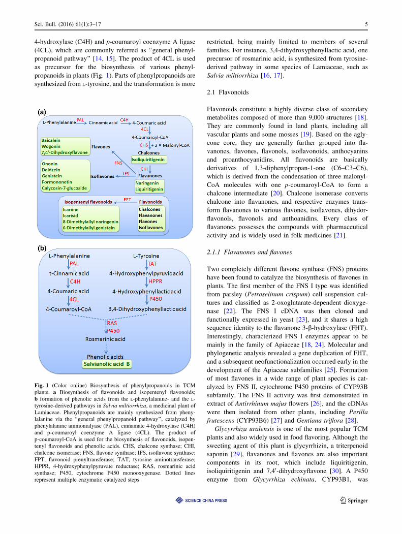
Fig. 1 (Color online) Biosynthesis of phenylpropanoids in TCM
plants. a Biosynthesis of flavonoids and isopentenyl flavonoids;
b formation of phenolic acids from the L-phenylalanine- and the L-
tyrosine-derived pathways in Salvia miltiorrhiza, a medicinal plant of
Lamiaceae. Phenylpropanoids are mainly synthesized from pheny-
lalanine via the ‘‘general phenylpropanoid pathway’’, catalyzed by
phenylalanine ammonialyase (PAL), cinnamate 4-hydroxylase (C4H)
and p-coumaroyl coenzyme A ligase (4CL). The product of
p-coumaroyl-CoA is used for the biosynthesis of flavonoids, isopen-
tenyl flavonoids and phenolic acids. CHS, chalcone synthase; CHI,
chalcone isomerase; FNS, flavone synthase; IFS, isoflavone synthase;
FPT, flavonoid prenyltransferase; TAT, tyrosine aminotransferase;
HPPR, 4-hydroxyphenylpyruvate reductase; RAS, rosmarinic acid
synthase; P450, cytochrome P450 monooxygenase. Dotted lines
represent multiple enzymatic catalyzed steps
Sci. Bull. (2016) 61(1):3–17 5
123

identified as flavanone 2-hydroxylase (F2H), a member of
FNS II [31]. The products, 2-hydroxyflavanones, were
transformed into flavones in vitro in acid treatment, sug-
gesting that an additional enzyme, probably a dehydratase,
was involved in catalyzing the formation of flavones. A
full-length cDNA of cytochrome P450 CYP93C2 was iso-
lated from the elicited G. echinata cells, which was shown
to encode 2-hydroxyisoflavanone synthase [32].
The flavones baicalin and wogonoside, as well as their
aglycones baicalein and wogonin, represent the dominant
flavonoids in Scutellaria baicalensis, a perennial species of
Lamiaceae and an important herb in Chinese traditional
and clinical-orientated medicine. The flavones, such as
baicalin and wogonin, are distinct for lacking a 40-OHgroup but having a 6-OH group on their A-ring [33]. Genes
encoding the upstream enzymes of the pathway, including
PAL, C4H, 4CL, chalcone synthase (CHS) and chalcone
isomerase (CHI), have been isolated [34, 35]. However, the
enzymes committed to the formation of the S. baicalensis-
type flavones remain unknown. It is also possible that
specific enzyme isoforms are involved in the formation of
cinnamoyl-CoA [36]. It has been reported that accumula-
tion of these flavones was enhanced by jasmonate (JA)
treatment, and a R2R3-MYB transcription factor,
SbMYB8, was found involved in the regulation [37, 38].
2.1.2 Isoflavones
The isoflavones are well studied for their substantial health
promoting benefits. They are found mainly in leguminous
plants and are the major bioactive ingredients in soybean,
Astragalus, Pueraria lobata [39]. Isoflavones are converted
from flavanones by the isoflavone synthase (IFS). By using
EST-based approach combined with enzymatic assays,
P450s of CYP93C subfamily from soybean were shown to
have such activities [40, 41].Members of this subfamilywith
IFS activity were also reported in other leguminous plants,
such as Lotus japonicus [42] and Trifolium pratense [43].
Astragalus membranaceus, a species of Fabaceae, has
been used in TCM for thousands of years. Astragaus is
considered an adaptogen because it is believed to help
protect the body against stresses, including those of phys-
ical, mental or emotional [44, 45]. In China, Astragalus has
been used to help patients with severe forms of heart dis-
ease in relieving symptoms, lowering cholesterol levels and
improving heart function. Constituents of the Astragalus
roots (radix astragali) include polysaccharides, triter-
penoids (astragalosides) and isoflavones [46, 47]. Iso-
flavones such as calycosin-7-glucoside and ononin are
considered the important active components in this medi-
cine. Hairy root system of Astragalus was developed a long
time ago to produce these ingredients [48, 49]. Research at
molecular level in this plant is limited, but will help reveal
the biosynthetic pathway in this leguminous medicinal
plant [50].
Pueraria lobata, also a species of Fabaceae, is com-
monly known as ‘‘kudzu’’. Puerariae radix, the dried root
of the kudzu, has been used in China as herbal medicine for
the prevention of cardiovascular disease and rehabilitation
of stroke patients [51]. The major secondary metabolites
accumulated in kudzu roots are isoflavones, including
daidzein, genistein, formononetin and their glucosides
Puerarin [52], among which the 8-C-glucoside of daidzein
is considered the major active compound [53]. The co-
occurrence of both O- and C-linked glycosides in root is of
particular interests and worthy of further investigation.
Using a functional genomics approach, He et al. identified
enzymes associated with the isoflavone biosynthesis in
kudzu roots, including 15 UDP-dependent glycosyltrans-
ferases (UGTs), among which one, GT04F14, exhibited the
in vitro activity of glycosylation of a wide range of sub-
strates, including coumarins, flavones, flavonols, and iso-
flavones. The isoflavones are converted region-specifically
to their 7-O-glucosides, whereas C-glycosylation might
take place at the 2,7,40-trihydroxyisoflavanone precursor ofdaidzein, rather than directly on daidzein. Conceivably the
intermediate 8-C-b-glucopyranosyl-2,7,40-trihydroxy-isoflavanone is converted to puerarin under in vivo con-
ditions by the action of 2-hydroxyisoflavanone dehydratase
(HID). A candidate gene encoding HID was identified from
the EST library of kudzu root [54]. In addition, a partially
purified preparation from kudzu root was shown to have
the C-glucosyltransferase activity that converted isoliquir-
itigenin (20,40,4-trihydroxychalcone) and UDP-Glc to
puerarin [55].
2.1.3 Isopentenyl flavonoids
Prenylation, the addition of prenyl groups, contributes to
the diversification of flavonoids, and the occurrence of
more than 1,000 prenylated flavonoids in plants has been
recorded [56]. This prenylation represents the coupling
process of the aromatic moiety from shikimate pathway
and the prenyl (isoprenoid) chain from the isoprenoid
pathways. Many prenylated flavonoids were identified as
active components in medicinal plants and thus are of
particular interests as lead compounds for drugs and
functional food ingredients [57].
Species Sophora, family Fabaceae, are widely dis-
tributed in Asia. Sophora flavescens has a long history of
use in China, and the root, known as Ku Shen, is a typical
TCM. It is used to dispel heat, dry dampness and eliminate
intestinal parasites. It is thus administered in formulas for
the treatment of dysentery and jaundice (damp-heat syn-
dromes), edema and dysuria (dampness syndromes), and
eczema and pruritis (damp-heat-wind syndromes). The S.
6 Sci. Bull. (2016) 61(1):3–17
123
flavescens prenyltransferase SfN8DT-1 is the first enzyme
identified to be responsible for the prenylation of narin-
genin at the 8-position, with dimethylallyl diphosphate
(DMAPP) as substrate [58]. Later, two new flavonoid
prenyltransferases (FPTs) were isolated from S. flavescens
at the molecular level: one is the isoflavone-specific
prenyltransferase (SfG6DT) for the prenylation of the
genistein at the 6-position and the other a chalcone-specific
prenyltransferase designated as isoliquiritigenin dimethy-
lallyltransferase (SfiLDT) [29].
Herba epimedii is prepared from the aerial parts of
Epimedium brevicornum or Epimedium sagittatum, species
of Berberidaceae. Herba epimedii contains various bioac-
tive components and has been utilized extensively in China
as the tonic and anti-rheumatic herb for thousands of years,
and in the treatments of diseases such as impotence, fre-
quency/urgency of urination, coronary heart disease,
chronic bronchitis and neurasthenia [59, 60]. The isopen-
tenyl flavonoids icariine and icarisid are the major active
compounds [61]; however, their biosynthesis remains
poorly understood [62]. Recently, Huang et al. isolated 12
structural genes and two putative transcription factors
(TFs) in the flavonoid pathway. Transcriptional analysis
revealed that two R2R3-MYB TFs (EsMYBA1 and
EsMYBF1), together with a bHLH TF (EsGL3) and WD40
protein (EsTTG1), are probably involved in coordinated
regulation of biosynthesis of the anthocyanins and the
flavonol-derived bioactive components [63].
2.2 Phenolic acids
Salvia miltiorrhiza is a perennial herb in the mint family
(Lamiaceae). Its dried root or rhizome is called Danshen in
TCM and was recorded in first pharmaceutical monograph
Shennong’s Classic of Materia Medica (A.D. 102-200). S.
miltiorrhiza has been cultivated throughout Eastern Asia
and used to prevent and cure cardiovascular, cerebrovas-
cular, hyperlipidemia and acute ischemic stroke diseases
[64]. Both the hydrophilic and lipophilic components in S.
miltiorrhiza are considered active ingredients. The hydro-
philic compounds are mainly phenolic acids including
rosmarinic acid, salvianolic acid B, lithospermic acid and
dihydroxyphenyllactic acid or Danshensu, and they may
also function as antioxidative, anti-bacterial and anti-viral
reagents [65, 66].
The biosynthetic pathway for phenolic acids in S. mil-
tiorrhiza is distinct and has attracted many interests.
Labeling experiments using [ring-(13)C]-phenylalanine
suggested two intermediates derived from the phenylala-
nine-derived general phenylpropanoid pathway and the
tyrosine-derived pathway, respectively (Fig. 1): 4-cou-
maroyl-CoA and 3,4-dihydroxyphenyllactic acid (DHPL),
which are coupled by a acyl-CoA-dependent
acyltransferase BAHD family enzyme rosmarinic acid
synthase (SmRAS) to form 4-coumaroyl-30,40-dihydrox-yphenyllactic acid (4C-DHPL). The 3-hydroxyl group is
introduced later in the pathway by a P450 monooxygenase
(SmCYP98A14) to form rosmarinic acid (RA) [16]. This
type of P450 was first reported in Coleus blumei (Lami-
aceae), and it catalyzes the 3-hydroxylation of 4-coumar-
oyl-30,40-dihydroxyphenyllactate and the 30-hydroxylationof caffeoyl-40- hydroxyphenyllactate, in both cases forming
rosmarinic acid [67]. Recent genome assembly to search
the putative enzymes involved in biosynthesis of phenolics
in S. miltiorrhiza revealed twenty-nine candidates, among
which 15 were predicted in the phenylpropanoid pathway,
seven in the tyrosine-derived pathway and six encoding
putative hydroxycinnamoyltransferases [17].
3 Terpenoids
Terpenoids are formed from sequential assembly of five-
carbon building blocks (C5H8) called isoprene units.
Accordingly, single or assemblies of two, three and four
units constitute hemiterpenes, monoterpenes, sesquiterpe-
nes and diterpenes, respectively. After the formation of the
basic carbon skeletons, subsequent modifications, such as
oxidation, reduction, isomerization and conjugation, lead to
enormous numbers of structures, which represent the most
abundant class of plant specialized metabolites, with more
than 36,000 individual compounds [68].
In plant cells, the common precursors of terpenoids,
isopentenyl diphosphate (IPP) and dimethylallyl diphos-
phate (DMAPP) are synthesized via two independent
pathways: the cytosolic mevalonic acid (MVA) pathway
that starts with the condensation of acetyl-CoA, and the
plastid-localized methylerythritol phosphate (MEP) path-
way that uses pyruvate and glyceraldehydes 3-phosphate as
substrates (Fig. 2). The IPP and DMAPP are condensed
into geranyl diphosphate (GPP, C10), farnesyl diphosphate
(FPP, C15) and geranylgeranyl diphosphate (GGPP, C20) by
the respective prenyltransferases and then converted to
terpenes by terpene synthases (TPSs), which catalyze the
critical step that determines the structures of terpen
skeletons [69].
Generally, the cytosolic MVA pathway provides the
precursor of FPP for the biosynthesis of sesquiterpenes and
triterpenes, whereas the plastid MEP pathway is responsi-
ble for the biosynthesis of GPP and GGPP for mono-, di-,
and tetra-terpenes [70]. Although cross-talk between these
two spatially separated IPP pathways is prevalent, partic-
ularly in a direction from plastid to cytosol, our under-
standing of the molecular mechanism behind remains
primitive.
Sci. Bull. (2016) 61(1):3–17 7
123
3.1 Sesquiterpenoids
Monoterpenoids (C10) and sesquiterpenoids (C15) are
widely distributed in plants, and they are the common
constituents of volatile compounds in flowers, fruits, stems
and leaves, playing important roles in plant–environment
interactions, many of them also possess great commercial
value and some are used in pharmaceuticals.
3.1.1 Artemisinin
One of the most famous plant-sourced medicines is arte-
misinin, an endoperoxide sesquiterpene lactone isolated
from Artemisia annua L., an annual herb of Asteraceae.
Due to its effectiveness against drug-resistant cerebral
malaria, it is the essential component of the combinational
therapies recommended by the World Health Organization
[8]. It has saved millions of lives globally, especially in
developing countries. The 2011 Lasker DeBakey Clinical
Research Award and the 2015 Nobel Prize in Physiology or
Medicine honor the Chinese scientist Youyou Tu who
made the important contribution to the discovery of arte-
misinin [71–73].
As a sesquiterpenoid, artemisinin is believed to be
synthesized from the cytosolic MVA pathway. However, a
recent report suggested that the MEP pathway may also
contribute to its biosynthesis. GPP, which is synthesized in
plastids, can be transported to cytoplasm, forming FPP with
the addition of another IPP unit [74]. The FPP is converted
to the artemisinin skeleton by amorpha-4,11-diene synthase
(ADS), a sesquiterpene synthase [75], and then oxidated by
the cytochrome P450 CYP71AV1. When expressed in
Saccharomyces cerevisiae, CYP71AV1 catalyzed the
continuous oxidation of amorpha-4,11-diene into artemi-
sinic alcohol and artemisinic aldehyde [76], with signifi-
cantly increased production of artemisinic acid and
artemisinic aldehyde when co-expressed with a cyto-
chrome b5 (CYB5) in yeast [8]. The artemisinic aldehyde
D11(13) reductase (Dbr2), a double-bond reductase, cat-
alyzes the formation of dihydroartemisinic aldehyde [77],
which is further converted into dihydroartemisinic acid by
aldehyde dehydrogenase 1 (ALDH1) [78]. Moreover, an
additional alcohol dehydrogenase (ADH1) was also found
to be involved in the oxidation of amorpha-4,11-diene to
artemisinic acid, with specificity toward artemisinic alco-
hol in A. annua plants [8].
Several transcription factors have been shown to par-
ticipate in the regulation of artemisinin biosynthesis [79].
Two jasmonate responsive AP2/ERF proteins, AaERF1
and AaERF2, were found to up-regulate the transcription of
ADS and CYP71AV1 genes, by binding to the
CRTDREHVCBF2 (CBF2) and RAV1AAT (RAA) motifs
present in their promoters [80]. A WRKY family tran-
scription factor, AaWRKY1, was demonstrated to be cap-
able of binding to the W-box in the ADS promoter and
involved in the regulation of artemisinin biosynthesis [81].
A deep sequencing on the transcriptome of A. annua to
identify genes and markers for fast-track breeding was per-
formed, and a detailed genetic map with nine linkage groups
was built. Replicated field trials resulted in a quantitative
trait loci (QTL)map that accounts for a significant amount of
the variation in key traits controlling artemisinin yield, and
positive QTLs in parents of new high-yielding hybrids were
enriched, which made it available to convert A. annua into a
robust crop [82]. Ma et al. [83] recently reported an inte-
grated approach combining metabolomics, transcriptomics
and gene function analyses to characterize gene-to-terpene
and terpene pathway scenarios in a self-pollinating variety of
A. annua. Forty-seven genes that mapped to the terpenes
biosynthesis pathway were identified by sequence mining,
and suchmetabolites-transcriptome network associated with
different tissues is fundamental to metabolic engineering to
artemisinin.
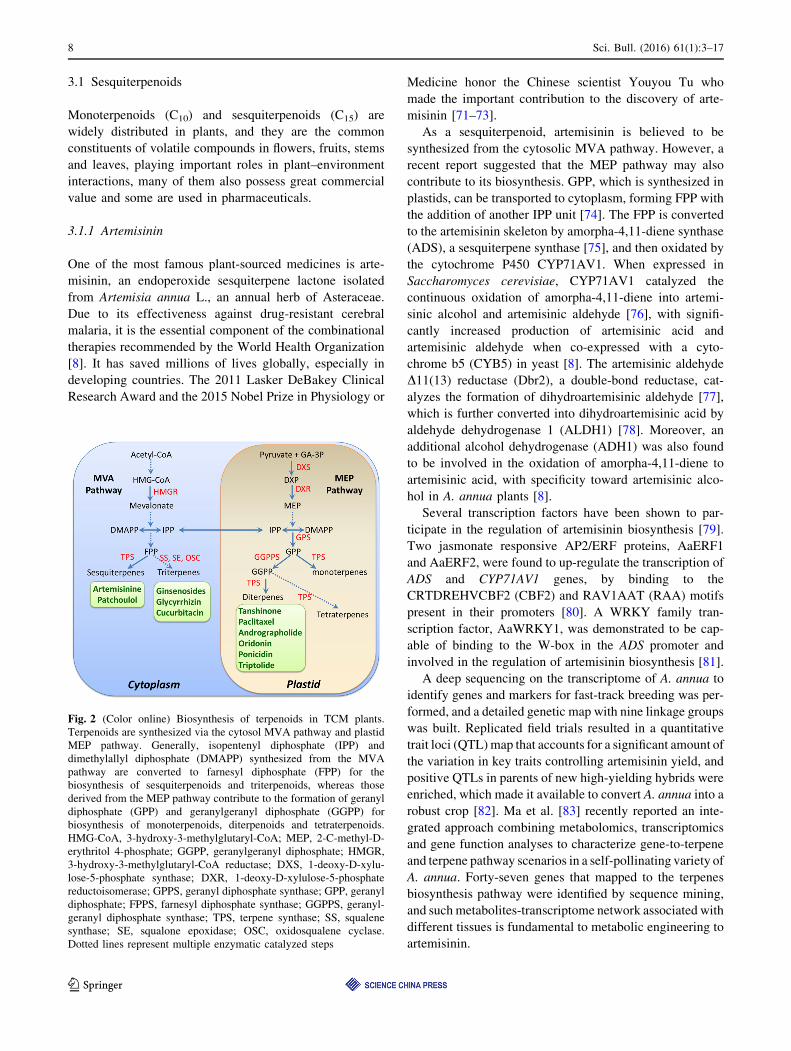
Fig. 2 (Color online) Biosynthesis of terpenoids in TCM plants.
Terpenoids are synthesized via the cytosol MVA pathway and plastid
MEP pathway. Generally, isopentenyl diphosphate (IPP) and
dimethylallyl diphosphate (DMAPP) synthesized from the MVA
pathway are converted to farnesyl diphosphate (FPP) for the
biosynthesis of sesquiterpenoids and triterpenoids, whereas those
derived from the MEP pathway contribute to the formation of geranyl
diphosphate (GPP) and geranylgeranyl diphosphate (GGPP) for
biosynthesis of monoterpenoids, diterpenoids and tetraterpenoids.
HMG-CoA, 3-hydroxy-3-methylglutaryl-CoA; MEP, 2-C-methyl-D-
erythritol 4-phosphate; GGPP, geranylgeranyl diphosphate; HMGR,
3-hydroxy-3-methylglutaryl-CoA reductase; DXS, 1-deoxy-D-xylu-
lose-5-phosphate synthase; DXR, 1-deoxy-D-xylulose-5-phosphate
reductoisomerase; GPPS, geranyl diphosphate synthase; GPP, geranyl
diphosphate; FPPS, farnesyl diphosphate synthase; GGPPS, geranyl-
geranyl diphosphate synthase; TPS, terpene synthase; SS, squalene
synthase; SE, squalone epoxidase; OSC, oxidosqualene cyclase.
Dotted lines represent multiple enzymatic catalyzed steps
8 Sci. Bull. (2016) 61(1):3–17
123
3.1.2 Patchoulol
Patchouli (Pogostemon cablin), a perennial herbaceous
species of Lamiaceae, is not only a fragrant plant produc-
ing patchouli oil for cosmetics industry, but also a
medicinal plant for the treatment of medical ailments, such
as removing dampness, relieving summer heat and exterior
syndrome, and serving as an anti-emetic and appetite
stimulant [84]. The patchouli oil is composed of
sesquiterpenoids dominated by (-)-patchoulol. The
sesquiterpene synthase, patchoulol synthase, was firstly
purified from patchouli leaves by chromatofocusing, anion
exchange, gel permeation and hydroxylapatite chromatog-
raphy [85]. Then, its cDNA was cloned and the recombi-
nant patchoulol synthase was shown to produce patchoulol
as the major product, plus at least 13 additional
sesquiterpenes [86].
Patchouli oil in leaves accumulates with plant age: The
content is low at juvenile stage and increases during plant
growth and reaches a high level in mature plant. The
microRNA156 (miR156)-targeted SQUAMOSA promoter
binding protein-like (SPL) factors, which function as the
major plant age cue in regulating developmental phase
transition and flowering, play a key role in the age-de-
pendent progressive up-regulation of the patchoulol syn-
thase gene expression, and the patchouli oil biosynthesis.
Interestingly, expression of a miR156-resistant form of
SPL not only accelerated plant maturation but also pro-
moted patchouli oil production [87].
3.2 Diterpenoids
Certain groups of diterpenoids (C20), such as gibberellins,
are regulators (phytohormones) of plant growth and
development. Many other specialized diterpenoids, like
tanshinone from Salvia miltiorrhiza and taxol from Taxus,
are highly valuable in medicine. A few more examples
include: stevioside, extracted from Stevia rebaudiana of
Asteraceae, is a natural sweetest [88–90]; adenanthin, from
the leaves of Rabdosia adenantha, induces differentiation
of acute promyelocytic leukemia (APL) cells [91]; ori-
donin, from Lamiaceae plants Isodon rubescens and Isodon
amethystoides, is a potential compound for molecular tar-
get-based therapy of leukemia [92]; and triptolide, a highly
oxygenated diterpene isolated from Tripterygium wilfordii,
was shown to have anti-leukemic activity [93].
3.2.1 Tanshinone
Besides the phenolic acids discussed above, tanshinones
are another class of active diterpenoid compounds of S.
miltiorrhiza, which include tanshinone I, tanshinone IIA,
cryptotanshinone and dihydrotanshinone I. They are all
abietane-type derivatives, among which tanshinone IIA is
considered to be an important bioactive component in
protecting cardiovascular system [94, 95], and tanshinone I
was reported to be an apoptosis inducer and display anti-
cancer activities [95].
As diterpenoid compounds, tanshinones are expected to
be traced to the plastid MEP pathway, and their biosyn-
thesis starts from the conversion of geranylgeranyl
diphosphate (GGPP) to ent-copalyl diphosphate (CPP) and
then to miltiradiene. The subsequent extensively structural
tailing converts miltiradiene to cryptotanshinone, tanshi-
none I, tanshinone IIA or tanshinone IIB [96].
Based on sequence homology, enzymes shared by other
diterpenoid biosynthesis have been characterized [96, 97].
To date, two enzymes specifically committed to the tan-
shinone biosynthetic pathway have been identified: the
kaurene synthase-like (SmKSL), a diterpene synthase that
utilizes CPP as substrate to produce miltiradiene [96], and
a P450 monooxygenase CYP76AH1 which transforms
miltiradiene to ferruginol [98], both representing the mile-
stone achievement in the research of TCM plant. Recently,
functional divergence of SmCPSs and SmKSLs was
reported, which specified the roles of individual CPSs in
tanshinone production in different tissues, including
SmCPS1 in roots and SmCPS2 in aerial part, and SmCPS4
and SmKSL2 were found to oxidize ent-13-epi-manoyl in
floral sepals, and the conserved SmCPS5 involved in the
plant growth hormone gibberellin biosynthesis. This study
is a typical example of how the evolutionary diversification
of diterpenoids in plants in molecular level [99].
With the rapid development of sequencing technologies,
several transcriptome datasets and the draft genome of S.
miltiorrhiza have been reported. For examples, the cDNA
library of whole plant contained 10,228 ESTs [100], the
transcriptome of nearly entire growing cycle generated by
Illumina revealed 56,774 unigenes [101], and the searching
of the draft genome resulted in 40 putative genes encoding
enzymes involved in the biosynthesis of universal isoprene
precursors of IPP and DMAPP [102]. Genes encoding
cytochrome P450 monooxygenases, dehydrogenases and
reductases, as well as several groups of transcription fac-
tors were predicted to be involved in tanshinone biosyn-
thesis by comparative analysis of transcriptomes generated
from different tissues [103]. Recently, next-generation
sequencing (NGS) and single-molecule real-time (SMRT)
sequencing were combined to generate a more com-
plete/full-length set of S. miltiorrhiza transcriptome, which
provides a valuable resource for further investigation of
tanshinone biosynthesis [104].
Organ- or tissue-specific patterns are common feature
observed in biosynthesis and accumulation of specialized
metabolites, as well as the expression patterns of corre-
sponding genes [105–107]. Tanshinones are actively
Sci. Bull. (2016) 61(1):3–17 9
123

synthesized and stored in roots, whereas only a low or trace
amount was detected in aerial organs like leaves [108].
Moreover, both the accumulation and the expression of the
related genes of tanshinones in hairy root cultures can be
induced by biotic elicitors, such as the carbohydrate frac-
tion of yeast extract, and phytohormones of salicylic acid
and jasmonate [97, 109–114]. Further investigation can be
directed to the characterization of the signaling compo-
nents and transcription factors that regulate the diterpenoid
biosynthesis in S. miltiorrhiza.
3.2.2 Taxol (paclitaxel)
Taxol (paclitaxel) is a diterpenoid isolated from the bark of
Taxus trees. The anti-mitotic and cytotoxic properties of
taxol are derived from its activity in disrupting normal
tubulin dynamics, leading to dysfunction of microtubules
[115]. Fourteen enzymes involved in taxol biosynthesis have
been identified, they are geranylgeranyl diphosphate syn-
thase [116], taxadiene synthase [117], taxadien-5a-ol-O-
acetyl transferase [118], taxane 2a-O-benzoyltransferase
[119], baccatin III: 3-animo-3phenylpropanoyltransferase
[120], 10-deacetylbacctin III-10-O-acetyltransferase [121],
30-N-debenzoyl-20-deoxytaxol N-benzoyltransferase [122],
taxane 5-alpha hydroxylase [123], taxane 10-alpha hydrox-
ylase [124], taxane 13-alpha hydroxylase [125], taxane
2-alpha hydroxylase [126], taxane 7-alpha hydroxylase
[127], taxane 14-alpha hydroxylase [128] and phenylalanine
aminomutase [129].
In addition to elucidation of the biosynthetic enzymes,
progresses have been made in identification of transcription
factors involved in taxol biosynthesis, which include mem-
bers of the AP2 and WRKY families [130]. A recent report
showed that the bHLH transcription factors of TcJAMYC1,
TcJAMYC2 and TcJAMYC4 act as negative regulators of
taxol biosynthesis in T. cuspidata cultured cells [131].
Due to the extremely low content of taxol (at ppm level)
in plant, it requires massive harvesting to obtain sufficient
amounts of the drug; thus, productions by total synthesis,
semi-synthesis, tissue or cell cultures, endophytic fungal
fermentation and more recently metabolic engineering and
synthetic biology have attracted great interests [132]. Pre-
cursors of taxol biosynthesis have been produced in
Escherichia coli [7] and Saccharomyces cerevisiae [123,
133], and the integration of parts (modules) of the whole
pathway in separate organisms cultured together led to the
combination of production of taxadiene in E. coli and
oxygenation of taxadiene by S. cerevisiae [9].
3.3 Triterpenoids
Triterpenoids are cyclization product of squalene which is
condensed by two molecules of FPP. In general,
triterpenoids are formed from MVA pathway in cytoplasm,
as sesquiterpenoids.
3.3.1 Ginsenosides
Ginseng, the root of Panax ginseng, is one of the oldest tra-
ditional medicines and is widely regarded as a tonic in East
Asia [88–90]. The principle bioactive constituents of Ginseng
are ginsenosides, a group of tetra- or pentacyclic triterpene
glycosides belonging to saponins [134]. The clinical and
pharmacological activities of ginsenosides include anti-dia-
betic, anticancer, anti-amestic hypoglycemic, radioprotec-
tive, immunomodulatory, neuroprotective and anti-stress
[135–139]. More than 40 ginsenosides have been isolated
from the white and the red ginseng, and they show different
biological activities based on their structural differences
[140]. Generally, the major pharmacologically active gin-
senosides belong to tetracyclic dammarane- and pentacyclic
oleanane-type triterpene saponins [141].
The common precursor of ginsenosides is squalene,
which is formed by condensation of two FPPs with squa-
lene synthase (SS) [135, 142, 143]. In Ginseng, squalene is
converted into dammarenediol-II by squalone epoxidase
(SE). The cyclization of 2,3-oxidosqualene can result in
two different type of triterpenoids: dammarane and olea-
nane type. Ginsenosides belonging to dammarane-type
triterpenoids are biosynthesized from 2,3-oxidosqualene by
dammarenediol synthase (DS) to form dammarenediol-II
[144], whereas the biosynthesis of oleanane-type ginseno-
sides is started by b-amyrin synthase (PNY1) that trans-
forms 2,3-oxidosqualene into b-amyrin [145, 146]. SS is
considered a rate-limiting enzyme in the pathway and
catalyzes the initial biosynthetic step for both steroids and
triterpenoids [147]. PgPDR, a member of ABC trans-
porters, was found to be involved in the ginsenosides
accumulation upon MeJA induction [148].
3.3.2 Cucurbitacins
Cucurbitacins, conferring a bitter taste in cucurbits such as
cucumber, melon, watermelon, squash, and pumpkin,
belong to a class of highly oxidized tetracyclic triter-
penoids mainly found in the plant of Cucurbitaceae family,
in which Gynostemma pentaphyllum, Hemsleya chinesis,
Siraitia grosvenorii and Bolbostemma paniculatum are
well-known TCM plants. Recent studies suggest that
cucurbitacins repress cancer cell progression [149] and
inhibit neuroblastoma cell proliferation through up-regu-
lation of PETN (phosphatase and tensin homolog) [150].
By genome-wide association study based on the genome
variation map of 115 diverse cucumber lines, the gene of
Csa6G088690 (Bi) encoding oxidosqualene cyclase is
found to be correlated to the cucurbitacin C (CuC)
10 Sci. Bull. (2016) 61(1):3–17
123

biosynthesis. Co-expression and co-regulation studies
revealed a 9-gene module responsible for CucC biosyn-
thesis, of which, four enzymes, including Bi, two P450s
and one ACT, were characterized. Moreover, two bHLH
transcription factors, Bl (bitter leaf) and Bt (bitter fruit),
were found to directly regulate the expression of 9-gene
module in cucumber leaf and fruit, respectively. During the
cucumber domestication, mutations occurred within Bt
promoter region which decreased its expression in the fruit
tissue which may have been selected and fixed and resulted
in nonbitter fruit we eat nowadays [151].
3.3.3 Glycyrrhizin
The roots and stolons of Glycyrrhiza plants (G.uralensis and
G. glabra) contain a large amount of oleanane-type triter-
penoid glycyrrhizin. It is not only used worldwide as a
natural sweetener and flavoring additive due to its sweet
taste, but also exhibit a wide range of pharmacological
activities, including anti-inflammatory [152], immunomod-
ulatory [153], anti-ulcer [154], anti-allergy [155], and anti-
viral activity [156–158].
From G. glabra, genes that encode enzymes responsible
for triterpene skeleton formation, including the squalene
synthase (SS) and b-amyrin synthase (bAS), were isolated
[159, 160]. Later biosynthesis steps of glycyrrhizin involve
a series of oxidative reactions at positions C-11 and C-30
and glucuronylation of the C-3 hydroxyl group. Enzymes
that catalyze the oxidation steps have been found to be
cytochrome P450 monooxygenases. One of them,
CYP88D6, was characterized to catalyze the sequential
two-step oxidation of b-amyrin at C-11 to produce 11-oxo-
b-amyrin by both in vitro assay with recombinant protein
and co-expression with b-amyrin synthase in yeast [161].
Another P450, CYP72A154, was identified to be respon-
sible for three sequential oxidations at C-30 to transform
11-oxo-b-amyrin to glycyrrhetinic acid, a glycyrrhizin
aglycone [162]. Both CYP88D6 and CYP72A154 tran-
scripts were detected in the roots and stolons, but not in the
leaves or stems, which is consistent with the accumulation
pattern of glycyrrhizin in planta [161, 162].
4 Alkaloids
Alkaloids are a group of nitrogen-containing compounds
with basic properties, most of which are derivatives of
amino acids [163–166]. Biosynthesis of alkaloids usually
starts from modification of amino acids, mostly decar-
boxylation or deamination, and undergoes further steps like
methylation, hydroxylation and oxidation, and/or coupled
with other compounds. There are over 12,000 alkaloids that
have been identified from plants. Although widely dis-
tributed in plants, they are particularly enriched in certain
families, such as Solanaceae, Manispermaceae, Papaver-
aceae, Berberidaceae and Fabaceae (Table 2).
It is noteworthy that the most of alkaloids display
bioactivities to certain degrees, often derived from their
nitrogen-containing properties. Not surprisingly, alkaloids
constitute the major portion of drugs both in history and
nowadays. The discovery and isolation of morphine from
the opium poppy (Papaver somniferum) by Friedrich Ser-
turner in 1806 is a milestone in the history of pharmacy.
Investigations of biosynthesis of natural alkaloids such as
morphinan, vindoline and noscapine have been intensive
and led to the complete elucidation of the pathway [167–
170], and increasing alkaloid biosynthesis in plant through
co-expression of enzymes genes was also reported [114].
Unfortunately, although alkaloids with TCM background
like camptothecin, higenamine, huperzine A and tetran-
drine have been used in pharmacy, reports of their
biosynthesis are relatively rare. We list in Table 3 several
Table 2 List of examples of TCM plants rich in terpenoids
Plant species Chinese name in Pin-yin Family Representative compounds
Pogostemon cablin Guanghuoxiang Lamiaceae Patchoulol
Artemisia annua Huanghuahao or Qinghao Asteraceae Artemisinine
Salvia miltiorrhiza Danshen Lamiaceae Tanshinone
Taxus chinensis Hongdoushan Taxaceae Paclitaxel
Andrographis paniculata Chuanxinlian Acanthaceae Andrographolide
Isodon rubescens Donglingcao Lamiaceae Oridonin, ponicidin
Isodon amethystoides Xiangchacai Lamiaceae Oridonin, ponicidin
Tripterygium wilfordii Leigongteng Celastraceae Triptolide
Panax ginseng Ginsen or Renshen Araliaceae Ginsenosides
Panax notoginseng Sanqi Araliaceae Notoginsenosides
Radix liquiritiae Gancao Fabaceae Glycyrrhizin
Dioscorea polystachya Shuyu Dioscoreaceae Dioscin
Sci. Bull. (2016) 61(1):3–17 11
123

typical alkaloids in TCM plants, and the relevant refer-
ences. Various aspects on the alkaloid biosynthesis, regu-
lation and metabolites trafficking can be found in review
articles [178–183]. Without doubt more efforts are needed
to study alkaloids in TCM plants to further explore their
biological activities and facilitate their usage.
5 Perspective
Unlike model plant or staple crops, medicinal plants often
lack a well-studied genetic background and a high-quality
genome sequence. Due to the recently developed high-
throughput sequencing technologies, it is possible to gen-
erate transcriptomic data of medicinal plants in a short time
at an affordable cost. Comparative analysis of chemical
constituents, transcriptomes and correlation of spatial and
temporal patterns of gene expressions with those of
metabolite accumulation have led to the identification of
candidate genes of the biosynthesis pathway [184]. GWS
combined with metabolomics analysis (mGWAS) provides
a powerful platform which screens a large number of
accessions simultaneously to understand genetic contribu-
tions to the metabolic diversity [185, 186].
Throughout the history, herbal plants are an integral part
of our lives. In addition to curing illness, they are grown in
elegant gardens, provide natural fragrance, delicate acces-
sories and stimulate appetite. The biosynthesis of
metabolites in medicinal plants is complex and specialized
and involves many sequence-similar but functionally
diverged enzymes. With the fast development of new
technologies of analytical chemistry, bioinformatics and
synthetic biology, more and more achievements will be
made in this genomic or post-genomic era and bring us
better life.
Acknowledgments This work was supported by the National Nat-
ural Science Foundation of China (31200222), and Special Fund for
Shanghai Landscaping Administration Bureau Program (F132424,
F112418 and G152421).
Conflict of interest The authors declare that they have no conflict
of interest.
Open Access This article is distributed under the terms of the
Creative Commons Attribution 4.0 International License (http://
creativecommons.org/licenses/by/4.0/), which permits unrestricted
use, distribution, and reproduction in any medium, provided you give
appropriate credit to the original author(s) and the source, provide a
link to the Creative Commons license, and indicate if changes were
made.
References
1. Harvey AL, Edrada-Ebel R, Quinn RJ (2015) The re-emergence
of natural products for drug discovery in the genomics era. Nat
Rev Drug Discov 14:111–129
2. Qiu J (2007) Traditional medicine: a culture in the balance.
Nature 448:126–128
3. Sakurai Y, Kolokoltsov AA, Chen CC et al (2015) Ebola virus.
Two-pore channels control Ebola virus host cell entry and are
drug targets for disease treatment. Science 347:995–998
4. Liu J, Lee J, Salazar Hernandez MA et al (2015) Treatment of
obesity with celastrol. Cell 161:999–1011
5. Chan E, Tan M, Xin J et al (2010) Interactions between tradi-
tional Chinese medicines and Western therapeutics. Curr Opin
Drug Discov Dev 13:50–65
6. Butelli E, Titta L, Giorgio M et al (2008) Enrichment of tomato
fruit with health-promoting anthocyanins by expression of select
transcription factors. Nat Biotechnol 26:1301–1308
7. Ajikumar PK, Xiao WH, Tyo KE et al (2010) Isoprenoid
pathway optimization for Taxol precursor overproduction in
Escherichia coli. Science 330:70–74
8. Paddon CJ, Westfall PJ, Pitera DJ et al (2013) High-level semi-
synthetic production of the potent antimalarial artemisinin.
Nature 496:528–532
9. Zhou K, Qiao K, Edgar S et al (2015) Distributing a metabolic
pathway among a microbial consortium enhances production of
natural products. Nat Biotechnol 33:377–383
10. Winkel BS (2004) Metabolic channeling in plants. Annu Rev
Plant Biol 55:85–107
11. Dixon RA, Achnine L, Kota P et al (2002) The phenylpropanoid
pathway and plant defence-a genomics perspective. Mol Plant
Pathol 3:371–390
12. Vogt T (2010) Phenylpropanoid biosynthesis. Mol Plant 3:2–20
13. Hartmann T (2007) From waste products to ecochemicals: fifty
years research of plant secondary metabolism. Phytochemistry
68:2831–2846
14. Naoumkina MA, Zhao Q, Gallego-Giraldo L et al (2010) Gen-
ome-wide analysis of phenylpropanoid defence pathways. Mol
Plant Pathol 11:829–846
15. Weisshaar B, Jenkins GI (1998) Phenylpropanoid biosynthesis
and its regulation. Curr Opin Plant Biol 1:251–257
16. Di P, Zhang L, Chen J et al (2013) (1)(3)C tracer reveals phe-
nolic acids biosynthesis in hairy root cultures of Salvia milti-
orrhiza. ACS Chem Biol 8:1537–1548
17. Wang B, Sun W, Li Q et al (2015) Genome-wide identification
of phenolic acid biosynthetic genes in Salvia miltiorrhiza. Planta
241:711–725
Table 3 List of examples of TCM plants rich in alkaloids
Plant species Chinese name in Pin-yin Family Representative compounds References
Camptotheca acuminate Xishu Cornaceae Camptothecin [171–173]
Coptis chinensis Huanglian Ranunculaceae Berberine [174, 175]
Isatis indigotica Songlan Brassicaceae Isatin, indigotin [176]
Baphicacanthus cusia Baalan Acanthaceae Isatin, indigotin [177]
12 Sci. Bull. (2016) 61(1):3–17
123
18. Martens S, Mithofer A (2005) Flavones and flavone synthases.
Phytochemistry 66:2399–2407
19. Williams CA, Grayer RJ (2004) Anthocyanins and other flavo-
noids. Nat Prod Rep 21:539–573
20. Ferrer JL, Jez JM, Bowman ME et al (1999) Structure of chal-
cone synthase and the molecular basis of plant polyketide
biosynthesis. Nat Struct Biol 6:775–784
21. Kuo SM (1997) Dietary flavonoid and cancer prevention: evi-
dence and potential mechanism. Crit Rev Oncog 8:47–69
22. Sutter A, Poulton J, Grisebach H (1975) Oxidation of flavanone
to flavone with cell-free extracts from young parsley leaves.
Arch Biochem Biophys 170:547–556
23. Martens S, Forkmann G, Matern U et al (2001) Cloning of
parsley flavone synthase I. Phytochemistry 58:43–46
24. Gebhardt YH, Witte S, Steuber H et al (2007) Evolution of
flavone synthase I from parsley flavanone 3beta-hydroxylase by
site-directed mutagenesis. Plant Physiol 144:1442–1454
25. Gebhardt Y, Witte S, Forkmann G et al (2005) Molecular evo-
lution of flavonoid dioxygenases in the family Apiaceae. Phy-
tochemistry 66:1273–1284
26. Stotz G, Forkmann G (1981) Oxidation of flavanones to flavones
with flower extracts of Antirrhinum majus (Snapdragon).
Z Naturforsch C 36:737–741
27. Kitada C, Gong Z, Tanaka Y et al (2001) Differential expression
of two cytochrome P450s involved in the biosynthesis of fla-
vones and anthocyanins in chemo-varietal forms of Perilla
frutescens. Plant Cell Physiol 42:1338–1344
28. Nakatsuka T, Nishihara M, Mishiba K et al (2005) Temporal
expression of flavonoid biosynthesis-related genes regulates
flower pigmentation in gentian plants. Plant Sci 168:1309–1318
29. Sasaki K, Tsurumaru Y, Yamamoto H et al (2011) Molecular
characterization of a membrane-bound prenyltransferase specific
for isoflavone from Sophora flavescens. J Biol Chem
286:24125–24134
30. Ma CJ, Li GS, Zhang DL et al (2005) One step isolation and
purification of liquiritigenin and isoliquiritigenin from Gly-
cyrrhiza uralensis Risch. using high-speed counter-current
chromatography. J Chromatogr A 1078:188–192
31. Akashi T, Aoki T, Ayabe S (1998) Identification of a cyto-
chrome P450 cDNA encoding (2S)-flavanone 2-hydroxylase of
licorice (Glycyrrhiza echinata L.; Fabaceae) which represents
licodione synthase and flavone synthase II. FEBS Lett
431:287–290
32. Akashi T, Aoki T, Ayabe S (1999) Cloning and functional
expression of a cytochrome P450 cDNA encoding 2-hydroxy-
isoflavanone synthase involved in biosynthesis of the iso-
flavonoid skeleton in licorice. Plant Physiol 121:821–828
33. Nurul Islam M, Downey F, Ng CY (2011) Comparative analysis
of bioactive phytochemicals from Scutellaria baicalensis,
Scutellaria lateriflora, Scutellaria racemosa, Scutellaria
tomentosa and Scutellaria wrightii by LC-DAD-MS. Metabo-
lomics 7:446–453
34. Xu H, Park NI, Li X et al (2010) Molecular cloning and char-
acterization of phenylalanine ammonia-lyase, cinnamate 4-hy-
droxylase and genes involved in flavone biosynthesis in
Scutellaria baicalensis. Bioresour Technol 101:9715–9722
35. Park NI, Xu H, Li X et al (2011) Enhancement of flavone levels
through overexpression of chalcone isomerase in hairy root cul-
tures of Scutellaria baicalensis. Funct Integr Genom 11:491–496
36. Schneider K, Hovel K, Witzel K et al (2003) The substrate
specificity-determining amino acid code of 4-coumarate: CoA
ligase. Proc Natl Acad Sci USA 100:8601–8606
37. Kuzovkina IN, Guseva AV, Alterman IE et al (2001) Flavonoid
production in transformed Scutellaria baicalensis roots and
ways of its regulation. Russ J Plant Physiol 48:448–452
38. Yuan Y, Qi L, Yang J et al (2015) A Scutellaria baicalensis
R2R3-MYB gene, SbMYB8, regulates flavonoid biosynthesis
and improves drought stress tolerance in transgenic tobacco.
Plant Cell Tissue Org 120:961–972
39. Veitch NC (2013) Isoflavonoids of the leguminosae. Nat Prod
Rep 30:988–1027
40. Steele CL, Gijzen M, Qutob D et al (1999) Molecular charac-
terization of the enzyme catalyzing the aryl migration reaction
of isoflavonoid biosynthesis in soybean. Arch Biochem Biophys
367:146–150
41. Jung W, Yu O, Lau SM et al (2000) Identification and expres-
sion of isoflavone synthase, the key enzyme for biosynthesis of
isoflavones in legumes. Nat Biotechnol 18:208–212
42. Shimada N, Akashi T, Aoki T et al (2000) Induction of iso-
flavonoid pathway in the model legume Lotus japonicus:
molecular characterization of enzymes involved in phytoalexin
biosynthesis. Plant Sci 160:37–47
43. Kim BG, Kim SY, Song HS et al (2003) Cloning and expression
of the isoflavone synthase gene (IFS-Tp) from Trifolium pra-
tense. Mol Cells 15:301–306
44. Shao BM, Xu W, Dai H et al (2004) A study on the immune
receptors for polysaccharides from the roots of Astragalus
membranaceus, a Chinese medicinal herb. Biochem Biophys
Res Commun 320:1103–1111
45. Matkovic Z, Zivkovic V, Korica M et al (2010) Efficacy and
safety of Astragalus membranaceus in the treatment of patients
with seasonal allergic rhinitis. Phytother Res 24:175–181
46. Xu Q, Ma X, Liang X (2007) Determination of astragalosides in
the roots of Astragalus spp. using liquid chromatography tan-
dem atmospheric pressure chemical ionization mass spectrom-
etry. Phytochem Anal 18:419–427
47. Wang D, Zhuang Y, Tian Y et al (2012) Study of the effects of
total flavonoids of Astragalus on atherosclerosis formation and
potential mechanisms. Oxid Med Cell Longev 2012:282383
48. Hirotani M, Zhou Y, Rui H et al (1994) Cycloartane triterpene
glycosides from the hairy root cultures of Astragalus mem-
branaceus. Phytochemistry 37:1403–1407
49. Cho H-J, Widholm J (2002) Improved shoot regeneration pro-
tocol for hairy roots of the legume Astragalus sinicus. Plant Cell
Tissue Org 69:259–269
50. Duan LX, Chen TL, Li M et al (2012) Use of the metabolomics
approach to characterize Chinese medicinal material Huangqi.
Mol Plant 5:376–386
51. Lukaczer D, Darland G, Tripp M et al (2005) Clinical effects of a
proprietary combination isoflavone nutritional supplement in
menopausal women: a pilot trial. Altern Ther Health Med 11:60–65
52. Rong H, Stevens JF, Deinzer ML et al (1998) Identification of
isoflavones in the roots of Pueraria lobata. Planta Med
64:620–627
53. Ohshima Y, Okuyama T, Takahashi K et al (1988) Isolation and
high performance liquid chromatography (HPLC) of iso-
flavonoids from the Pueraria root. Planta Med 54:250–254
54. He X, Blount JW, Ge S et al (2011) A genomic approach to
isoflavone biosynthesis in kudzu (Pueraria lobata). Planta
233:843–855
55. Chen G, Wu X, Zhou WL et al (2010) Preparation and assay of
C-glucosyltransferase from roots of Pueraria lobata. J Environ
Biol 31:655–660
56. Yazaki K, Sasaki K, Tsurumaru Y (2009) Prenylation of aro-
matic compounds, a key diversification of plant secondary
metabolites. Phytochemistry 70:1739–1745
57. Kitaoka M, Kadokawa H, Sugano M et al (1998) Prenyl-
flavonoids: a new class of non-steroidal phytoestrogen (Part 1).
Isolation of 8-isopentenylnaringenin and an initial study on its
structure-activity relationship. Planta Med 64:511–515
Sci. Bull. (2016) 61(1):3–17 13
123
58. Sasaki K,MitoK,OharaK et al (2008) Cloning and characterization
of naringenin 8-prenyltransferase, a flavonoid-specific prenyltrans-
ferase of Sophora flavescens. Plant Physiol 146:1075–1084
59. Yu L, Li H, Huang G et al (1992) Clinical observations on
treatment of 120 cases of coronary heart disease with herba
epimedii. J Tradit Chin Med 12:30–34
60. Li C, Li Q, Mei Q et al (2015) Pharmacological effects and
pharmacokinetic properties of icariin, the major bioactive
component in Herba Epimedii. Life Sci 126:57–68
61. Islam NM, Yoo HH, Lee MW et al (2008) Simultaneous
quantitation of five flavonoid glycosides in Herba Epimedii by
high-performance liquid chromatography-tandem mass spec-
trometry. Phytochem Anal 19:71–77
62. Zeng S, Xiao G, Guo J et al (2010) Development of a EST
dataset and characterization of EST-SSRs in a traditional Chi-
nese medicinal plant, Epimedium sagittatum (Sieb. Et Zucc.)
Maxim. BMC Genom 11:94
63. Huang W, Zeng S, Xiao G et al (2015) Elucidating the
biosynthetic and regulatory mechanisms of flavonoid-derived
bioactive components in Epimedium sagittatum. Front Plant Sci
6:689
64. Zhou L, Zuo Z, Chow MS (2005) Danshen: an overview of its
chemistry, pharmacology, pharmacokinetics, and clinical use.
J Clin Pharmacol 45:1345–1359
65. Zhang HS, Wang SQ (2006) Salvianolic acid B from Salvia
miltiorrhiza inhibits tumor necrosis factor-alpha (TNF-alpha)-
induced MMP-2 upregulation in human aortic smooth muscle
cells via suppression of NAD(P)H oxidase-derived reactive
oxygen species. J Mol Cell Cardiol 41:138–148
66. Wu WY, Wang YP (2012) Pharmacological actions and thera-
peutic applications of Salvia miltiorrhiza depside salt and its
active components. Acta Pharmacol Sin 33:1119–1130
67. Eberle D, Ullmann P, Werck-Reichhart D et al (2009) cDNA
cloning and functional characterisation of CYP98A14 and
NADPH:cytochrome P450 reductase from Coleus blumei involved
in rosmarinic acid biosynthesis. Plant Mol Biol 69:239–253
68. Gershenzon J, Dudareva N (2007) The function of terpene
natural products in the natural world. Nat Chem Biol 3:408–414
69. Chen F, Tholl D, Bohlmann J et al (2011) The family of terpene
synthases in plants: a mid-size family of genes for specialized
metabolism that is highly diversified throughout the kingdom.
Plant J 66:212–229
70. Oldfield E, Lin FY (2012) Terpene biosynthesis: modularity
rules. Angew Chem Int Ed 51:1124–1137
71. Cooperative Research Group on Artemisinin Structure (1977)
Artemisinin—a new sesquiterpene lactone. Chin Sci Bull 22:142
72. Liu JM, Ni MY, Fan JF et al (1979) Structure and reaction of
Artemisinin. Acta Chim Sin 37:129–143
73. Miller LH, Su X (2011) Artemisinin: discovery from the Chi-
nese herbal garden. Cell 146:855–858
74. Schramek N, Wang H, Romisch-Margl W et al (2010) Artemi-
sinin biosynthesis in growing plants of Artemisia annua.
A13CO2 study. Phytochemistry 71:179–187
75. Mercke P, Bengtsson M, Bouwmeester HJ et al (2000) Molec-
ular cloning, expression, and characterization of amorpha-4,11-
diene synthase, a key enzyme of artemisinin biosynthesis in
Artemisia annua L. Arch Biochem Biophys 381:173–180
76. Teoh KH, Polichuk DR, Reed DW et al (2006) Artemisia annua
L. (Asteraceae) trichome-specific cDNAs reveal CYP71AV1, a
cytochrome P450 with a key role in the biosynthesis of the
antimalarial sesquiterpene lactone artemisinin. FEBS Lett
580:1411–1416
77. Zhang Y, Teoh KH, Reed DW et al (2008) The molecular
cloning of artemisinic aldehyde Delta 11(13) reductase and its
role in glandular trichome-dependent biosynthesis of artemisinin
in Artemisia annua. J Biol Chem 283:21501–21508
78. Teoh KH, Polichuk DR, Reed DW et al (2009) Molecular
cloning of an aldehyde dehydrogenase implicated in artemisinin
biosynthesis in Artemisia annua. Botany 87:635–642
79. Afrin S, Huang J-J, Luo Z-Y (2015) JA-mediated transcriptional
regulation of secondary metabolism in medicinal plants. Sci Bull
60:1062–1072
80. Yu ZX, Li JX, Yang CQ et al (2012) The jasmonate-responsive
AP2/ERF transcription factors AaERF1 and AaERF2 positively
regulate artemisinin biosynthesis in Artemisia annua L. Mol
Plant 5:353–365
81. Ma D, Pu G, Lei C et al (2009) Isolation and characterization of
AaWRKY1, an Artemisia annua transcription factor that regu-
lates the amorpha-4,11-diene synthase gene, a key gene of
artemisinin biosynthesis. Plant Cell Physiol 50:2146–2161
82. Graham IA, Besser K, Blumer S et al (2010) The genetic map of
Artemisia annua L. identifies loci affecting yield of the anti-
malarial drug artemisinin. Science 327:328–331
83. Ma DM, Wang Z, Wang L et al (2015) A genome-wide scenario
of terpene pathways in self-pollinated Artemisia annua L. Mol
Plant. doi:10.1016/j.molp.2015.07.004
84. Swamy MK, Sinniah UR (2015) A comprehensive review on the
phytochemical constituents and pharmacological activities of
Pogostemon cablin benth: an aromatic medicinal plant of
industrial importance. Molecules 20:8521–8547
85. Munck SL, Croteau R (1990) Purification and characterization
of the sesquiterpene cyclase patchoulol synthase from Po-
gostemon cablin. Arch Biochem Biophys 282:58–64
86. Deguerry F, Pastore L, Wu S et al (2006) The diverse
sesquiterpene profile of patchouli, Pogostemon cablin, is cor-
related with a limited number of sesquiterpene synthases. Arch
Biochem Biophys 454:123–136
87. Yu ZX, Wang LJ, Zhao B et al (2015) Progressive regulation of
sesquiterpene biosynthesis in Arabidopsis and Patchouli (Po-
gostemon cablin) by the miR156-targeted SPL transcription
factors. Mol Plant 8:98–110
88. Court WE (2000) Ginseng: the history of an insignificant plant.
Pharm Hist (Lond) 30:38–44
89. Sathiyamoorthy S, In JG, Gayathri S et al (2010) Gene ontology
study of methyl jasmonate-treated and non-treated hairy roots of
Panax ginseng to identify genes involved in secondary meta-
bolic pathway. Russ J Genet 46:932–939
90. Zhu GY, Li YW, Hau DK et al (2011) Protopanaxatriol-type
ginsenosides from the root of Panax ginseng. J Agric Food
Chem 59:200–205
91. Liu CX, Yin QQ, Zhou HC et al (2012) Adenanthin targets
peroxiredoxin I and II to induce differentiation of leukemic
cells. Nat Chem Biol 8:486–493
92. Zhen T, Wu CF, Liu P et al (2012) Targeting of AML1-ETO in
t(8;21) leukemia by oridonin generates a tumor suppressor-like
protein. Sci Transl Med 4:127ra38
93. Shamon LA, Pezzuto JM, Graves JM et al (1997) Evaluation of
the mutagenic, cytotoxic, and antitumor potential of triptolide, a
highly oxygenated diterpene isolated from Tripterygium wil-
fordii. Cancer Lett 112:113–117
94. Li YI, Elmer G, Leboeuf RC (2008) Tanshinone IIA reduces
macrophage death induced by hydrogen peroxide by upregu-
lating glutathione peroxidase. Life Sci 83:557–562
95. Dong Y, Morris-Natschke SL, Lee KH (2011) Biosynthesis, total
syntheses, and antitumor activity of tanshinones and their analogs
as potential therapeutic agents. Nat Prod Rep 28:529–542
96. Gao W, Hillwig ML, Huang L et al (2009) A functional geno-
mics approach to tanshinone biosynthesis provides stereo-
chemical insights. Org Lett 11:5170–5173
97. Kai GY, Liao P, Xu H et al (2012) Molecular mechanism of
elicitor-induced tanshinone accumulation in Salvia miltiorrhiza
hairy root cultures. Acta Physiol Plant 34:1421–1433
14 Sci. Bull. (2016) 61(1):3–17
123
98. Guo J, Zhou YJ, Hillwig ML et al (2013) CYP76AH1 catalyzes
turnover of miltiradiene in tanshinones biosynthesis and enables
heterologous production of ferruginol in yeasts. Proc Natl Acad
Sci USA 110:12108–12113
99. Cui G, Duan L, Jin B et al (2015) Functional divergence of
diterpene syntheses in the medicinal plant Salvia miltiorrhiza
Bunge. Plant Physiol. doi:10.1104/pp.15.00695
100. Yan Y, Wang Z, Tian W et al (2010) Generation and analysis of
expressed sequence tags from the medicinal plant Salvia milti-
orrhiza. Sci China Life Sci 53:273–285
101. Wenping H, Yuan Z, Jie S et al (2011) De novo transcriptome
sequencing in Salvia miltiorrhiza to identify genes involved in
the biosynthesis of active ingredients. Genomics 98:272–279
102. Ma Y, Yuan L, Wu B et al (2012) Genome-wide identification
and characterization of novel genes involved in terpenoid
biosynthesis in Salvia miltiorrhiza. J Exp Bot 63:2809–2823
103. Yang L, Ding G, Lin H et al (2013) Transcriptome analysis of
medicinal plant Salvia miltiorrhiza and identification of genes
related to tanshinone biosynthesis. PLoS One 8:e80464
104. Xu Z, Peters RJ, Weirather J et al (2015) Full-length tran-
scriptome sequences and splice variants obtained by a combi-
nation of sequencing platforms applied to different root tissues
of Salvia miltiorrhiza and tanshinone biosynthesis. Plant J
82:951–961
105. Xu YH, Wang JW, Wang S et al (2004) Characterization of
GaWRKY1, a cotton transcription factor that regulates the
sesquiterpene synthase gene (?)-delta-cadinene synthase-A.
Plant Physiol 135:507–515
106. Murata J, Roepke J, Gordon H et al (2008) The leaf epidermome
of Catharanthus roseus reveals its biochemical specialization.
Plant Cell 20:524–542
107. Wang H, Penmetsa RV, Yuan M et al (2012) Development and
characterization of BAC-end sequence derived SSRs, and their
incorporation into a new higher density genetic map for culti-
vated peanut (Arachis hypogaea L.). BMC Plant Biol 12:10
108. Li JT, Dong JE, Liang ZS et al (2008) Distributional difference
of fat-soluble compounds in the roots, stems and leaves of four
Salvia plants. Fen Zi Xi Bao Sheng Wu Xue Bao 41:44–52
109. Zhang C, Yan Q, Cheuk WK et al (2004) Enhancement of
tanshinone production in Salvia miltiorrhiza hairy root culture
by Ag? elicitation and nutrient feeding. Planta Med 70:147–151
110. Ge X, Wu J (2005) Induction and potentiation of diterpenoid
tanshinone accumulation in Salvia miltiorrhiza hairy roots by
beta-aminobutyric acid. Appl Microbiol Biotechnol 68:183–188
111. Ge X, Wu J (2005) Tanshinone production and isoprenoid
pathways in Salvia miltiorrhiza hairy roots induced by Ag? and
yeast elicitor. Plant Sci 168:487–491
112. Shi M, Kwok KW, Wu JY (2007) Enhancement of tanshinone
production in Salvia miltiorrhiza Bunge (red or Chinese sage)
hairy-root culture by hyperosmotic stress and yeast elicitor.
Biotechnol Appl Biochem 46:191–196
113. Yan Q, Wu J, Liu R (2011) Modeling of tanshinone synthesis
and phase distribution under the combined effect of elicitation
and in situ adsorption in Salvia miltiorrhiza hairy root cultures.
Biotechnol Lett 33:813–819
114. Hao X, Shi M, Cui L et al (2015) Effects of methyl jasmonate
and salicylic acid on tanshinone production and biosynthetic
gene expression in transgenic Salvia miltiorrhiza hairy roots.
Biotechnol Appl Biochem 62:24–31
115. Schiff PB, Fant J, Horwitz SB (1979) Promotion of microtubule
assembly in vitro by taxol. Nature 277:665–667
116. Hefner J, Ketchum RE, Croteau R (1998) Cloning and func-
tional expression of a cDNA encoding geranylgeranyl diphos-
phate synthase from Taxus canadensis and assessment of the
role of this prenyltransferase in cells induced for taxol produc-
tion. Arch Biochem Biophys 360:62–74
117. Wildung MR, Croteau R (1996) A cDNA clone for taxadiene
synthase, the diterpene cyclase that catalyzes the committed step
of taxol biosynthesis. J Biol Chem 271:9201–9204
118. Walker K, Schoendorf A, Croteau R (2000) Molecular cloning
of a taxa-4(20),11(12)-dien-5alpha-ol-O-acetyl transferase
cDNA from Taxus and functional expression in Escherichia
coli. Arch Biochem Biophys 374:371–380
119. Walker K, Croteau R (2000) Taxol biosynthesis: molecular
cloning of a benzoyl-CoA: taxane 2alpha-O-benzoyltransferase
cDNA from Taxus and functional expression in Escherichia
coli. Proc Natl Acad Sci USA 97:13591–13596
120. Walker K, Fujisaki S, Long R et al (2002) Molecular cloning
and heterologous expression of the C-13 phenylpropanoid side
chain-CoA acyltransferase that functions in Taxol biosynthesis.
Proc Natl Acad Sci USA 99:12715–12720
121. Walker K, Croteau R (2000) Molecular cloning of a
10-deacetylbaccatin III-10-O-acetyl transferase cDNA from
Taxus and functional expression in Escherichia coli. Proc Natl
Acad Sci USA 97:583–587
122. Walker K, Long R, Croteau R (2002) The final acylation step in
taxol biosynthesis: cloning of the taxoid C13-side-chain
N-benzoyltransferase from Taxus. Proc Natl Acad Sci USA
99:9166–9171
123. Jennewein S, Long RM, Williams RM et al (2004) Cytochrome
P450 taxadiene 5alpha-hydroxylase, a mechanistically unusual
monooxygenase catalyzing the first oxygenation step of taxol
biosynthesis. Chem Biol 11:379–387
124. Schoendorf A, Rithner CD, Williams RM et al (2001) Molecular
cloning of a cytochrome P450 taxane 10 beta-hydroxylase
cDNA from Taxus and functional expression in yeast. Proc Natl
Acad Sci USA 98:1501–1506
125. Jennewein S, Rithner CD, Williams RM et al (2001) Taxol
biosynthesis: taxane 13 alpha-hydroxylase is a cytochrome
P450-dependent monooxygenase. Proc Natl Acad Sci USA
98:13595–13600
126. Chau M, Croteau R (2004) Molecular cloning and characteriza-
tion of a cytochrome P450 taxoid 2alpha-hydroxylase involved in
Taxol biosynthesis. Arch Biochem Biophys 427:48–57
127. Chau M, Jennewein S, Walker K et al (2004) Taxol biosyn-
thesis: molecular cloning and characterization of a cytochrome
P450 taxoid 7 beta-hydroxylase. Chem Biol 11:663–672
128. Jennewein S, Rithner CD, Williams RM et al (2003) Taxoid
metabolism: taxoid 14beta-hydroxylase is a cytochrome P450-
dependent monooxygenase. Arch Biochem Biophys 413:262–270
129. Walker KD, Klettke K, Akiyama T et al (2004) Cloning,
heterologous expression, and characterization of a phenylalanine
aminomutase involved in Taxol biosynthesis. J Biol Chem
279:53947–53954
130. Li S, Zhang P, Zhang M et al (2013) Functional analysis of a
WRKY transcription factor involved in transcriptional activation
of the DBAT gene in Taxus chinensis. Plant Biol 15:19–26
131. Lenka SK, Nims NE, Vongpaseuth K et al (2015) Jasmonate-
responsive expression of paclitaxel biosynthesis genes in Taxus
cuspidata cultured cells is negatively regulated by the bHLH
transcription factors TcJAMYC1, TcJAMYC2, and TcJA-
MYC4. Front Plant Sci 6:115
132. Ajikumar PK, Tyo K, Carlsen S et al (2008) Terpenoids:
opportunities for biosynthesis of natural product drugs using
engineered microorganisms. Mol Pharm 5:167–190
133. Hefner J, Rubenstein SM, Ketchum RE et al (1996) Cytochrome
P450-catalyzed hydroxylation of taxa-4(5),11(12)-diene to taxa-
4(20),11(12)-dien-5alpha-ol: the first oxygenation step in taxol
biosynthesis. Chem Biol 3:479–489
134. Shibata S (2001) Chemistry and cancer preventing activities of
ginseng saponins and some related triterpenoid compounds.
J Korean Med Sci 16(Suppl):S28–S37
Sci. Bull. (2016) 61(1):3–17 15
123
135. Wang BX, Zhou QL, Yang M et al (2003) Hypoglycemic
activity of ginseng glycopeptide. Acta Pharmacol Sin 24:50–54
136. Sritularak B, Morinaga O, Yuan CS et al (2009) Quantitative
analysis of ginsenosides Rb1, Rg1, and Re in American ginseng
berry and flower samples by ELISA using monoclonal anti-
bodies. J Nat Med 63:360–363
137. Xie JT, Wang CZ, Zhang B et al (2009) In vitro and in vivo
anticancer effects of American ginseng berry: exploring repre-
sentative compounds. Biol Pharm Bull 32:1552–1558
138. Joo KM, Lee JH, Jeon HY et al (2010) Pharmacokinetic study of
ginsenoside Re with pure ginsenoside Re and ginseng berry extracts
in mouse using ultra performance liquid chromatography/mass
spectrometric method. J Pharm Biomed Anal 51:278–283
139. Cho IH (2012) Effects of Panax ginseng in neurodegenerative
diseases. J Ginseng Res 36:342–353
140. Chan PC, Huff J (2012) Hexane fraction of American ginseng
suppresses colitis and colon cancer. Cancer Prev Res (Phila)
5:982
141. Cao H, Nuruzzaman M, Xiu H et al (2015) Transcriptome
analysis of methyl jasmonate-elicited Panax ginseng adventi-
tious roots to discover putative ginsenoside biosynthesis and
transport genes. Int J Mol Sci 16:3035–3057
142. Sathiyamoorthy S, In JG, Gayathri S et al (2010) Generation and
gene ontology based analysis of expressed sequence tags (EST)
from a Panax ginseng C. A. Meyer roots. Mol Biol Rep
37:3465–3472
143. Kim YJ, Jeon JN, Jang MG et al (2014) Ginsenoside profiles and
related gene expression during foliation in Panax ginseng
Meyer. J Ginseng Res 38:66–72
144. Tansakul P, Shibuya M, Kushiro T et al (2006) Dammarenediol-
II synthase, the first dedicated enzyme for ginsenoside biosyn-
thesis, in Panax ginseng. FEBS Lett 580:5143–5149
145. Luo HM, Song JY, Li XY et al (2013) Cloning and expression
analysis of a key device of HMGR gene involved in ginsenoside
biosynthesis of Panax ginseng via synthetic biology approach.
Yao Xue Xue Bao 48:219–227
146. Han JY, Kwon YS, Yang DC et al (2006) Expression and RNA
interference-induced silencing of the dammarenediol synthase
gene in Panax ginseng. Plant Cell Physiol 47:1653–1662
147. Lee MH, Jeong JH, Seo JW et al (2004) Enhanced triterpene and
phytosterol biosynthesis in Panax ginseng overexpressing
squalene synthase gene. Plant Cell Physiol 45:976–984
148. Zhang R, Huang J, Zhu J et al (2013) Isolation and characteri-
zation of a novel PDR-type ABC transporter gene PgPDR3 from
Panax ginseng C. A. Meyer induced by methyl jasmonate. Mol
Biol Rep 40:6195–6204
149. Gao Y, Islam MS, Tian J et al (2014) Inactivation of ATP citrate
lyase by Cucurbitacin B: a bioactive compound from cucumber,
inhibits prostate cancer growth. Cancer Lett 349:15–25
150. Shang Y, Guo XX, Li WW et al (2014) Cucurbitacin-B inhibits
neuroblastoma cell proliferation through up-regulation of PTEN.
Eur Rev Med Pharmacol Sci 18:3297–3303
151. Shang Y, Ma Y, Zhou Y et al (2014) Plant science. Biosynthesis,
regulation, and domestication of bitterness in cucumber. Science
346:1084–1088
152. Matsui S, Matsumoto H, Sonoda Y et al (2004) Glycyrrhizin and
related compounds down-regulate production of inflammatory
chemokines IL-8 and eotaxin 1 in a human lung fibroblast cell
line. Int Immunopharmacol 4:1633–1644
153. Takahara T, Watanabe A, Shiraki K (1994) Effects of gly-
cyrrhizin on hepatitis B surface antigen: a biochemical and
morphological study. J Hepatol 21:601–609
154. He JX, Akao T, Nishino T et al (2001) The influence of com-
monly prescribed synthetic drugs for peptic ulcer on the phar-
macokinetic fate of glycyrrhizin from Shaoyao–Gancao–Tang.
Biol Pharm Bull 24:1395–1399
155. Park HY, Park SH, Yoon HK et al (2004) Anti-allergic activity
of 18beta-glycyrrhetinic acid-3-O-beta-D-glucuronide. Arch
Pharm Res 27:57–60
156. Ito M, Nakashima H, Baba M et al (1987) Inhibitory effect of
glycyrrhizin on the in vitro infectivity and cytopathic activity of
the human immunodeficiency virus [HIV (HTLV-III/LAV)].
Antivir Res 7:127–137
157. Ito M, Sato A, Hirabayashi K et al (1988) Mechanism of inhi-
bitory effect of glycyrrhizin on replication of human immun-
odeficiency virus (HIV). Antivir Res 10:289–298
158. Cinatl J, Morgenstern B, Bauer G et al (2003) Glycyrrhizin, an
active component of liquorice roots, and replication of SARS-
associated coronavirus. Lancet 361:2045–2046
159. Hayashi H, Hirota A, Hiraoka N et al (1999) Molecular cloning
and characterization of two cDNAs for Glycyrrhiza glabra
squalene synthase. Biol Pharm Bull 22:947–950
160. Hayashi H, Huang P, Kirakosyan A et al (2001) Cloning and
characterization of a cDNA encoding beta-amyrin synthase
involved in glycyrrhizin and soyasaponin biosyntheses in
licorice. Biol Pharm Bull 24:912–916
161. Seki H, Ohyama K, Sawai S et al (2008) Licorice beta-amyrin
11-oxidase, a cytochrome P450 with a key role in the biosyn-
thesis of the triterpene sweetener glycyrrhizin. Proc Natl Acad
Sci USA 105:14204–14209
162. Seki H, Sawai S, Ohyama K et al (2011) Triterpene func-
tional genomics in licorice for identification of CYP72A154
involved in the biosynthesis of glycyrrhizin. Plant Cell
23:4112–4123
163. Bush LP, Wilkinson HH, Schardl CL (1997) Bioprotective
alkaloids of grass-fungal endophyte symbioses. Plant Physiol
114:1–7
164. Laurent P, Braekman JC, Daloze D et al (2002) In vitro pro-
duction of adaline and coccinelline, two defensive alkaloids
from ladybird beetles (Coleoptera: Coccinellidae). Insect Bio-
chem Mol Biol 32:1017–1023
165. Kajii S, Nishikawa T, Isobe M (2008) Synthetic studies and
biosynthetic speculation on marine alkaloid chartelline. Chem
Commun 3121–3123
166. Joosten L, van Veen JA (2011) Defensive properties of pyrro-
lizidine alkaloids against microorganisms. Phytochem Rev
10:127–136
167. Winzer T, Gazda V, He Z et al (2012) A Papaver somniferum
10-gene cluster for synthesis of the anticancer alkaloid
noscapine. Science 336:1704–1708
168. Winzer T, Kern M, King AJ et al (2015) Morphinan biosynthesis
in opium poppy requires a P450-oxidoreductase fusion protein.
Science 349:309–312
169. Qu Y, Easson ML, Froese J et al (2015) Completion of the
seven-step pathway from tabersonine to the anticancer drug
precursor vindoline and its assembly in yeast. Proc Natl Acad
Sci USA 112:6224–6229
170. Lau W, Sattely ES (2015) Six enzymes from mayapple that
complete the biosynthetic pathway to the etoposide aglycone.
Science 349:1224–1228
171. Sun Y, Luo H, Li Y et al (2011) Pyrosequencing of the
Camptotheca acuminata transcriptome reveals putative genes
involved in camptothecin biosynthesis and transport. BMC
Genom 12:533
172. Lorence A, Medina-Bolivar F, Nessler CL (2004) Camptothecin
and 10-hydroxycamptothecin from Camptotheca acuminata
hairy roots. Plant Cell Rep 22:437–441
173. Valletta A, Trainotti L, Santamaria AR et al (2010) Cell-specific
expression of tryptophan decarboxylase and 10-hydroxygeraniol
oxidoreductase, key genes involved in camptothecin biosyn-
thesis in Camptotheca acuminata Decne (Nyssaceae). BMC
Plant Biol 10:69
16 Sci. Bull. (2016) 61(1):3–17
123

174. Mao T, Yan Q, Zhou F et al (1997) Isolation and identification
of berberine from cell cultures of Coptis chinensis. Chin J
Biotechnol 13:193–199
175. Liu XF, Xia YM, Fang Y (2005) Effect of metal ions on the
interaction between bovine serum albumin and berberine chlo-
ride extracted from a traditional Chinese Herb Coptis chinensis
franch. J Inorg Biochem 99:1449–1457
176. Zou P, Koh HL (2007) Determination of indican, isatin,
indirubin and indigotin in Isatis indigotica by liquid chro-
matography/electrospray ionization tandem mass spectrometry.
Rapid Commun Mass Spectrom 21:1239–1246
177. Xiaojing W, Liang Z, Chen HB et al (2013) Identification of
Daqingye and Banlangen including crude drugs and decoction
dregs from three plant species by normal light and fluorescence
microscopy. Microsc Res Tech 76:774–782
178. Ziegler J, Facchini PJ (2008) Alkaloid biosynthesis: metabolism
and trafficking. Annu Rev Plant Biol 59:735–769
179. Cordell GA (2013) Fifty years of alkaloid biosynthesis in Phy-
tochemistry. Phytochemistry 91:29–51
180. Yamada Y, Sato F (2013) Transcription factors in alkaloid
biosynthesis. Int Rev Cell Mol Biol 305:339–382
181. Panjikar S, Stoeckigt J, O’Connor S et al (2012) The impact of
structural biology on alkaloid biosynthesis research. Nat Prod
Rep 29:1176–1200
182. Schafer H, Wink M (2009) Medicinally important secondary
metabolites in recombinant microorganisms or plants: progress
in alkaloid biosynthesis. Biotechnol J 4:1684–1703
183. Facchini PJ (2006) Regulation of alkaloid biosynthesis in plants.
Alkaloids Chem Biol 63:1–44
184. Goossens A (2015) It is easy to get huge candidate gene lists for
plant metabolism now, but how to get beyond? Mol Plant 8:2–5
185. Chan EK, Rowe HC, Hansen BG et al (2010) The complex
genetic architecture of the metabolome. PLoS Genet 6:e1001198
186. Riedelsheimer C, Lisec J, Czedik-Eysenberg A et al (2012)
Genome-wide association mapping of leaf metabolic profiles for
dissecting complex traits in maize. Proc Natl Acad Sci USA
109:8872–8877
Sci. Bull. (2016) 61(1):3–17 17
123