received date: may 23, 2014, accepted date: published date ... · research annex, room 513, new...
TRANSCRIPT
Vigabatrin Retinal Toxicity First Detected with Electroretinographic Changes: ACase ReportDianne Barrett1, Jin Yang2, Tharikarn Sujirakul2, and Stephen H. Tsang2-4*
1College of Physicians & Surgeons, Columbia University, New York, NY 10032, USA2Edward S. Harkness Eye Institute, Columbia University, New York, NY 10032, USA3Bernard & Shirlee Brown Glaucoma Laboratory, Departments of Ophthalmology, Pathology & Cell Biology, Columbia University, New York, NY 10032, USA4New York-Presbyterian Hospital/Columbia University Medical Center, New York, NY 10032, USA*Corresponding author: Stephen H. Tsang, Edward S. Harkness Eye Institute, New York-Presbyterian/Columbia University Medical Center, 160 Fort Washington Ave.Research Annex, Room 513, New York, NY 10032, USA, Tel: +212 3421189; Fax: +212 3054987; E-mail: [email protected], [email protected]
Received date:May 23, 2014, Accepted date:October 14, 2014Published date:October 17, 2014
Copyright:©2014Barrett D, et al. This is an open-access article distributed under the terms of the Creative Commons Attribution License, which permits unrestricteduse, distribution, and reproduction in any medium, provided the original author and source are credited
Abstract
Vigabatrin is an effective antiepileptic drug (AED) typically used in the treatment of refractory partial seizures andinfantile spasms. Its use, however, is limited due to the concern of retinal toxicity and subsequent visual fielddefects. Herewith in we describe a case of vigabatrin toxicity that illustrates electroretinographic (ERG) changesoccur before imaging and visual field deterioration. Decrease in maximal ERG b: a ratio was observed beforethinning of the retinal nerve fiber layer (RNFL) on optical coherence tomography (OCT).
Keywords: Vigabatrin; Retinal toxicity
IntroductionGamma-aminobutyric acid (GABA) is a major inhibitory
neurotransmitter in the CNS. Vigabatrin (VGB) produces itsantiepileptic effects by irreversibly inhibiting GABA transaminase, anenzyme responsible for GABA degradation. This inhibition leads todecreased breakdown of endogenous GABA, higher GABA levels anddecreased neuronal activity [1]. Clinically, VGB is often used incombination with other antiepileptic drugs as third or fourth lineefforts to address pharmacoresistant complex partial seizures and firstline for infantile spams. As to its effectiveness, European, Canadianand American groups have found significant responder rates i.e.seizure reduction and even elimination typically within two weeks ofstarting the medication [2]. In 1997, VGB-induced retinal damage wasfirst reported in three individuals with severe peripheral visual fielddefect (VFD). Today, the prevalence of VGB-induced peripheral VFDis estimated at 25-50% in adults, 15% in children and 15-31% ininfants. Typically, patients with VGB- induced retinal toxicity areasymptomatic, though there have been reports of oscillopsia, blurredvision, tunnel vision and difficulty in navigation [3].
Moreover, most patients with VGB- induced retinal toxicity havenormal visual acuity and color vision [4]. Abnormal fundoscopicfindings include: optic nerve pallor, retinal wrinkling, peripheralretinal arteriolar narrowing and subtle macular reflex changes [5].Furthermore, two studies using OCT to evaluate vigabatrin retinaltoxicity found evidence of retinal nerve fiber layer (RNFL) thinningwith associated visual field loss [6]. On examination of the visual field,patients usually present with bilateral nasal wedge defects that canprogress to concentric, bilateral field loss. The degree of visual fieldloss is typically symmetric in both eyes [2]. Electrophysiological testing
in patients with VGB-induced retinal toxicity demonstrateabnormalities in electroretinogram (ERG) and electrooculargraphy(EOG). On ERG, the most consistent presentation is a reduction incone b-wave amplitude. Additional findings can include, rod b-wavereduction, implicit time delay and reduced oscillatory potential [7,8].On EOG, patients have a reduced Arden Index which can improve tonormal after cessation of the medication. Notably, the majority ofpatients have persistent visual field defects despite improvement ofERG and EOG after discontinuation of the drug [9]. Most screeningguidelines on VGB retinal toxicity rely heavily on monitoring visualfields as evidence of toxicity [2]. However, in this case we report ERGabnormalities occurring years in advance of imaging and visual fielddefects.
Case ReportThe patient is a 46 year old woman with symptomatic generalized
epilepsy with periventricular gray matter heterotopias. She beganhaving seizures at 14 years old. They have been very difficult tocontrol, thus managed with a variety of antiepileptic regimes. She wasreferred by her neurologist to establish an ophthalmologic baselineprior to starting vigabatrin therapy. The patient denied visualcomplaints. Visual acuity was 20/20 OU, tonometry was normal,extraocular movements were intact and visual fields full toconfrontation. Anterior and posterior segment exams, ERG,Humphrey 24-2 visual field were grossly normal. She was started on2,000mg of vigabatrin twice a day for a total dose of 4,000 mg daily.She returned two months later; exam and testing were all withinnormal limits. However, on her second follow up visit, 5 months afterstarting vigabatrin therapy, the ERG was notable for decreased b:aratio, but OCT and visual fields were unremarkable (Figure 1, 2 and 3).She had no visual complaints at this time.
Barrett et al., J Clin Exp Ophthalmol 2014, 5:5 DOI: 10.4172/2155-9570.1000363
Case Report Open Access
J Clin Exp OphthalmolISSN:2155-9570 JCEO, an open access journal
Volume 5 • Issue 5 • 1000363
Journal of Clinical & Experimental OphthalmologyJo
urna
l of C
linica
l & Experimental Ophthalmology
ISSN: 2155-9570

Figure 1: Optical Coherence Tomography (OCT). Images are labeled according to the date taken; each date corresponds to one set of left andright eye images. 2011.06 and 2012.11 depict normal retinal layers throughout. The left and right eye images taken 2014.03 are notable for lefteye greater than right eye thinning of the retinal nerve fiber layer, ganglion cell layer, inner plexiform and inner nuclear layers with loss of thefoveal pit in the left eye.
Figure 2: Goldmann perimetry field. Images show constricted visual fields. Notably, the field dated 2014.03, is moderately improved from thevisual field taken four months earlier - 2013.12.
Due to the abnormal findings on ERG the dose was halved to 2,000mg per day. On the next follow up visit 3 months later, ERG findingspersisted, however visual field and OCT were again within normallimits. The dose was again halved, now to 1,000 mg daily. Eleven
months after starting VGB, the Humphrey 24-2 visual field wasnotable for mild peripheral superior field defects in both eyes. Thedecreased maximal b:a ratio first seen 6 months ago on ERG persisted(Figure 3).
Citation: Barrett D, Yang J, Sujirakul T, Tsang SH (2014) Vigabatrin Retinal Toxicity First Detected with Electroretinographic Changes: A CaseReport. J Clin Exp Ophthalmol 5: 363. doi:10.4172/2155-9570.1000363
Page 2 of 5
J Clin Exp OphthalmolISSN:2155-9570 JCEO, an open access journal
Volume 5 • Issue 5 • 1000363
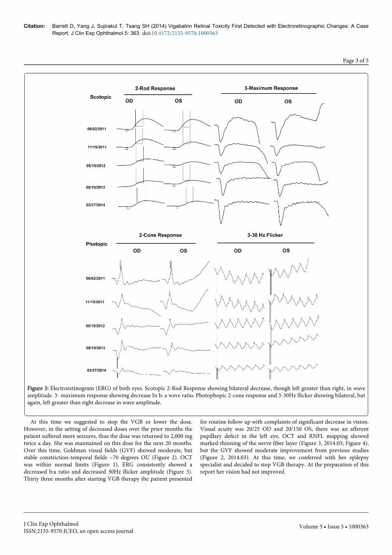
Figure 3: Electroretinogram (ERG) of both eyes. Scotopic 2-Rod Response showing bilateral decrease, though left greater than right, in waveamplitude. 3- maximum response showing decrease In b: a wave ratio. Photophopic 2-cone response and 3-30Hz flicker showing bilateral, butagain, left greater than right decrease in wave amplitude.
At this time we suggested to stop the VGB or lower the dose.However, in the setting of decreased doses over the prior months thepatient suffered more seizures, thus the dose was returned to 2,000 mgtwice a day. She was maintained on this dose for the next 20 months.Over this time, Goldman visual fields (GVF) showed moderate, butstable constriction-temporal fields ~70 degrees OU (Figure 2). OCTwas within normal limits (Figure 1). ERG consistently showed adecreased b:a ratio and decreased 30Hz flicker amplitude (Figure 3).Thirty three months after starting VGB therapy the patient presented
for routine follow up with complaints of significant decrease in vision.Visual acuity was 20/25 OD and 20/150 OS, there was an afferentpupillary defect in the left eye, OCT and RNFL mapping showedmarked thinning of the nerve fiber layer (Figure 3, 2014.03; Figure 4),but the GVF showed moderate improvement from previous studies(Figure 2, 2014.03). At this time, we conferred with her epilepsyspecialist and decided to stop VGB therapy. At the preparation of thisreport her vision had not improved.
Citation: Barrett D, Yang J, Sujirakul T, Tsang SH (2014) Vigabatrin Retinal Toxicity First Detected with Electroretinographic Changes: A CaseReport. J Clin Exp Ophthalmol 5: 363. doi:10.4172/2155-9570.1000363
Page 3 of 5
J Clin Exp OphthalmolISSN:2155-9570 JCEO, an open access journal
Volume 5 • Issue 5 • 1000363
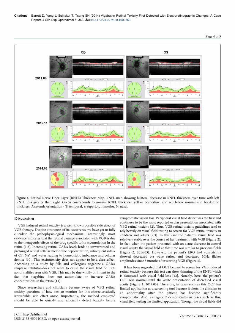
Figure 4: Retinal Nerve Fiber Layer (RNFL) Thickness Map. RNFL map showing bilateral decrease in RNFL thickness over time with leftRNFL loss greater than right. Green corresponds to normal RNFL thickness, yellow borderline, and red below normal and borderlinethickness. Anatomic orientation - T: temporal, S: superior, I: inferior, N: nasal.
DiscussionVGB induced retinal toxicity is a well-known possible side effect of
VGB therapy. Despite awareness of its occurrence we have yet to fullyelucidate the pathophysiological mechanism. Interestingly, mostevidence indicates that the retinal damage associated with VGB is dueto the therapeutic effects of the drug specific to its accumulation in theretina [1,6]. Increasing retinal GABA levels leads to unwarranted andprolonged retinal cellular membrane depolarization, subsequent influxof Cl-, Na+ and water leading to homeostatic imbalance and cellulardemise [10]. This excitotoxicity does not appear to be a class effect.According to a study by Sills and colleagues tiagabine-a GABAreuptake inhibitor-does not seem to cause the visual field or ERGabnormalities seen with VGB. This may be due wholly or in part to thefact that tiagabine does not accumulate or increase GABAconcentrations in the retina [11].
Since researchers and clinicians became aware of VBG retinaltoxicity questions of how best to monitor for this characteristicallyirreversible side effect arose. Importantly, the method employedshould be able to quickly and efficiently detect toxicity before
symptomatic vision loss. Peripheral visual field defect was the first andcontinues to be the most reported ocular presentation associated withVBG retinal toxicity [2]. Thus, VGB retinal toxicity guidelines tend torely heavily on visual field testing to screen for VGB retinal toxicity inchildren and adults [2,3]. In this case the patient’s visual field wasrelatively stable over the course of her treatment with VGB (Figure 2).In fact, when the patient presented with an acute decrease in centralvisual acuity the visual field at that time was similar to previous fields(Figure 2, 2014.03). However, the patient’s ERG had consistentlyshowed decreased b:a wave ratios, and decreased 30Hz flickeramplitudes since 5 months after starting VGB (Figure 3).
It has been suggested that OCT be used to screen for VGB inducedretinal toxicity because this test can show thinning of the RNFL whichis associated with visual field loss [12]. Notably, here, the patient’sOCT was normal until the acute presentation of decreased visualacuity (Figure 1, 2014.03). Therefore, in cases such as this OCT haslimited application as a screening tool because it alerts the clinician toan abnormality after the patient has become significantlysymptomatic. Also, as Figure 2 demonstrates in cases such as this,visual field testing has limited application. Though the visual fields did
Citation: Barrett D, Yang J, Sujirakul T, Tsang SH (2014) Vigabatrin Retinal Toxicity First Detected with Electroretinographic Changes: A CaseReport. J Clin Exp Ophthalmol 5: 363. doi:10.4172/2155-9570.1000363
Page 4 of 5
J Clin Exp OphthalmolISSN:2155-9570 JCEO, an open access journal
Volume 5 • Issue 5 • 1000363

show some constriction it was mild and unreliable as the patient hadsomewhat improved visual fields when she presented with an acutedeterioration in visual acuity (Figure 2). Moreover, this case is uniquein that the patient’s visual symptoms were much more pronounced inthe left eye when most cases report bilateral involvement. Because weare in the infancy of understanding VGB induced retinal toxicity thedata is limited on how to limit toxicity in patients who cannot stop themedication. Some groups using animal studies have suggested thatsupplementation of an amino acid- taurine may help to prevent ortreat VGB retinal toxicity given that taurine plays a role in retinalexcitability and free radical generation [6]. However, the authorsconcede more studies are necessary before a widespreadrecommendation can be supported. Moreover, other groups havedemonstrated the role of phototransduction in enhancing vigabatrintoxicity. In this study, mice with altered phototransduction pathways-thus a decreased ability to perceive light-have decreased incidence ofVGB retinal toxicity [1]. This observation suggests decreasing lightexposure i.e by wearing sunglasses or avoiding excess light might helptreat and/or prevent VGB retinal toxicity.
In sum, this report details a case of VGB retinal toxicity which wasfirst identified via ERG abnormalities. It provides a snapshot of thecomplex world of retinal physiology and the drugs that can affect theretina. This case also affirms that monitoring for VGB retinal toxicityshould not be a one-size-fits-all approach. Here, as a single modalityERG demonstrated retinal dysfunction early-before OCT andperimetry-and before the patient was visually symptomatic. Thistestifies to the usefulness of this exam to screen for and monitor VGBinduced retinal toxicity. Ideally, we recommend cessation of VGBwhen ERG changes occur. However, we understand that may not beeasily done or possible for some patients whose seizures return orworsen-as it was in this case-with decreased doses or cessation of VGB.Moreover, in patients with visual field or visual acuity abnormalitieswe agree with current guidelines that an evaluation of the risks andbenefits of decreased dose or cessation of VGB must be weighedagainst the effect of the vision changes on the patient’s functionality.More work is necessary to better carve out the exact mechanism ofVGB retinal toxicity, how to best monitor for retinal damage beforesymptomatic visual loss and how to treat and even prevent this sideeffect.
AcknowledgmentThe research is supported by NIH core grants 5P30CA013696,
5P30EY019007 and unrestricted funds from Research to PreventBlindness, New York, New York, R01EY018213, New York State(N09G-302), and the Foundation Fighting Blindness New YorkRegional Research Center Grant (C-NY05-0705-0312).
References1. Yang J, Naumann MC, Tsai YT, Tosi J, Erol D, et al. (2012) Vigabatrin-
induced retinal toxicity is partially mediated by signaling in rod and conephotoreceptors. PLoS One 7: e43889.
2. Willmore LJ, Abelson MB, Ben-Menachem E, Pellock JM, Shields WD(2009) Vigabatrin: 2008 update. Epilepsia 50: 163-173.
3. Hawker MJ, Astbury NJ (2008) The ocular side effects of vigabatrin(Sabril): information and guidance for screening. Eye (Lond) 22:1097-1098.
4. Nousiainen I, Kälviäinen R, Mäntyjärvi M (2000) Color vision in epilepsypatients treated with vigabatrin or carbamazepine monotherapy.Ophthalmology 107: 884-888.
5. Krauss GL, Johnson MA, Miller NR (1998) Vigabatrin-associated retinalcone system dysfunction: electroretinogram and ophthalmologicfindings. Neurology, 50: 614-618.
6. Heim MK, Gidal BE (2012) Vigabatrin-associated retinal damage:potential biochemical mechanisms. Acta Neurol Scand 126: 219-228.
7. Audo I, Warchol ME (2012) Retinal and cochlear toxicity of drugs: newinsights into mechanisms and detection. Curr Opin Neurol 25: 76-85.
8. Lima LH, Cella W, Brue C, Tsang SH (2010) Unilateral electronegativeERG in a presumed central retinal artery occlusion. Clin Ophthalmol 4:1311-1314.
9. Graniewski-Wijnands HS, van der Torren K (2002) Electro-ophthalmological recovery after withdrawal from vigabatrin. DocOphthalmol 104: 189-194.
10. Wang Y, Qin ZH (2010) Molecular and cellular mechanisms ofexcitotoxic neuronal death. Apoptosis 15: 1382-1402.
11. Sills GJ, Patsalos PN, Butler E, Forrest G, Ratnaraj N, et al. (2001) Visualfield constriction: accumulation of vigabatrin but not tiagabine in theretina. Neurology 57: 196-200.
12. Kjellstrom U, Andreasson S, Ponjavic V (2014) Attenuation of the retinalnerve fibre layer and reduced retinal function assessed by opticalcoherence tomography and full-field electroretinography in patientsexposed to vigabatrin medication. Acta Ophthalmologica, 92: 149-157.
Citation: Barrett D, Yang J, Sujirakul T, Tsang SH (2014) Vigabatrin Retinal Toxicity First Detected with Electroretinographic Changes: A CaseReport. J Clin Exp Ophthalmol 5: 363. doi:10.4172/2155-9570.1000363
Page 5 of 5
J Clin Exp OphthalmolISSN:2155-9570 JCEO, an open access journal
Volume 5 • Issue 5 • 1000363