rcr_4_2_79_82-4
DESCRIPTION
nnnTRANSCRIPT
Respir Case Rep 2015;4(2):79-82 DOI: 10.5505/respircase.2015.65487
CASE REPORT OLGU SUNUMU
79
Ümran Toru1, Mehmet Sait Incebıyık
2, Mehmet Suat Patlakoğlu
3, Tuncay Vatansever
2
It is a known fact that the risk of iatrogenic pneumo-
thorax is high after invasive procedures in patients
with emphysema. However, pneumothorax is rarely
seen as a complication of a non-invasive procedure
such as spirometry, which is the key method for the
diagnosis and follow-up of chronic obstructive pul-
monary disease. Herein, we present a case of pneu-
mothorax that developed as a complication of spi-
rometry in a patient with emphysema.
Key words: Chronic obstructive pulmonary disease,
emphysema, spirometry, pneumothorax.
Chronic obstructive pulmonary disease (COPD) is
characterized by the gradual progression of irre-
versible airflow obstruction (1). Pulmonary function
tests (PFTs) provide objective and quantifiable
measures of lung function (2). Spirometry, which is
a part of PFTs, is the key for the diagnosis and
management of COPD (3).
Pneumothorax is the presence of air in the pleural
cavity (4). According to the etiology, pneumotho-
races are classified as spontaneous, traumatic,
Amfizemli hastalarda invazif prosedürlerden sonra
iatrojenik pnömotoraks riskinin yüksek olduğu bilinen
bir gerçektir. Hâlbuki kronik obstrüktif akciğer hasta-
lığının erken tanısı ve takibi için anahtar yöntem olan
spirometri gibi non-invazif bir prosedürün komplikas-
yonu olarak pnömotoraks nadiren görülmektedir. Biz
burada amfizemli bir hastada spirometrinin kompli-
kasyonu olarak gelişen pnömotoraks olgusunu sun-
duk.
Anahtar Sözcükler: Kronik obstrüktif akciğer hastalığı,
amfizem, spirometri, pnömotoraks.
and iatrogenic (5). Although iatrogenic pneumo-
thorax commonly occurs due to invasive diagnostic
and treatment methods, it can also occur as a
possible sequel to an abrupt increase in transpul-
monary pressure due to the spirometry (6,7).
Herein, we report a 71-year-old patient who was
hospitalized for COPD exacerbation and devel-
oped pneumothorax after undergoing spirometry
in the stable period.
1Department of Chest Diseases, Dumlupınar University Faculty of
Medicine, Kütahya, Turkey
2Department of Chest Diseases, Kütahya Evliya Çelebi Training
and Research Hospital, Kütahya, Turkey
3Department of Thoracic Surgery, Kütahya Evliya Çelebi Training
and Research Hospital, Kütahya, Turkey
1Dumlupınar Üniversitesi Tıp Fakültesi, Göğüs Hastalıkları
Ana Bilim Dalı, Kütahya
2Kütahya Evliya Çelebi Eğitim ve Araştırma Hastanesi, Göğüs
Hastalıkları Kliniği, Kütahya
3Kütahya Evliya Çelebi Eğitim ve Araştırma Hastanesi, Göğüs
Cerrahisi Kliniği, Kütahya
Submitted (Başvuru tarihi): 21.09.2014 Accepted (Kabul tarihi): 20.11.2014
Correspondence (İletişim): Ümran Toru, Department of Chest Diseases, Dumlupınar University Faculty of Medicine, Kütahya, Turkey
e-mail: [email protected]
RES
PIR
ATO
RY
CA
SE R
EPO
RTS
Respiratory Case Reports
Cilt - Vol. 4 Sayı - No. 2 80 80
CASE
A 71-year old male patient was admitted to our emer-
gency services with complaints of dyspnea. The postero-
anterior (PA) chest x-ray was consistent with emphysema
(Figure 1), so the patient was hospitalized with a pre-
diagnosis of COPD exacerbation. Upon follow-up, the
clinical condition of the patient improved and he under-
went spirometry in the stable period. After spirometry he
complained of sudden-onset left-sided chest pain. In the
physical examination, breath sounds were diminished in
the left lung. Electrocardiogram and cardiac enzymes
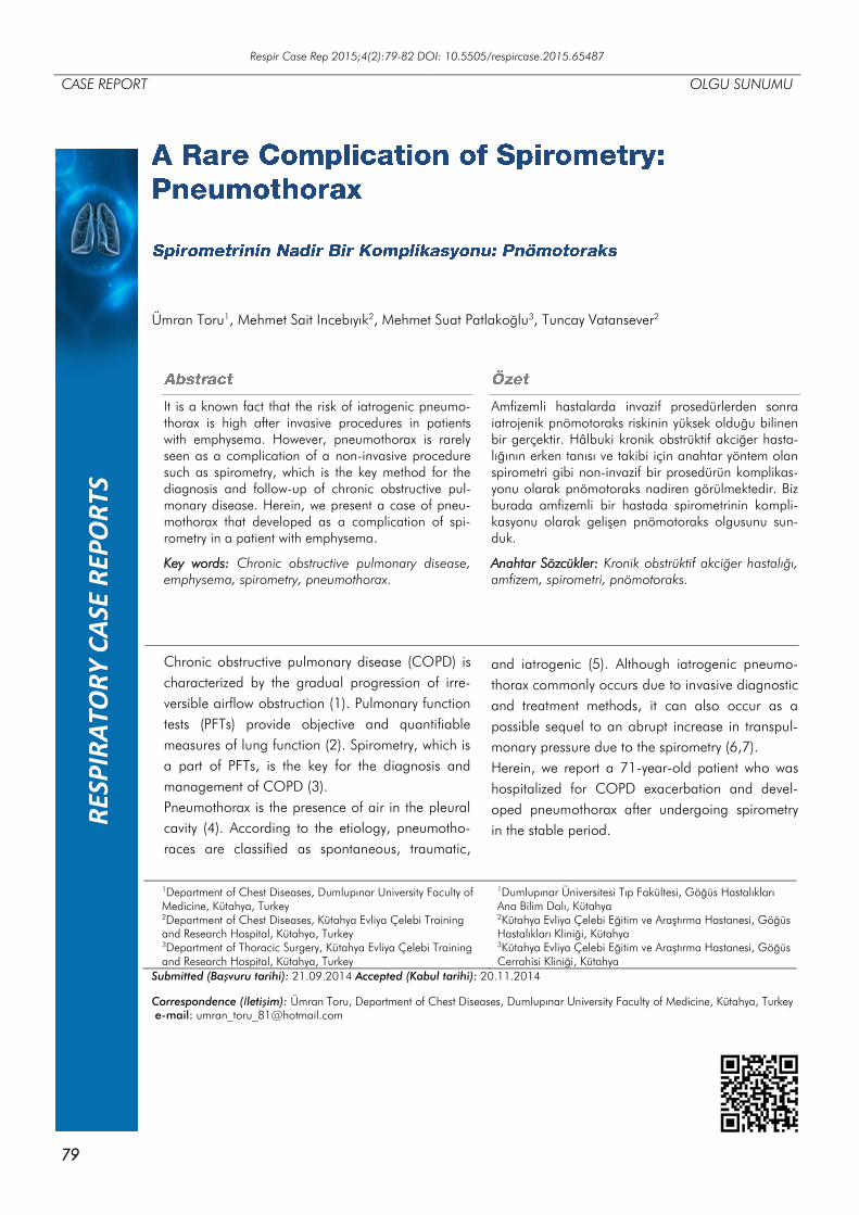
were detected as normal. The PA chest x-ray showed
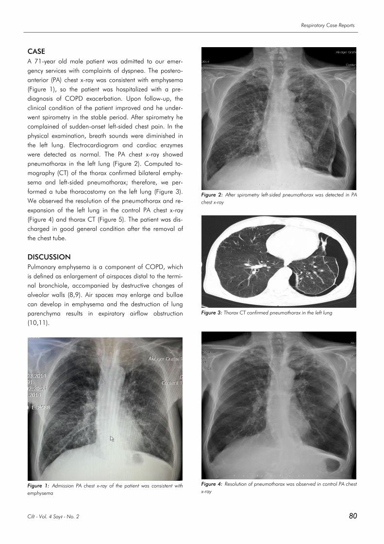
pneumothorax in the left lung (Figure 2). Computed to-
mography (CT) of the thorax confirmed bilateral emphy-
sema and left-sided pneumothorax; therefore, we per-
formed a tube thoracostomy on the left lung (Figure 3).
We observed the resolution of the pneumothorax and re-
expansion of the left lung in the control PA chest x-ray
(Figure 4) and thorax CT (Figure 5). The patient was dis-
charged in good general condition after the removal of
the chest tube.
DISCUSSION
Pulmonary emphysema is a component of COPD, which
is defined as enlargement of airspaces distal to the termi-
nal bronchiole, accompanied by destructive changes of
alveolar walls (8,9). Air spaces may enlarge and bullae
can develop in emphysema and the destruction of lung
parenchyma results in expiratory airflow obstruction
(10,11).
Figure 1: Admission PA chest x-ray of the patient was consistent with
emphysema
Figure 2: After spirometry left-sided pneumothorax was detected in PA
chest x-ray
Figure 3: Thorax CT confirmed pneumothorax in the left lung
Figure 4: Resolution of pneumothorax was observed in control PA chest
x-ray

A Rare Complication of Spirometry: Pneumothorax | Toru et al.
81 www.respircase.com
The airflow obstruction in patients with suspected emphy-
sema can be assessed by spirometry, which is the best
diagnostic test to evaluate lung function in COPD (1). To
date, spirometry remains the most effective means of
identification and assessment of the course of COPD and
responses to therapy (3). In our case, the patient was
hospitalized with a pre-diagnosis of COPD exacerbation.
After medical treatment, in the stable period of the patient,
we aimed to assess his response to therapy and measure
the degree of airflow obstruction by spirometry.
Pneumothorax, which is the presence of air in the pleural
cavity, may be traumatic, spontaneous with or without
underlying lung disease, or iatrogenic (4). Spontaneous
pneumothoraces that occur without recognized lung dis-
ease are called primary spontaneous pneumothoraces
(PSP), whereas secondary spontaneous pneumothorax
(SSP) occurs as a complication of an underlying lung
disease like COPD (4,5,12). It was reported that emphy-
sema and bullae formation in COPD may lead to the
occurrence of SSP (13,14).
The clinical diagnosis of pneumothorax is based on the
anamnesis, physical examination, and radiological inves-
tigations (13,15,16). The clinical results are dependent
on the degree of collapse on the affected lung (17). Alt-
hough 10% of pneumothoraces are asymptomatic, the
symptoms due to the symptomatic pneumothorax consist
of acute chest pain, breathlessness, tachypnea, and tach-
ycardia (18). Additionally, a shift of the mediastinum and
haemodynamic instability may occur if the pneumothorax
is significant (17).
Figure 5: Control Thorax CT showed re-expansion of the left lung after
tube thoracostomy
Iatrogenic pneumothorax commonly occurs due to the
invasive diagnostic and treatment methods such as trans-
thoracic needle aspiration, subclavian vein catheterization,
thoracentesis, transbronchial lung biopsy, pleural biopsy,
and mechanical ventilation (6). Additionally, iatrogenic
pneumothorax may occur due to non-invasive diagnostic
methods, such as spirometry. Breathing maneuvers with a
spirometer decreases pleural pressure, increases
transpulmonary pressure, and produces large negative
swings in intrathoracic pressures, which is similar to the
Müller maneuver (19,20). A possible complication of an
abrupt increase in transpulmonary pressure is pneumo-
thorax secondary to the barotrauma (7). The clinical pro-
file of iatrogenic pneumothorax is affected by any under-
lying diseases, the health conditions of the patient, and
the etiology of pneumothorax. For instance, a patient in
poor general condition or with an underlying disease may
experience severe symptoms, even from a small volume
of pneumothorax (6). In our case, the patient underwent
spirometry and after the procedure he complained of
acute left-sided chest pain. With the sudden onset of
chest pain, we performed PA chest x-ray and thorax CT,
and detected left-sided pneumothorax. Although there
was partial pneumothorax in the left lung, he became
symptomatic immediately due to the underlying emphy-
sema. We were aware that there was an underlying dis-
ease like emphysema, but pneumothorax did not occur
spontaneously. Therefore, we explained the etiology of
pneumothorax as iatrogenic, which is a complication of
spirometry. To our knowledge, the number of case re-
ports presenting pneumothorax as a complication of
spirometry is very limited in the literature.
Depending on the clinical profile or underlying disease of
the patient, non-invasive treatment methods including
observation, simple aspiration, or chest tube placement
using a small catheter are recommended (6). However,
underwater seal drainage is the classical treatment meth-
od that is used for the treatment of pneumothorax; in
particular there is a 99 % rate of success reported in
iatrogenic pneumothorax by tube thoracostomy (21). In
the current case, we performed tube thoracostomy on our
patient and achieved a good clinical response.
As a conclusion, the sudden occurrence of respiratory
symptoms after spirometry, especially in patients with
underlying COPD, should be evaluated carefully in terms
of pneumothorax.
CONFLICTS OF INTEREST
None declared.
AUTHOR CONTRIBUTIONS
Concept - Ü.T., M.S.İ., M.S.P., T.V.; Planning and Design
- Ü.T., M.S.İ., M.S.P., T.V.; Supervision - Ü.T., M.S.İ.,
M.S.P., T.V.; Funding - Ü.T., M.S.İ., M.S.P., T.V.; Materi-

Respiratory Case Reports
Cilt - Vol. 4 Sayı - No. 2 82 82
als - Ü.T., M.S.İ.; Data Collection and/or Processing -
Ü.T., M.S.İ., M.S.P.; Analysis and/or Interpretation - Ü.T.,
T.V.; Literature Review - Ü.T.; Writing - Ü.T.; Critical
Review - Ü.T., M.S.İ., M.S.P., T.V.
YAZAR KATKILARI
Fikir - Ü.T., M.S.İ., M.S.P., T.V.; Tasarım ve Dizayn - Ü.T.,
M.S.İ., M.S.P., T.V.; Denetleme - Ü.T., M.S.İ., M.S.P.,
T.V.; Kaynaklar - Ü.T., M.S.İ., M.S.P., T.V.; Malzemeler -
Ü.T., M.S.İ.; Veri Toplama ve/veya İşleme - Ü.T., M.S.İ.,
M.S.P.; Analiz ve/veya Yorum - Ü.T., T.V.; Literatür Tara-
ması - Ü.T.; Yazıyı Yazan - Ü.T.; Eleştirel İnceleme - Ü.T.,
M.S.İ., M.S.P., T.V.
REFERENCES
1. Dewar M, Curry RW Jr. Chronic obstructive pulmonary
disease: diagnostic considerations. Am Fam Physician
2006; 73:669-76.
2. Crapo RO. Pulmonary-function testing. N Engl J Med
1994; 331:25-30. [CrossRef]
3. Petty TL. The history of COPD. Int J Chron Obstruct
Pulmon Dis 2006; 1:3-14. [CrossRef]
4. Sahn SA, Heffner JE. Spontaneous pneumothorax. N Engl
J Med 2000; 342:868–74. [CrossRef]
5. Haynes D, Baumann MH. Pleural controversy: aetiology
of pneumothorax. Respirology 2011; 16:604-10.
[CrossRef]
6. Choi WI. Pneumothorax. Tuberc Respir Dis (Seoul) 2014;
76:99-104. [CrossRef]
7. West JB. Invited review: pulmonary capillary stress failure.
J Appl Physiol (1985) 2000; 89:2483-9.
8. Pauwels RA, Rabe KF. Burden and clinical features of
chronic obstructive pulmonary disease (COPD). Lancet
2004; 364:613–20. [CrossRef]
9. American Thoracic Society. Chronic bronchitis, asthma,
and pulmonary emphysema: statement by the Committee
on Diagnostic Standards for Nontuberculous Respiratory
Disease. Am Rev Respir Dis 1962; 85:762–8.
10. Tuddenham WJ. Glossary of terms for thoracic radiology:
recommendations of the Nomenclature Committee of the
Fleishner Society. AJR Am J Roentgenol 1984; 143:509–
17. [CrossRef]
11. Mohebbi I, Hassani E, Salarilak S, Bahrami AR. Do bul-
lae and emhysema increase the risk of pneumothorax in
silicosis? J Occup Med Toxicol 2007; 2:8. [CrossRef]
12. Gupta D, Hansell A, Nichols T, Duong T, Ayres JG, Stra-
chan D. Epidemiology of pneumothorax in England.
Thorax 2000; 55:666–71. [CrossRef]
13. Al-Qudah A. Treatment options of spontaneous pneumo-
thorax. Indian J Chest Dis Allied Sci 2006; 48:191–200.
14. Davis SG. Silicosis. In: Hendrick DJ, Burge PS, Becket WS,
Churg A, eds. Occupational Disorders of the Lung
Recognition, Management and Prevention Vol 1. London:
WB Saunders; 2002:105–127.
15. Strobel SL. Pathologic quiz case: recurrent spontaneous
pneumothorax in an industrial worker. Arch Pathol Lab
Med 2002; 126:749–50.
16. Weill H, Jones RN, Parkes WR. Silicosis and related dis-
eases. In: Parkes WR, edt. Occupational Lung Disorders.
Oxford, UK: Butterworth-Heinemann; 1994:285–339.
17. Dimitroulis G. A simple classification of orthognathic sur-
gery complications. Int J Adult Orthodon Orthognath
Surg 1998; 13:79–87.
18. Bertossi D, Malchiodi L, Turra M, Bondi V, Albanese M,
Lucchese A, et al. Bilateral pneumothorax after orthog-
natic surgery. Dent Res J (Isfahan) 2012; 9:S242-5.
[CrossRef]
19. Toumpanakis D, Kastis GA, Zacharatos P, Sigala I,
Michailidou T, Kouvela M, et al. Inspiratory resistive
breathing induces acute lung injury. Am J Respir Crit
Care Med 2010; 182:1129-36. [CrossRef]
20. Kenny JE, Kuschner WG. Pneumothorax caused by ag-
gressive use of an incentive spirometer in a patient with
emphysema. Respir Care 2013; 58:e77-9. [CrossRef]
21. Martin T, Fontana G, Olak J, Ferguson M. Use of pleural
catheter for the management of simple pneumothorax.
Chest 1996; 110:1169-72. [CrossRef]