rapid spectrophotometric method for determining …yzuo/documents/8-xinru-2014.pdfrapid...
TRANSCRIPT
Rapid Spectrophotometric Method for Determining Surface FreeEnergy of Microalgal CellsXinru Zhang,†,‡,⊥ Zeyi Jiang,*,†,§ Mengyin Li,† Xinxin Zhang,†,∥ Ge Wang,⊥ Aihui Chou,† Liang Chen,†
Hai Yan,# and Yi Y. Zuo*,‡
†School of Mechanical Engineering, University of Science and Technology Beijing, Beijing, 100083, China‡Department of Mechanical Engineering, University of Hawaii at Manoa, Honolulu, Hawaii 96822, United States§Engineering Research Centre for Energy Saving and Environmental Protection, University of Science and Technology Beijing,Beijing, 100083, China∥Beijing Key Laboratory for Energy Saving and Emission Reduction of Metallurgical Industry, University of Science and TechnologyBeijing, Beijing, 100083, China⊥School of Materials Science and Engineering, University of Science and Technology Beijing, Beijing, 100083, China#School of Chemistry and Biological Engineering, University of Science and Technology Beijing, Beijing, 100083, China
*S Supporting Information
ABSTRACT: Microalgae are one of the most promising renewable energysources with environmental sustainability. The surface free energy of microalgalcells determines their biofouling and bioflocculation behavior and hence playsan important role in microalgae cultivation and harvesting. To date, the surfaceenergetic properties of microalgal cells are still rarely studied. We developed anovel spectrophotometric method for directly determining the surface freeenergy of microalgal cells. The principles of this method are based on analyzingcolloidal stability of microalgae suspensions. We have shown that this methodcan effectively differentiate the surface free energy of four microalgal strains, i.e.,marine Chlorella sp., marine Nannochloris oculata, freshwater autotrophicChlorella sp., and freshwater heterotrophic Chlorella sp. With advantages ofhigh-throughput and simplicity, this new spectrophotometric method has the potential to evolve into a standard method formeasuring the surface free energy of cells and abiotic particles.
Microalgae are one of the most promising renewable energysources due to their fast growth rate, low nutrient
requirement, and ability to accumulate high lipid andcarbohydrate contents.1,2 Moreover, microalgae cultivation hasbeneficial environmental impacts, including fast carbon dioxidemitigation and effective wastewater treatment.3 To optimizemicroalgae cultivation and harvesting, there is an urgent need tounderstand the physicochemical and rheological properties ofmicroalgal cells and their culturing medium, such as theircolloidal viscosity, thermophysical properties, and surfaceproperties.4,5
The surface free energy of microalgal cells plays a crucial role intheir cultivation and harvesting. First, the surface free energydetermines the formation and development of microalgalbiofilms.5,6 While cultivating in a photobioreactor, microalgalcells tend to form highly productive biofilms over solid surfaces.This microalgal biofouling can significantly lower the perform-ance of the photobioreactor and hinder cell proliferation in theculture medium.6,7 Second, the surface free energy of microalgalcells determines their self-aggregation and sedimentation, thusdirectly influencing microalgae harvesting.5,8,9 Hence, under-standing the surface free energy of microalgal cells can have aprofound impact on the design of more effective photo-
bioreactors for large-scale algal cultivation and on theoptimization of algal harvesting and downstream processingtechniques toward a more cost-effective production of biofuels.Despite its importance, the surface free energy of microalgal
cells is rarely reported.5,10 One reason is the lack of easy-to-usemethods for determining the surface free energy of live cells. Todate, nearly all quantitative determinations of surface free energyof particles or cells (mainly bacterial cells) rely on the contactangle method.11−15 In this method, contact angles are measuredon filtrated cell lawns using the sessile drop technique, followedby theoretical interpretation that indirectly determines thesurface free energy of cell layers, which is in turn assumed to beidentical to the surface free energy of individual cells. Although itis a plausible method, it is well-known that contact anglemeasurement on live cells and its energetic interpretation are nottrivial tasks, which can introduce significant methodologicalcomplications for nonexperts.16−19
In this paper, we have developed a novel easy-to-usespectrophotometric method for directly determining surface
Received: May 24, 2014Accepted: August 13, 2014
Article
pubs.acs.org/ac
© XXXX American Chemical Society A dx.doi.org/10.1021/ac501940h | Anal. Chem. XXXX, XXX, XXX−XXX

free energy of microalgae cells. We first verified the accuracy ofthis method by studying polystyrene microbeads with knownsurface free energy. Subsequently, we used this method tomeasure the surface free energy of four widely used microalgalstrains in biofuel production, i.e., marine Chlorella sp., marineNannochloris oculata, freshwater autotrophic Chlorella sp., andfreshwater heterotrophic Chlorella sp. We showed that thisspectrophotometric method is highly sensitive in differentiatingthe surface free energy of various microalgal strains. Withcombined advantages of high-throughput and simplicity, thisspectrophotometric method has the potential to evolve into astandard method for measuring surface free energy of cells andabiotic particles.
■ EXPERIMENTAL SECTIONPrinciples ofMeasurement. Fundamental principles of this
spectrophotometric method follow the sedimentation volumemethod developed by Neumann and co-workers in 1980s.20−22
Without loss of generality, as demonstrated in Figure 1, the
colloidal stability of a particle suspension is determined by thebalance between the electrostatic repulsion and the van derWaals attraction, as predicted by the DLVO theory.23 Thethermodynamic free energy for particle coagulation can beexpressed as
Δ = + −E E E E211 22 12 (1)
where 1 denotes the suspending medium and 2 denotes theparticles.The contribution of van der Waals interactions in this
coagulation process can be attributed to the determination ofthe interaction Hamaker constant A212
= + −A A A A2212 11 22 12 (2)
Applying the geometrical mean combining rule, A12 =(A11A22)
1/2, the Hamaker constant of two particles interactingacross the suspending medium is
= −A A A( )212 11 222
(3)
It should be noted that this geometrical mean combining ruleis derived from the Lifshitz theory and has been widely used insolution theory to calculate heterogeneous interactions.23
However, it is applicable only when dispersion forces dominatethe interactions but breaks down when applied to media withhigh dielectric constants.23,24
Relating the Hamaker constant to surface tensions (γ) using A= 24πD2γ,23,24 it yields
π γ γ= −A D24 ( )2122
1 22
(4)
where D is the minimum separation distance between twosurfaces.Equation 4 implies that when the surface tension of the
suspending medium (denoted by 1) and the surface free energyof the dispersed particles (denoted by 2) are equal to each other,the van der Waals attractions between particles (across themedium) are reduced to a minimum. Consequently, the particlesare expected to be largely dispersed in the medium withoutsignificant aggregation, thus resulting in a minimum sedimenta-tion.To implement this principle, one can disperse particles in a
series of liquids with known surface tension values that cover thepossible surface free energy of the particles. When measuring thesedimentation volume or mass of the particles, as in the originalmethod developed by Neumann and co-workers,20−22 aminimum is expected at which the surface tension of the liquidis equal to the surface free energy of the particles (see Figure 1).However, the measurement of sedimentation volume/mass in aseries of suspending liquids is a time-consuming procedure thatrequires careful handling of individual samples with highlysensitive balances. These difficulties have rendered thesedimentation volume/mass method unpopular in the pastthree decades.To circumvent these difficulties, here we propose a novel
approach of implementing the sedimentation principles. Insteadof analyzing the sediment, we focus on the supernatant andmeasure the absorbance of transmitted light through thesupernatant after particle sedimentation. Since the amount ofsedimentation should be inversely related to the amount of freeparticles suspended in the supernatant, we hypothesize that amaximum optical density (OD) should coincide with theminimum sedimentation volume/mass (see Figure 1). Thisspectrophotometric method allows us to take advantage of thehighly sensitive and high throughput capacity of a microplatereader to quickly determine the surface free energy of particles.
Algal Cultivation and Sample Preparation. The algalstrains used in this study were marine Chlorella sp., marineNannochloris oculata, freshwater autotrophic Chlorella sp., andfreshwater heterotrophic Chlorella sp. The marine Chlorella sp.and marine Nannochloris oculata were both cultivated in the f/2medium. The freshwater autotrophic Chlorella sp. was cultivatedin Bold’s Basal medium. The freshwater heterotrophic Chlorellasp. was cultivated in the Fitzgerald medium. All culture mediawere prepared on the basis of the procedures described byAndersen.25 All microalgal strains were grown in 500 mL flaskscontaining 100 mL of culture medium at 25 ± 1 °C under
Figure 1. Schematic of the experimental principles of the sedimentationmethod. Colloidal stability of a particle suspension is determined by thebalance between the electrostatic repulsion and van derWaals attraction,as predicted by the DLVO theory. Dispersing particles in a liquid ofsurface tension similar to the surface free energy of the particlesminimizes the van der Waals attraction between particles across thesuspending liquid, thus resulting in a minimum amount of particlesedimentation. Since the amount of sedimentation is inversely related tothe amount of free particles suspended in the supernatant, a maximumlight absorbance should coincide with the minimum sedimentationvolume/mass. Therefore, the surface tension of the liquid that results ina minimum sedimentation volume/mass or a maximum optical density(OD) should be close to the surface free energy of the particles.
Analytical Chemistry Article
dx.doi.org/10.1021/ac501940h | Anal. Chem. XXXX, XXX, XXX−XXXB

continuous irradiance of 100 μmol/m2·s. The suspensions werecultured for about 7 days before cells were harvested.Samples used in this study were prepared from actively
growing cultures during their exponential growth phase. Afterharvesting, the cultivated microalgal cell suspensions werecentrifuged at 4000 rpm for 3 min at room temperature toremove cell debris, followed by washing with phosphate buffersolution. After three rounds of centrifugation and washing, themicroalgal cells were resuspended in phosphate buffer solution toprepare the concentrated samples used in our experiments.Before surface free energy measurements, the concentratedmicroalgae suspensions were ultrasonically vibrated for 2 min tobreak aggregates and to remove air bubbles.
Algal Cell Characterization. Before determining theirsurface free energies, the microalgal cells were characterizedwith their colloidal properties, including primary size, hydro-dynamic size, and surface charge. The primary size wasdetermined by scanning electron microscopy (SEM) with theparticle size analyzed by ImageJ (NIH). The hydrodynamic sizewas determined by a laser particle sizer (S3500SI, Microtrac Inc.,Largo, FL). The surface charge was measured by a zeta potentialanalyzer (Nanotrac wave, Microtrac Inc.).
Implementation of the Spectrophotometric Method.Aseries of suspending liquids was prepared with binary mixtures ofethanol and ultrapure water at predefined mixing ratios. Surfacetensions of these binary liquids range from ∼22 mJ/m2 (for pureethanol) to∼73mJ/m2 (for pure water), which were determined
Table 1. Characterization of Microalgal Cells with Their Primary Diameter (DP) Determined by SEM, Hydrodynamic Diameter(DH) Determined by a Particle Sizer, and Zeta Potential (ζ)
Figure 2. Proof-of-principle: determination of the surface free energy of polystyrene microbeads. (a) Sedimentation mass as a function of time afteradding the polystyrene microbeads into suspending liquids of surface tensions 28.0, 33.6, and 39.6 mJ/m2, respectively. (b) Optical density (OD) as afunction of time in these three suspending liquids. (c) Particle size distributions in these three suspending liquids. (d) Sedimentation mass and OD as afunction of the surface tensions of a series of suspending liquids. After peak fitting, both the locations of the minimum sedimentation mass and themaximumOD consistently indicate that the surface free energy of the polystyrene microbeads is around 32−33mJ/m2, which is in good agreement withthe literature values.
Analytical Chemistry Article
dx.doi.org/10.1021/ac501940h | Anal. Chem. XXXX, XXX, XXX−XXXC

at room temperature with a Wilhelmy plate surface tensiometer(K100, Kruss GmbH, Germany). (Density and viscosity of theseliquids were also determined and listed in Table S1 of theSupporting Information.) This liquid surface tension rangecovers the surface free energy of most polymeric particles andbiological cells.12,24 When studying unknown particles/cells,coarse measurements with suspending liquids spanning a largesurface tension range were performed followed by finemeasurements with liquids in a narrower range of surfacetensions.Specifically, a trace amount of condensed microalgal cells (5
μL at 50−100 g/L) was added to a series of 10 to 12 clearlylabeled suspending liquids, each at 1 mL, and fully mixed. Toaccelerate the sedimentation process, the cell-containingsuspensions were centrifuged at 1000 rpm for 1 min. Aftercentrifugation, 200 μL of supernatant of each suspending liquidwas transferred from centrifuge tubes to a 96-well microplate.Optical densities of these supernatants were measured with amicroplate reader (Epoch, BioTek, Winooski, VT) at acharacteristic wavelength of 690 nm, determined in a separateexperiment. (See Figure S1 of the Supporting Information.) Themeasured OD690 was plotted against the surface tensions of thesuspending liquids. A maximum was determined by peak fittingexperimental points with extreme function in statistics usingOriginLab.26 Each measurement was repeated for at least 5 timesand averaged. For the purpose of comparison, we conductedparallel measurements of the sedimentation mass using theanalytical balance associated with the Kruss K100 surfacetensiometer.
■ RESULTS AND DISCUSSIONCharacterization of Microalgal Cells. The four microalgal
cells were characterized with their primary size, hydrodynamicsize, and surface charge. As shown in Table 1, all four microalgalcells have a nearly spherical shape with a mean primary diameter(DP) close to 3 μm, determined by SEM. The meanhydrodynamic diameter (DH) of these microalgal cells,determined with the particle sizer, ranges from 3.8 ± 0.8 μmfor freshwater autotrophic Chlorella sp. to 5.1 ± 0.9 μm forfreshwater heterotrophicChlorella sp. All four microalgal cells arenegatively charged with the zeta potential (ζ) ranges from −14.8± 2.1 mV for freshwater heterotrophic Chlorella sp. to −42.8 ±3.8 mV for marine Chlorella sp. These characteristic surfaceproperties ensure the applicability of the DLVO theory and ourmeasurement principles to these microalgal cells.Proof-of-Principle: Measuring Surface Free Energy of
Polystyrene Microbeads. Prior to measuring microalgal cells,we first validated our method by studying polystyrene microbe-ads with the similar size and surface charge as our microalgal cells.The studied polystyrene microbeads (Aladding Reagents Co.,Shanghai, China) have a mean diameter of ∼3 μm and a zetapotential of approximately −30 mV, both comparable to ourmicroalgal cells (Table 1). The surface free energy of polystyrenehas been well studied and reported from 30 to 33 mJ/m2 in theliterature.27,28
Figure 2 illustrates the experimental results pertinent tomeasuring the surface free energy of polystyrene microbeads.Figure 2a shows the change of the sedimentation mass within thefirst 7 min after adding the microbeads into three representativesuspending liquids whose surface tensions are 28.0, 33.6, and39.6 mJ/m2, respectively. It should be noted that, in ourexperiments, a series of 10 to 12 liquids with a range of surfacetensions from∼22 to∼50 mJ/m2 was used as suspending liquids
(see Table S1, Supporting Information, and Figure 2d).However, for the sake of clarity, only experimental results ofthree representative liquids are shown in Figure 2a−c. Surfacetensions of these three suspending liquids are lower than, closeto, and higher than the expected surface free energy of thepolystyrene microbeads.Figure 2a clearly shows that the sedimentation mass increases
steeply within the first minute after adding the microbeads intothe suspending liquids. After 1 min, sedimentation reachesequilibrium. The sedimentation mass in the liquid with theintermediate surface tension (i.e., 33.6 mJ/m2) was significantlylower than that in the other two suspending liquids. A similartrend was found with ODmeasurements. As shown in Figure 2b,the OD value levels off after 1 min of centrifugation. Theequilibrium OD value of the 33.6 mJ/m2 suspending liquid ishigher than that of the other two liquids.To verify that the different behaviors in sedimentation and
light absorbance (Figure 2a,b) are indeed due to the differentparticle aggregations as predicted by the theory shown in Figure1, we directly measured the particle size distribution in thesethree suspending liquids using a particle sizer. As shown in Figure2c, more than 50% of particles in the liquids of surface tension28.0 and 39.6 mJ/m2 has a mean diameter larger than 1000 μm,indicating significant particle aggregation/agglomeration. Theadditional 40% of particles in these two liquids is free particleswith a size of about 3 μm, plus a small portion (∼10%) minoragglomeration at∼20 μm. However, when dispersing particles ina liquid of surface tension of 33.6 mJ/m2, i.e., a liquid surfacetension close to the surface free energy of the polystyrenemicrobeads, a majority of the particles (87%) remains dispersedat ∼3 μm, with only a small portion (13%) of moderatelyaggregated particles at ∼20 μm. This direct characterization ofparticle size distributions validates our principles of measure-ment and explains the different behaviors of sedimentation andlight absorbance in these three representative suspending liquids(Figure 2a,b).Figure 2d shows the sedimentation mass and OD in a range of
suspending liquids of different surface tensions, measured at 5min after adding the polystyrene microbeads (i.e., at theequilibrium sedimentation). As predicted by theory (Figure 1),a minimum appears for the sedimentation mass which coincideswith a maximum OD. Using peak fitting, the minimumsedimentation mass is located at 32.4 mJ/m2 and the maximumOD is located at 32.7 mJ/m2. With one-way ANOVA analysis,these two measurements were not statistically significant (p >0.05). This indicates that the spectrophotometric method has acomparable accuracy as the sedimentation mass method. Bothmethods determine the surface free energy of polystyrenemicrobeads to be around 32−33 mJ/m2, which is in goodagreement with the literature values.It should be noted that, although it has a comparable accuracy,
the spectrophotometric method is much quicker and moresensitive than the sedimentation mass method. Taking advantageof the high throughput processing of a microplate reader, thespectrophotometric method can simultaneously implement ODmeasurements in all suspending liquids. Hence, the entiremeasurement can be completed within 10 min. Moreover, sincethe OD measurement is much more sensitive than measuringsedimentation mass or volume, the spectrophotometric methodonly uses less than 1% of the sample required for thesedimentation measurement.
Measuring Surface Free Energy of Microalgal Cells.Figure 3 shows the experimental results for determining the
Analytical Chemistry Article
dx.doi.org/10.1021/ac501940h | Anal. Chem. XXXX, XXX, XXX−XXXD
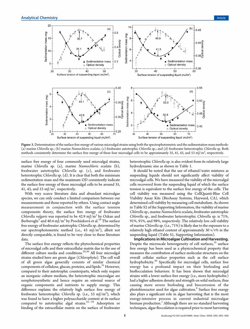
surface free energy of four commonly used microalgal strains,marine Chlorella sp. (a), marine Nannochloris oculata (b),freshwater autotrophic Chlorella sp. (c), and freshwaterheterotrophic Chlorella sp. (d). It is clear that both the minimumsedimentation mass and the maximum OD consistently indicatethe surface free energy of these microalgal cells to be around 35,41, 43, and 53 mJ/m2, respectively.With very scarce literature data and abundant microalgae
species, we can only conduct a limited comparison between ourmeasurements and those reported by others. Using contact anglemeasurement in conjunction with the surface tensioncomponents theory, the surface free energy of freshwaterChlorella vulgaris was reported to be 42.9 mJ/m2 by Ozkan andBerberoglu5 and 40.4 mJ/m2 by Prochazkova et al.10 The surfacefree energy of freshwater autotrophic Chlorella sp. determined byour spectrophotometric method (i.e., 43 mJ/m2), albeit notdirectly comparable, is found to be very close to these literaturedata.The surface free energy reflects the phytochemical properties
of microalgal cells and their extracellular matrix due to the use ofdifferent culture media and conditions.29,30 All four microalgalstrains studied here are green algae (Chlorophyta). The cell wallof all green algae generally consists of similar chemicalcomponents of cellulose, glucan, proteins, and lipids.31 However,compared to their autotrophic counterparts, which only requirean inorganic culture medium, the heterotrophic microalgae arenonphotosynthetic and hence require an external source oforganic components and nutrients to supply energy. Thisdifference explains the relatively high surface free energy offreshwater heterotrophic Chlorella sp. (i.e., 53 mJ/m2), whichwas found to have a higher polysaccharide content at its surfacecompared to autotrophic algal strains.32−34 Adsorption orbinding of the extracellular matrix on the surface of freshwater
heterotrophic Chlorella sp. is also evident from its relatively largehydrodynamic size as shown in Table 1.It should be noted that the use of ethanol/water mixtures as
suspending liquids should not significantly affect viability ofmicroalgal cells. We have measured the viability of the microalgalcells recovered from the suspending liquid of which the surfacetension is equivalent to the surface free energy of the cells. Thecell viability was measured using the CellQuanti-Blue CellViability Assay Kits (BioAssay Systems, Hayward, CA), whichdetermined cell viability by measuring cell metabolism. As shownin Table S2 of the Supporting Information, the viability of marineChlorella sp., marineNannochloris oculata, freshwater autotrophicChlorella sp., and freshwater heterotrophic Chlorella sp. is 71%,91%, 91%, and 98%, respectively. The relatively low cell viabilityof marineChlorella sp. (i.e., 71%) is likely due to the exposure to arelatively high ethanol content of approximately 30 v/v% in thesuspending liquid (Table S1, Supporting Information).
Implications inMicroalgae Cultivation andHarvesting.Despite the microscale heterogeneity of cell surfaces,35 surfacefree energy has been used as a physicochemical property thatmeasures the contribution of surface molecular structures on theoverall cellular surface properties such as the cell surfacehydrophobicity.36 Specifically for microalgal cells, surface freeenergy has a profound impact on their biofouling andbioflocculation behaviors. It has been shown that microalgalstrains with a lower surface free energy (i.e., more hydrophobic)had a higher adhesion density and strength on solid surfaces, thuscausing more severe biofouling and biocorrosion of thephotobioreactor used for algae cultivation.6 Surface free energyalso plays a significant role in algae harvesting that is the mostenergy-intensive process in current industrial microalgaebiomass production.1 Although there are no standard harvestingtechniques, algae flocculation is required prior to most harvesting
Figure 3.Determination of the surface free energy of various microalgal strains using both the spectrophotometric and the sedimentationmassmethods:(a) marine Chlorella sp.; (b) marine Nannochloris oculata; (c) freshwater autotrophic Chlorella sp.; and (d) freshwater heterotrophic Chlorella sp. Bothmethods consistently determine the surface free energy of these four microalgal cells to be approximately 35, 41, 43, and 53 mJ/m2, respectively.
Analytical Chemistry Article
dx.doi.org/10.1021/ac501940h | Anal. Chem. XXXX, XXX, XXX−XXXE

methods such as filtration, floatation, or sedimentation.1 It hasbeen shown that microalgal strains with a high surface free energy(i.e., hydrophilic) tend to form a planktonic state withoutflocculation due to the difficulty of excluding water between algalcells.6 Most of the current methods use the addition offlocculants such as multivalent cations or cationic polymers(e.g., chitosan) to neutralize the negative charge of microalgae,thus promoting flocculation.1 Our present study demonstratesthat algae flocculation can also be induced by increasing van derWaals attractions (see Figure 1 for schematic), albeit to a lesserdegree of commercial feasibility compared to the approach ofreducing the electrostatic repulsion. Nevertheless, understandingthe surface free energy of microalgal cells can help predict andmanipulate the fluccolation and sedimentation behavior ofmicroalgal cells, thus optimizing microalgae cultivation andharvesting techniques, reducing energy cost, and increasingenvironmental sustainability of microalgae production.
■ CONCLUSIONWe have developed a novel easy-to-use spectrophotometricmethod for directly determining the surface free energy ofmicroalgal cells. This method is a modified sedimentationtechnique based on the DLVO theory of colloidal stability,aggregation, and sedimentation. In comparison with the originalsedimentation method, the spectrophotometric method is muchquicker, more sensitive, and comparably accurate. We haveshown that this spectrophotometric method can effectivelydifferentiate the surface free energy of various microalgal strains.With combined advantages of high-throughput and simplicity,this method has the potential to evolve into a standard methodfor measuring surface free energy of cells and abiotic particles.
■ ASSOCIATED CONTENT*S Supporting InformationAdditional information as noted in the text. This material isavailable free of charge via the Internet at http://pubs.acs.org.
■ AUTHOR INFORMATIONCorresponding Authors*Phone/Fax: +86-10-62334971. E-mail: [email protected].*Phone: 808-956-9650. Fax: 808-956-2373. E-mail: [email protected].
NotesThe authors declare no competing financial interest.
■ ACKNOWLEDGMENTSThis work was supported by the 111 Project No. B13004 (X.Z.and Z.J.), the Doctoral Scientific Fund Project of the Ministry ofEducation of China Grant No. 20110006130002 (X.Z. and Z.J.),and the NSF Grant No. CBET-1254795 (Y.Y.Z.).
■ REFERENCES(1) Brennan, L.; Owende, P. Renewable Sustainable Energy Rev. 2010,14, 557.(2) Chisti, Y. Biotechnol. Adv. 2007, 25, 294.(3) Wang, L. A.; Min, M.; Li, Y. C.; Chen, P.; Chen, Y. F.; Liu, Y. H.;Wang, Y. K.; Ruan, R. Appl. Biochem. Biotechnol. 2010, 162, 1174.(4) Zhang, X.; Jiang, Z.; Chen, L.; Chou, A.; Yan, H.; Zuo, Y. Y.; Zhang,X. Bioresour. Technol. 2013, 139, 209.(5) Ozkan, A.; Berberoglu, H. Colloids Surf., B: Biointerfaces 2013,112C, 287.(6) Ozkan, A.; Berberoglu, H. Biofouling 2013, 29, 469.
(7) Harris, L.; Tozzi, S.; Wiley, P.; Young, C.; Richardson, T.M.; Clark,K.; Trent, J. D. Bioresour. Technol. 2013, 144, 420.(8) Schilp, S.; Kueller, A.; Rosenhahn, A.; Grunze, M.; Pettitt, M. E.;Callow, M. E.; Callow, J. A. Biointerphases 2007, 2, 143.(9) Grima, E. M.; Belarbi, E. H.; Fernandez, F. G. A.; Medina, A. R.;Chisti, Y. Biotechnol. Adv. 2003, 20, 491.(10) Prochazkova, G.; Safarík, I.; Branyik, T. Procedia Eng. 2012, 42,1778.(11) Absolom, D. R.; Lamberti, F. V.; Policova, Z.; Zingg, W.; van Oss,C. J.; Neumann, A. W. Appl. Environ. Microbiol. 1983, 46, 90.(12) Sharma, P. K.; Rao, K. H. Adv. Colloid Interface Sci. 2002, 98, 341.(13) Busscher, H. J.; Weerkamp, A. H.; van der Mei, H. C.; van Pelt, A.W.; de Jong, H. P.; Arends, J. Appl. Environ. Microbiol. 1984, 48, 980.(14) Mozes, N.; Rouxhet, P. G. J. Microbiol. Methods 1987, 6, 99.(15) Djeribi, R.; Boucherit, Z.; Bouchloukh, W.; Zouaoui, W.;Latrache, H.; Hamadi, F.; Menaa, B. Colloids Surf., B: Biointerfaces2013, 102, 540.(16) Vanoss, C. J.; Chaudhury, M. K.; Good, R. J. Chem. Rev. 1988, 88,927.(17) Kwok, D. Y.; Neumann, A. W. Adv. Colloid Interface Sci. 1999, 81,167.(18) David, R.; Neumann, A. W. Adv. Colloid Interface Sci. 2014, 206C,46.(19) van der Mei, H. C.; Bos, R.; Busscher, H. J. Colloids Surf., B 1998,11, 213.(20) Vargha-Butler, E.; Zubovits, T.; Hamza, H.; Neumann, A. J.Dispersion Sci. Technol. 1985, 6, 357.(21) Absolom, D. R.; Policova, Z.; Bruck, T.; Thomson, C.; Zingg, W.;Neumann, A. W. J. Colloid Interface Sci. 1987, 117, 550.(22) Vargha-Butler, E.; Neumann, M. A. Colloids Surf. 1987, 24, 315.(23) Israelachvili, J. N. Intermolecular and surface forces, Revised Thirded.; Academic Press: New York, 2011.(24) Zuo, Y.; Li, D.; Neumann, A. W. In Applied SurfaceThermodynamics, Second ed.; Neumann, A. W., Robert, D., Zuo, Y.,Eds.; CRC Press: Boca Raton, FL, 2010; p 599.(25) Andersen, R. A. Algal culturing techniques; Academic Press: NewYork, 2005.(26) Coles, S. An Introduction to Statistical Modeling of Extreme Values;Springer: London, 2001.(27) Kwok, D. Y.; Lam, C. N. C.; Li, A.; Zhu, K.; Wu, R.; Neumann, A.W. Polym. Eng. Sci. 1998, 38, 1675.(28) Butler, T. I.; Ealer, G. E.; Marks, S. B.; Oliver, G. D.; Perdikoulias,J.; TAPPI Press: Atlanta, GA, 2005.(29) Blumreisinger, M.; Meindl, D.; Loos, E. Phytochemistry 1983, 22,1603.(30) Abo-Shady, A. M.; Mohamed, Y. A.; Lasheen, T. Biol. Plant 1993,35, 629.(31) Popper, Z. A.; Michel, G.; Herve, C.; Domozych, D. S.;Willats, W.G.; Tuohy, M. G.; Kloareg, B.; Stengel, D. B. Annu. Rev. Plant Biol. 2011,62, 567.(32) Perez-Garciaa, O.; Escalante, F. M.; de-Bashan, L. E.; Bashan, Y.Water Res. 2011, 45, No. e3.(33) Boonaert, C. J.; Rouxhet, P. G. Appl. Environ. Microbiol. 2000, 66,2548.(34) Latała, A.; Nędzi, M.; Stepnowski, P. Green Chem. 2009, 11, 1371.(35) Lee, D. H.; Bae, C. Y.; Han, J. I.; Park, J. K. Anal. Chem. 2013, 85,8749.(36) Hermansson, M. Colloids Surf., B 1999, 14, 105.
Analytical Chemistry Article
dx.doi.org/10.1021/ac501940h | Anal. Chem. XXXX, XXX, XXX−XXXF