rapid detection of multiple myeloma using a microfluidic ... · rapid detection of multiple myeloma...
TRANSCRIPT

Rapid Detection of Multiple Myeloma using a microfluidic platform
G. Kaigala*, J. VanDijken**, B.J. Taylor**, C.J. Backhouse*, L.M. Pilarski**
* Applied Miniaturization Laboratory, Department of Electrical and Computer Engineering, University of
Alberta, Edmonton, Canada T6G 2V4. {govind, chrisb}@ece.ualberta.ca
**Cross Cancer Institute and Department of Oncology, 11560 University Avenue, Edmonton, AB,
Canada T6G 1Z2. {jaron, bjtaylor, lpilarsk} @ualberta.ca
ABSTRACT
Diagnosis platforms incorporating microfluidic chips
enable sensitive, rapid and accurate genetic analysis at low
cost that could facilitate customized therapies tailored to
match the vulnerabilities of each individual cancer clone.
Multiple Myeloma (MM) is characterized by a distinct
immunoglobulin gene rearrangement, and by IgH
translocations that enable unequivocal identification of the
MM clone. Here, microfluidic chip based approaches for
genetic amplification via PCR and chip-based
electrophoretic detection is demonstrated. Further
development of the microfluidic platform could facilitate
monitoring of response to therapy, detection of residual
cancer cells that lead to relapse and potential prognostic
capabilities in myeloma.
Keywords: Microfluidics, polymerase chain reaction (PCR),
capillary electrophoresis (CE), multiple myeloma (MM).
1 INTRODUCTION
We are developing microfluidic platforms that offer rapid,
inexpensive and sensitive detection of molecular
characteristics of cancer and other diseases. Disposable
inexpensive and reusable microfluidic chips are being
developed to meet clinical needs for real-time testing. This
study utilizes disposable polymer-based microchips.
Multiple Myeloma (MM) is an incurable cancer of the
immune system localized to the blood and bone marrow,
with a median survival rate of 3-4 years post-diagnosis. For
each patient, the malignant clone is characterized by a
unique immunoglobulin gene rearrangement termed the
clonotypic IgH VDJ [1] and often by IgH translocations,
that enable unequivocal identification of the MM clone [2].
These molecular signatures, which remain constant
throughout the course of disease, identify all cells that are
part of the MM clone, independent of changes in
morphology, differentiation markers or other genetic
changes that arise as disease progresses. Furthermore, the
detection of genomic IgH VDJ provides a quantitative
measure of tumor burden because each cancer cell has only
one copy of the rearranged IgH gene. This facilitates
monitoring of response to therapy and early warning for
relapse. Currently this type of testing is complex and
extremely expensive for routine clinical monitoring.
Implementation, on microfluidic chips, of testing for unique
molecular signatures and quantitative real-time monitoring
of disease burden on a routine basis, would thus improve
patient care while reducing the costs of testing. In this
study, a microchip-based approach is demonstrated using
two types of molecular signature as “proof-of-concept” for
use in MM diagnosis and monitoring. The intent is to port
to chips numerous other clinically valuable molecular tests
that inform prognosis and/or treatment decisions, in the
context of multiparameter molecular testing within the
clinic. Procedures developed for testing in MM are readily
applicable to other types of cancers and diseases, with only
minor changes in reagents and testing protocols [3].
1.1 Monitoring clonotypic signatures
One informative method for monitoring MM evaluates the
extent to which the cells with the clonotypic IgH VDJ
dominate the normal immune system [1]. The normal
immune system is comprised of many individual clones,
characterized by extensive diversity of clonal signatures.
When MM is progressing, the MM clone inhibits the
normal immune system to the extent that the MM clone
becomes dominant and the polyclonal population of
immune cells becomes undetectable. On molecular
analysis, this results in a profile with one dominant
signature. When MM is in “remission” the normal
diversity returns and many signatures are detected by PCR,
indicating that the normal immune system has undergone
some degree of restoration.
1.2 Chromosomal translocations
MM is characterized by extensive and complex
chromosomal abnormalities. Recurrent translocations
involving the immunoglobin heavy chain gene on
chromosome 14 and a partner chromosome are found in 70-
80% of myeloma patients [2]. Patients having the t(4;14)
translocation have reduced survival and respond poorly to
conventional chemotherapy. Clinical monitoring for the
t(4;14) translocation would enable more informed treatment
decisions. This translocation can be detected by an RT-
PCR assay for hybrid transcripts created by the
translocation [2].
NSTI-Nanotech 2006, www.nsti.org, ISBN 0-9767985-7-3 Vol. 2, 2006 49

PCR/RT-PCR based detection approaches lend themselves
to the use of an automated microfluidic-based platform.
Heterogeneity of MM necessitates a multi-parameter
analysis for different signatures and subsequent linkage
analysis. Using the microfluidic platform, analysis of ex-
vivo cancer cells is performed in an automated, rapid, low-
volume (µL and sub-µL) regime, leading to accelerated
thermal kinetics and faster testing of MM patents, risk
stratification of patients with associated genetic
abnormalities, and the design of customized therapy
targeted to the characteristics of each MM clone.
2 MATERIALS AND METHODS
2.1 Microchip fabrication and chip-based PCR
PCR is performed in a hybrid polymer/glass microchip
comprising of wells and channels moulded in
poly(dimethyl)siloxane (PDMS) and fabricated using the
soft-lithography replica molding approach [4]. The chips
are comprised of a 1.2 mm thick layer of molded PDMS
(Sylgard 184, Dow Corning, NC, USA) and a 1.1 mm thick
borofloat glass (Paragon Optical Company, PA, USA)
substrate. The glass and PDMS are irreversibly bonded
after an oxygen plasma exposure of the mating surfaces.
Further details are reported in [5].
After informed consent, bone marrow samples were
obtained at diagnosis or relapse from patients with multiple
myeloma (from the Cross Cancer Institute, Edmonton).
Bone marrow was processed as previously described [6],
using ficoll hypaque density gradient centrifugation. All
samples were confirmed as having the t(4;14) translocation
by detection of hybrid IgH-MMSET transcripts as
previously described [2, 7].
The on-chip PCR protocol was as follows: All PCR
reactions were prepared in a total volume of 25 µl. The
PCR mixture included 2.5 µl 10X PCR buffer (20 mM Tris-
HCl (pH 8.4), 50 mM final concentration of KCl), 0.5 µl
MgCl2 (2mM), 1ul dNTP mix (0.4 mM), 1.5 µl of each of
the forward and reverse primers (0.2 µM), and 0.5 µl
Platinum Taq (0.5 U) (Invitrogen Life Technology), 2 µl
cDNA template, 2.5 µl of BSA (1 mg/ml), and 12.5 µl of
double distilled water. Chip thermal cycling conditions for
the Peltier system were 94 °C for 5 minutes, 35 cycles of 94
°C for 30 s, 60 °C for 30 s, 72 °C for 30 s, and a final
extension of 72 °C for 10 minutes, subsequently stored at 4
°C.
2.2 Microchip thermal cycler and fluid handling
Thermal cycling for PCR was performed in a custom-
made instrument used in conjunction with the fluidic
handling system. A dual-Peltier within a custom-built
physical housing performs rapid thermal cycling by the
control of current flow into the system using a micro-
controller. Rapid temperature transitions result with both
during heating (5-6 °C/s) and cooling (3-4 °C/s) along with
stable hold temperatures (± 0.50 °C of the set point). More
details can be found in [8].
To eliminate the influence on electrokinetic fluid handling
of variations in physical parameters of bodily fluids, a
servo-motor based diaphragm pumping and pinch-off
valving method was developed. This approach accurately
controls and manipulates fluids, and is suitable for
immobilization of reaction mixtures at elevated
temperatures [9].
Both the fluidics and the thermal system are reusable.
Both are external to the assay, eliminating contact with the
PCR mixture and thus minimizing potential cross-
contamination, thereby allowing for efficient and effective
inter-run cleaning. This ensures a single system can be
used for multiple runs on multiple chips.
2.3 Microchip capillary electrophoresis (CE)
Fragment analysis for PCR product detection and sizing is
performed within the cross-channel CE section of the
microchip using a modified procedure employed for the
glass-based microfluidic chips [10]. Fragment analysis
(CE) of the amplified PCR mix is performed using a
microfluidic tool kit (µTK, Micralyne, Edmonton, Canada).
The µTK provides the optical detection and high voltages
needed to perform CE with confocal laser-induced
fluorescence (LIF) detection. The LIF system uses
excitation at 532 nm and detection at 578 nm. PCR product
was analyzed using on-chip capillary electrophoresis (CE)
within the glass chips. GeneScan® polymer (Applied
Biosystems, Foster City, CA) polymer was used as the
sieving matrix in the CE chip. Sizing was performed by
simultaneously loading 0.3 µl of a DNA ladder GeneScan®
500 TAMRA (Applied Biosystems, Foster City, CA) in the
CE chip along with the PCR product to be analyzed.
Further details could be found in [11].
To perform “gold standard” verification fragment analysis
was performed on the ABI 3100 Genetic Analyzer (Applied
Biosystems, Foster City, CA) with POP4 polymer (ABI) for
size verification. 1 µl of the PCR product was mixed with
12 µl of HiDi formamide (ABI) and 0.2 µl of GS500 and
after denaturing for 4 minutes at 96 °C and snap cooled on
ice for 5 minutes were run on the ABI 3100 with 15 kV
voltage.
PCR and CE functionality on a microchip provides proof-
of-concept for rapid, large-scale and inexpensive MM
molecular signature screening on integrated microfluidic
chips.
NSTI-Nanotech 2006, www.nsti.org, ISBN 0-9767985-7-3 Vol. 2, 200650
3 RESULTS AND DISCUSSION
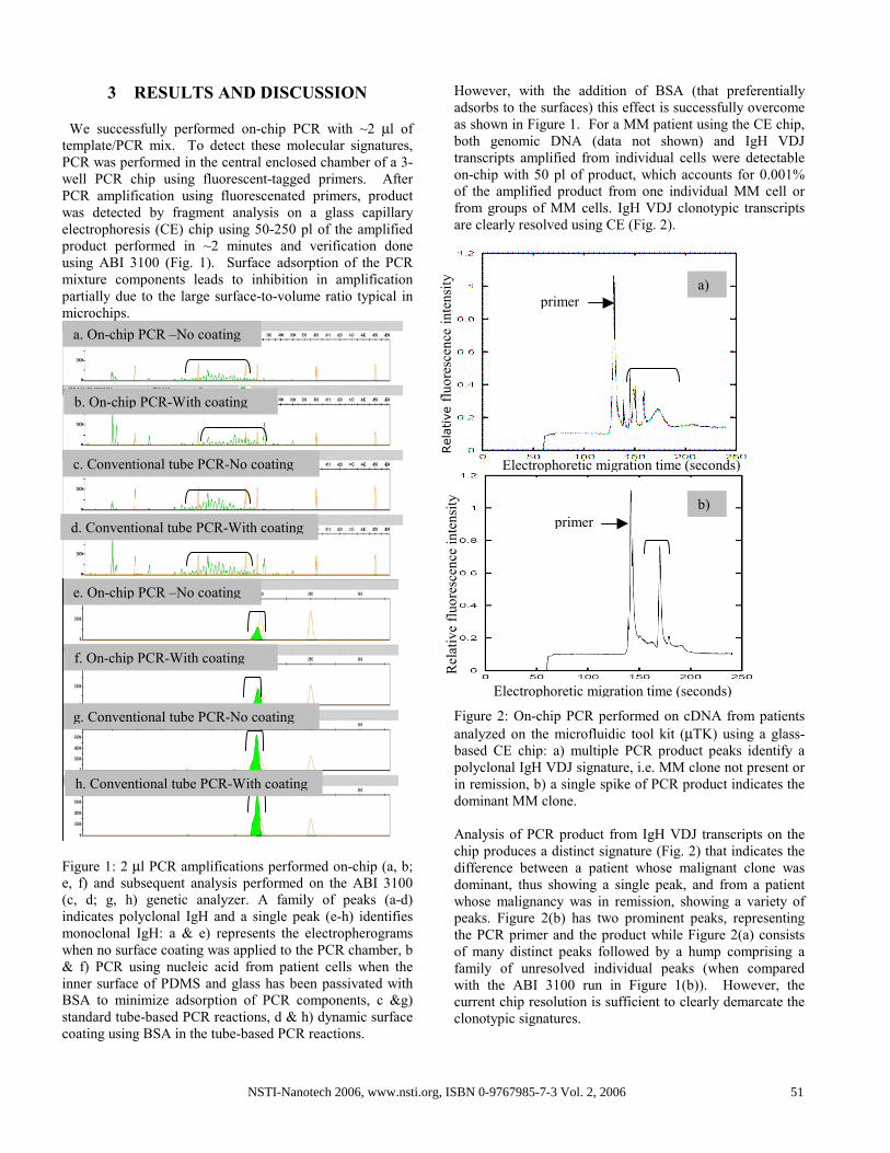
We successfully performed on-chip PCR with ~2 µl of
template/PCR mix. To detect these molecular signatures,
PCR was performed in the central enclosed chamber of a 3-
well PCR chip using fluorescent-tagged primers. After
PCR amplification using fluorescenated primers, product
was detected by fragment analysis on a glass capillary
electrophoresis (CE) chip using 50-250 pl of the amplified
product performed in ~2 minutes and verification done
using ABI 3100 (Fig. 1). Surface adsorption of the PCR
mixture components leads to inhibition in amplification
partially due to the large surface-to-volume ratio typical in
microchips.
Figure 1: 2 µl PCR amplifications performed on-chip (a, b;
e, f) and subsequent analysis performed on the ABI 3100
(c, d; g, h) genetic analyzer. A family of peaks (a-d)
indicates polyclonal IgH and a single peak (e-h) identifies
monoclonal IgH: a & e) represents the electropherograms
when no surface coating was applied to the PCR chamber, b
& f) PCR using nucleic acid from patient cells when the
inner surface of PDMS and glass has been passivated with
BSA to minimize adsorption of PCR components, c &g)
standard tube-based PCR reactions, d & h) dynamic surface
coating using BSA in the tube-based PCR reactions.
However, with the addition of BSA (that preferentially
adsorbs to the surfaces) this effect is successfully overcome
as shown in Figure 1. For a MM patient using the CE chip,
both genomic DNA (data not shown) and IgH VDJ
transcripts amplified from individual cells were detectable
on-chip with 50 pl of product, which accounts for 0.001%
of the amplified product from one individual MM cell or
from groups of MM cells. IgH VDJ clonotypic transcripts
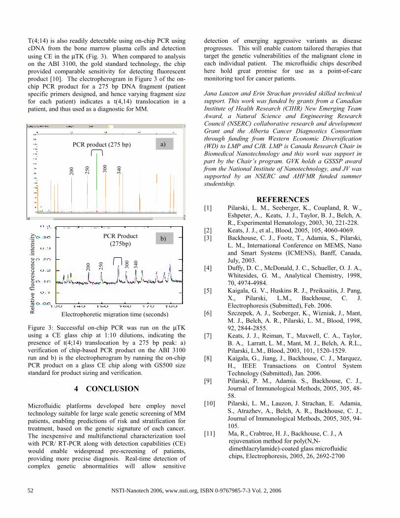
are clearly resolved using CE (Fig. 2).
Figure 2: On-chip PCR performed on cDNA from patients
analyzed on the microfluidic tool kit (µTK) using a glass-
based CE chip: a) multiple PCR product peaks identify a
polyclonal IgH VDJ signature, i.e. MM clone not present or
in remission, b) a single spike of PCR product indicates the
dominant MM clone.
Analysis of PCR product from IgH VDJ transcripts on the
chip produces a distinct signature (Fig. 2) that indicates the
difference between a patient whose malignant clone was
dominant, thus showing a single peak, and from a patient
whose malignancy was in remission, showing a variety of
peaks. Figure 2(b) has two prominent peaks, representing
the PCR primer and the product while Figure 2(a) consists
of many distinct peaks followed by a hump comprising a
family of unresolved individual peaks (when compared
with the ABI 3100 run in Figure 1(b)). However, the
current chip resolution is sufficient to clearly demarcate the
clonotypic signatures.
a. On-chip PCR –No coating
e. On-chip PCR –No coating
b. On-chip PCR-With coating
f. On-chip PCR-With coating
c. Conventional tube PCR-No coating
g. Conventional tube PCR-No coating
d. Conventional tube PCR-With coating
h. Conventional tube PCR-With coating
a)
primer
Electrophoretic migration time (seconds)
Relative fluorescence intensity
b)
primer
Electrophoretic migration time (seconds)
Relative fluorescence intensity
NSTI-Nanotech 2006, www.nsti.org, ISBN 0-9767985-7-3 Vol. 2, 2006 51
T(4;14) is also readily detectable using on-chip PCR using
cDNA from the bone marrow plasma cells and detection
using CE in the µTK (Fig. 3). When compared to analysis
on the ABI 3100, the gold standard technology, the chip
provided comparable sensitivity for detecting fluorescent
product [10]. The electropherogram in Figure 3 of the on-
chip PCR product for a 275 bp DNA fragment (patient
specific primers designed, and hence varying fragment size
for each patient) indicates a t(4,14) translocation in a
patient, and thus used as a diagnostic for MM.
Figure 3: Successful on-chip PCR was run on the µTK
using a CE glass chip at 1:10 dilutions, indicating the
presence of t(4;14) translocation by a 275 bp peak: a)
verification of chip-based PCR product on the ABI 3100
run and b) is the electropherogram by running the on-chip
PCR product on a glass CE chip along with GS500 size
standard for product sizing and verification.
4 CONCLUSION
Microfluidic platforms developed here employ novel
technology suitable for large scale genetic screening of MM
patients, enabling predictions of risk and stratification for
treatment, based on the genetic signature of each cancer.
The inexpensive and multifunctional characterization tool
with PCR/ RT-PCR along with detection capabilities (CE)
would enable widespread pre-screening of patients,
providing more precise diagnosis. Real-time detection of
complex genetic abnormalities will allow sensitive
detection of emerging aggressive variants as disease
progresses. This will enable custom tailored therapies that
target the genetic vulnerabilities of the malignant clone in
each individual patient. The microfluidic chips described
here hold great promise for use as a point-of-care
monitoring tool for cancer patients.
Jana Lauzon and Erin Strachan provided skilled technical
support. This work was funded by grants from a Canadian
Institute of Health Research (CIHR) New Emerging Team
Award, a Natural Science and Engineering Research
Council (NSERC) collaborative research and development
Grant and the Alberta Cancer Diagnostics Consortium
through funding from Western Economic Diversification
(WD) to LMP and CJB. LMP is Canada Research Chair in
Biomedical Nanotechnology and this work was support in
part by the Chair’s program. GVK holds a GSSSP award
from the National Institute of Nanotechnology, and JV was
supported by an NSERC and AHFMR funded summer
studentship.
REFERENCES [1] Pilarski, L. M., Seeberger, K., Coupland, R. W.,
Eshpeter, A., Keats, J. J., Taylor, B. J., Belch, A.
R., Experimental Hematology, 2003, 30, 221-228.
[2] Keats, J. J., et al., Blood, 2005, 105, 4060-4069.
[3] Backhouse, C. J., Footz, T., Adamia, S., Pilarski,
L. M., International Conference on MEMS, Nano
and Smart Systems (ICMENS), Banff, Canada,
July, 2003.
[4] Duffy, D. C., McDonald, J. C., Schueller, O. J. A.,
Whitesides, G. M., Analytical Chemistry, 1998,
70, 4974-4984.
[5] Kaigala, G. V., Huskins R. J., Preiksaitis, J. Pang,
X., Pilarski, L.M., Backhouse, C. J.
Electrophoresis (Submitted), Feb. 2006.
[6] Szczepek, A. J., Seeberger, K., Wizniak, J., Mant,
M. J., Belch, A. R., Pilarski, L. M., Blood, 1998,
92, 2844-2855.
[7] Keats, J. J., Reiman, T., Maxwell, C. A., Taylor,
B. A., Larratt, L. M., Mant, M. J., Belch, A. R.L.,
Pilarski, L.M., Blood, 2003, 101, 1520-1529.
[8] Kaigala, G., Jiang, J., Backhouse, C. J., Marquez,
H., IEEE Transactions on Control System
Technology (Submitted), Jan. 2006.
[9] Pilarski, P. M., Adamia. S., Backhouse, C. J.,
Journal of Immunological Methods, 2005, 305, 48-
58.
[10] Pilarski, L. M., Lauzon, J. Strachan, E. Adamia,
S., Atrazhev, A., Belch, A. R., Backhouse, C. J.,
Journal of Immunological Methods, 2005, 305, 94-
105.
[11] Ma, R., Crabtree, H. J., Backhouse, C. J., A
rejuvenation method for poly(N,N-
dimethlacrylamide)-coated glass microfluidic
chips, Electrophoresis, 2005, 26, 2692-2700
PCR Product
(275bp) b)
PCR product (275 bp) a)
200
250
300
340
200
250
300
340
Relative fluorescence intensity
Electrophoretic migration time (seconds)
NSTI-Nanotech 2006, www.nsti.org, ISBN 0-9767985-7-3 Vol. 2, 200652