rapid detection of human pathogenic … 9760, grov strain co h 35211, gerv strain sa ar 1050, and...
TRANSCRIPT
JOURNAL OF CLINICAL MICROBIOLOGY, July 2003, p. 3299–3305 Vol. 41, No. 70095-1137/03/$08.00�0 DOI: 10.1128/JCM.41.7.3299–3303.2003Copyright © 2003, American Society for Microbiology. All Rights Reserved.
Rapid Detection of Human Pathogenic OrthobunyavirusesManfred Weidmann,1* Veronique Rudaz,1 Marcio R. T. Nunes,2
Pedro F. C. Vasconcelos,2 and Frank T. Hufert1
Department of Virology, Institute of Medical Microbiology and Hygiene, University of Freiburg, 79104 Freiburg,Germany,1 and Department of Arbovirus, Instituto Evandro Chagas, 66090-000 Belem, PA, Brazil2
Received 30 September 2002/Returned for modification 22 December 2002/Accepted 12 April 2003
Modern detection and identification tools can help to provide answers to urgent questions about the inci-dence, prevalence, and epidemiology of currently emerging diseases. We developed highly sensitive one-stepTaqMan reverse transcription-PCR assays with sensitivities ranging from 104 to 101 molecules for 11 humanpathogens of the orthobunyaviruses. We compared the performances of these assays on three currently avail-able cyclers (ABI-PRISM 7700, LightCycler, and SmartCycler). The assay for Oropouche virus (OROV) wastested using sera collected from days 1 to 5 after onset of OROV disease and was found to be greatly superiorto an established nested PCR system. A mean copy number of 1.31 � 107 OROV RNA/ml of serum was detected.Diagnostic RNA detection can be used as early as day 1 after onset of OROV disease. The use of a mobileSmartCycler and a hands-on time of less than 3 h could help to intensify outbreak surveillance and control,especially in field studies.
The Bunyaviridae family of viruses is subdivided into fivegenera. Except for the plant pathogenic genus Tospovirus, theOrthobunyavirus, Hantavirus, Nairovirus, and Phlebovirus gen-era are composed of enzootic viruses, some of which causezoonotic disease in humans. As is the case with many zoonoses,humans act as accidental dead-end hosts of a zoonotic trans-mission cycle oscillating between mammals (mostly rodents)and arthropods (Orthobunyavirus, Nairovirus, and Phlebovirus)or among rodents via aerosols and bite wounds (Hantavirus).There are 330 known viruses in these four genera, and 174 arelisted as belonging to the genus Orthobunyavirus, including halfof the approximately 60 Bunyaviridae viruses causing disease inhumans (10, 11).
The diseases elicited by Orthobunyavirus range from typicalviral diseases with flu-like symptoms (e.g., caused by Tahynavirus [TAHV]) (34) to febrile arthralgia (e.g., caused by Oro-pouche virus [OROV]) (26), encephalitis (e.g., caused by LaCrosse virus [LACV]) (35), and, as recently reported, evenhemorrhagic fever (caused by Garissa virus) (4). Althoughmost infections have a rather mild outcome, some can bedeadly (15, 33).
Many orthobunyaviruses are underestimated with regard totheir potential prevalence and distribution. Snowshoe Harevirus (SSHV), Inkoo virus (INKV), TAHV, and Batai virus(BATV), originally isolated in the United States (14), Finland(30), the Czech Republic (3), and Slovakia (2), respectively,have all been isolated in Siberia (20–22). TAHV and BATVhave been isolated in several Western European countries(19), and the isolation of BATV has also been reported inSudan (24) and India (12).
Some orthobunyaviruses have gradually become accepted asetiological agents of growing disease problems. LACV and
related viruses from the California serogroup cause about 100cases of encephalitis per annum in the United States (6). Oro-pouche fever caused by OROV has developed into the secondmost common arboviral disease, next to Dengue fever, in Bra-zil (27).
In recent decades, evidence has accumulated indicating that,especially in developing countries, the complex interaction offactors such as the growth of the human population, the ac-companying demographic and rapid socioeconomic changes,urbanization, and ecological upheaval contribute to the emer-gence of new infectious diseases (36).
As an initial measure to counter the development of thesediseases into major public health problems, it is essential togather basic clinical and epidemiological data (8). Moderndiagnostic detection and identification tools can help to pro-vide answers to urgent questions about the incidence, preva-lence, and epidemiology of currently emerging diseases andabout pathogens that have the potential to emerge in thefuture. They can also help researchers pick up the trail ofseveral viruses isolated 20 to 30 years ago (Bunyamwera vi-rus [BUNV], Germiston virus [GERV], and Guaroa virus[GROV]) and used to study the molecular biology of the Bu-nyaviridae but for which essential information regarding publichealth issues are almost completely missing.
Our aim was to devise rapid diagnostic tools for humanpathogens of the genus Orthobunyavirus. Therefore, we devel-oped one-step TaqMan reverse transcription (RT)-PCR assaysfor all viruses for which sequence information was availableand established quantification standards for each virus. A sin-gle set of reaction conditions and one temperature profile wasused for all TaqMan RT-PCR assays. Here we describe theTaqMan RT-PCR protocols and present data from the testingof OROV serum samples collected during an outbreak in Bra-zil.
MATERIALS AND METHODS
Virus strains and patient material. California encephalitis virus (CEV) strainBFS 283, Jamestown Canyon virus (JCV) strain 61V2235, SSHV, OROV strain
* Corresponding author. Mailing address: Department of Virology,Institute of Medical Microbiology and Hygiene, University of Frei-burg, Hermann-Herder-Str. 11, 79104 Freiburg, Germany. Phone: 49761 203 6610. Fax: 49 761 203 6608. E-mail: [email protected].
3299
on April 29, 2019 by guest
http://jcm.asm
.org/D
ownloaded from

TR 9760, GROV strain Co H 35211, GERV strain SA Ar 1050, and INKV werereceived as lyophilisates from R. E. Shope, University of Texas, Galveston, Tex.;LACV Thompson was obtained from Rayu Ramasamy, Meharry Medical Col-lege, Nashville, Tenn.; TAHV was obtained from J. Pilaski, Medizinisches Insti-tut fur Umwelthygiene, Dusseldorf, Germany; and BUNV and a pUC18 plasmidcontaining the S segment of BATV were provided by R. Elliot, University ofGlasgow, Glasgow, Scotland. The patient material was collected from April toMay 1996 during an outbreak of OROV disease in Oriximina, Para State, Brazil.Samples consisted of 30 sera from OROV-infected patients collected from days1 to 5 after onset of disease and diagnosed by virus isolation at the InstitutoEvandro Chagas (IEC), Belem, Brazil.
Virus culture and RNA preparation. Viruses were grown in BHK-21 cells(INKV, TAHV, GROV, SSHV, and LACV), VeroE6 cells (JCV, Californiaencephalitis virus [CALV], and OROV), or both (GERV) in 95% Dulbecco’smodified Eagle medium-5% fetal calf serum in 175-cm2 flasks at 37°C in anatmosphere of 5% CO2. Each strain was passaged three times. RNA was pre-pared using RNeasy columns (Qiagen, Hilden, Germany) according to the man-ufacturer’s instructions.
RT-PCR and cloning of S segments. One-step RT-PCR was performed usingthe RT enzyme RAV-2 (Amersham Pharmacia, Freiburg, Germany) and thepolymerase Tth (Roche, Mannheim, Germany) as recommended by Kuno (17).Briefly, 1 �M concentrations of the S segment primers (Table 1) were used forRT-PCR amplification from viral RNA by using the following profile: RT at 53°Cfor 30 min and 30 cycles of PCR at 95°C for 60 s, 60°C for 60s, and 72°C for 60s.The annealing temperature differed for BUNV (55°C). Reaction mixtures con-tained 1 U of RAV-2, 1 U of Tth, and 500 �M concentrations of deoxynucleosidetriphosphates (dNTPs) in 10 mM Tris-HCl (pH 8.9), 100 mM KCl, 3 to 5 mMMgCl2, 50 �g of bovine serum albumin (BSA)/ml, and 0.05% Tween 20. Betaine(1 �M) was added to amplify the S segments of CALV and OROV. Ampliconswere ligated into pCRII, and ligations were transformed into Escherichia coliinv� cells by using a TA cloning kit (Invitrogen, Breda, The Netherlands). Clonescarrying a positively oriented DNA copy of the S segment were propagated in50-ml overnight cultures, and plasmids were prepared by using a Maxiprep kit(Bio-Rad, Munchen, Germany). The plasmids were sequenced using an ABI 377sequencer to confirm the sequence identity.
In vitro transcription and quantification of transcribed RNA. Runoff tran-scription was performed from the T7 promoter downstream of the pCRII mul-tiple cloning site to yield negative single-stranded RNA (�ssRNA). In prepara-tion for T7 runoff transcription, 10 to 15 �g of plasmids was linearized andrestriction enzymes were removed by phenol-chloroform extraction. The�ssRNA was transcribed in a total volume of 100 �l for 1 h at 37°C from 2 �gof linearized plasmid by using 40 U of T7 (Roche) in 40 mM Tris-HCl (pH 8.0),6 mM MgCl2, 15 mM dithiothreitol, 2 mM spermidine, 0.05 mg of BSA/ml, 200U of RNasin (Promega, Mannheim, Germany), and 100 �M concentrations ofeach ribonucleoside triphosphate. To remove the template DNA, the reactionmixtures were incubated twice with 10 U of DNase I (Roche) at 37°C for 10 min.After the first round of digestion, the RNAs were purified with the RNeasyclean-up kit (Qiagen). After the second round, a Trizol (Invitrogen) extractionwas used to completely remove DNase. The efficiency of the DNase digestionimproved markedly when, prior to incubation at 37°C, the reaction mixtures weresequentially incubated at 95°C for 1 min and in ice water for 1 min to melt downDNA-RNA hybrids. After two rounds of DNase treatment, template DNAscould no longer be detected in 1 �l of the undiluted RNA standards by species-specific PCR using a Fast Start DNA master kit (Roche), 300 nM concentrationsof primers, 200 nM concentrations of probes, and 40 cycles of PCR at 95°C for5 s and 60°C for 15 s on the LightCycler. Quantification of RNA was performedusing the fluorescent dye RiboGreen (Molecular Probes, Eugene, Oreg.), whichspecifically binds to ssRNA. Briefly, RNA samples and standard RNA in therange of 20 to 1,000 ng/ml were mixed with RiboGreen in Tris-EDTA buffer.Fluorescent emission of samples and standards at 525 nm was measured usingthe plate read mode of the SDS1.6.3 software of ABI-PRISM 7700, and thequantities of the sample RNAs were calculated. ABI-PRISM 7700 has to becalibrated for the use of RiboGreen; alternatively, the emission spectrum ofSybrGreen can be used for RiboGreen fluorescence detection. Copy numbers ofthe RNA transcripts were calculated from their molecular weight, and molecules,ranging from 107 to 101, were diluted in 100 �g of tRNA/ml (Sigma, Munchen,Germany).
RT-PCR amplicon design. Amplicons were placed into conserved regions ofsequence alignments done with the Megalign software (DNAstar; Lasergene).Primers were designed for an annealing midpoint temperature (Tm) of 60°C byusing the PCR document window of the Primer-Express software (AppliedBiosystems), which operates by using the algorithm developed by Rychlik et al.(28). Species-specific amplicons were designed in conserved regions. Primer Tm
ranged between 58 and 60°C, and the Tm of the 5�FAM- and 3�TAMRA-taggedprobes ranged from 68 to 70°C. Species-specific primers were designed in refer-ence to sequences with the following accession numbers: K00108 and K00610(LACV); U12797 (CEV); U12796 and U12799 (JCV); J02390 and U12800(SSHV); Z68497, U47142, and X73468 (TAHV); Z68496, U47137, and U47138(INKV); D00353 (BUNV); M19420 (GERV); X37466 (GROV); and AF164531-58 (OROV). Nested primers were designed to hybridize to the S segment ofOROV.
RT-PCR conditions. The RT-PCR conditions for the ABI-PRISM 7700 (Ap-plied Biosystems) were as follows: 53°C for 30 min and 40 cycles of 95°C for 15 sand 60°C for 60 s [reaction mixtures in a total volume of 25 �l contained 2.5 Uof RAV-2–2.5 U of Tth, 500 �M dNTPs, 10 U of RNasin, and 2 �M Rox in 50mM Bicine (pH 8.2), 115 mM potassium acetate (KOAc), 5 mM Mn(OAc)2, 8%glycerol, 1 �M concentrations of primers, and 600 nM concentrations of probes].
TABLE 1. Primers for S segments and species-specific RT-PCR
Primer or probea Sequence
S segmentBUN�.....................AGTAGTGTACTCCACBUN�.....................AGTAGTGTGCTCCACLACSFUP ..............AGTAGTGTACTCCACTTGAATACTTTGALACSFDP ..............AGTAGTGTGCTCCACTGAATACATTTAHSFUP..............AGTAGTGTACTCCACTTGAATACTTTGAATAHSFDP..............AGTAGTGTGCTCCACTGAATACCTTBUNSFUP..............AGTAGTGTACTCCACACTACAAACTTGBUNSFDP..............AGTAGTGTGCTCCACCTAAAACTTAGERSFUP..............GTAGTGTACTCCACGCATAAAACTTTGERSFDP..............AGTAGTGTGCTCCACCTTAAACTTAAOROSFUP .............AGTAGTGTACTCCACTATOROSFDP .............AGTAGTGTGGCTCCACATGROSFUP .............AGTAGTGTACTCCACACTACATACCAATGROSFDP .............AGTAGTGTGCTCCACCTAATACCTATA
Species specificLAC FP ..................CCAGATGGGTCCTTGATCALAC P.....................CGAGAATGATGATGAGTCTCAGCACGLAC RP..................CAATTGGGTTGATAATAGTTGTTCTGCAL FP ..................GCGGAGTCAAATGGCATCAL P.....................AGAATGGTGCAGAAATTTATTTGGCATTCCAL RP..................GGTAAAATTTGAAAGTTTCCAAGAAJC FP ......................GGATATCTAGCCAGATGGGTTCTJC P.........................CTCAGATGATGACGAGTCTCAGAGAGAACTCJC RP......................ATTGGATTCTGCAATTGGATTTSSH FP ...................GGATATTTAGCCAGATGGGTTCSSH P......................AGAGAATGAAGACGAGTCTCGGCGSSH RP...................TTGATGATTGTTGTCTTGATCAATAH FP..................CAAAGCTGCTCTCGCTCGTAH P ....................CCGGAGAGGAAGGCTAGTCCTAAATTTGGATAH RP .................TTCCAGGAAAATGATWATTGACGAINK FP ...................CATTGGAACAATGGCCCINK P......................TCCCAGGAACAGAAATGTTTCTAGAAGTTTTCINK RP...................AGGATCCATCATACCATGCTTGER FP..................TGTACTCAATACGAATTTCCCTGGGER P ....................AGGAACAATGCAGTGCCTGACTACGGTCGER RP .................TCCACTGATACGGTGGAAGGTABUN FP..................AATTTTCCTGGCAACCGGABUN P ....................AACCCAGTTCCTGACGATGGTCTTACCCBUN RP .................AAGGAATCCACTGAGGCGGGRO FP .................GAGGCTGGAGAATTCCTGTGRO P....................TAATACACATTTTCCTGGAAACCGGAGRO RP.................GAATCATCGAGGACTGGACTORO FP .................CATTTGAAGCTAGATACGGACAAORO P....................CAATGCTGGTGTTGTTAGAGTCTTCTTCCTORO RP.................CCATGGGCCTCGATGBAT FP ..................GACGCGAGATTAAAACTAGTCTCTCBAT P.....................AAGTGAATGGGAGGTTACGCTTAACCTTGBAT RP..................AGGAAAATTTGTATTAAATACAGTAACCTTC
NestedORO N5 .................AAAGAGGATCCAATAATGTCAGAGTTCATTTORO N3 .................GTGAATTCCCACTATATGCCAATTCCGAATT
a SFUP, S-segment upstream primer; SFDP, S-segment downstream primer;SFSP, S-segment single primer; FP, forward primer; RP, reverse primer; P,TaqMan probe. All sequences are given in the 5�-to-3� orientation.
3300 WEIDMANN ET AL. J. CLIN. MICROBIOL.
on April 29, 2019 by guest
http://jcm.asm
.org/D
ownloaded from
To increase sensitivity, 2 �g of the single-strand binding protein GP32 (Roche)was added per reaction (32). RT-PCR conditions for the LightCycler (Roche)were as follows: RT at 61°C for 20 min, activation at 95°C for 5 min, and 40 cyclesof PCR at 95°C for 5 s and 60°C for 15 s. We used a RNA master hybridizationprobes kit with 500 nM concentrations of primers and 200 nM concentrations ofprobes. This kit includes an aptamer-blocked Tth which performs both RT andhotstart PCR amplification. RT-PCR conditions for the SmartCycler (Cepheid)were as follows: RT at 53°C for 5 min and 40 cycles of PCR at 95°C for 5 s and60 to 63°C for 15 s [reaction mixtures in a 25-�l total volume contained 1 U ofRAV-2–1 U of Tth, 500 �M dNTPs, 500 nM concentrations of primers, and 200nM concentrations of probes in 50 mM Bicine (pH 8.2), 115 mM KOAc, 5 mMMn(OAc)2, 8% glycerol, and SmartCycler additive reagent (200 mM Tris-HCl,pH 8.0, 200 ng of BSA/ml, 0.15 M trehalose, 0.2% Tween 20)]. To increasesensitivity, 2 �g of the single-strand binding protein GP32 was added per reac-tion. We had to adapt hybridization temperatures to 62°C for BUNV and JCVand 63°C for GERV, JCV, SSHV, INKV, and TAHV, but they remained at 60°Cfor GROV, CEV, and BATV. The nested PCR conditions were as describedabove for the amplification of S segments, with 25 cycles for each round andhybridization at 52°C. Outside primers OROSFUP and OROSFDP and nestedprimers ORO N5 and ORO N3 have been described previously (29) (Table 1).
RNA preparation of patient samples. RNA was extracted from 125 �l of serumwith Trizol LS (Invitrogen) according to the manufacturer’s instructions and wasresuspended in a volume of 20 �l.
RESULTS
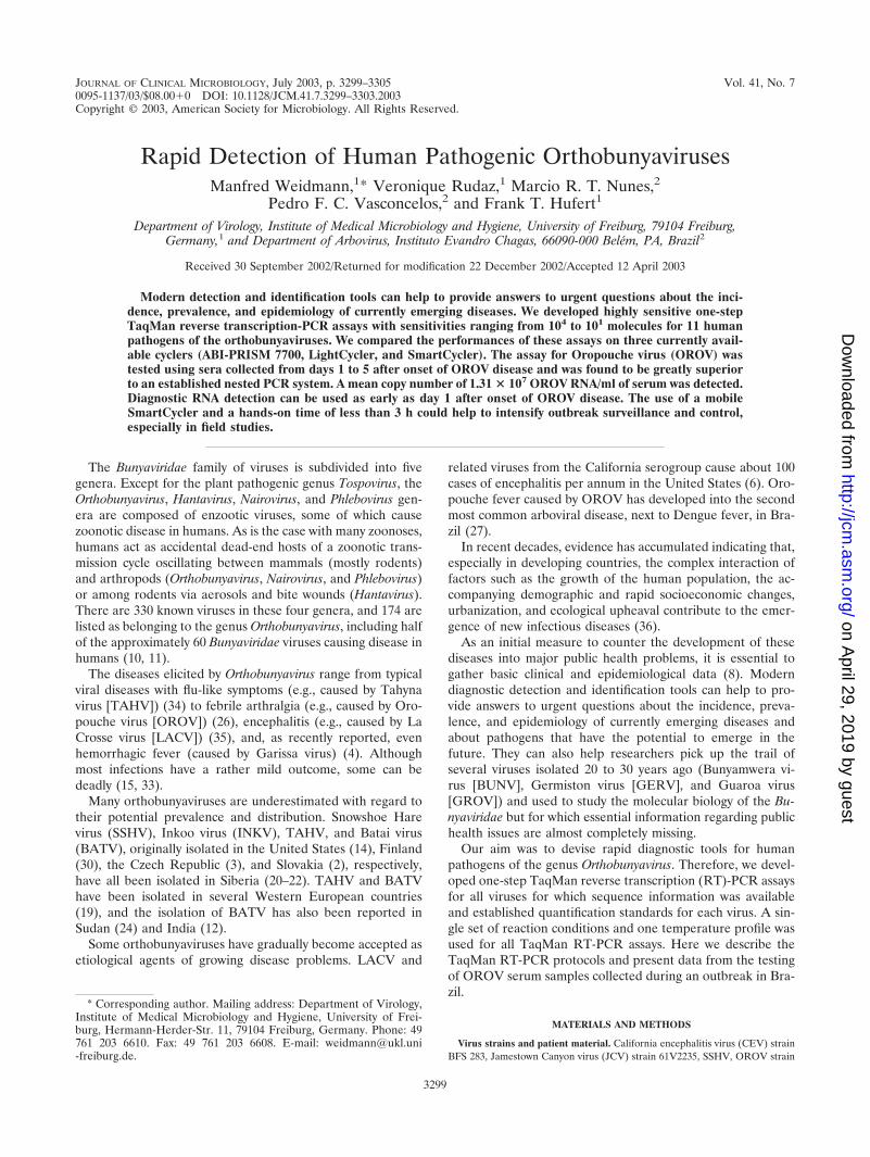
Primer design. The majority of sequences published for Bun-yaviridae species are S-segment sequences coding for the nu-cleocapsid. Primers and probes were placed into blocks ofconserved sequences across sequence alignments of variantspecies isolates. The advantage of the TaqMan chemistry (twoprimers and one probe) is that three blocks of conserved se-quences across the species variants are needed as opposed tofour blocks for the hydprobe chemistry (two primers and twoprobes). We designed the primers by using the ARMS (ampli-fication refractory mutation system) principle, which is basedon the observation that at high stringency (60°C), the thirdnucleotide upstream of the 3-prime end of a primer is mostdecisive for the specificity of the overall 3-prime hybridizationof a primer (25). This principle was adhered to wherever pos-sible in order to design primers able to specifically distinguishbetween closely related sequences, as exemplified by the prim-ers designed for some of the closely related California groupOrthobunyavirus sequences (Fig. 1). Cross-reactivity was testedwith 107 molecules of the respective standard RNA. Apartfrom the species-specific amplicon for LACV which showed aslight cross amplification with GROV (Fig. 2), we observed nocross amplification between any of the amplicons (primers andprobes) and the RNA standards of all 11 viruses.
Cell culture, cloning of S segments, and synthetic RNAstandards. We successfully cultured 10 orthobunyaviruses(BATV was the exception). They all showed marked cyto-pathic effects on BHK-21 or Vero E6 cells beginning from thesecond 3-day passage after inoculation. The presence of theviruses was confirmed by species-specific RT-PCR. We cul-tured all viruses sequentially and never in parallel to avoidlaboratory cross-contamination of the strains. We performedthe amplification of the S segments by using a one-step RT-PCR approach as suggested by Kuno (17) with a combinationof the reverse transcriptase RAV-2 (Amersham) and the poly-merase Tth. This worked remarkably well when we used S-segment primers designed with a Tm of about 60°C (Table 1).With the exception of JCV and CALV, for which primersBUN� and BUN� were applied (9), species-specific S-seg-ment primers were used to avoid false amplification. All clonedamplicons were confirmed by sequencing. The use of full-length negative-sense RNA from the cloned S segments asRNA standards enabled us to come as close as possible to thenatural behavior of the viral RNA in the RT-PCR. We suc-cessfully produced synthetic S-segment RNA standards for all11 viruses.
One-step TaqMan RT-PCR. The performance of the ampli-cons was assessed on a standard range of 107 to 101 moleculesof in vitro-transcribed �ssRNA. The sensitivities achieved withthe amplicons on ABI-PRISM 7700 using RAV-2 and Tth arelisted in Table 2. The addition of the single-strand bindingprotein GP32 of the T4 phage has been shown to increase thesensitivity of PCRs (32). The addition of GP32 increased theslope of the kinetic curves of the fluorescent signal produced inour TaqMan assays, especially in the lower standard range of103 to 101 molecules. The increase in slope of the kineticreaction curves allowed the threshold to be raised and placedinto the elongated exponential phases of the low-copy-numberstandard curves of the range. This improved the correlationcoefficient of the whole standard range, providing a solid basisfor quantification at a higher sensitivity. We tested the stan-dards on the LightCycler with an RNA master amplificationhybridization probes kit (Roche). The results shown in Table 2indicate that, in comparison with the results with the ABI-PRISM 7700, we found increased sensitivities of 1 to 2 logs forfive species-specific TaqMan RT-PCR assays when using theRoche kit on the LightCycler. We could not generate compa-rable sensitivities using the Roche kit on the ABI PRISM 7700
FIG. 1. Primer design for California group Orthobunyavirus. According to the ARMS principle, the third nucleotide from the 3-prime end ofa primer is the most decisive for specific binding. Shown is an alignment of the target area (sequence accession numbers) and the amplicons (FP,forward primer; P, probe; RP, reverse primer) of LACV, SSHV, and JCV. Variant nucleotides at the 3-prime ends of primers are highlighted inblue, and nonmatching nucleotides in nontarget sequences are printed in red. All sequences are given in sense orientation.
VOL. 41, 2003 RAPID DETECTION OF PATHOGENIC ORTHOBUNYAVIRUSES 3301
on April 29, 2019 by guest
http://jcm.asm
.org/D
ownloaded from

(data not shown). Finally, we adapted all TaqMan RT-PCRassays to the SmartCycler. We again used the RAV-2–Tthenzyme mix we already had used on the ABI-PRISM 7700. Wefound that we had to increase the reaction temperatures toobtain good sensitivities, which on the whole were reduced by1 to 0.5 logs compared with the performance on the LightCy-cler (Table 2). With our RNA standard, the nested assay forOROV showed a sensitivity of 105 RNA molecules detected in
the first round of PCR and 103 RNA molecules detected in thesecond round of PCR in ethidium bromide stained gel analysis.
Testing patient material. In a retrospective analysis, we pre-pared RNA from 30 serum samples from which OROV hadbeen isolated at the IEC. A nested PCR assay established atthe IEC laboratory detected OROV RNA in 8 of 30 samples(26.6%). The TaqMan RT-PCR assay detected viral RNA in28 of the 30 samples (93.3%), and the number of RNA mol-
FIG. 2. Sensitivity and specificity of the LACV amplicon. (A) Amplification blot of the LACV RNA standard from 107 to 101 molecules (fromleft to right: 107, blue; 106, green; 105, red; 104, black; 103, red; 102, green; and 102, blue). (B) The LACV species-specific assay was tested with107 copies of the standard RNA of all of the other Orthobunyavirus species. The LACV amplicon showed minor cross amplification of 107 copiesof the GROV standard RNA (red curve) but no cross amplification of the standard RNA of any other strain (all other colors).
3302 WEIDMANN ET AL. J. CLIN. MICROBIOL.
on April 29, 2019 by guest
http://jcm.asm
.org/D
ownloaded from

ecules per milliliter of serum ranged from 5.42 � 104 to 1.65 �108, with a mean copy number of 1.31 � 107 (median, 2.36 �106). Two samples were negative but also scored negative inthe nested PCR assay. TaqMan RT-PCR positive samples hadbeen drawn from patients at days 1 (two samples), 2 (ninesamples), 3 (seven samples), 4 (five samples), and 5 (five sam-ples), and negative samples had been drawn at days 2 and 3after onset of disease (Fig. 3).
DISCUSSION
In recent decades, emerging infections have been a steadycompanion of the accelerated growth of the human populationand its concomitant impact, including deforestation and urban-ization, on inhabited ecosystems (36). The development of
FIG. 3. Detection of OROV in patient sera. (A) OROV RNA standard range from 107 to 101 molecules (as established on the SmartCycler).(B) Results are given as the number of copies of viral RNA detected per milliliter of serum obtained from each of 28 patients sampled on days1 to 5 after onset of disease. Two patient samples drawn on days 2 and 3 after onset of disease tested negative. RNA was Trizol extracted from125 �l of serum. OROV infection had been previously confirmed by virus isolation.
TABLE 2. TaqMan RT-PCR sensitivities
Species
Sensitivity (lowest no. of molecules ofRNA standard detected)
ABI-PRISM 7700 LightCycler SmartCycler
BUNV 102 101 102
GERV NDa 102 103
GROV 104 102 103
OROV 102 102 102
BATV ND 101 102
CEV ND 102 102
JCV 102 102 103
LACV 103 101 101
SSHV ND 104 104
INKV ND 103 103
TAHV ND 102 103
a ND, not done.
VOL. 41, 2003 RAPID DETECTION OF PATHOGENIC ORTHOBUNYAVIRUSES 3303
on April 29, 2019 by guest
http://jcm.asm
.org/D
ownloaded from

modern rapid diagnostics offers researchers the chance to col-lect clinical and epidemiological data on the etiological agentsof these diseases (13, 16). We have developed a set of 11fluorescence one-step TaqMan RT-PCR assays for humanpathogens of the orthobunyaviruses.
Most fluorescence PCR approaches settle for cloning thetarget area of the amplicon, since the fluorescence PCR simplymeasures the ratio of the fluorescent signal to the copy num-bers of target molecules. The nature of the target molecule issecondary. RNA target molecules, however, tend to form com-plex secondary structures that reduce accessibility of the targetmolecule to PCR and thus influence the sensitivity of RT-PCRassays (18). In recognition of this effect, we decided to use thecomplete viral S segment for our RNA standard. The T4 phagesingle-strand binding protein GP32 has been shown to increasethe sensitivity of PCR due to breakdown of secondary struc-tures in single-stranded DNA and RNA (1, 7). GP32 indeedincreased the sensitivity of our TaqMan RT-PCR assays by upto 2 logs. The RNA standards were tested on the ABI-PRISM7700 in a Bicine buffer using RAV-2, Tth as the polymerase(23), and the additive GP32. We obtained high sensitivities formost assays. The sensitivity of the SSHV assay, however, wasvery low (104 to 106 molecules). One reason for this may be theextremely stable secondary structures in the S segment ofSSHV. We were able to increase the sensitivities of severalassays when using a RNA master hybridization probes kit onthe LightCycler. One ingredient of this mixture is an aptamer-blocked Tth which allows separation of RT activity from thepolymerase activity of the enzyme. The sensitivity of the LACVassay improved markedly, i.e., down to 10 molecules, when thismixture was used on the LightCycler. Compared with the useof RAV-2–Tth, the Roche mixture improved the sensitivity byjust 1 additional log on the ABI-PRISM 7700. We attribute thesuperior efficiency of the RT-PCR in the LightCycler to theminimal transition phases between the temperatures of thePCR steps due to the fast (20°C/s) heating and cooling rates ofthe LightCycler.
The TaqMan amplicons performed well on the SmartCycler,although slightly reduced sensitivities compared with those forthe LightCycler were obtained. Again, this may be due to theslightly reduced performance in heating (10°C/s) and cooling(2.5°C/s) of the SmartCycler compared with that of the Light-Cycler.
Thanks to the ARMS principle, the specificity of the assayswas very good, as no cross amplification was observed when wecross tested the 11 assays with very high copy numbers of theRNA standards (107 molecules) of each of the 11 viruses. Onlythe LACV assay showed a minor cross amplification of theGROV RNA. However, the crossing point on the threshold forthis cross amplification was very late and the fluorescenceintensity was very low, i.e., the cross amplification was not veryefficient. It can be ruled out by raising the threshold above afluorescence intensity value of 0.1.
In recent years, OROV has developed into an urban diseasein Amazonia and it bears all the hallmarks of an emergingdisease that could quickly spread beyond its present reach (27).The OROV TaqMan RT-PCR assay showed a higher sensitiv-ity than the established nested RT-PCR assay. It detected viralRNA in 93.3% of the 30 patient samples collected from days 1to 5 after onset of disease, compared with 26.6% for the nested
RT-PCR assay. The most likely reason for the decreased sen-sitivity of the nested RT-PCR assay for OROV compared withthe that of the TaqMan RT-PCR assay is the differences in theamplicon sizes produced by each assay (754 and 693 bp for thefirst and second rounds, respectively, of the nested RT-PCRassay versus 97 bp for the TaqMan RT-PCR assay). The highmean viral load of 1.31 � 107 molecules/ml of serum is typicalfor infections with Bunyaviridae. In sheep, Rift Valley fevervirus, for example, produces a high viremia of up to 105 virus-es/ml of serum in the early days of an infection (31). An earlydiagnostic window for this virus is therefore amenable to RT-PCR and has also been documented for Crimean-Congo hem-orrhagic fever virus (5).
The higher sensitivity of our assay allowed detection andidentification of OROV in two samples collected at day 1 andin nine samples collected at day 2 of the disease. This meansthat patients with typical symptoms can be confirmed to beinfected with OROV at a very early stage of the disease. Byusing our assay, rapid detection of OROV infection shouldindeed be possible within a total hands-on time (sampling,preparation, and PCR) of less than 3 h. This method shouldtherefore greatly facilitate the investigation of an OROV out-break in time to differentiate it from a dengue outbreak forwhich it might be mistaken. To confirm this, we are now en-deavoring to collect sequential serum samples from individualpatients during the next outbreak.
The combination of easy RNA extraction, mobile cyclerssuch as the SmartCycler TD system, and the highly sensitiveand rapid one-step RT-PCRs presented here could become anideal tool for outbreak surveillance, epidemiological screening,or detection of released biological agents in the field.
Since detection with TaqMan probes is very specific, estab-lished amplicons can only be as good as the latest sequencinginformation available. Especially when trying to detect RNAviruses, one should always bear in mind that a negative resultonly means that the sequence the amplicon has been designedfor was not detected in the sample. It does not formally excludethe possible presence of a variant of the particular virus. Virusisolation and sequencing is very important to keeping fluores-cent nucleic acid detection up to date. Consequently, mobilesurveillance work for pathogenic RNA viruses should not reston mobile nucleic acid detection alone. Although mobile PCRcan facilitate outbreak investigation and control, it does notobviate the necessity for serology assays.
ACKNOWLEDGMENTS
We thank Robert E. Shope for providing the majority of strains fromhis collection. Without this generous support, the project would nothave been possible. We are indebted to Melanie Riemer for perfecttechnical assistance.
This work was supported by grants InSanI 0598-V4301 and InSanI030-V4304 from the Bundesministerium fur Verteidigung, Germany.
REFERENCES
1. Abu Al-Soud, W., and P. Radstrom. 2000. Effects of amplification facilitatorson diagnostic PCR in the presence of blood, feces, and meat. J. Clin. Mi-crobiol. 38:4463–4470.
2. Bardos, V., and C. E. Cupkova. 1962. The calovo virus—the second virusisolated from mosquitos in Czechoslovakia. J. Hyg. Epidemiol. Microbiol.Immunol. 6:186–192.
3. Bardos, V., and D. V. Danielova. 1959. The Tahyna virus—a virus isolatedfrom mosquitos in Czechoslovakia. J. Hyg. Epidemiol. Microbiol. Immunol.3:264–276.
3304 WEIDMANN ET AL. J. CLIN. MICROBIOL.
on April 29, 2019 by guest
http://jcm.asm
.org/D
ownloaded from

4. Bowen, M. D., S. G. Trappier, A. J. Sanchez, R. F. Meyer, C. S. Goldsmith,S. R. Zaki, L. M. Dunster, C. J. Peters, T. G. Ksiazek, and S. T. Nichol. 2001.A reassortant bunyavirus isolated from acute hemorrhagic fever cases inKenya and Somalia. Virology 291:185–190.
5. Burt, F. J., P. A. Leman, J. F. Smith, and R. Swanepoel. 1998. The use of areverse transcription-polymerase chain reaction for the detection of viralnucleic acid in the diagnosis of Crimean-Congo haemorrhagic fever. J. Virol.Methods 70:129–137.
6. Calisher, C. H. 1994. Medically important arboviruses of the United Statesand Canada. Clin. Microbiol. Rev. 7:89–116.
7. Chandler, D. P., C. A. Wagnon, and H. Bolton. 1998. Reverse transcriptase(RT) inhibition of PCR at low concentrations of template and its implica-tions for quantitative RT-PCR. Appl. Environ. Microbiol. 64:669–677.
8. DaSilva, E., and M. Iaccarino. 1999. Emerging diseases: a global threat.Biotechnol. Adv. 17:363–384.
9. Dunn, E. F., D. C. Pritlove, and R. M. Elliott. 1994. The S RNA genomesegments of Batai, Cache Valley, Guaroa, Kairi, Lumbo, Main Drain andNorthway bunyaviruses: sequence determination and analysis. J. Gen. Virol.75:597–608.
10. Elliot, R. M. (ed.). 1996. The Bunyaviridae. Plenum Press, New York. N.Y.11. Elliott, R. M., M. Bouloy, C. H. Calisher, R. Goldbach, J. T. Moyer, S. T.
Nichol, R. Pettersson, A. Plyusnin, and C. S. Schmaljohn. 2000. FamilyBunyaviridae. In M. H. V. van Regenmortel et al. (ed.), Virus taxonomy:classification and nomenclature of viruses. Seventh report of the Interna-tional Committee on Taxonomy of Viruses. Academic Press, San Diego,Calif.
12. Geevarghese, G., N. Y. Prasanna, P. G. Jacob, Hanumaiah, and H. R. Bhat.1994. Isolation of Batai virus from sentinel domestic pig from Kolar districtin Karnataka State, India. Acta Virol. 38:239–240.
13. Heymann, D. L., and G. R. Rodier. 2001. Hot spots in a wired world:W. H. O. surveillance of emerging and re-emerging infectious diseases.Lancet Infect. Dis. 1:345–353.
14. Hoff, G. L., R. O. Anslow, J. Spalatin, and R. P. Hanson. 1971. Isolation ofMontana snowshoe hare serotype of California encephalitis virus group froma snowshoe hare and Aedes mosquitoes. J. Wildl. Dis. 7:28–34.
15. Huang, C., W. H. Thompson, N. Karabatsos, L. Grady, and W. P. Campbell.1997. Evidence that fatal human infections with La Crosse virus may beassociated with a narrow range of genotypes. Virus Res. 48:143–148.
16. Hughes, J. M. 2001. Emerging infectious diseases: a CDC perspective.Emerg. Infect. Dis. 7:494–496.
17. Kuno, G. 1998. Universal diagnostic RT-PCR protocol for arboviruses. J. Vi-rol. Methods 72:27–41.
18. Kuo, K. W., M. F. Leung, and W. C. Leung. 1997. Intrinsic secondarystructure of human TNFR-I mRNA influences the determination of geneexpression by RT-PCR. Mol. Cell. Biochem. 177:1–6.
19. Lundstrom, J. O. 1999. Mosquito-borne viruses in western Europe: a review.J. Vector Ecol. 24:1–39.
20. L’Vov, D. K., V. L. Gromashevskii, T. M. Skvortsova, V. A. Aristova, L. V.Kolobukhina, T. N. Morozova, I. V. Galkina, A. M. Butenko, M. S. Nedi-alkova, I. M. Selivanov, et al. 1998. Circulation of viruses of the Californiaserocomplex (Bunyaviridae, Bunyavirus) in the central and southern parts ofthe Russian plain. Vopr. Virusol. 43:10–14. (In Russian.)
21. L’Vov, S. D., V. L. Gromashevskii, I. V. Voropanov, V. P. Andreev, and T. M.
Skvortsova. 1989. Isolation of viruses of antigenic complexes of Californiaencephalitis and Bunyamwera (Bunyaviridae, Bunuavirus) from mosquitoesin northeast Asia. Vopr. Virusol. 34:333–338. (In Russian.)
22. Mitchell, C. J., S. D. Lvov, H. M. Savage, C. H. Calisher, G. C. Smith, D. K.Lvov, and D. J. Gubler. 1993. Vector and host relationships of Californiaserogroup viruses in western Siberia. Am. J. Trop. Med. Hyg. 49:53–62.
23. Myers, T. W., and D. H. Gelfand. 1991. Reverse transcription and DNAamplification by a Thermus thermophilus DNA polymerase. Biochemistry30:7661–7666.
24. Nashed, N. W., J. G. Olson, and A. el-Tigani. 1993. Isolation of Batai virus(Bunyaviridae:Bunyavirus) from the blood of suspected malaria patients inSudan. Am. J. Trop. Med. Hyg. 48:676–681.
25. Newton, C. R., A. Graham, L. E. Heptinstall, S. J. Powell, C. Summers, N.Kalsheker, J. C. Smith, and A. F. Markham. 1989. Analysis of any pointmutation in DNA. The amplification refractory mutation system (ARMS).Nucleic Acids Res. 17:2503–2516.
26. Pinheiro, F. P., A. P. Travassos da Rosa, J. F. Travassos da Rosa, R. Ishak,R. B. Freitas, M. L. Gomes, J. W. LeDuc, and O. F. Oliva. 1981. Oropouchevirus. I. A review of clinical, epidemiological, and ecological findings. Am. J.Trop. Med. Hyg. 30:149–160.
27. Pinheiro, F. P., A. P. A. Travassos da Rosa, and P. F. C. Vasconcelos (ed.).1998. An overview of Oropouche fever epidemics in Brazil and neighbouringcountries. Instituto Evandro Chagas, Belem, Brazil.
28. Rychlik, W., W. J. Spencer, and R. E. Rhoads. 1990. Optimization of theannealing temperature for DNA amplification in vitro. Nucleic Acids Res.18:6409–6412.
29. Saeed, M. F., H. Wang, M. Nunes, P. F. Vasconcelos, S. C. Weaver, R. E.Shope, D. M. Watts, R. B. Tesh, and A. D. Barrett. 2000. Nucleotide se-quences and phylogeny of the nucleocapsid gene of Oropouche virus. J. Gen.Virol. 81:743–748.
30. Saikku, P., C. H. von Bonsdorff, M. Brummer-Korvenkontio, and A. Vaheri.1971. Isolation of non-cubical ribonucleoprotein from Inkoo virus, a Bun-yamwera supergroup arbovirus. J. Gen. Virol. 13:335–337.
31. Sall, A. A., J. Thonnon, O. K. Sene, A. Fall, M. Ndiaye, B. Baudez, C.Mathiot, and M. Bouloy. 2001. Single-tube and nested reverse transcriptase-polymerase chain reaction for detection of Rift Valley fever virus in humanand animal sera. J. Virol. Methods 91:85–92.
32. Schwarz, K., T. Hansen-Hagge, and C. Bartram. 1990. Improved yields oflong PCR products using gene 32 protein. Nucleic Acids Res. 18:1079.
33. Sexton, D. J., P. E. Rollin, E. B. Breitschwerdt, G. R. Corey, S. A. Myers,M. R. Dumais, M. D. Bowen, C. S. Goldsmith, S. R. Zaki, S. T. Nichol, C. J.Peters, and T. G. Ksiazek. 1997. Life-threatening Cache Valley virus infec-tion. N. Engl. J. Med. 336:547–549.
34. Simkova, A., and F. Sluka. 1973. Isolation of Tahyna virus from the blood ofa case of influenza-like disease. Acta Virol. 17:94.
35. Sokol, D. K., M. B. Kleiman, and B. P. Garg. 2001. LaCrosse viral enceph-alitis mimics herpes simplex viral encephalitis. Pediatr. Neurol. 25:413–415.
36. Vasconcelos, P. F., A. P. Travassos da Rosa, S. G. Rodrigues, E. S. Travassosda Rosa, N. Degallier, and J. F. Travassos da Rosa. 2001. Inadequatemanagement of natural ecosystem in the Brazilian Amazon region results inthe emergence and reemergence of arboviruses. Cad. Saude Publica 17:155–164.
VOL. 41, 2003 RAPID DETECTION OF PATHOGENIC ORTHOBUNYAVIRUSES 3305
on April 29, 2019 by guest
http://jcm.asm
.org/D
ownloaded from