rapid and persistent delivery of antigen by lymph node...
TRANSCRIPT

Rapid and Persistent Delivery of Antigen by Lymph Node TargetingPRINT Nanoparticle Vaccine Carrier To Promote Humoral ImmunitySarah N. Mueller,† Shaomin Tian,‡,§ and Joseph M. DeSimone*,†,§,∥,⊥,#
†Department of Chemistry, ‡Department of Microbiology & Immunology, §Lineberger Comprehensive Cancer Center, and ∥Institutefor Nanomedicine, University of North Carolina at Chapel Hill, Chapel Hill, North Carolina 27599, United States#Department of Chemical and Biomolecular Engineering, North Carolina State University, Raleigh, North Carolina 27695, UnitedStates⊥Sloan-Kettering Institute for Cancer Research, Memorial Sloan-Kettering Cancer Center, New York, New York 10021, United States
*S Supporting Information
ABSTRACT: Nanoparticle delivery of subunit vaccines may increase vaccine efficacy, leading to a wide variety of safe andeffective vaccines beyond those available through dosing inactivated or live, attenuated whole pathogens. Here we present aversatile vaccine delivery platform based on PRINT hydrogels made of biocompatible hydroxy-poly(ethylene glycol) (PEG) thatis able to activate the complement system by the alternative pathway. These lymph node targeting nanoparticles (NPs) promotethe immunogenicity of a model antigen, ovalbumin, showing comparable adjuvant effect to alum. We demonstrate that anantigen-specific humoral response is correlated with antigen delivery to the draining lymph nodes, in particular, B cell rich regionsof the lymph nodes. 80 × 180 nm cylindrical NPs were able to sustain prolonged antigen presentation to antigen presenting cells(APCs) and elicit a stronger immune response than nondraining 1 × 1 μm NPs or rapidly clearing soluble antigen. The 80 × 180nm NPs also show high levels of uptake by key APCs and efficiently stimulate CD4+ helper T cell proliferation in vivo, furtherpromoting antibody production. These features together produce a significant humoral immune response, superior to thatproduced by free antigen alone. The simplicity of the chemistries used in antigen conjugation to PRINT NPs confers versatilityto this antigen delivery platform, allowing for potential application to many infectious diseases.
KEYWORDS: particulate vaccine, PRINT, lymphatic trafficking, PEGylation
■ INTRODUCTION
Draining lymph nodes (LNs) are the primary site of action forinitiating adaptive immunity, where T and B cells, major celltypes involved in a humoral immune response, meet antigen orantigen-loaded antigen presenting cells (dendritic cells, macro-phages).1,2 To activate B cells and generate a robust humoralresponse, two signals are required: direct cross-linking of B cellreceptors by antigens, and costimulatory signals from CD4+ Tcells (e.g., cytokines and CD40/CD40L binding).3,4 Antigenpresenting cells, especially dendritic cells (DCs), are critical inpriming T cells to provide helper signals to B cells.4−9 Becauseof the myriad activities of the immune system that take place inthe lymph nodes, recent literature has focused on deliveringvaccines directly to the draining LNs.4,6,7,9,10 By targeting thedraining LNs, vaccine uptake by antigen presenting cells(APCs), APC maturation, and antigen presentation to T and B
cells may all occur in close proximity, thus increasing thepotency of the resulting response.Utilizing purified and synthetic pathogen subunits (peptides,
polysaccharides, lipids, DNA, etc.) for vaccination has becomean increasingly attractive option due to significantly improvedsafety profiles compared to whole pathogen-based vaccines.Subunit vaccines have gained significant clinical success in somediseases, e.g., HPV vaccines (Gardasil from Merck and Co.,Cervarix from GlaxoSmithKline), seasonal influenza vaccines,pneumococcus, HBV, diphtheria, pertussis, etc. However, thereis still a tremendous need for new strategies to improve subunitvaccines and expand their application to a wider variety of
Received: September 2, 2014Revised: January 12, 2015Accepted: March 28, 2015
Article
pubs.acs.org/molecularpharmaceutics
© XXXX American Chemical Society A DOI: 10.1021/mp500589cMol. Pharmaceutics XXXX, XXX, XXX−XXX
diseases. Since purified pathogen subunits used in subunitvaccines are usually poor immunogens, experiencing non-specific degradation and metabolism in vivo and subject torapid clearance from the body,10−13 different strategies are usedto improve the immunogenicity of these subunit antigens suchas the addition of adjuvants or incorporation into differentvaccine delivery vehicles. Particle mediated delivery has showngreat potential for subunit vaccine development and has gainedincreasing attention.6,10−12,14−23 Size, shape, and surfaceproperties of particle vectors can be manipulated in order totarget key APCs and promote cell uptake of antigens viaphagocytosis, or facilitate self-drainage and direct delivery ofvaccine components to lymph node-resident immunecells.10−13,24 Surface display of antigens on particle carriersmay allow multivalent interaction with B cells, mimickingpresentation by natural pathogens and enabling more efficientcross-linking of cognate B cell receptors, thereby increasingpotency of these agents10,17,25 and achieving dose sparingeffects.14,25
Many parameters of particulate vaccine carriers (charge, size,and surface properties) may all contribute to the quality of theresulting immune response. Previous work on the effects oflymphatic trafficking and efficacy of particulate vaccines hasexplored delivery vehicles such as liposomes, polymericparticles, and albumin hitchhiking molecules, amongothers.2,16,19,20,23,26,27 There is a narrow particle size rangethat appears to harness lymphatic flow to the lymph nodewithout becoming trapped at the site of injection; however,within the range of 20−100 nm NPs, the optimal particle sizeappears to be widely system dependent.19,21,28,29
Herein we present a versatile vaccine delivery platform basedon hydrogel particles made of hydroxy-poly(ethylene glycol)(PEG), fabricated via PRINT technology (particle replicationin nonwetting templates),30−32 a unique mold-based particlefabrication process. The highly tunable nature of PRINT allowsfor a great degree of control over NP size, aspect ratio, charge,and surface functionality, facilitating a systematic study of theseeffects on NP trafficking through the lymphatic system and thesubsequent immune response. We establish that this vaccinecarrier has the capacity to deliver subunit vaccine componentsto the draining LNs in a sustained manner and elicit asignificant antigen-specific humoral immune response.
■ MATERIALS AND METHODS
Materials. DyLight 680 maleimide and maleimide-PEG(500)-NHS were purchased from Thermo FisherScientific, Inc. Alexa Fluor 488 maleimide was purchasedfrom Invivogen. Maleimide-PEG(5k)-NHS and NHS-PEG(260)-OH were purchased from Creative PEGworks.EndoGrade Ovalbumin (98% purity, <1 EU/mg) waspurchased in bulk from Hyglos GmbH and tested periodicallyfor endotoxin contamination. Alexa Fluor 555 conjugatedovalbumin was purchased from Life Technologies. Tetra-ethylene glycol monoacrylate (HP(250)A) was synthesized inhouse. PRINT molds were supplied by Liquidia Technologies.All other chemicals and reagents were obtained from Sigma-Aldrich, Inc., unless otherwise noted.Animals. Female Balb/c, C57BL/6, and OT-II mice were
purchased from Jackson Laboratory and used at age 6−12weeks. All experiments involving the mice were carried out inaccordance with an animal use protocol approved by theUniversity of North Carolina Animal Care and Use Committee.
Fabrication of Hydrogel NPs via the PRINT Process.The fabrication of nano- and micron-sized NPs was achieved bymold-based PRINT particle fabrication technology31−33 using acomposition shown in Table S1 in the Supporting Information.Briefly, cure-site monomer (CSM) solutions were prepared at2.5 wt % solids in dry methanol. NPs used in trafficking studiesincluded 2 wt % of a fluorescent dye, either DyLight 680maleimide or AlexaFluor 488 maleimide, covalently cross-linked into the particle matrix. The film-split technique forpreparing NPs was performed as described in the following:using a #5 Mayer rod, 350 μL of CSM solution was cast on asheet of corona treated poly(ethylene terephthalate) (PET),followed by brief evaporation of solvent with a heat gun to yielda transparent film (delivery sheet). Patterned Fluorocur PRINTmolds (Liquidia Technologies) were laminated against thedelivery sheet with moderate pressure (40 psi) and delaminatedat the same pressure. The filled mold was laminated againstcorona-treated PET and subsequently cured in a UV-LEDchamber (Phoseon, λmax =395 nm) for 3.5 min under anitrogen environment. After photocuring, the mold wasremoved to reveal an array of NPs on PET. NPs weremechanically harvested off the PET with sterile water (1 mL/40in.2). NPs were washed via centrifugation (30 min, 14,000 rpm,4 °C), removal of supernatant, and resuspension in fresh, sterilesolvent. NP yield was determined by thermogravimetricanalysis (Q5000IR, TA Instruments). To conjugate ovalbumin(OVA) to the NPs, particles were first PEGylated withheterobifunctional PEG, maleimide-PEG(5k)-NHS, or malei-mide-PEG(500)-NHS, by reacting 1 mg of NPs with 1.6 μmolof PEG plus triethylamine (100 μL) in DMF at a finalconcentration of 1 mg of NPs in 1.4 mL.33 The reaction wasrun at room temperature overnight with shaking at 1400 rpm.NPs were then washed with fresh DMF. Residual amine groupson the surface of NPs were quenched with 13.5 μmol of NHS-PEG(260)-OH (Creative PEGworks) following the samePEGylation procedure above, or with 150 μmol of succinicanhydride, reacted in the presence of 186 μmol of pyridine for30 min with agitation at 1400 rpm. NPs were then washed intowater. OVA was conjugated to the free maleimide groups byreacting NPs and OVA in a 1:1 weight ratio at a NPconcentration of 4 mg/mL in borate buffer pH 9.5 with 0.1 wt% PVOH, MW 2 kDa, overnight at room temperature withshaking at 1400 rpm. NPs were washed with buffer to removeunbound protein and washed with water to remove residualsalt. For PEG(0) NPs, OVA was conjugated to the NP surfaceby first reacting the free amines on the NPs with succinicanhydride as used in quenching above, followed by reactionwith OVA, EDC (1-ethyl-3-(3-(dimethylamino)propyl)-carbodiimide), and sulfo-NHS according to protocol byThermo Scientific. NPs were washed with buffer to removeunbound protein and washed thoroughly with water to removeresidual salt. Antigen conjugation levels were controlled basedon the ratio of NP:OVA during the conjugation reaction,varying from 1:1 to 4:1, with a constant total volume of 1 mLfor each reaction.
NP Characterization. Scanning electron microscopy(SEM) enabled imaging of hydrogel NPs that were dispersedon a silicon wafer, dehydrated, and coated with approximately1.5 nm of Au/Pd (Hitachi S-4700, FEI Helios Nanolab 600).Size and ζ-potential measurements were conducted on ∼20μg/mL NP dispersions in water using a Zetasizer Nano ZSparticle analyzer (Malvern Instruments Inc.). OVA conjugationwas measured using a standard BCA Assay (Fisher).
Molecular Pharmaceutics Article
DOI: 10.1021/mp500589cMol. Pharmaceutics XXXX, XXX, XXX−XXX
B

Lymphatic Drainage Studies. Mice were dosed with 50μg of fluorescent NPs in 20 μL of isotonic solution,subcutaneously in the rear right footpad. To monitor OVAdrainage, 5 μg of OVA labeled with AlexaFluor 555 (Sigma),soluble or conjugated to NPs, was injected into the footpad.Mice were sacrificed at the indicated time points, and drainingpopliteal LNs (PLNs) from both the dosed and contralateralcontrol sides were resected. Resected PLNs were imaged fortotal fluorescence and/or homogenized into a single cellsuspension for analysis of NP distribution in various cell typesby flow cytometry. Additional dosed PLNs were frozen at −80°C in OCT medium (TissueTek) for histological analysis. Thepercent-injected dose was calculated as (mass of NPs in PLN)÷ (50 μg injected dose) × 100. Mass of NPs in PLN wascalculated as (fluorescence of dosed PLN) − (fluorescence ofcontrol PLN) and compared to a standard curve of NPs. NoNP fluorescence was found in other lymph nodes or majororgans (liver, kidney, spleen, lung, heart) at any time point.Ex Vivo Imaging. Imaging of resected LNs was done using
an IVIS-Lumina II (PerkinElmer, Inc. Hopkinton, MA) withanalysis done on Living Image software, version 3.2(PerkinElmer, Inc. Hopkinton, MA). For optimal performanceof the DyLight 680 dye, excitation and emission filters were setto 675 and 720 nm, respectively.Flow Cytometry. Draining LNs were resected at indicated
time points postsubcutaneous injections of 50 μg of dye-labeledNPs. Single cell suspensions of LNs were made physically withfrosted slides. Cells were stained with CD11c-eFluor450, F4/80-FITC, B220-PE, and PDCA-PerCP-eFluor710, all fromeBioscience. Cells were then examined with Cyan ADP (Dako)and analyzed with Summit software. For DC subset analysis,LN cells were stained with CD11c-eFluor450, CD8-FITC, andDEC205-PE (eBioscience).In Vivo CD4+ T Cell Proliferation. CD4+ T cells
recognizing OVA323−339 were isolated from spleens of OT-II transgenic mice with a kit (Miltenyi Biotech). The purified Tcells were labeled with 4 μM CFSE fluorescent dye for 10 minat 37 °C, and 10 million cells were adoptively transferred intoeach C57BL/6 mouse intravenously. On the next day, micewere subcutaneously immunized with 1 μg of OVA, soluble orNP-loaded. Spleens were harvested 4 days later, and single cellsuspensions were stained with CD4-PE-Cy7 and Valpha2-eFluor450 (eBioscience). Cells were then examined with CyanADP (Dako) and analyzed with Summit software.Complement Activation. A C3a sandwich ELISA was
performed to measure complement activation in mouse serumfollowing incubation with NPs. EIA plates (Corning 9018)were coated with an anti-mouse C3a monoclonal antibody (BDBiosciences, clone I87-1162) diluted 1:250 in coating buffer(eBioscience) overnight at 4 °C. Mouse serum was incubated1:1 with either PBS or NPs at 37 °C for 50 min. Serial dilutionsof purified mouse C3a protein (BD Biosciences) were includedin each ELISA plate to establish a standard curve. Serumsamples were added to wells in duplicate (50 μL total volume)and incubated for 3 h. Anti-C3a-biotinylated detection antibody(BD Biosciences, clone I87-419) was used at a 1:500 dilution in1× assay diluent, and incubated for 40 min. Streptavidin-HRP(BD) was diluted 1:250 in 1× assay diluent for 30 min. 1×TMB substrate solution (eBioscience) was added to developcolor. The reaction was stopped with 0.2 N H2SO4, andabsorbance was read at 450 nm with a reference wavelength of570 nm on a SpectraMax (Molecular Devices) plate reader.
Confocal Microscopy. Resected draining LNs were frozenin OCT medium without fixation. 10 μm sections were madewith Leica cryostat, fixed with ice cold acetone, and stained withpurified anti-B220 (eBioscience) coupled with goat anti-ratIgG-Alexa Fluor 488 or -Alexa Fluor 647 (Invitrogen), anti-B220-biotin (eBioscience) coupled with Streptavidin-AlexaFluor 555 (Invitrogen), or CD11c-biotin (eBioscience) coupledwith Streptavidin-Alexa Fluor 633 (Invitrogen). Sections wereexamined with a Zeiss 710 confocal microscope.
Immunizations and Antibody ELISA. C57BL/6 mice, 6−8 weeks old, were immunized with soluble OVA or NP-conjugated OVA at 5 μg per mouse, subcutaneously in theflank. Mice were primed on day zero and boosted on day 21.Plasma samples were collected by bleeding mice submandib-ularly on day 28, and OVA-specific antibody production wasexamined by ELISA. Briefly, EIA plates (Corning) were coatedwith 10 μg/mL OVA in ELISA coating buffer (eBioscience) at4 °C overnight. The wells were washed and blocked with 200μL per well of 3% BSA in PBST (PBS with 0.05% Tween 20)for 2 h. Plasma samples were diluted in blocking buffer andincubated for 2 h. The wells were washed extensively withPBST, and anti-OVA IgG was detected using HRP conjugatedgoat anti-mouse IgG (Invitrogen) and was visualized by adding100 μL of TMB (eBioscience) to each well. The reaction wasstopped after 11 min with 50 μL of 0.2 M H2SO4. Opticaldensities (OD) were read at 450 and 570 nm. The antibodytiter was determined as the highest dilutions with OD (450−570 nm) > 0.1.
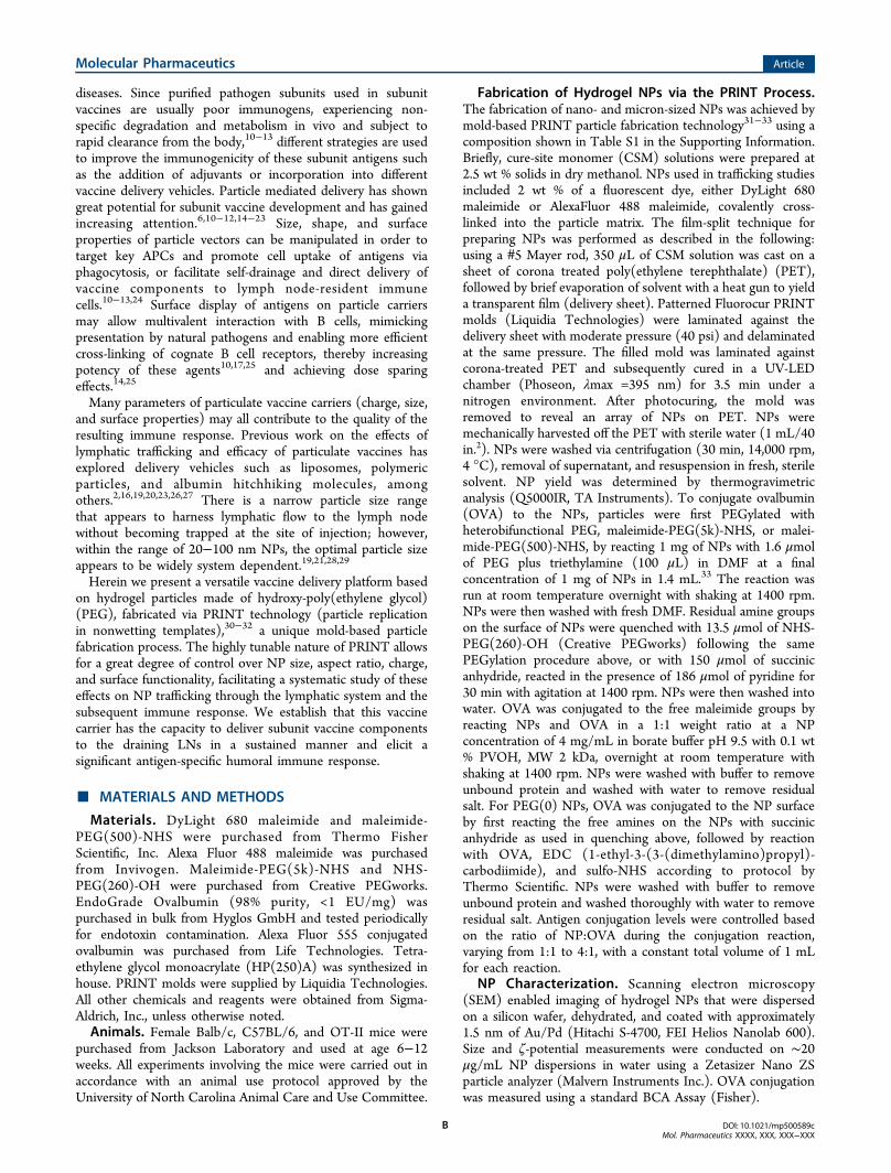
■ RESULTS AND DISCUSSIONNanoparticle delivery of protein subunit vaccines to the lymphnodes allows antigens to interact directly with the immunesystem. Additionally, surface display of protein antigens similarto antigen presentation by natural pathogens may boosttherapeutic efficacy of these subunits to the levels associatedwith whole pathogen vaccines with an improved safetyprofile.4,8,34 This study aimed to evaluate PRINT nanoparticles(NPs) of various size, aspect ratio, and surface characteristicsfor their ability to traffic through the lymphatic system and toexplore the use of these NPs for antigen delivery in vaccineapplications.A delivery vector that traffics quickly and efficiently to the
draining lymph nodes would be beneficial for deliveringantigens to B cells and other antigen presenting cells (APCs)resident in the lymph nodes. A panel of rod/cylindrical PRINTNPs of different size, aspect ratio, and surface charge (Table S2in the Supporting Information) were injected subcutaneously inmice to screen for the ability to drain to the popliteal lymphnodes (PLNs) (Figure 1). While NPs larger than 100 nm insize do not traditionally drain well through the lymphaticssystem as shown in the literature,19,21,28,29 rod shaped NPs withtwo dimensions under 100 nm may be sufficiently small totraffic through the extracellular matrix of the lymphatic systemin order to drain to the lymph nodes while maintaining benefitsover traditional spherical NPs in terms of cellular uptake as wellas an increased surface area for cargo loading.31 Ex vivo imagingof resected PLNs revealed that, within 2 h of injection, anionic80 × 180 nm rod NPs were visible in the PLN, with a timedependent accumulation over 48 h. In contrast, all other NPs,regardless of size and charge, generally remained at the site ofinjection with less than 0.2% (0.1 μg NPs) of the injected dosetrafficking to the PLN. In terms of trafficking abilities, NPsurface charge appears to be the most important determinant
Molecular Pharmaceutics Article
DOI: 10.1021/mp500589cMol. Pharmaceutics XXXX, XXX, XXX−XXX
C
when selecting a self-draining NP delivery vehicle, followed byNP size. Anionic 80 × 180 nm NPs, the best self-drainingparticle type of the particles surveyed, were chosen for furthervaccine delivery studies.Surface display of antigens greatly increases the chances of
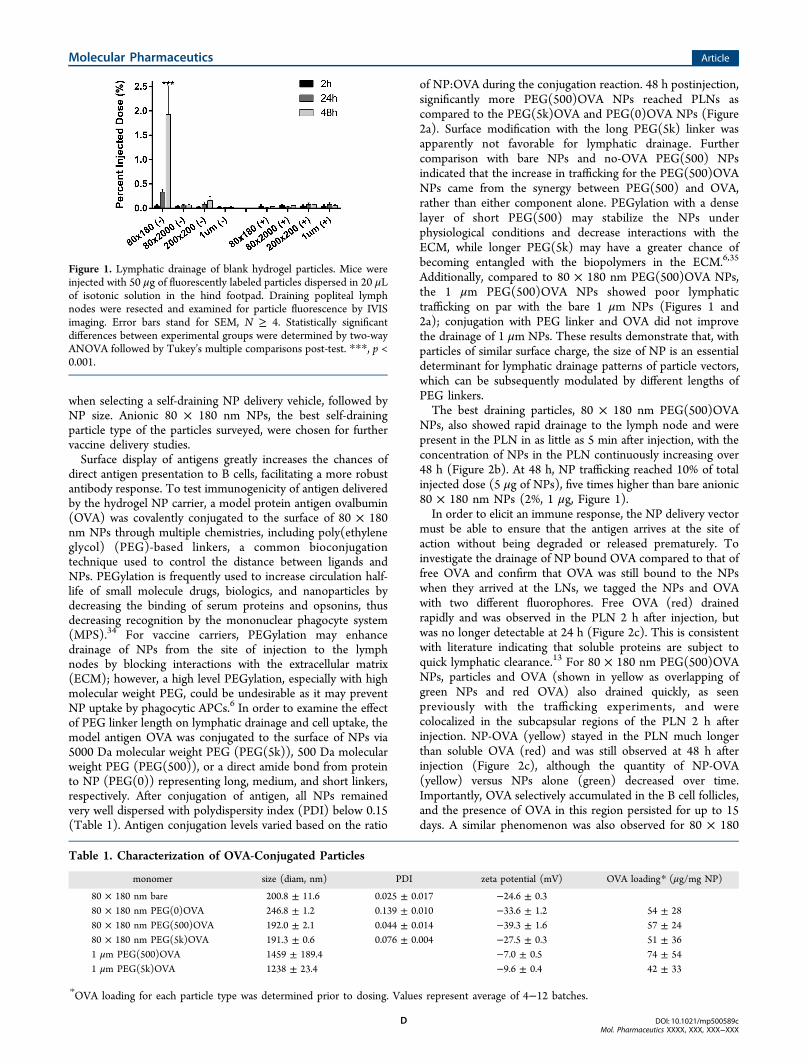
direct antigen presentation to B cells, facilitating a more robustantibody response. To test immunogenicity of antigen deliveredby the hydrogel NP carrier, a model protein antigen ovalbumin(OVA) was covalently conjugated to the surface of 80 × 180nm NPs through multiple chemistries, including poly(ethyleneglycol) (PEG)-based linkers, a common bioconjugationtechnique used to control the distance between ligands andNPs. PEGylation is frequently used to increase circulation half-life of small molecule drugs, biologics, and nanoparticles bydecreasing the binding of serum proteins and opsonins, thusdecreasing recognition by the mononuclear phagocyte system(MPS).34 For vaccine carriers, PEGylation may enhancedrainage of NPs from the site of injection to the lymphnodes by blocking interactions with the extracellular matrix(ECM); however, a high level PEGylation, especially with highmolecular weight PEG, could be undesirable as it may preventNP uptake by phagocytic APCs.6 In order to examine the effectof PEG linker length on lymphatic drainage and cell uptake, themodel antigen OVA was conjugated to the surface of NPs via5000 Da molecular weight PEG (PEG(5k)), 500 Da molecularweight PEG (PEG(500)), or a direct amide bond from proteinto NP (PEG(0)) representing long, medium, and short linkers,respectively. After conjugation of antigen, all NPs remainedvery well dispersed with polydispersity index (PDI) below 0.15(Table 1). Antigen conjugation levels varied based on the ratio
of NP:OVA during the conjugation reaction. 48 h postinjection,significantly more PEG(500)OVA NPs reached PLNs ascompared to the PEG(5k)OVA and PEG(0)OVA NPs (Figure2a). Surface modification with the long PEG(5k) linker wasapparently not favorable for lymphatic drainage. Furthercomparison with bare NPs and no-OVA PEG(500) NPsindicated that the increase in trafficking for the PEG(500)OVANPs came from the synergy between PEG(500) and OVA,rather than either component alone. PEGylation with a denselayer of short PEG(500) may stabilize the NPs underphysiological conditions and decrease interactions with theECM, while longer PEG(5k) may have a greater chance ofbecoming entangled with the biopolymers in the ECM.6,35
Additionally, compared to 80 × 180 nm PEG(500)OVA NPs,the 1 μm PEG(500)OVA NPs showed poor lymphatictrafficking on par with the bare 1 μm NPs (Figures 1 and2a); conjugation with PEG linker and OVA did not improvethe drainage of 1 μm NPs. These results demonstrate that, withparticles of similar surface charge, the size of NP is an essentialdeterminant for lymphatic drainage patterns of particle vectors,which can be subsequently modulated by different lengths ofPEG linkers.The best draining particles, 80 × 180 nm PEG(500)OVA
NPs, also showed rapid drainage to the lymph node and werepresent in the PLN in as little as 5 min after injection, with theconcentration of NPs in the PLN continuously increasing over48 h (Figure 2b). At 48 h, NP trafficking reached 10% of totalinjected dose (5 μg of NPs), five times higher than bare anionic80 × 180 nm NPs (2%, 1 μg, Figure 1).In order to elicit an immune response, the NP delivery vector
must be able to ensure that the antigen arrives at the site ofaction without being degraded or released prematurely. Toinvestigate the drainage of NP bound OVA compared to that offree OVA and confirm that OVA was still bound to the NPswhen they arrived at the LNs, we tagged the NPs and OVAwith two different fluorophores. Free OVA (red) drainedrapidly and was observed in the PLN 2 h after injection, butwas no longer detectable at 24 h (Figure 2c). This is consistentwith literature indicating that soluble proteins are subject toquick lymphatic clearance.13 For 80 × 180 nm PEG(500)OVANPs, particles and OVA (shown in yellow as overlapping ofgreen NPs and red OVA) also drained quickly, as seenpreviously with the trafficking experiments, and werecolocalized in the subcapsular regions of the PLN 2 h afterinjection. NP-OVA (yellow) stayed in the PLN much longerthan soluble OVA (red) and was still observed at 48 h afterinjection (Figure 2c), although the quantity of NP-OVA(yellow) versus NPs alone (green) decreased over time.Importantly, OVA selectively accumulated in the B cell follicles,and the presence of OVA in this region persisted for up to 15days. A similar phenomenon was also observed for 80 × 180
Figure 1. Lymphatic drainage of blank hydrogel particles. Mice wereinjected with 50 μg of fluorescently labeled particles dispersed in 20 μLof isotonic solution in the hind footpad. Draining popliteal lymphnodes were resected and examined for particle fluorescence by IVISimaging. Error bars stand for SEM, N ≥ 4. Statistically significantdifferences between experimental groups were determined by two-wayANOVA followed by Tukey’s multiple comparisons post-test. ***, p <0.001.
Table 1. Characterization of OVA-Conjugated Particles
monomer size (diam, nm) PDI zeta potential (mV) OVA loading* (μg/mg NP)
80 × 180 nm bare 200.8 ± 11.6 0.025 ± 0.017 −24.6 ± 0.380 × 180 nm PEG(0)OVA 246.8 ± 1.2 0.139 ± 0.010 −33.6 ± 1.2 54 ± 2880 × 180 nm PEG(500)OVA 192.0 ± 2.1 0.044 ± 0.014 −39.3 ± 1.6 57 ± 2480 × 180 nm PEG(5k)OVA 191.3 ± 0.6 0.076 ± 0.004 −27.5 ± 0.3 51 ± 361 μm PEG(500)OVA 1459 ± 189.4 −7.0 ± 0.5 74 ± 541 μm PEG(5k)OVA 1238 ± 23.4 −9.6 ± 0.4 42 ± 33
*OVA loading for each particle type was determined prior to dosing. Values represent average of 4−12 batches.
Molecular Pharmaceutics Article
DOI: 10.1021/mp500589cMol. Pharmaceutics XXXX, XXX, XXX−XXX
D

nm PEG(0)OVA (Figure 2c). This separation of antigen fromthe delivery vehicle is consistent with findings by Catron et al.,who observed the cleavage of a model antigen from aparticulate delivery vehicle upon trafficking to the lymphnodes.36 They determined that this cleavage occurred in aprotease-dependent manner over a course of several hours,allowing antigen to accumulate in B cell follicles without theneed for the particles themselves to be taken up by APCs.Overall this result indicates that in general 80 × 180 nmhydrogel NPs are able to efficiently deliver antigen to B cells inthe LNs, supporting sustained antigen retention in B cell richregions. The longer residence time of NP-conjugated OVA inthe PLN may help increase the interaction between antigen andB cells and LN-resident APCs compared to free OVA, resultingin an enhanced antibody response.In addition to the delivery of antigens to B cells and cross-
linking of cognate B cell receptors, eliciting a potent humoralresponse and B cell memory also requires help from CD4+ T
cells;24 therefore good vaccine carriers need to be able todeliver antigens to APCs and prime T cells efficiently. Analysisof cells from draining LNs by flow cytometry showed that, 48 hpostsubcutaneous dosing, 80 × 180 nm hydrogels with OVAlinked through all three linker lengths reached 10−20% of theDCs, and 10−35% of the macrophages in the PLNs, while 1μm PEG(500)OVA NPs were found in less than 2% of DCs ormacrophages (Figure 3a), indicating that the 80 × 180 nm NPsmay efficiently deliver antigens to key APCs. Although the totaldrainage to LNs of these three NPs with various linker lengths(Figure 2a) did not directly correlate with the uptake of NPs bycells in the PLNs, both results suggest that a long PEG linker isless favorable for antigen delivery to immune cells. Thecolocalization of the 80 × 180 nm PEG(500)OVA NPs withDCs was also observed by confocal microscopy analysis ofsectioned draining LNs (Figure S1 in the SupportingInformation), indicating that these NPs are able to access allregions of the PLNs where B cell and T cell activation can
Figure 2. Drainage of OVA-loaded hydrogel NPs to lymph nodes. (a) Total drainage of NPs in lymph nodes. 50 μg of fluorescently labeled 80 × 180nm hydrogel NPs was subcutaneously injected into footpads of C57BL/6 mice, and draining popliteal LNs were collected at 48 h and imaged withIVIS Lumina. Mass of NPs administered was held constant with minor variations in OVA dosage. Statistically significant differences betweenexperimental groups were determined by one-way ANOVA followed by Tukey’s multiple comparisons test. *, p < 0.05; ***, p < 0.001. Error barsstand for SEM. N = 4−14. (b) 80 × 180 nm PEG(500)OVA NPs drained rapidly to the lymph nodes accumulated over 48 h. Error bars stand forSEM. N = 4−8. (c) Persistent delivery of antigen to B cells by hydrogels. Blue, B220 (B cells); green, NPs; red, OVA-AF555. Scale bar: 200 μm.
Molecular Pharmaceutics Article
DOI: 10.1021/mp500589cMol. Pharmaceutics XXXX, XXX, XXX−XXX
E

occur, facilitating activation of both humoral and cellularimmune responses. While B cells did take up significantly more
80 × 180 nm PEG(500)OVA and 80 × 180 nm PEG(0)OVANPs compared to the 80 × 180 nm PEG(5k) and 1 μm
Figure 3. Delivery of antigen to APCs and T cell priming by OVA-loaded hydrogel NPs. (a) 80 × 180 nm NPs are efficiently taken up by keyantigen presenting cells (DCs, macrophages, and B cells respectively) in LNs 48 h postsubcutaneous injection, as analyzed by flow cytometry. N ≥ 4.(b) Uptake of NPs by various DC subsets in draining LNs, with an increase in the percentage of migratory DCs over time. (c) In vivo CD4+ OT-II Tcell proliferation. (d) Quantitative analysis of OT-II T cell proliferation. The percentage of proliferation represents the ratio of cell numbers in R5and R4 shown in panel c (R5/R4). N = 3−4. Hydrogel-mediated delivery of antigen is more efficient in stimulating CD4+ T cell proliferation thansoluble antigen. Statistically significant differences between experimental groups were determined by one-way ANOVA followed by Tukey’s multiplecomparisons test. Error bars stand for SEM. *, p < 0.05; **, p < 0.01, ***, p < 0.001.
Molecular Pharmaceutics Article
DOI: 10.1021/mp500589cMol. Pharmaceutics XXXX, XXX, XXX−XXX
F
PEG(500)OVA NPs, less than 5% of B cells took up particlesfor all groups (Figure 3a). This is not surprising: B cells are notphagocytic cells, unlike macrophages and DCs, which arespecialized for taking up particulate matter. Our own andothers’ work has shown that nonphagocytic cells like epithelialcells are much less efficient in internalizing neutral andnegatively charged NPs.37 However, anionic NPs can still betaken up efficiently through receptor-mediated endocytosiswhen a targeting ligand is available on NPs.38 Activation of Bcells requires recognition and uptake of antigens through theircognate B cell receptors. The accumulation of NPs in B cellregions would still greatly increase the chance of encounteringan antigen by B cells carrying its cognate BCRs for initiation ofan immune response. In addition, as seen in Figure 2c, overtime proteases in the lymphatic fluid cleave antigen from theNPs, also allowing for the antigen to interact with B cellswithout the NPs being taken up.36
Lymph nodes are home to a large population of DCs,especially CD8α+ DCs, which have been shown to be the mostefficient DCs in antigen cross-presentation.6,9,39 In addition,there are other major DC subsets including migratoryLangerhans cells and dermal DCs, normally resident in distalareas of the body, as well as LN resident double negative DCsas defined by surface markers CD8 and DEC20522 (Figure S2in the Supporting Information). Subsequent analysis of DCsfrom draining LNs showed that initially 80 × 180 nmPEG(500)OVA NPs distributed in all four different subsetsof DCs somewhat evenly with an increase in the percentage ofLN resident CD8α+ DCs over a 30 min period (Figure 3b).This suggests that these NPs are indeed self-draining, not fullydependent on uptake by migratory APCs to reach the LNs.This is further verified by the presence of NPs in the PLNs at asearly as 5 min postinjection (Figure 3b): cell-mediated deliveryof NPs has been shown to occur over several hours to days.3,40
At 27 h after injection, the percentage of NP+ LN resident DCsdecreases and the percentage of NP+ migratory dermal DCsincreases, likely due to continuous uptake of NPs by dermalDCs at the injection site followed by cell mediated transport toPLNs. These results demonstrate that the 80 × 180 nm
hydrogel NPs can traffic to the PLNs both through self-drainingand through cell mediated delivery and are able to accessvarious DC subsets, with a high percentage of CD8α+ DCs anddermal DCs internalizing NPs, potentially preparing them for Tcell priming.The T cell priming ability of the 80 × 180 nm
PEG(500)OVA NPs was examined with an in vivo proliferationassay using CD4+ OT-II cells that recognize peptide epitopeOVA323−339. As displayed in Figure 3c, immunizations with 80× 180 nm PEG(500)OVA NPs loaded with just 1 μg of OVAeffectively stimulated the proliferation of CFSE-labeled CD4+
OT-II T cells, causing a dilution of the fluorescent dye. Thedividing cells reached about 60% of total CFSE-labeled cells in3 days (Figure 3d). On the other hand, minimum proliferationwas seen in mice that were untreated or dosed with 1 μg ofsoluble OVA. Together with the flow cytometry data, we candeduce that the 80 × 180 nm PEG(500)OVA NPs areeffectively taken up by APCs, where they can deliver antigencargo and activate helper T cells.The complement system not only acts as the first line of
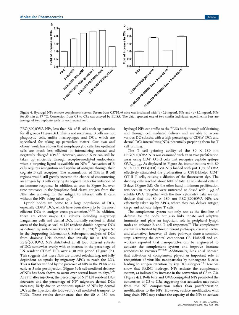
defense for the body but also links innate and adaptiveimmunity and plays an important role in peripheral lymphnodes to enhance B and T cell responses.41 The complementsystem is activated by three different pathways: classical, lectin,and alternative; however, all three pathways share a commonstep: activating the central component C3. Hubbell and co-workers reported that nanoparticles can be engineered toactivate the complement system and improve immuneresponses to vaccines.16,42,43 Additionally, Link et al. showedthat activation of complement played an important role inrecognition of virus-like nanoparticles by noncognate B cells,leading to antigen retention by key DC subtypes.44 Here weshow that PRINT hydrogel NPs activate the complementsystem, as indicated by increase in the conversion of C3 to C3a(Figure 4a). Both bare and OVA-conjugated NPs promoted theconversion of C3 to C3a, suggesting that activation may resultfrom the NP composition rather than postfabricationmodifications to the NPs. However, surface modification withlong chain PEG may reduce the capacity of the NPs to activate
Figure 4. Hydrogel NPs activate complement system. Serum from C57BL/6 mice was incubated with (a) 0.5 mg/mL NPs and (b) 1.2 mg/mL NPsfor 50 min at 37 °C. Conversion from C3 to C3a was assayed by ELISA. The data represent one of two similar individual experiments; bars areaverage of two replicate wells in each experiment.
Molecular Pharmaceutics Article
DOI: 10.1021/mp500589cMol. Pharmaceutics XXXX, XXX, XXX−XXX
G

the complement system, possibly due to a higher degree ofshielding of the NP surface groups that would otherwiseinteract with components in the complement system.Furthermore, EDTA but not EGTA blocked the conversionof C3 to C3a (Figure 4b), indicative of complement activationvia the alternative pathway rather than the classical pathway.These results demonstrate that, in addition to the efficient LNtargeted delivery of antigen, PRINT hydrogel NP vaccinevectors may potentially improve immune responses byactivating the complement system.Immunogenicity of antigen delivered by this NP vector was
tested by vaccinating mice against OVA delivered either insoluble form or conjugated to NPs as described previously. Thedisplay of antigen on the NP surface may increase the chance ofdirect presentation of antigen to B cells, although this strategymay be less protective to the antigen than encapsulationtechniques. The immune response to free versus particulateOVA was evaluated following a prime-boost regimen. Sevendays after the boost dose, mice immunized with 80 × 180 nmPEG(500)OVA NPs showed a 10-fold increase in OVA-specific
IgG production compared to free OVA and free OVA + bareNPs (p < 0.05, Figure 5a), whereas the NPs that werecoinjected with free OVA did not augment the immuneresponse. This data suggests that covalent conjugation to theNP vector is necessary for enhanced immunity. NP-OVA wascompared to free OVA plus the adjuvant alum, the standard ofcare for adjuvanted vaccines.45 Free OVA + alum elicited higherantibody titers than NP-OVA; however, NP-OVA + alum gavea significant increase in antibody response compared to freeOVA + alum (Figure 5b), indicating that this NP-based vectorfor antigen delivery may be able to further improve theantibody response against protein antigen in adjuvantedvaccines. Previous work has shown that the PRINT hydrogelNPs induce no inflammatory response on their own;46
therefore the major advantage of the NP vector over alumcomes from its efficient delivery of antigen to immune systemin addition to direct immunomodulation through complementactivation.The correlation between trafficking and immune response
was examined by comparing the anti-OVA IgG antibody
Figure 5. NP conjugated OVA elicits higher antibody titers than soluble administration. (a) OVA conjugated to NPs elicits higher response thansoluble OVA or soluble OVA admixed with NPs, indicating that conjugation to NPs is necessary for increased immunogenicity. (b) OVA deliveredvia NPs elicits higher antibody titers than soluble antigen when both groups are delivered with or without alum adjuvant. Mice were immunized onday 0 and again on day 21 with 5 μg of OVA, soluble or conjugated to PRINT hydrogel NPs. OVA-specific IgG in plasma was examined by ELISA.Statistically significant differences between experimental groups were determined by one-way ANOVA followed by Tukey’s multiple comparisonstest. *, p < 0.05; **, p < 0.01; ***, p < 0.001. Error bars stand for SEM. N = 5.
Figure 6. Size rather than PEG linker length dramatically influences IgG response. (a) Length of PEG linker for OVA conjugation does not affectIgG response. (b) Large 1 μm NPs elicit lower IgG production than soluble administration or smaller 80 × 180 nm NPs. Mice were immunized as inFigure 5, and plasma IgG was evaluated by ELISA. Statistically significant differences between experimental groups were determined by one-wayANOVA followed by Tukey’s multiple comparisons test. *, p < 0.05. Error bars stand for SEM. Data represent two or three individual experiments ofN = 4.
Molecular Pharmaceutics Article
DOI: 10.1021/mp500589cMol. Pharmaceutics XXXX, XXX, XXX−XXX
H
production after OVA delivery via 80 × 180 nm NPs withvarious PEG linker lengths as well as the 1 μm NPs.Interestingly, despite the influence PEG linker length had onNP trafficking (Figure 2a), PEG linker length appeared to haveno statistical effect on antigen-specific IgG production (Figure6a). All linker lengths showed a 10-fold increase in OVA-specific IgG production compared to free OVA, but the IgGlevels were equivalent among the NP groups. However, the sizeof the NPs used to deliver OVA appeared to have a moredramatic effect on the total IgG. The antibody response againstthe 80 × 180 nm PEG(500)OVA NPs was over 1000 timeshigher than the response to the 1 μm PEG(500)OVA NPs (p <0.05, Figure 6b). Remarkably, IgG response to 1 μmPEG(500)OVA NPs was even lower than that for solubleOVA, strongly suggesting that drainage of vaccine carrier andantigen interaction with LN-resident B cells are crucial toeliciting a humoral response. It is likely that there is a thresholdamount of antigen needed in the lymph nodes for initiating ahumoral immune response. This level may be sufficientlyreached by the 80 × 180 nm NPs, including the relatively lowself-draining 80 × 180 nm PEG(5k)OVA NPs, while the 1 μmNPs do not appear to deliver enough antigen to the LNs to doso.
■ CONCLUSION
In conclusion, we have designed and optimized a versatilevaccine delivery platform based on PRINT NPs. Wedemonstrate that the size, aspect ratio, charge, and surfacecharacteristics of NPs are all important in improving thelymphatic trafficking of NPs and their subsequent uptake bykey APCs. Anionic hydrogel NPs, with dimensions smaller than100 nm, loaded with a model antigen showed high levels of self-drainage and were able to efficiently deliver antigen to B cellsand major APCs, inducing antigen-specific humoral and cellularresponses superior to free antigen alone. The simplicity of thechemistries used in antigen conjugation confers versatility tothis delivery platform, allowing for potential application tomany infectious diseases. Increasing the efficacy of subunitvaccines through a particulate delivery platform is of greatinterest and may lead to a wide variety of safe and effectivevaccines based on dosing pathogen subunits.
■ ASSOCIATED CONTENT
*S Supporting InformationCharacterization and trafficking data for prescreened particlesof various size and charge. This material is available free ofcharge via the Internet at http://pubs.acs.org.
■ AUTHOR INFORMATION
Corresponding Author*Department of Chemistry, University of North Carolina atChapel Hill, CB#3290, 257 Caudill Laboratories, Chapel Hill,NC 27599-3290. Tel: (919) 962-2166. E-mail: [email protected].
NotesThe authors declare the following competing financialinterest(s): The research reported in this paper received partialfinancial support from a venture capital-backed company that J.M. DeSimone cofounded, Liquidia Technologies (www.liqui-dia.com). Currently he has personal financial interests inLiquidia Technologies.
■ ACKNOWLEDGMENTS
The authors thank Dr. Ashish Pandya for the synthesis ofHP(250)A, the University of North Carolina Animal StudiesCore for their assistance with animal experiments, and theChapel Hill Analytical and Nanofabrication Laboratory(CHANL) for support with NP imaging. This work wassupported by the NIH Director’s Pioneer Award (5-DP1-CA174425-04) and Liquidia Technologies.
■ ABBREVIATIONS USED
NP, nanoparticle; PRINT, particle replication in nonwettingtemplates; APC, antigen presenting cell; PLN, popliteal lymphnode; LN, lymph node; DC, dendritic cell; OVA, ovalbumin
■ REFERENCES(1) Moon, J. J.; Huang, B.; Irvine, D. J. Engineering Nano- andMicroparticles to Tune Immunity. Adv. Mater. 2012, 24, 3724−3746.(2) Liu, H.; Moynihan, K. D.; Zheng, Y.; Szeto, G. L.; Li, A. V.;Huang, B.; Van Egeren, D. S.; Park, C.; Irvine, D. J. Structure-BasedProgramming of Lymph-Node Targeting in Molecular Vaccines.Nature 2014, 507, 519−522.(3) Bachmann, M. F.; Jennings, G. T. Vaccine Delivery: A Matter ofSize, Geometry, Kinetics and Molecular Patterns. Nat. Rev. Immunol.2010, 10, 787−796.(4) Pal, I.; Ramsey, J. D. The Role of the Lymphatic System inVaccine Trafficking and Immune Response. Adv. Drug Delivery Rev.2011, 63 (10−11), 909−922.(5) Belz, G.; Smith, C.; Bharadwaj, M.; Rice, A.; Jackson, D. DCs asTargets for Vaccine Design. Cytotherapy 2004, 6, 88−98.(6) Hubbell, J. A.; Thomas, S. N.; Swartz, M. A. MaterialsEngineering for Immunomodulation. Nature 2009, 462, 449−460.(7) Cruz, L. J.; Tacken, P. J.; Rueda, F.; Domingo, J. C.; Albericio, F.;Figdor, C. G. Targeting Nanoparticles to Dendritic Cells forImmunotherapy, 1st ed.; Elsevier Inc.: 2012; Vol. 509, pp 143−163.(8) De Temmerman, M.-L.; Rejman, J.; Demeester, J.; Irvine, D. J.;Gander, B.; De Smedt, S. C. Particulate Vaccines: On the Quest forOptimal Delivery and Immune Response. Drug Discovery Today 2011,16, 569−582.(9) Swartz, M. A.; Hubbell, J. A.; Reddy, S. T. Lymphatic DrainageFunction and Its Immunological Implications: From Dendritic CellHoming to Vaccine Design. Semin. Immunol. 2008, 20, 147−156.(10) Ferreira, S. A.; Gama, F. M.; Vilanova, M. Polymeric Nanogelsas Vaccine Delivery Systems. Nanomedicine 2013, 9, 159−173.(11) Xiang, S. D.; Scholzen, A.; Minigo, G.; David, C.;Apostolopoulos, V.; Mottram, P. L.; Plebanski, M. PathogenRecognition and Development of Particulate Vaccines: Does SizeMatter? Methods 2006, 40, 1−9.(12) Storni, T.; Kundig, T. M.; Senti, G.; Johansen, P. Immunity inResponse to Particulate Antigen-Delivery Systems. Adv. Drug DeliveryRev. 2005, 57, 333−355.(13) Porter, C. J.; Charman, S. A. Lymphatic Transport of Proteinsafter Subcutaneous Administration. J. Pharm. Sci. 2000, 89, 297−310.(14) Kasturi, S. P.; Skountzou, I.; Albrecht, R. A.; Koutsonanos, D.;Hua, T.; Nakaya, H. I.; Ravindran, R.; Stewart, S.; Alam, M.; Kwissa,M.; Villinger, F.; Murthy, N.; Steel, J.; Jacob, J.; Hogan, R. J.; García-Sastre, A.; Compans, R.; Pulendran, B. Programming the Magnitudeand Persistence of Antibody Responses with Innate Immunity. Nature2011, 470, 543−547.(15) Moon, J.; Suh, H.; Bershteyn, A.; Stephan, M. Interbilayer-Crosslinked Multilamellar Vesicles as Synthetic Vaccines for PotentHumoral and Cellular Immune Responses. Nat. Mater. 2011, 10, 243−251.(16) Reddy, S. T.; van der Vlies, A. J.; Simeoni, E.; Angeli, V.;Randolph, G. J.; O’Neil, C. P.; Lee, L. K.; Swartz, M. A.; Hubbell, J. A.Exploiting Lymphatic Transport and Complement Activation inNanoparticle Vaccines. Nat. Biotechnol. 2007, 25, 1159−1164.
Molecular Pharmaceutics Article
DOI: 10.1021/mp500589cMol. Pharmaceutics XXXX, XXX, XXX−XXX
I
(17) Rice-Ficht, A. C.; Arenas-Gamboa, A. M.; Kahl-McDonagh, M.M.; Ficht, T. A. Polymeric Particles in Vaccine Delivery. Curr. Opin.Microbiol. 2010, 13, 106−112.(18) John, A. L. S.; Chan, C. Y.; Staats, H. F.; Leong, K. W.;Abraham, S. N. Synthetic Mast-Cell Granules as Adjuvants to Promoteand Polarize Immunity in Lymph Nodes. Nat. Mater. 2012, 11, 1−8.(19) Fifis, T.; Gamvrellis, A.; Crimeen-Irwin, B.; Pietersz, G. A.; Li, J.;Mottram, P. L.; McKenzie, I. F. C.; Plebanski, M. Size-DependentImmunogenicity: Therapeutic and Protective Properties of Nano-Vaccines against Tumors. J. Immunol. 2004, 173, 3148−3154.(20) Zhuang, Y.; Ma, Y.; Wang, C.; Hai, L.; Yan, C.; Zhang, Y.; Liu,F.; Cai, L. PEGylated Cationic Liposomes Robustly Augment Vaccine-Induced Immune Responses: Role of Lymphatic Trafficking andBiodistribution. J. Controlled Release 2012, 159, 135−142.(21) Oussoren, C.; Storm, G. Liposomes to Target the Lymphaticsby Subcutaneous Administration. Adv. Drug Delivery Rev. 2001, 50,143−156.(22) Zhan, X.; Tran, K. K.; Shen, H. Effect of the Poly(ethyleneGlycol) (PEG) Density on the Access and Uptake of Particles byAntigen-Presenting Cells (APCs) after Subcutaneous Administration.Mol. Pharmaceutics 2012, 9 (12), 3442−3451.(23) Kaur, R.; Bramwell, V. W.; Kirby, D. J.; Perrie, Y. Manipulationof the Surface Pegylation in Combination with Reduced Vesicle Size ofCationic Liposomal Adjuvants Modifies Their Clearance Kinetics fromthe Injection Site, and the Rate and Type of T Cell Response. J.Controlled Release 2012, 164, 331−337.(24) Johansen, P.; Mohanan, D.; Martínez-Gomez, J. M.; Kundig, T.M.; Gander, B. Lympho-Geographical Concepts in Vaccine Delivery. J.Controlled Release 2010, 148, 56−62.(25) Bershteyn, A.; Hanson, M. C.; Crespo, M. P.; Moon, J. J.; Li, A.V.; Suh, H.; Irvine, D. J. Robust IgG Responses to Nanograms ofAntigen Using a Biomimetic Lipid-Coated Particle Vaccine. J.Controlled Release 2012, 157, 354−365.(26) Al Kobiasi, M.; Chua, B. Y.; Tonkin, D.; Jackson, D. C.;Mainwaring, D. E. Control of Size Dispersity of Chitosan BiopolymerMicroparticles and Nanoparticles to Influence Vaccine Trafficking andCell Uptake. J. Biomed. Mater. Res., Part A 2012, 100, 1859−1867.(27) Moghimi, S. M. The Effect of Methoxy-PEG Chain Length andMolecular Architecture on Lymph Node Targeting of Immuno-PEGLiposomes. Biomaterials 2006, 27, 136−144.(28) Reddy, S. T.; Berk, D. A.; Jain, R. K.; Swartz, M. A. A Sensitivein Vivo Model for Quantifying Interstitial Convective Transport ofInjected Macromolecules and Nanoparticles. J. Appl. Physiol. 2006,101, 1162−1169.(29) Reddy, S. T.; Rehor, A.; Schmoekel, H. G.; Hubbell, J. A.;Swartz, M. A. In Vivo Targeting of Dendritic Cells in Lymph Nodeswith Poly(propylene Sulfide) Nanoparticles. J. Controlled Release 2006,112, 26−34.(30) Rolland, J. P.; Maynor, B. W.; Euliss, L. E.; Exner, A. E.;Denison, G. M.; DeSimone, J. M. Direct Fabrication and Harvesting ofMonodisperse, Shape-Specific Nanobiomaterials. J. Am. Chem. Soc.2005, 127, 10096−10100.(31) Gratton, S. E. A.; Ropp, P. A.; Pohlhaus, P. D.; Luft, J. C.;Madden, V. J.; Napier, M. E.; DeSimone, J. M. The Effect of ParticleDesign on Cellular Internalization Pathways. Proc. Natl. Acad. Sci.U.S.A. 2008, 105, 11613−11618.(32) Gratton, S. E. A.; Williams, S. S.; Napier, M. E.; Pohlhaus, P. D.;Zhou, Z.; Wiles, K. B.; Maynor, B. W.; Shen, C.; Olafsen, T.; Samulski,E. T.; Desimone, J. M. The Pursuit of a Scalable NanofabricationPlatform for Use in Materials and Life Science Applications. Acc. Chem.Res. 2008, 41, 1685−1695.(33) Perry, J. L.; Reuter, K. G.; Kai, M. P.; Herlihy, K. P.; Jones, S.W.; Luft, J. C.; Napier, M.; Bear, J. E.; DeSimone, J. M. PEGylatedPRINT Nanoparticles: The Impact of PEG Density on ProteinBinding, Macrophage Association, Biodistribution, and Pharmacoki-netics. Nano Lett. 2012, 12, 5304−5310.(34) Wilson, J. T.; Keller, S.; Manganiello, M. J.; Cheng, C.; Lee, C.-C.; Opara, C.; Convertine, A.; Stayton, P. S. pH-Responsive
Nanoparticle Vaccines for Dual-Delivery of Antigens and Immunos-timulatory Oligonucleotides. ACS Nano 2013, 7 (5), 3912−3925.(35) Swartz, M. A. The Physiology of the Lymphatic System. Adv.Drug Delivery Rev. 2001, 50, 3−20.(36) Catron, D. M.; Pape, K. A.; Fife, B. T.; van Rooijen, N.; Jenkins,M. K. A Protease-Dependent Mechanism for Initiating T-Dependent BCell Responses to Large Particulate Antigens. J. Immunol. 2010, 184,3609−3617.(37) Gratton, S. E. A.; Napier, M. E.; Ropp, P. A.; Tian, S.;DeSimone, J. M. Microfabricated Particles for Engineered DrugTherapies: Elucidation into the Mechanisms of Cellular Internalizationof PRINT Particles. Pharm. Res. 2008, 25, 2845−2852.(38) Wang, J.; Tian, S.; Petros, R. A.; Napier, M. E.; Desimone, J. M.The Complex Role of Multivalency in Nanoparticles Targeting theTransferrin Receptor for Cancer Therapies. J. Am. Chem. Soc. 2010,132, 11306−11313.(39) Wilson, N. S.; El-Sukkari, D.; Belz, G. T.; Smith, C. M.; Steptoe,R. J.; Heath, W. R.; Shortman, K.; Villadangos, J. A. Most LymphoidOrgan Dendritic Cell Types Are Phenotypically and FunctionallyImmature. Blood 2003, 102, 2187−2194.(40) Manolova, V.; Flace, A.; Bauer, M.; Schwarz, K.; Saudan, P.;Bachmann, M. F. Nanoparticles Target Distinct Dendritic CellPopulations according to Their Size. Eur. J. Immunol. 2008, 38,1404−1413.(41) Carroll, M. C. The Complement System in Regulation ofAdaptive Immunity. Nat. Immunol. 2004, 5, 981−986.(42) Thomas, S. N.; van der Vlies, A. J.; O’Neil, C. P.; Reddy, S. T.;Yu, S. S.; Giorgio, T. D.; Swartz, M. A.; Hubbell, J. A. EngineeringComplement Activation on Polypropylene Sulfide Vaccine Nano-particles. Biomaterials 2011, 32, 2194−2203.(43) Fine, D. P.; Marney, S. R.; Colley, D. G.; Sergent, J. S.; Prez, R.M. Des. C3 Shunt Activation in Human Serum Chelated with EGTA.J. Immunol. 1972, 109, 807−809.(44) Link, A.; Zabel, F.; Schnetzler, Y.; Titz, A.; Brombacher, F.;Bachmann, M. F. Innate Immunity Mediates Follicular Transport ofParticulate but Not Soluble Protein Antigen. J. Immunol. 2012, 188,3724−3733.(45) Baylor, N. W.; Egan, W.; Richman, P. Aluminum Salts inVaccinesUS Perspective. Vaccine 2002, 20 (Suppl. 3), S18−S23.(46) Roberts, R. A.; Shen, T.; Allen, I. C.; Hasan, W.; DeSimone, J.M.; Ting, J. P. Y. Analysis of the Murine Immune Response toPulmonary Delivery of Precisely Fabricated Nano- and MicroscaleParticles. PLoS One 2013, 8, e62115.(47) Robbins, G. R.; Roberts, R. A.; Guo, H.; Reuter, K.; Shen, T.;Sempowski, G. D.; McKinnon, K. P.; Su, L.; DeSimone, J. M.; Ting, J.P. Y. Analysis of human innate immune responses to PRINTfabricated nanoparticles with cross validation using a humanizedmouse model. Nanomedicine 2015, 11, 589−599.
■ NOTE ADDED AFTER ASAP PUBLICATIONThis paper was published ASAP on April 8, 2015. Thefollowing additional information and ref 47 were added onApril 14, 2015: Complement activation by PEG hydrogelnanoparticles may be dose and composition-dependent, as arecent study from our lab indicated that complement activationdid not occur with low doses of hydrogel particles with differentsurface properties (Robbins et al., 2015).47
Molecular Pharmaceutics Article
DOI: 10.1021/mp500589cMol. Pharmaceutics XXXX, XXX, XXX−XXX
J