radiotherapy with or without hyperthermia treatment of superficial localized.pdf
TRANSCRIPT

Int. J. Radiation Oncology Biol. Phys., Vol. 35, No. 4, pp. 73 l-744, 1996 Copyright 0 1996 Elsevier Science Inc. hinted in the USA. All tights reserved
036Q-3016/96 $15.00 + 00
ELSEVIER PII: SO360-3016(%)00154-X
l Hypertherrnia Original Contribution
RADIOTHERAPY WITH OR WITHOUT HYPERTHERMIA IN THE TREATMENT OF SUPERFICIAL LOCALIZED BREAST CANCER:
RESULTS FROM FIVE RANDOMIZED CONTROLLED TRIALS
INTERNATIONAL COLLABORATIVE HYPERTHERMIA GROUP:
UK Medical Research Council: CLARE C. VERNON, M.A., F.R.C.R.,* JEFFREY W. HAND, PH.D., D.Sc.,* STANLEY B. FIELD, PH.D.,*
DAV~DMACHIN, M.Sc., PH.D.+ AND JILLB. WHALEY, B.V.M.&St
European Society for Hyperthermic Oncology and the Dutch Hyperthermia Group: JACOBA VAN DER ZEE, M.D., PH.D.,* WIM L. J. VAN PUT-TEN, M.Sc.,*
GERARD C. VAN RHOON, PH.D.,$ JAN D. P. VAN DIJK, PH.D.” AND
DIONISIO GONZALEZ GONZALEZ, M.D., PH.D.§
Princess Margaret HospitaVOntario Cancer Institute: FEI-FEI LIU, M.D., F.R.C.P.C.,j’ PHYLLIS GOODMAN, M.Sc! AND MICHAEL SHERAR, F%.D!
*MRC Cyclotron Unit, Hammersmith Hospital, London, United Kingdom; +MRC Cancer Trials Office, Cambridge, United Kingdom; *Departments of Hyperthermia and Statistics, Daniel den Hoed Cancer Center, Rotterdam,The
Netherlands; “Department of Radiotherapy, Academic Medical Center, Amsterdam,The Netherlands; apartments of Radiation Oncology, Biostatistics, and Clinical Physics, Princess Margaret Hospital/Ontario Cancer Institute,
University of Toronto, Canada
purpose: Claims for the value of hyperthermia as an adjunct to radiotherapy in the treatment of cancer have mostly been based on small Phase I or II trials. To test the benefit of thii form of treatment, randomized phase III trials were needed. Methods and Materials: Five randomized trials addressing this question were started between 1988 and 1991. In these trials, patients were eligible if they had advanced prhnary or recurrent breast cancer, and local radiotherapy was indicated in preference to surgery. In addition, heating of the lesions and treatment with a prescribed (re)irradiation schedule had to be feasible and informed consent was obtained. The primary endpoint of ail trials was local complete response. Slow recruitment led to a decision to collaborate and combii the trial rest&s in one analysis, and report them simuhaneously in one publication. Interim analyses were carried out and the trials were closed to recruitment when a previously agreed statistically sign&ant difference in complete response rate was observed in the two larger trials. Results: We report on pretreatment characteristics, the treatments received, the local response observed, duration of response, time to local failure, distant progression and survival, and treatment toxicity of tbe 306 patients randomized. The overali CR rate for RT alone was 41% and for the combined treatment arm was 59%, giving, after stratification by trial, an odds ratio of 2.3. Not alI trials demonstrated an advantage for the combined treatment, although tbe 95% confidence intervals of the different trials all contain the pooled odds ratio. The greatest effect was observed in patients with recurrent lesions in previously irradiated areas, where further irradiation was limited to low doses. Conclusion: The combined result of the five trials has demonstrated the efficacy of hyperthemia as an adjunct to radiotherapy for treatment of recurrent breast cancer. The implication of these encouraging results is that hyperthermia appears to have an important role in the clinical management of this disease, and there should be no doubt that further studies of the use of hypertbermia are warranted.
Breast cancer, Hyperthermia, Radiotherapy, Randomized trial.
Reprint requests to: Jill B. Whaley, MRC Cancer Trials Office, 5 Shaftesbury Road, Cambridge CB2 2BW UK
Accepted for publication 6 March 1996.
731

732 1. J. Radiation Oncology 0 Biology 0 Physics Volume 35, Number 4, 1996
INTRODUCTION
Early clinical applications of hyperthermia (HT) include those of Coley (5) and Westermark (34) whereas the or- igins of the use of HT as a radiosensitizing agent are to be found in the early years of the present century (20,26). Although there was periodic interest through the interven- ing years, progress in the clinical application of HT was largely frustrated by a lack of adequate techniques for heating tumors. Development of a biological rationale for the use of heat began during the 1960s and, in recent years, the considerable effort applied to the physics and engi- neering problems associated with clinical HT has led to the development of acceptable techniques for treatment of superficial tumors (10). Sufficient knowledge has also been gained about methods of applying HT, with respect to fractionation and combination with other modalities, to result in a safe and possibly effective clinical treatment.
As a consequence, a large number of clinical Phase I and II studies (11, 15, 24, 27, 32, 33) have been carried out and their results indicate that HT may be of value in cancer treatment when given as an adjunct to either radio- therapy (RT) or chemotherapy. A few randomized clinical trials have also been performed (9,25,31) but, in most of these, the numbers of patients recruited were too small or the lesions were not properly heated. It became clear that properly conducted prospectively randomized trials were needed to define the role of HT in cancer management. A trial in patients with malignant melanoma reported by Ov- ergaard et al. in 1995 (21) was undertaken for the same reasons.
For patients with breast cancer, previous studies have indicated that local treatment does not affect survival in patients with recurrent disease, and that distant metastases will be detected ultimately in 75-93% of patients (1, 4, 28). The median survival time for these patients ranges from 12-53 months, depending on tumor characteristics, and 21-50% will survive 5 years or longer (4). Local recurrence causes pain, bleeding, and ulceration in over 60% of patients (3), in addition to the psychological dis- tress of watching a tumor grow. For both nonirradiated and previously irradiated recurrences, durable local con- trol decreases with increasing size of the lesion and, for the latter group of patients, the radiation dose that can be administered safely is lower than that considered effective. Chemotherapy is also less effective in areas that have pre- viously been irradiated. Thus, the use of a local treatment that can provide durable local tumor control for the re- maining lifespan of these patients would be considered worthwhile.
The optimum HT regimen with regard to temperature or number of treatments is not known. However, a small number of I-IT fractions may be as effective as a larger number because of the development of thermotolerance (14). The need to select appropriate patients and tumors and the importance of compliance with appropriate quality assurance guidelines when delivering HT treatment has
been highlighted by the results of previous clinical trials, especially Perez et aZ. (25).
The key question addressed by the randomized trials reported here was whether or not the addition of HT treat- ment to RT increased the complete response (CR) rate in patients suffering from recurrent or inoperable primary breast cancer.
METHODS AND MATERIALS
Trial design The five randomized trials, with individual patient data
combined in this analysis, were each planned indepen- dently with their own design, sample size requirements and, in some cases, stopping rules. All trials examined the effect of the addition of HT treatment to RT for treatment of breast cancer and the primary endpoint of each of the five trials was the local tumor response. The trials were performed by four collaborating groups: the Dutch Hy- perthermia Group at the Academic Medical Center in Am- sterdam and the Daniel den Hoed Cancer Center in Rot- terdam (trial DHG), the Medical Research Council (MRC) hyperthermia group at the Hammersmith Hospital, Lon- don, UK, (trials MRC BrI and MRC BrR), the European Society of Hyperthermic Oncology (ESHG), (trial ESHO), and the hyperthermia group at the Princess Mar- garet Hospital/Ontario Cancer Institute, Toronto, Canada (trial PMH). The groups in Rotterdam and London coor- dinated multicenter trials.
At the design stage of each trial, sample sizes were cal- culated on the basis of the anticipated CR rate to RT alone and the anticipated increased response rates, S, for RT + HT together with test size (Y = 0.05 and power 1-p = 0.8 (18). The basis of these calculations is summarised for the individual trials in Table 1 together with the correspond- ing recruitment targets.
The decision to combine the information from the ESHO and two MRC trials was made in October 1990, because it had become clear that the accrual rate of the separate trials was too low to reach their individual design targets. In particular, without the prospect of combining the results, the MRC trials would have been closed to re- cruitment, leaving, at best, considerable uncertainty about the real effect of HT. Given the similarities in design of the ESHO and MRC trials, it was possible to plan to com- bine prospectively the analysis of these three trials without altering the design or data management procedures of the individual trials. The MRC trials had included a third group, within the same protocol, of patients with head and neck nodes, which recruited only 9 patients and which was closed at the time of the decision to combine with the ESHO group. These patients are not included in this re- port. Just prior to this stage, following an interim analysis and a review of more recent literature, the required num- ber of patients was recalculated for the MRC trials, as- suming a two-tailed test size and power, but an odds ratio (OR) for obtaining a local CR equal to 2, corresponding

Hypezthermia forbreastcancer l INTERNATIONALCOLLABORATIVE HYPERTHERMIA GROUP 733
Table 1. Anticipated complete response (CR) rates to radiotherapy (RT), anticipated benefit by the addition of hyperthermia (HT), planned trial size, date of opening, and final patient accrual
Trial
Anticipated CR %
RT RT + HT
Anticipated benefit S
(%) odds Total Date trial ratio planned opened
Final accrual
RT RT + HT
DHG 30 60 30* 3.5 80 May ‘88 19 19 MRC BrI MRC BrR I
45 55 lo+ 1.5 800’ Jan ‘89 71 108
ESHO 20-60 40-80 20* 2.25 152 Ott ‘89 29 27 PMH 35-50 55-75 20+ 2.5 234 July ‘91 16 17
Total
* One-sided a = 0.05. + Two-sided (Y = 0.05.
1266 135 171
t Originally planned sample size 800 for all MRC trials combined, with a 60:40 randomization in favor of the HT arm. Recalculation in September 1990 following an interim analysis based on the first 67 patients, assuming a larger treatment effect (6
= 17%), led to a reduced target recruitment of 280.
to a difference in CR rates of S = 17%. This gave a revised target recruitment of 280 patients. After including the ESHO trial, this target was retained for the combined anal- ysis. Subsequently, the opportunity arose to include the DHG and PMH trials within the collaboration in 1992, retaining the combined recruitment target of 280. For pur- poses of this report, the trials are ordered in the sequence of the dates that the trial was opened (Table 1).
Patient eligibility criteria The common eligibility criteria of the five trials in-
cluded measurable breast cancer lesions where local ther- apy was indicated and surgery was not feasible. In addi- tion, treatment with a prescribed (re)irradiation schedule and HT according to the ESHO or Radiation Therapy On- cology Group (RTOG) guidelines (8, 12) were both fea- sible, and informed consent was obtained. In the MRC trials, patients were included if they were already on che- motherapeutic or hormone treatment, provided that their cancers were progressing locally. In the DHG, ESHO, and PMH trials, those on systemic chemotherapy were not el- igible but patients already on hormonal treatment were, if their local disease had progressed and required local intervention.
Trial specific details are:
(a) DHG Patients with breast cancer recurrences in previously irradiated areas, patients with recurrences in nonirra- diated areas, for whom shortened fractionation sched- ules were considered appropriate in view of poor per- formance status or long traveling distances, and others with inoperable recurrences in previously nonirra- diated areas who were considered fit for a high dose fractionated radiation schedule. After the ESHO trial had been opened, patients with recurrences in previ- ously irradiated areas were entered into the ESHO trial.
(b) MRC BrI Patients with primary advanced (T3 or T4) disease that was deemed inoperable (13).
(c) MRC BrR Patients with recurrent disease, with or without pre- vious irradiation.
(d) ESHO Patients with recurrent disease within a previously ir- radiated area.
(e) PMH Patients with postmastectomy recurrences with or without previous irradiation.
Thus, within the combined trials, three groups of pa- tients can be distinguished. These are patients with un- treated primary inoperable breast cancer, those with re- current tumors in sites that had no previous irradiation, and those with recurrences in previously irradiated areas.
Disease assessment Lesions were measured at entry to the trials and were
classified as single or multiple, depending on whether or not there was more than one discrete area of tumor within the intended treatment area. The area of a single tumor was calculated as the product of the maximum diameter of the lesion and its perpendicular diameter. The area of multiple disease was the product of the maximum length and width of the area of disease to be treated. The depth was defined as the maximum tumor depth in the treatment area. The majority of these and subsequent measurements were verified independently by personnel other than the clinical coordinators.
The presence or history of metastatic disease at the time of randomization was also recorded, although PMH was the only trial to conduct comprehensive staging prior to entry.
Randomization In all trials, randomization was conducted by telephone
call to a central office. In the DHG trial, stratification was

734 I. J. Radiation Oncology 0 Biology 0 Physics
by participating center, whether or not previous RT had been given, and the preferred RT schedule. The two MRC studies were randomized, 40% to radiation only and 60% to the combined treatment, to provide more information on thermal parameters, which will be reported elsewhere. In the other trials, 50% of patients were allocated to each treatment arm. In the ESHO trial, stratification was by participating center and the diameter of the lesions (I or > 3 cm). In the PMH trial, patients were stratified ac- cording to whether or not previous RT had been admin- istered, area of disease (I or > 25 cm’), and tumor depth (5 or > 1 cm).
In the ESHO and DHG trials, multiple lesions of one patient could be separately randomized and evaluated. However, for the purposes of this report, only one lesion per patient, the first randomized, is included.
Radiotherapy and hyperthemia schedules Radiation was applied using either high voltage photons
or electrons through one or multiple ports. Within the DHG, MRC BrR, and PMH trial protocols, radical and palliative schedules of RT were defined. Radical treat- ments were used where tumors occurred in areas that had not received previous radiation therapy. Palliative treat- ment was used for recurrences in previously treated areas. Details of radiation doses used in each trial are given in Table 2; the doses administered were the same, regardless of the outcome of randomization.
To compare the various RT treatments given by the different groups, “effective doses’ ’ have been computed for each of the treatments, based on the linear quadratic formula with a/P taken as 10 and the correction for re- population based on 0.5 Gy per day, for a fraction size of 2 Gy. These values have been converted to equivalent RT doses as given in 30 fractions over 6 weeks. The relation- ship between these doses is not very sensitive to the choice of value for a//3. The topic has been thoroughly reviewed by Steel (30).
Hyperthermia treatments were given in accordance with quality assurance guidelines drawn up under the auspices of ESHO (12) or, in the case of PMH, RTOG (8). For HT treatment to be administered, patients were positioned on a couch. Thermometry probes were inserted into catheters that had been introduced into the tumorous area under local anesthesia and, also, placed on the tumor surface and on normal skin. Hyperthermia was induced using various externally applied electromagnetic applicators, most of which operated at 434 MHz. Current sheet applicators were used at Hammersmith, and water-filled waveguides in Amsterdam, Rotterdam, and Latina, Italy. In addition, dielectrically loaded waveguide applicators were used in Utrecht (custom-built), Trento ‘, Warsaw ‘, Graz ‘, Cam- bridge ‘, and Saarbrucken’. Warsaw and PMH used com- mercial dielectrically loaded waveguide applicators ‘, op-
Volume 35, Number 4, 1996
erating at 915 MHz. The only HT treatment given at Sheffield involved a custom-built mechanically scanned 2450 MHz air-filled waveguide. Except for this single treatment using the air-filled waveguide applicator, all treatments involved the use of a temperature-controlled water bolus that was either contained in a flexible bag or was an integral part of the applicator.
In the DHG, MRC, and ESHO trials, the aim of each HT treatment was to achieve a minimum temperature of 43°C at all sensors located within the tumor and to main- tain this for a period of 60 min. The hyperthermal treat- ment was considered to commence either 10 min after the electromagnetic fields were applied or from the time at which all sensors within the tumor recorded at least 43°C (if this was less than 10 mm), and to finish 60 min later. In the DHG trial, the total treatment time was 60 min. In the PMH trial, the intention was to reach a minimum tem- perature of 42.5”C at monitored locations in the tumor within 15 min and to maintain this for 30 min.
Three measures of the actual HT treatments delivered were calculated for the MRC and PMH trials, and the treatments conducted in Rotterdam for the ESHO and DHG trials. These were the lower 90th percentile of all intratumor temperatures recorded during a treatment (T,,), the 50th percentile of all intratumor temperatures recorded during a treatment (T&, and the maximum intratumor temperature recorded during a treatment (Tmax,,,). Tem- peratures were recorded every 20 s at all sensors during the duration of treatment. In contrast to the other trials, in which stationary multisensor temperature probes were used, the PMH trial employed a thermal mapping tech- nique with generally two intratumoral sensors scanning 5 mm continuously, through target volume, plus six surface sensors.
Endpoints Local response. Local response was assessed according
to the WHO criteria of objective response in measurable disease (35). Complete response of the treated area re- quired confirmation by a second consecutive observation at least 4 weeks after the first. Following this confirmation, the date of the CR was defined as the date of the first observation without evidence of tumor within the treat- ment area.
Patients who either received no treatment or who died before response could be evaluated were classified as treatment failures (no CR). Death without a previously confirmed CR counted as a failure. Patients who achieved local complete regression only after the addition of a (new) systemic therapy were also classified as failures.
Progressive disease was defined as a 25% increase in the size of measured lesions, or the appearance of new lesions within the treated area. Local progression was also deemed to have occurred if additional local treatment had
’ BSD Corporation 2Lund Science, Buchler

Hyperthermia for breast cancer 0 INTERNATIONAL COLLABORATIVE HYPERTHERMIA GROUP 735
Table 2. Treatment schedules and effective radiation doses for (re)irradiation plus hypertherxnia in the 5 trials
DHG MRC BrI, PMH MRC BrR MRC BrR
(Palliative) (Radical) (Palliative) (Radical) ESHO (Palliative) (Radical)
Radiotherapy total dose (Gy) 32 40.5-50 28.8 50 32 32 50 fraction size (Gy) 4 2-3 3.6 2 4 1.8 2 overall time (weeks) 4 3-5 2 5 4 3.5 5 boost (Gy) - lo-20 in - 1.5 in 5 - loin5
5-10 fractions fractions fractions
Effective radiation dose* (GY) 44.8 60.5-69.3 47.2 66.3 44.8 39.8 60.0
Hyperthermia technique (MHz) 434 434 434 434.2450 loo- 1000 915 915 allowed depth (cm) 54 =z4 54 54 54 52.5 12.5 maximum number of
applicators used simultaneously l-5 l-5 l-4 l-4 l-5 1 1
margin around 23 cm 23 cm 50% SAR’ at 50% SAR at 50% SAR at 70% SAR at 70% SAR at macroscopic 10 mm depth 10 mm depth 5 mm depth 10 mm depth’ 10 mm depth* tumour
HT-HT interval (days) 23 23 I 7 23 14 21 Number of treatments,
including boost 4-8 4-8 3 6 4-8 2 2 Duration per treatment
(min) 60 60 (10) + 60 (10) + 60 (10) + 60 (15) + 30 (15) + 30 Target temperature “C 43 43 43 43 43 42.5 42.5 RT-HT interval (mm) 30-60 30-60 290 290 30-60 <30 <30
* Relative to 60 Gy given in 30 fractions in 6 weeks. + Specific absorption rate. $ Determined by thermographic image.
been given, whether or not a CR had previously been obtained.
Time to local failure and distant progression. For pa- tients not reaching a CR, the time to local failure was set at zero, even if the patient initially showed a partial re- sponse or stable disease. For patients with a CR, time to local failure was the time to local progression from the date of randomization. Patients dying in local CR, or in continuing local CR at last follow-up, were censored at the date of death or last follow-up.
The time to development of distant metastatic disease was recorded.
Survival. Overall survival was calculated from the date of randomization to death or was censored at the date last known to be alive.
Side effects. Acute and late toxicities for both RT and HT treatments were documented. Tolerance and patient acceptability of the treatments were also recorded for the MRC trials by means of a self-reported questionnaire completed by the patients. A quality of life study was also conducted at PMH.
Data management. A common data set was defined for the combined interim and final analyses, although this un- derwent some revision toward the end of the trials. These data were abstracted from the data files of the individual trials. All data were sent to Rotterdam, the statistical cen- ter for the ESHO and DHG trials, for merging and anal- ysis. Data management of the DHG and ESHO trials used
dBase III plus. COMPACT was used for data management of the MRC trials (6). For the PMH trial, the SAS database package was used. For analysis of the data, the statistical package STATA (29) was used. In COMPACT, a module was developed to export data into STATA format.
Interim analysis and data monitoring. After the deci- sion to combine the trials had been made, with a combined recruitment target of 280 patients, subsequent interim analyses were planned once a year, with emphasis on the monitoring of the trials, in particular, the accrual. A formal stopping rule based on the interim results of the combined data was not defined, because this could interfere with the design objectives of the individual trials. However, the following pragmatic guideline based on the intention to treat principle was adopted (17):
‘ ‘At interim analyses tests of differences would be per- formed for each trial on an annual basis. The results would not be disclosed to participants unless both the ESHO trial and the MRC BrR trial showed a statistically significant difference (two-sided test) in the CR rate between treat- ments withp < 0.05 and the combined analysis of all trials would be statistically significant with p < 0.001.”
On this basis, the decision to stop or continue each trial was left to the specific coordinating committee.
Statistical analysis. Logistic regression stratified by trial was used for the evaluation of the differences between treatments in CR rate and for the calculation of ORs and associated 95% confidence intervals (CI), an OR < 1 in-

736 I. J. Radiation Oncology l Biology 0 Physics
dicating a benefit of the addition of HT (2, page 269). The sizes of the boxes in Fig. 1 are proportional to the standard error of log OR, and give an indication of the relative precision of the estimate of the OR for each trial. Time to local failure and survival were analysed using Kaplan- Meier curves, the logrank test and the Cox proportional hazards model. For these analyses, the relative efficacy of the two treatments was assessed by the hazard ratio (HR) and the associated 95% CIs, a HR < 1 indicating a benefit from the addition of HT (22).
RESULTS
There were 3 17 lesions randomized in 307 patients. Of these, one patient was excluded because she had micro- scopic nonmeasurable disease and, therefore, had been randomized in error. She was treated with RT only, ac- cording to randomization, and maintained local control to the time of this analysis 5 years later. Ten secondary le- sions are not included in the analysis. Eight of these were from patients with multiple lesions that were separately randomized at entry to the trials, and two were MRC BrR patients who had previously been entered into MRC BrI. Of these 10 lesions, 7 were randomized to combined ther- apy, of which 3 achieved CR and, of these, none relapsed locally. Two patients were still alive after 3 years. Of the 3 randomized to RT only, none achieved CR.
The remainder of this report refers to the remaining 306 lesions in 306 patients.
Following interim analyses in 1991 and 1992, the third interim analysis, carried out in July 1993, fulfilled the cri-
Trial
No of CRslNo
randomised
RT RT+HT OR 95% Cl
Volume 35, Number 4, 1996
teria for disclosure and, at the same time, recruitment had reached 269 patients, nearing the target number of 280. At a meeting of all the participants concerned, it was de- cided to continue accrual until the end of 1993, and then close the trials.
The final analysis was conducted in June 1994, ensuring a minimum follow-up of 5 months for all patients.
Patient characteristics The pretreatment characteristics including age, disease
status, previous irradiation, systemic therapy prior to ran- domization, presence or history of distant metastases, lo- cation, size and extent of the lesions of the 306 patients recruited are summarized in Table 3. There are some clear differences between trials that reflect the different eligi- bility criteria within the respective protocols. For example, patients with chest wall and multiple lesions are included in all trials except MRC BrI, and nodal disease was treated in all except the MRC trials.
In 152 patients (50%), there was no evidence or history of distant disease but, as already indicated, comprehensive staging prior to randomization was only carried out for the PMH patients. Two hundred and sixteen (71%) lesions were on the chest wall and 79 (26%) in breast tissue. One hundred and fifty-nine (52%) patients had single lesions. One patient in the MRC BrR trial was male and received RT only.
In the combined treatment arm, there was a higher pro- portion of patients who had received chemotherapy prior to randomization and the median lesion size was greater. There appears to be no obvious explanation other than
Odds Ratio I
1
1 DHG 14119 14119 1.0 0.2 1 - 4.2 ri I I 1
MRC Brl 8/l 2 1 O/l 8 0.6 0.1 - 2.9 0
I n 1 I I I
MRCBrR 17159 51190 3.2 1.6 - 6.5 , I
I
ALL 55/l 35 101/?71 2.3’ 1.4 - 3.8 +il+
I I I I I I l obtained after stratification by trial “4 14 1 2 4 a 16
HT worse HT better
Fig. 1. The OR for a CR by trial, with associated 95% confidence intervals.

Tabl
e 3.
Pat
ient
an
d tu
mor
ch
arac
teris
tics
on a
dmis
sion
to
eac
h tri
al
DH
G
MR
C
BrI
MR
C
BrR
ES
HO
PM
H TO
TAL
RT
RT+H
T RT
RT
+HT
RT
RT+H
T RT
RT
+HT
RT
RT+H
T RT
RT
+HT
12
18
Tota
l Ag
e (y
ears
) M
ean
(SD
) Lo
catio
n of
lesio
n br
east
ch
est w
all
lym
ph n
ode
Num
ber
of le
sion
s sin
gle
mul
tiple
un
clear
Ar
ea (
cm*)
M
edia
n (R
ange
) D
epth
(cm
) M
edia
n (R
ange
) Pr
esen
ce o
r hi
stor
y of
m
etas
tatic
di
seas
e at
ra
ndom
izatio
n no
ye
s no
t kno
wn
Prim
ary/
recu
rrent
di
seas
e st
atus
pr
imar
y re
t no
RT
ret
with
RT
Pr
evio
us s
yste
mic
tre
atm
ent
no
horm
ones
ch
emot
hera
py
both
no
t kno
wn
Cur
rent
sys
tem
ic
treat
men
t no
ho
rmon
es
chem
othe
rapy
no
t kno
wn
19
19
59
90
61 (
14)
60 (
13)
17
25
42
65
- -
29
27
61 (
10)
59 (
12)
2 1
27
24
- 2
16
56 (
11)
17
59 (
13)
135
61 (
13)
1 -
34
15
16
96
- 1
5
171
5:
61 (
13)
2 “3
z 45
El
12
0 5’
6 B o-
88
i;l
83
8
- D
3 56
(0.
5-40
0)
E .
60 (
16)
58 (
13)
2 12 5 6 13
-
1 15 3 7 12
67 (
11)
70 (
13)
12
18
- -
12
18
- -
- -
34
48
24
42
1 -
14
10
15
17
5 11
5 71
12
63
-
1
36 (
0.3-
143)
39
(4-
270)
20
(9-
56)
33 (
8-14
0)
40 (
1.5-
460)
69
(4-
400)
29
(I-4
00)
60 (
2-35
7)
32 (
1.5-
360)
69
(0.
5-28
8)
33 (
0.3-
460)
2 (0
.5-6
) 2
(l-6)
3
(2-3
) 3
(2-4
) 2
(0.5
-4)
2 (0
.5-5
) 1
(0.5
-g)
1 (0
.5-4
) l (
O.l-
2)
0.4
(0.1
-3)
2 (0
.1-g
) 2 2
(0.1
-6)
(0.1
-6)
5 5 a a
87
87
E E
83
83
i?
i?
1 1 F F 8 8
18
18
5 5 33
33
Is
Is
120
120
ri ri 3 3 36
36
8 8
51
51
25
25
F F
58
58
0 0
1 1 2 2 -0
-0
12
11
7 8
22
41
35
48
2 1
19
15
10
12
- -
7 10
65
9
7 68
-
- 2
12
18
- -
-
- 12
6
7 33
10
10
90
- 16
15
3 4
- -
11
11
48
79
- -
- -
29
27
8 7
5 5
2 3
4 4
- -
8 15
13
8
- -
22
30
3 3
3 12
1
20
39
- 1
1
9 5
I 1
6 8
4 8
3 4
- 3
11
10
11
5 -
- -
-
39
36
10
47 1
16
15
3 2
3 4
9 16
18
40
19
21
37
48
9
6 3
1 1
- 1
1 -
8 12
8
5 64
64
4 1
90
78 1 1
- -
-

738 I. J. Radiation Oncology 0 Biology 0 Physics
chance for these imbalances, but they are adjusted for in the analysis presented below.
Treutment received Ninety-five percent of patients received the treatment
to which they were randomized. Details of the treatments administered are summarized in Tables 4 and 5.
There were 11 patients (5 randomized to RT and 6 to RT + HT) who received no treatment because of disease progression. One patient allocated to RT only refused treatment and one allocated to RT only received combined treatment (five sessions of HT). The majority of patients received palliative doses of RT because they had been previously radically irradiated, although the ESHO trial was the only one in which all patients had been previously irradiated.
Although the intention in all HT treatments was to raise intratumor temperatures to a minimum of 43°C (42.5”C in the case of PMH), this target was not achieved in the ma- jority of treatments. The results of an analysis of the actual temperatures achieved, in terms of three thermal param- eters previously shown to be reasonable descriptors of HT treatment (7), are given in Table 5. In general, the duration of treatments actually delivered was similar to that in- tended (see Table 2).
Local response The number of patients and CRs by trial and treat-
ment group are given in Table 6, together with the me- dian time to CR and details of disease progression and survival.
Fifty-five of the 135 patients randomized to RT alone (4 1%) and 101 of the 17 1 of the patients randomized to
Volume 35. Number 4. 1996
RT + HT (59%) had a CR. This difference, following logistic regression analysis stratified by trial, is statisti- cally significant with p < 0.001, and translates into an OR Stratified = 2.3 (95% CI 1.4 to 3.8). However, as is shown in Table 6, there is considerable variation in the CR rates, and the corresponding ORs, observed in the five trials. As we have already indicated, the two largest studies (ESHO and MRC BrR) both show a statistically significant @ = 0.004 and 0.001, respectively) advantage for the addition of HT, whereas the other three trials do not show a benefit (ORs < 1). Such variation in the ORs may be explained by the small patient numbers in these trials, because a formal test for interaction indicates that there is not a sta- tistically significant difference in HT effect among the 5 trials @ = 0.14). Even though the individual trial results differ, they are not inconsistent with an advantage for HT, as is indicated by Fig. 1.
Univariate analysis of the effect of baseline character- istics on the CR rate showed that it depended strongly on the size of the tumor (CR rate 70% for lesions with area < 16 cm2 compared to 45% for lesions with area z 16 cm2), the depth of the lesion (CR rate 60% for lesions with a depth < 3 cm, vs. 38% for lesions with depth 2 3 cm), and on a history or presence of metastatic disease outside the treatment area (CR rate 39% vs. 63%). This last effect is caused by the higher death rate of patients with a history of metastatic disease. These patients had a higher risk of dying from progression elsewhere, and the tumor in the treated area had not yet had the time to dis- appear completely. In the multiple logistic regression analysis, stratified by trial and adjusted for the baseline characteristics that were individually prognostic for CR (maximum diameter, area of lesions, and systemic dis- ease), the benefit of the addition of HT to RT was con-
Table 4. Summary of radiotherapy treatment received in each trial
Total
Treatment deviations Allocated RT
No treatment RT + HT
Allocated RT + HT No treatment
Radiotherapy, total dose (Gy) (actual) No RT <28 28-32 33-40 41-50 51-60 61-70
Dose in relation to previous RT:Mean (SD)
primary recurrent no RT recurrent RT
DHG
38
- -
1
1 1
11 - 1 6
18
59 (15) 31 (3)
MRC BrI MRC BrR
30 149
- 4 - 1
1 3
1 7 1 9
- 105 2 14 3 6 9 6
14 2
58 (12) - - 47 (12) - 29 (2)
ESHO
56
1 -
1
2 2
52 - - - -
-
3 lj3)
PMH
33
- -
-
- 1
19 - - 13 -
58 (8) 33 (8)
Total
306
5 1
6
11 14
187 16 10 34 34
58 (12) 55 (14) 30 (4)

Hyperthermiaforbreastcancer 0 INTEFWATIONALCOLLABORATIVEHYPERTHERMIAGROUP 739
Table 5. Summary of hyperthermia treatment received in each trial
DHG MRC BrI MRC BrR ESHO PMH Total
Hyperthermia, (number of treatments)
0 1 2 3 4 5 6 8
Thermal parameters, median (range)
T90 ("Cl TAO ("'3 Tmax,, (“C)
Duration of each treatment (min), median (range)
No of intra-tumor sensors, median (range)
1 1 3 - - 2 - - 5 1 1 73 1 4 4 9 1 3’ 1 11 - 6 - -
1 - 6 - - 2
17 22 1 - 76
- - 9 5 - 18 1 - 13
19 - 25
t t
39.0 (35.7-41.1) 40.4 (37.6-42.7) 40.7 (34.6-43.3) 39.5 (37.6-41.5) 40.7 (39.5-43.0) 40.7 (39.3-42.2) 42.3 (39.2-44.4) 42.5 (40.2-44.7) 41.1 (38.5-42.9) 42.2 (41.0-43.6) 43.5 (41.9-50.7) 45.1 (41.0-47.5) 45.6 (42.0-49.1) 43.3 (39.8-44.7) 44.6 (43.4-46.5)
60 (55-61) 60 (30-60) 60 (17-65) 60 (60-60) 28 (22-36)
7 (4-13) 10 (4-20) 10 (3-36) 6 (O-11) 28 (7-52)”
* In addition, one patient allocated RT received RT plus 5 HT treatments. ’ Thermal parameters for Rotterdam patients only. ’ Thermal mapping.
firmed and enhanced, ORAdjusted = 3.0, 95% CI 1.7 to 5.1 the lesions (80%) in areas without previous radiation re- 0, = 0.0001). ceived radical treatment with doses over 40 Gy.
Primary lesions, or recurrent lesions in an area not pre- Even though the differences in treatment effects be- viously irradiated, had a higher CR rate (61%) than re- tween the studies are not inconsistent with an overall ben- current lesions in a previously irradiated area (46%). Le- efit of HT, as measured by a combined OR, the differences sions in an area exposed to previous radiation received could be caused by clinically relevant differences in pa- palliative doses of radiation around 28 Gy, and most of tient characteristics and, associated with this, in radiation
Table 6. Percentage of CR, median time to CR, disease progression, and survival, by trial and treatment
DHG MRC BrI MRC BrR ESHO PMH TOTAL
RT RT+HT RT RT+HT RT RT+HT RT RT+HT RT RT+HT RT RT+HT
Total 19 19 12 18 59 90 29 27 16 17 135 171 CR 14 14 8 10 17 51 11 21 5 5 55 101
% 74 74 67 56 29 57 38 78 31 29 41 59 Difference 0 -11 28 40 -2 18 OR 1 .oo 0.65 3.23 5.73 0.92 2.30* Median time to CR
(days) 105 77 399 149 84 77 90 70 127 91 101 81 Local recurrence after
CR 6 3 - 1 6 3 4 8 1 2 17 17 Progression elsewhere 9 11 2 3 12 42 7 18 1 1 31 75 Dead 2 6 2 4 9 36 3 6 - - 16 52
No CR 5 5 4 8 42 39 18 8 11 12 80 70 Progression elsewhere 5 3 4 5 33 31 14 5 10 11 66 55 Dead 4 3 4 5 35 36 9 4 6 6 58 54
All Patients, % Actuarial survival at 2 year (SE) 65 (12) 62 (13) 48 (15) 44 (12) 32 (6) 21 (5) 42 (11) 68 (10) 46 (17) 59 (13) 41 (5) 36 (4)
* Estimated using logistic regression stratified by trial.
740 1. J. Radiation Oncology l Biology l Physics Volume 35, Number 4, 1996
1.00 -
I
RTtHT f--l
0.00 - ’ d
I 15
I 2b
1
Time to local failure [months1
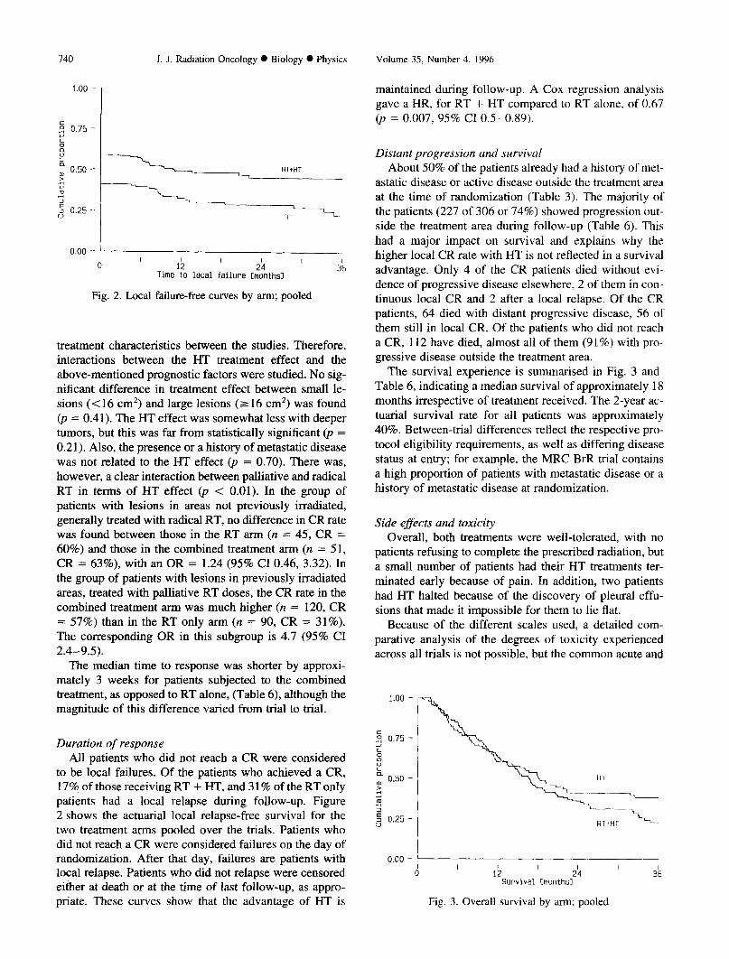
Fig. 2. Local failure-free curves by arm; pooled
treatment characteristics between the studies. Therefore, interactions between the HT treatment effect and the above-mentioned prognostic factors were studied. No sig- nificant difference in treatment effect between small le- sions (< 16 cm’) and large lesions (Z 16 cm’) was found (JJ = 0.41). The HT effect was somewhat less with deeper tumors, but this was far from statistically significant @ = 0.21). Also, the presence or a history of metastatic disease was not related to the HT effect (p = 0.70). There was, however, a clear interaction between palliative and radical RT in terms of HT effect (p < 0.01). In the group of patients with lesions in areas not previously irradiated, generally treated with radical RT, no difference in CR rate was found between those in the RT arm (n = 45, CR = 60%) and those in the combined treatment arm (n = 5 1, CR = 63%), with an OR = 1.24 (95% CI 0.46, 3.32). In the group of patients with lesions in previously irradiated areas, treated with palliative RT doses, the CR rate in the combined treatment arm was much higher (n = 120, CR = 57%) than in the RT only arm (n = 90, CR = 31%). The corresponding OR in this subgroup is 4.7 (95% CI 2.4-9.5).
The median time to response was shorter by approxi- mately 3 weeks for patients subjected to the combined treatment, as opposed to RT alone, (Table 6), although the magnitude of this difference varied from trial to trial.
Duration of response All patients who did not reach a CR were considered
to be local failures. Of the patients who achieved a CR, 17% of those receiving RT + HT, and 3 1% of the RT only patients had a local relapse during follow-up. Figure 2 shows the actuarial local relapse-free survival for the two treatment arms pooled over the trials. Patients who did not reach a CR were considered failures on the day of randomization. After that day, failures are patients with local relapse. Patients who did not relapse were censored either at death or at the time of last follow-up, as appro- priate. These curves show that the advantage of HT is
maintained during follow-up. A Cox regression analysis gave a HR, for RT + HT compared to RT alone, of 0.67 (p = 0.007, 95% CI 0.5-0.89).
Distant progression and survival About 50% of the patients already had a history of met-
astatic disease or active disease outside the treatment area at the time of randomization (Table 3). The majority of the patients (227 of 306 or 74%) showed progression out- side the treatment area during follow-up (Table 6). This had a major impact on survival and explains why the higher local CR rate with HT is not reflected in a survival advantage. Only 4 of the CR patients died without evi- dence of progressive disease elsewhere. 2 of them in con- tinuous local CR and 2 after a local relapse. Of the CR patients, 64 died with distant progressive disease, 56 of them still in local CR. Of the patients who did not reach a CR, 112 have died, almost all of them (91%) with pro- gressive disease outside the treatment area.
The survival experience is summarised in Fig. 3 and Table 6, indicating a median survival of approximately 18 months irrespective of treatment received. The 2-year ac- tuarial survival rate for all patients was approximately 40%. Between-trial differences reflect the respective pro- tocol eligibility requirements, as well as differing disease status at entry; for example, the MRC BrR trial contains a high proportion of patients with metastatic disease or a history of metastatic disease at randomization.
Side effects and toxicity Overall, both treatments were well-tolerated, with no
patients refusing to complete the prescribed radiation, but a small number of patients had their HT treatments ter- minated early because of pain. In addition, two patients had HT halted because of the discovery of pleural effu- sions that made it impossible for them to lie flat.
Because of the different scales used, a detailed com- parative analysis of the degrees of toxicity experienced across all trials is not possible, but the common acute and
d I lb
I 2b
I 3i
Survival [months1
Fig. 3. Overall survival by arm; pooled

Hyperthermia for breast cancer 0 INTERNATIONAL COLLABORATIVE HYPERTHERMIA GROUP 141
late side effects of therapy are summarized in Table 7 by treatments received, for those patients for whom this in- formation is available.
In terms of acute reactions, there is little difference in erythema and desquamation between treatments but, as expected, more blistering occurred with the addition of HT (11%) as compared to RT alone (2%). This excess is noted in all trials except MRC BrI, where no blistering occurred in either treatment arm. Similarly, acute effects of HT were 7% ulceration and 7% necrosis in the com- bined arm, compared with 2% and l%, respectively in the RT-only arm. These were greatest in the MRC BrR trial, where 10% of patients receiving HT suffered some early necrosis. In general, the acute effects of HT treatment tended to occur in areas of reduced sensitivity and healed with conservative treatment, with little impact on patient well-being.
Severe late reactions occurred: 1 each of bone necrosis, bone fracture, and brachial plexus lesion, all in the com- bined arm of the ESHO trial. The late effects of pigmen-
tation, telangiectasia, and fibrosis show very little varia- tion between treatments.
Thus, HT, as delivered in these trials, was well tol- erated and did not significantly add to either the clini- cally relevant acute or long-term toxicity over irradia- tion alone, even in those patients who had received prior radical RT.
DISCUSSION
The trials presented in this paper result from a mul- ticenter international collaboration, and the analysis has demonstrated a statistically significant benefit from the use of HT, in addition to radiation, in superficial breast cancer.
In view of the need for quality assurance for HT treat- ment highlighted in previous trials (23), an effort was made to ensure that only tumors that could be heated sat- isfactorily would be entered into the trials. For this reason, quality assurance guidelines based on general experience
Table 7. Summary of recorded treatment-related toxicity by trial and treatment
DHG MRC BrI MRC BrR ESHO PMH TOTAL
RT RT + HT RT RT + HT RT RT + HT RT RT + HT RT RT + HT RT RT + HT
Erythema (mild/mod) No II Yes 2
Erythema (severeldesquamation)
No 5 Yes 8
Blister No 13 Yes 0
Ulceration No 13 Yes 0
Necrosis No 13 Yes 0
Fibrosis No 6* Yes 4
Telangiectasia No 8* Yes 2
Pigmentation No 4* Yes 6
9 7 11 21 46 11 10 7 5 7 5 6 33 42 16 15 9 12
8 12 15 48 77 18 17 10 9 8 0 2 6 11 9 8 6 8
15 12 17 53 82 27 20 15 13 3 0 0 1 6 0 6 1 4
16 12 16 52 85 27 22 16 16 2 0 1 2 3 0 4 0 1
17 12 16 53 78 27 26 16 1 0 1 1 10 0 0 0
5” 4
4 8
7 10
10 7
14 3
24* 24* 3’ 14 35 4
9* 8 0 4
27* 11
6* 1
3* 9 6 3
22* 16
39* 20
29* 30
1* 6
9* 3
10* 2
4* 8
9 7
- -
11 5
17 0
10 7
- -
12 5
57 81 65 82 53% 50%
93 126 29 37 24% 23%
120 2 2%
147 19 11%
119 3 2%
155 11 7%
121 1 1%
154 12 7%
46 55 37 59 45% 52%
49 68 18 29 27% 30%
47 62 36 52 43% 46%
* Confined to patients with at least 1 year of follow-up. - Not recorded.

742 1. J. Radiation Oncology 0 Biology 0 Physics Volume 35, Number 4. 1996
and consensus within the HT communities in Europe and North America (8, 12) were adopted. In addition, quality assurance programs in some institutes were assessed dur- ing site visits carried out under the auspices of ESHO (16).
Superficial breast cancer was chosen for the trials be- cause, even within the limitations of available HT equip- ment, it was felt by the individual trial groups that it was feasible to heat the relatively shallow lesions adequately and to obtain satisfactory measurements of the tempera- ture distribution at several locations within the treated field.
The combined therapy was well tolerated and did not result in major toxicity. There were, however, differences in outcome between the individual trials with two, MRC BrR and ESHO, illustrating an advantage for HT. In the DHG trial, there was no apparent difference between the treatments and MRC BrI and PMH indicated a small ad- vantage for RT alone. Note, however, that all of the 95% CIs for the odds ratios (OR) from the five trials are not inconsistent with a substantial benefit from HT (Fig. 1).
Overall survival did not differ markedly between the two treatment arms, although the pooled data suggest that the group receiving additional HT has a marginally infe- rior survival, as shown in Fig. 2. This may be caused by lesion size, which has been shown to be of prognostic value for overall survival (4), and was larger in the com- bined treatment arm.
We have shown that size and depth of lesions, distant metastatic disease, and RT dose are important factors that affect CR rate. The patients in these trials are heteroge- neous in these respects and this accounts, in part, for the variable CR rates between the trials. However, following adjustment for these factors, there remains a statistically significant difference in CR rate in favor of HT. The num- bers of patients in the individual trials are small and any inferences drawn from the inter-trial differences may, as a consequence, be unreliable. Nevertheless, these differ- ences do raise some interesting questions with regard to the possible differences in efficacy of varying RT and HT regimens. which are testable hypotheses for future trials.
There are differences within the five trials that may be important. All the MRC BrI patients, approximately two thirds of the DHG patients, and a smaller proportion in the MRC BrR and PMH trials, received radical RT. The MRC BrI patients had primary breast cancer that was deemed inoperable because of disease extent, and these lesions were probably more difficult to heat adequately. The depths of these tumors were estimated clinically and it is possible that this may have been underestimated. These patients, therefore, may not have complied with the guidelines for HT. The lesion sizes of the DHG patients were smaller. and we would expect all such patients to achieve a higher CR rate. The treatments delivered also varied between trials. Estimates of the equivalent radiation doses showed considerable differences between the pal- liative schedules, although the radical doses were less variable. Bedwinek et al. (3) have defined ‘adequate’ RT
doses for recurrent tumors of different sizes and depths. However. tumors in previously irradiated areas cannot be adequately treated by RT alone because of the effect on normal tissues, and because the hypoxia induced by pre- vious RT renders the tumor less sensitive to the effects of radiation. We believe that it is important to give the max- imum tolerated dose of radiation, even in those patients who have received previous radiation. to achieve the high- est possible CR rates. There was significant variation in the prescription of heat treatments in the different trials. hence, making the establishment of a heat-response rela- tionship difficult. It would appear, however, that the PMH trial had the lowest number and duration of heat treat- ments, with the lowest CR rate achieved for the combined treatment arm. A number of possibilities may account for this outcome, such as the small number of patients, vari- ation in clinical characteristics, and low RT dose. but the possibility that this may have been influenced by the heat treatments cannot be excluded.
The combined results reported here have, we suggest, confirmed the view that the role of HT in the treatment of breast cancer is as an adjunct to a palliative dose of RT in patients with tumor recurrence following a radical course of treatment.
These trials do not establish the beneficial use of HT for patients who are able to receive a full dose of radiation, but the numbers of patients treated radically in these trials is small and the apparent lack of success with the addition of HT may be explicable by other factors, as already dis- cussed. Biologically, there is no reason why HT should not be of benefit in the radical situation, and future trials should look at these patients in greater numbers.
The randomized trial reported by Overgaard et al. (21) in patients with recurrent or metastatic malignant melanoma treated by RT with or without HT, has shown CR rates of 46% for the combined arm and 28% for the radiation-only arm, which are similar to our own. This was a small trial based on only 71 patients with 134 lesions, and the form of analysis used leads to some doubt as to how reliable these estimates of CR really are (19). How- ever, both this trial and our own, with the CR rate at 2 years of 59% for irradiation plus HT vs. 41% for irradi- ation alone, is in keeping with other Phase III randomized trials. Following the results of our trials, we could rec- ommend the consideration of HT for patients with recur- rent breast cancer to be retreated with irradiation. We hope that these results will encourage the use of HT in clinical practice and, also, further study into its use in other tumor types, as well as the best scheduling of HT and radiation. Further research is also required to assess the benefit of HT on those patients for whom radical radiation is planned.
We would emphasize that, without the international col- laboration, each of these five trials would have been too small to contribute meaningful data on the role of HT in superficial breast cancer. This must have important impli- cations for the planning of future trials.

Hyperthermiafor breast cancer 0 INTERNATIONAL COLLABORATIVEHYPERTHERMIAGROUP 743
ACKNOWLEDGEMENTS
Medical Research Council Trials We acknowledge the financial support of the hyper-
thermia program at the Cyclotron Unit provided by the Medical Research Council (MRC; United Kingdom); the expertise of M. V. Prior, G. R. Forse, J. Khudhail, and Y. Robinson in administering hyperthermia; the contribu- tions to assessment made by J. Flanders and F. Paice; the secretarial support of V. Jackson and V. Ellen; and com- puter support from L. Jenkin.
Dutch Hyperthermia Group and European Society Hyperthermic Oncology Trials
We acknowledge the contributions of P. J. van Assen- delft in data management, of all involved in the adminis- tration of the hyperthermia treatments, and of J. Overgaard for contribution to protocol design for the European So- ciety Hyperthermic Oncology (ESHO) trial.
We also acknowledge the financial support of The Netherlands Cancer Foundation “Koningin Wilhelmina Fonds” (several grants for the development of clinical hyperthermia application), the Willem Kriiger Stichting, the Maurits and Anna de Kock Stichting, and the Stichting Bevordering Volkskracht, for hyperthermia equipment.
The contributions of J. D. P. van Dijk, C. J. Schneider and G. Lamaitre of the Dept of Radiotherapy, Academic Medical Center, Amsterdam, in carrying out the hyper- thermia QA site visit program under the auspices of the ESHO Technical Committee and with financial support of the Concerted Action program (COMAC BME contracts M4*/0204/B and 0306/B) within the European Commis- sion’s 4th Medical and Health Research Programme are also acknowledged.
The following clinicians entered patients into the Dutch Hyperthermia Group (DHG) and ESHO trials: G. van Tienhoven, M. C. C. M. Hulshof, L. E. C. M. Blank, J. G. J. Letschert, F. Oldenburger and A. L. J. Schuster- Uitterhoeve, Academic Medical Center, Amsterdam, The Netherlands (41); A. D. Treumiet-Donker, W. A. M. Mel- link, P. C. M. Koper, B. A. Reichgelt, P. A. Helle, A. Slot, A. J. Wijnmaalen, J. J. Seldenrath, P. L. A. van den Ende and P. J. C. M. Nowak, Dr. Daniel den Hoed Cancer Cen- ter, Rotterdam, The Netherlands (27); M. Amichetti and
C. Graiff, Oncology Center, San Chiara Hospital, Trento, Italy (8); J. Fijuth, Institute of Oncology, Warsaw, Poland (7); P. Hoffman and H. K. Wijrdeman, University Hos- pital, Utrecht, The Netherlands (3); P. F. Steindorfer and W. Amann, University Hospital Graz, Austria (3); G. Ar- cangeli, San Maria Goretti Hospital, Latina, Italy (2); and M. Treitz, Kliniken der Stadt Saarbriicken, Germany (1).
The following clinicians entered patients into the MRC trials: C. Vernon, H. Lambert, C. Mackenzie, J. Waxman, J. Stewart, A. Munro, K. Sikora, P. Price, A. Epenetos, Hammersmith Hospital, London (87); J. Maher, M. Saun- ders, Mount Vernon Hospital, Northwood (18); C. Coul- ter, M. Spittle, A. Cassoni, Middlesex Hospital, London (18); A. Jones, C. Alcock, D. Cole, Churchill Hospital, Oxford (9); C. Harmer, J. Yarnold, Royal Marsden Hos- pital, London (7); J. Dobbs, King’s College Hospital, Lon- don (6); M. Ghilchick, St. Mary’s Hospital, London (6); A. Timothy, St. Thomas’ Hospital, London (4); D. Tong, Guy’s Hospital, London (4); I. Kunkler, Weston Park Hospital, Sheffield (3); D. Pickering, Maidstone Hospital, Maidstone, Kent (2): J. Tobias, University College Hos- pital, London (2); P. Plowman, St Bartholomew’s Hos- pital (2), London; R. Beaney, J. Mould, Queen Elizabeth Hospital, Birmingham (2); N. Bleehen, Addenbrooke’s Hospital, Cambridge (2); C. Keen, T. Maughan, Univer- sity Hospital of Wales, Cardiff (2); H. Smedley, Kent and Canterbury Hospital, Canterbury, Kent (1); B. Southcott, Charing Cross Hospital, London (1); P. Murray, Essex County Hospital, Colchester (1); A. Hong, Royal Devon and Exeter Hospital, Exeter (1); A. Folkes, Royal Surrey County Hospital, Guildford (1); V. Svoboda, St Mary’s Hospital, Portsmouth (1); and H. Hope-Stone, Royal Lon- don Hospital, London (1).
Princess Margaret Hospital Trial We acknowledge the clinical support of W. Levin, L.
Manchul, P. Kirkbride, G. Rawlings, and L. Yeoh; the scientific support of R. P. Hill and J. W. Hunt; the tech- nical support of B. Cooper, D. Newcombe, and W. Taylor; and the statistical support of E. Rawlinson and M. Pintilie. The financial contributions of the Princess Margaret Hos- pital Trust Foundation, the Canadian Breast Cancer Foun- dation, and the Ontario Cancer Treatment and Research Foundation are also acknowledged.
REFERENCES
1. Aberizk, W. J.; Silver, B.; Henderson, I. C.; Cady, B.; Harris, J. R. The use of radiotherapy for treatment of locoregional recurrence of breast carcinoma after mastectomy. Cancer 58:1214-1218; 1986.
2. Altman, D. G. Practical statistics for medical research. Lon- don: Chapman and Hall; 1991.
3. Bedwinek, J. M.; Fineberg, B.; Lee, J.; Ocwieza, M. Anal- ysis of failures following local treatment of isolated local- regional recurrence of breast cancer. Int. J. Radiat. Oncol. Biol. Phys. 7:581-585; 1981.
4. Bedwinek, J. M.; Lee, J.; Fineberg, B.; Ocwieza, M. Prog-
nostic indicators in patients with isolated local-regional re- currence of breast cancer. Cancer 472232-2235: 1981.
5. Coley, W. C. The treatment of malignant tumours by re- peated inoculations of erysipelas; with a report of 10 original cases. Am. J. Med. Sci. 105:486-511; 1893.
6. COMPACT Steering Committee. Improving the quality of clinical trials in cancer. Br. J. Cancer 63:412-415; 1991.
7. Cox, R. S.; Kapp, D. S. Correlation of thermal parameters with outcome in combined radiation therapy-hyperthermia trials. Int. J. Hyperthermia 8:719-732; 1992.
8. Dewhirst, M. W.: Phillips, T. L.; Samulski, T. V.; Staufter,

744 I. J. Radiation Oncology l Biology l Physics Volume 35, Number 4, 19%
P.; et al. RTOG quality assurance guidelines for clinical tri- als using hyperthermia. Int. J. Radiat. Oncol. Biol. Phys. 18:1249-1259; 1990.
9. Egawa, S.; Tsukiyama, 1.; Watanabe, S.; Ohno, Y.; Mor- ita, K.; Tomingawa, S.; Onoyama, Y.; Hashimoto, S.; Yanagawa, S.; Uehara, S.; Abe, M.; Mochizuki, S.; Su- giyama, A.; Inoue, T. A randomised clinical trial of hy- perthermia and radiation vs. radiation alone for superfi- cially located cancers. J. Jpn. Sot. Ther. Radiol. Oncol. 1:135-140; 1989.
10. Field, S. B.: Hand, J. W., eds. An introduction to thepractical aspects of clinical hyperthermia. London: Taylor & Francis; 1990
11. Gonzalez Gonzalez, D.; van Dijk, J. D. P.; Blank, L. E. C. M. Chestwall recurrences of breast cancer: Results of combined treatment with radiation and hyperthermia. Radiother. Oncol. 12:95-103; 1988.
12. Hand, J. W.; Lagendijk, J. J. W.; Anderson, J. B .; Bolomey, J. C. Quality assurance guidelines for ESHO protocols. Int. J. Hyperthermia 5:421-428; 1989.
13. International Union Against Cancer. TNM classification of malignant tumours, 4th ed. Berlin: Springer-Verlag; 1992.
14. Kapp, D. S.: Petersen, I. A.; Cox, R. S.; Hahn, G. M.; Fes- senden. P.; Prionas, S. D.; Lee, E. R.; Meyer, J. L.; Samulski, T. V.; Bagshaw, M. A. Two or six hyperthennia treatments as an adjunct to radiation therapy yield similar tumour re- sponses: Results of a randomised trial. Int. J. Radiat. Oncol. Biol. Phys. 19:1481-1495; 1990.
15. Kjellen, E.; Lindholm, C.-E.; Nilsson, P. Radiotherapy in combination with hyperthermia in recurrent or metastatic mammary carcinomas. In: Sugahara, T.; Saito, M., eds. Hy- perthermic Oncology, Vol. 1. London and New York: Taylor & Francis; 1988:426-429.
16. Lamaitre, G.; Postma. A. J.: van Dijk, J. D. P. In: The COMAC-BME site visit program for superficial heating de- vices: Preliminary results (Abstr.). Book of abstracts. 13th ESHO conference. Brussels: 1993.
17. Lewis, J. A.; Machin, D. Intention to treat-who should use ITT. Br. J. Cancer 68:647-650; 1994.
18. Machin, D.; Campbell. M. J. Statistical tables for the design of clinical trials. Oxford: Blackwell; 1987.
19. Machin, D.; Parmar, M. K. B. Hyperthermia in cancer treat- ment. Lancet 345: 1635-1636; 1995.
20. Mtiller. C. Eine neue behandlungsmethode bbsartiger gesch- wtilste. Munch. Med. Wochenschr 28: 1490-1493; 1910.
21. Overgaard, J.; Gonzalez Gonzalez, D.; Hulshof, M. C. C. M.; Arcangeli, G.; Dahl, 0.; Mella, 0.; Bentzen, S. M., for the European Society of Hyperthermic Oncology. Randomised trial of hyperthermia as adjuvant to radiotherapy for recurrent or metastatic malignant melanoma. Lancet 345:540-543; 1995.
22. Parmar, M. K. B.; Machin, D. Survival analysis: A practical approach. Chichester, UK: Wiley: 1995.
23. Perez, C. A.; Gillespie, B.; Pajak, T.; Hornback. N. B.; Emami, B.; Rubin, P. Quality assurance problems in clinical hyperthermia and their impact on therapeutic outcome: a re- port by the Radiation Therapy Oncology Group. ht. J. Ra- diat. Oncol. Biol. Phys. 16:551-558; 1989.
24. Perez, C. A.; Kuske, R. R.; Emami, B.: Fineberg. B. Irradi- ation alone or combined with hyperthermia in the treatment of recurrent carcinoma of the breast in the chest wall: A nonrandomized comparison. Int. J. Hyperther. 2: 179-l 87: 1986.
25. Perez, C. A.; Pajak. T.; Emami, B.; Hornback. N. B.; Tup- thong, L.; Rubin, P. Randomized Phase III study comparing irradiation and hyperthermia with irradiation alone in super- ficial measurable tumors. Am. J. Clin. Oncol. (CCT) 14:133- 141; 1991.
26. Schmidt, W. E. Zur Rontganbehandlung trefliegander tu-
moren. Fortsor. Roengenstr. 14: 134; 1909. 27. Scott, R. S.; Johnson, R. J. R.; Story, K. V.; Clay, L. Local
hyperthermia in combination with definitive radiotherapy: Increased tumour clearance, reduced recurrence rate in ex- tended follow-up. Int. J. Radiat. Oncol. Biol. Phys. IO:21 19- 2123; 1984.
28. Stadler, B.; Kogelnik, D. H. Local control and outcome of patients irradiated for isolated chest wall recurrences of breast cancer. Radiother. Oncol. 8: 105-l 1 I: 1987.
29. Stata reference manual: release 3.1, 6th ed. Santa Monica. CA; Stata Corporation. 1993
30. Steel, G. G., ed. Basic clinical radiobiology. Sevenoaks, UK: Edward Arnold; 1993
3 1. Valdagni, R.; Amichetti, M.; Pani, G. Radical radiation alone vs. radical radiation plus microwave hyperthermia for N3 (TNM-UICC) neck nodes: A prospective randomized clini- cal trial. Int. J. Radiat. Oncol. Biol. Phys. 15:13-24; 1988.
32. Valdagni, R.; Liu, F.-F.; Kapp, D. S. Important prognostic factors influencing outcome of combined radiation and hy- perthermia. Int. J. Radiat. Oncol. Biol. Phys. 15:959-972; 1988.
33. van der Zee. J.; Treumiet-Donker, A. D.; The, S. K.; Helle, P. A.; Seldenrath, J. J.; Meerwaldt, J. H.; Wijnmaalen, A. J.; van den Berg, A. P.; van Rhoon, G. C.; Broekmeyer-Reu- rink, M. P.; Reinhold, H. S. Low dose reirradiation in com- bination with hyperthermia: a palliative treatment for pa- tients with breast cancer recurring in previously irradiated areas. Int. J. Radiat. Oncol. Biol. Phys. 15:1407-1413; 1988.
34. Westermark, F. Uber die Behandlung des Ulcerierenden Cer- vixcarcinoms mittels konstanter warme. Zbl. Gynakol. 22:1335-1339; 1898.
35. WHO handbook for reporting results of cancer treatment. WHO Offset Publication No. 48. Geneva: WHO: 1979.