radiotherapy manual (euro net-phl-c1) - skion - home · pdf fileeuronet-phl-c1: radiotherapy...
TRANSCRIPT
EuroNet-PHL-C1
EuroNet-Paediatric Hodgkin’s Lymphoma Group
First international Inter-Group Study for classical Hodgkin’s Lymphoma in Children and
Adolescents
• No radiotherapy in patients with adequate response at first restaging after
two cycles of chemotherapy
• Randomised comparison of Procarbazine versus Dacarbazine (within
COPP versus COPDAC) in patients in intermediate and advanced stages
• Standardised risk- and response-adapted salvage strategy
EudraCT-No.: 2006-000995-33
Radiotherapy Manual
Inter-group chairpersons
Prof. Dr. Dieter Körholz (Coordinating investigator) Zentrum für Kinderheilkunde Universitätsklinik und Poliklinik für Kinder- und Jugendmedizin Ernst-Grube-Straße 40, 06120 Halle (Saale) Germany
Dr. W. Hamish Wallace Royal Hospital for Sick Children,
Sciennes Road, Edinburgh EH9 1LF,
Scotland, UK.
Prof. Dr. Judith Landman-Parker Service d’hématologie et d’oncologie pédiatrique
Hôpital Trousseau AP-HP Paris France
Date of Version: 11.08.2006
Version: Final
EuroNet-PHL-C1: Radiotherapy manual Final Version 2006-08-11
RESPONSIBILITIES
Reference radiation oncologists of the central review board Halle/Leipzig
Prof. Dr. Rolf-Dieter Kortmann, Prof. Dr. Friedrich Hugo Kamprad, Dr. Kirsten Papsdorf
Universitätsklinik und Poliklinik für Strahlentherapie und Radioonkologie, Universitätsklinikum Leipzig Stephanstr. 9A , D-04103 Leipzig, Germany Tel.: +49 341- 97 18 400, Fax: +49 341- 97 18 409 Email: [email protected] [email protected] [email protected]
Radiation oncologists (writing committee of the Radiotherapy Manual)
Prof. Dr. Karin Dieckmann (coordinator of the writing committee of the Manual) Prof. Dr. R. Pötter
Universitätsklinik für Strahlentherapie und Strahlenbiologie Medizinische Universität Wien, Allgemeines Krankenhaus der Stadt Wien Währingergürtel 18 – 21, A -1090 Wien, Tel.: +43-1-40400-2692 Email: [email protected]
Dr. Ursula Rühl Vivantes Klinikum im Friedrichshain Standort Moabit Klinik für Strahlentherapie 10559 Berlin
Dr. Helen Lucraft NCCT, Newcastle General Hospital Newcastle upon Tyne NE4 6BE Tel.: +44 1912563565 (secretary) +44 1912563963 (direct line) Fax: +44 1912563674 Email: [email protected] Dr. Christian Carrie
Centre Léon Berard 28 rue Laennec F 69373 Lyon cedex 08 Tel : +33 4 78 78 26 52 Fax: +33 4 78 78 51 40
Email: [email protected]
- 1 -

EuroNet-PHL-C1: Radiotherapy manual Final Version 2006-08-11
CCOONNTTEENNTTSS
1 General introduction to Radiotherapy in paediatric Hodgkin´s Disease according to
GPOH experience ........................................................................................................3 2 Indications for Radiotherapy.........................................................................................8 3 Timing of Radiotherapy ..............................................................................................10 4 Volume and Dose of Radiotherapy.............................................................................10 5 Medical Aspects of Treatment Planning .....................................................................11 6 Target Volume ............................................................................................................12 7 Organs at risk: effects of radiotherapy on normal tissues in the growing child...........28 8 Radiotherapy in relapsed patients ..............................................................................32 9 Central review.............................................................................................................33 10 Technical and physics aspects of treatment planning and delivery............................33 11 Treatment delivery......................................................................................................37 12 Acute side effects .......................................................................................................38
- 2 -
EuroNet-PHL-C1: Radiotherapy manual Final Version 2006-08-11
11 GGEENNEERRAALL IINNTTRROODDUUCCTTIIOONN TTOO RRAADDIIOOTTHHEERRAAPPYY IINN PPAAEEDDIIAATTRRIICC
HHOODDGGKKIINN´́SS DDIISSEEAASSEE AACCCCOORRDDIINNGG TTOO GGPPOOHH EEXXPPEERRIIEENNCCEE
1.1 First study generation DAL-HD 78
Already the first study generation DAL-HD 78 set the general therapeutic paradigm:
Chemotherapy starting with 2 courses of intense and effective OPPA (followed by COPP consolidation in intermediate and advanced stages) plus radiotherapy. In 1978
radiotherapy consisted of 36 – 40 Gy to the involved field and 18 – 20 Gy in the adjacent
fields (Schellong 1986A, 21). In this study extended field radiotherapy was performed.
1.2 Second study generation DAL-HD 82
In the second study generation DAL-HD 82 (Schellong et al., 1986A, 21), patients for the first
time were divided into three treatment groups based on stage: TG-1: stage I/IIA TG-2: stage IIB/IIIA, IE/IIEA TG-3: stage IIIB/IV, stage IIEB/IIIE. The number of consolidation COPP cycles was scaled according to treatment group (0, 2, 4
respectively). Treated volume was reduced from extended to involved field radiotherapy. Indication for splenectomy was limited and the number of splenectomies dropped to about
40%. Radiation doses were reduced to 35 – 30 – 25 Gy in TG-1, TG-2, TG-3, respectively.
In case of insufficient response to chemotherapy the radiation dose was increased by 5 – 10
Gy (boost).
5-year-EFS rates were achieved with 99%, 96% and 90% in TG-1, TG-2 and TG-3,
respectively (Schellong et al., 1988B, 23). Due to these excellent results DAL-HD 82 for a
long time has been regarded as the gold standard.
1.3 Third study generation DAL-HD 85
(Chemotherapy Study) After the gonadotoxic effect of Procarbazine became apparent this drug was completely
eliminated from OPPA–COPP chemotherapy in the DAL-HD 85 study generation (23).
Chemotherapy was OPA-COMP, so that in the first two cycles only three agents were
administered and Procarbazine was replaced by Methotrexate in consolidation. Involved field
radiotherapy was dosed according to TG with 35, 30 or 25 Gy (Schellong et al., 1988B, 23).
By eliminating Procarbazine fertility in boys indeed was preserved (Brämswig et al., 1990, 2;
- 3 -
EuroNet-PHL-C1: Radiotherapy manual Final Version 2006-08-11
Hassel et al., 1991, 10), but treatment efficacy was compromised: For patients with early
stages (TG-1) the 10-year EFS rate dropped to 85%, however practically all patients could
be salvaged by relapse therapy and an overall survival rate of 98% after 10 years was seen
(Schellong et al., 1994A, 24). For patients in intermediate (TG-2) and advanced stages (TG-
3) the 3 year EFS rate dropped to unacceptable levels of 59% and 62%, respectively
(Schellong et al., 1988A, 22).
1.4 Fourth study generation DAL-HD 87
Therefore in the fourth study generation DAL-HD 87 Procarbazine was reintroduced into the
COPP cycles while it was still not administered in the OPA. In addition, the radiation dose was further reduced to 30-25-20 Gy for TG-1, TG-2 and TG-3, respectively (Schellong et
al., 1994B, 1996; 25, 26). Indication for splenectomy was further restricted so that only in
29% of the patients the spleen was removed. The 7-year EFS and overall survival (OS) rates
for all patients (85% and 97% respectively) were better than in DAL-HD 85 but still clearly
worse than those of the DAL-HD 82 study generation and were felt to be unsatisfactory.
1.5 Fifth study generation DAL-HD 90
Therefore initial therapy in the DAL-HD 90 study was re-intensified. All girls got OPPA again. Boys got OEPA, i.e. OPPA with Procarbazine replaced by 500 mg/m2 Etoposide
given over 4 days, in the hope that this would preserve fertility (Schellong et al., 1999, 1988;
29, 22; Kreuser et al., 1987, 14). Splenectomy was abandoned and the radiotherapy dose was further reduced to 25 – 25 - 20 Gy for TG-1-3 respectively. Modified involved field radiotherapy (MIF) was introduced (Schellong et al., 2004, 30).
With this strategy, a 5-year EFS rate of 91% was achieved with OPPA and 89% with OEPA.
Overall survival after 5 years was 98% in both groups. The results were comparable with the
excellent results of the DAL-HD 82 study although therapy intensity was clearly reduced
(Schellong et al., 1999, 29).
By introducing Etoposide the infertility rate of boys was significantly reduced (Gerres et al.,
1998, 9) in TG-1, while about half of the male patients in TG-2 and TG-3 still showed
abnormal FSH values after the COPP cycles.
In the study generation DAL-HD 90 a real-time upfront central review process for all patients
was established (Dieckmann et al., 2002, 6). Staging, therapy group assignment and
response assessment for all patients was performed centrally, assessing the clinical data
and reviewing all images. In about 20% of the patients the stage had to be revised by central
appraisal. 11.7% of patients were assigned to a higher therapy group while 1.6% were down
staged to a lower therapy group (Dieckmann et al., 2002, 6). About 50% of all patients were
- 4 -
EuroNet-PHL-C1: Radiotherapy manual Final Version 2006-08-11
treated with modified involved field radiotherapy (Pötter et al. 1995, 19). Lymph node
recurrence was in 29/578 patients (5%) and was not related to reduced radiation field size
(MIF).
1.6 Sixth study generation GPOH-HD 95
A major concern apart from infertility in boys has been the development of treatment related secondary malignancies. The rate of secondary haematological malignancies, which occur mostly 1 – 10 years after
therapy, is very low. The estimated risk after 15 years is about 1% for the patients in the
studies DAL-HD 78 to DAL-HD 90 (Schellong et al., 1997, 27). After the introduction of
Etoposide, no leukemias have been reported so far (Schellong 1998, 28; Schellong and
Riepenhausen 2004, 30).
On the other hand, the number of non-hematological secondary tumours still increases after
a latency period of 20 and more years (Schellong and Riepenhausen 2004, 30). The
cumulative risk of secondary solid tumours for the DAL / GPOH-HD study patients is 5.7%
after 20 years (standard error (SE) 1.5%), which is low compared to other groups (e.g. LESG
(Meadows et al. 1989, 15). Secondary solid tumours (SST) are the main cause for this late
increase. The most important risk factor for the development of SST seems to be radiation
therapy combined with carcinogenic chemotherapy (e.g. Mustargen, Meadows et al., 1989,
15). 22 of the overall 25 SST observed within the 6 generations of GPOH paediatric HD
studies have occurred in or at the border of the radiation field.
Therefore in the GPOH-HD 95 study generation the dose of radiotherapy was reduced to 20 Gy in all treatment groups and modified involved field radiotherapy was
systematically considered for all patients. In addition, radiotherapy was omitted in patients with complete remission (CR) at the end of chemotherapy in order to reduce the number
of patients at risk for radiotherapy associated secondary solid tumours.
Event free survival after 5 years was 88% for all patients; overall survival 97% (Dörffel et al.,
2003, 8).
In TG-1 there was no significant EFS difference between patients with (94%) and without
(97%) radiotherapy. Therefore omission of radiotherapy has been adopted as standard
treatment for patients in TG-1 who achieved CR after two cyles of chemotherapy.
However, in TG-2 and TG-3 omission of radiotherapy for CR patients led to a significant
decrease in EFS: 79% without versus 91% with radiotherapy. Therefore radiotherapy for all
patients in TG-2 and TG-3 was re-introduced as standard in the following study.
In patients treated with modified involved field radiotherapy, lymph node recurrence was in
15 out of 611 (2,5%) (Rühl et al. 2001, 20) which is comparable to the results as achieved in
DAL-HD 90. - 5 -
EuroNet-PHL-C1: Radiotherapy manual Final Version 2006-08-11
1.7 GPOH-HD 2002 Pilot study
In GPOH-HD 2002 Pilot study, all boys received an intensified OE*PA therapy (*20% more
Etoposide) and COPDAC instead of the COPP cycles (Dacarbazine instead of Procarbazine)
(Körholz et al., 2004, 13). Radiotherapy has remained as in GPOH-HD 1995 with modified
involved field technique and doses of 20 Gy in all treatment groups and boost doses of 10
and 15 Gy.
In conclusion: In the German-Austrian Studies on Hodgkin`s Disease in children and
adolescents radiation therapy was applied locally, initially using a classical involved field
technique (HD 82), later on (since HD 90/95) a modified involved field technique which
included the initially involved nodes and an appropriate safety margin taking into account the
involved lymph node area.
Standard dose has been reduced step by step to 20 Gy, regardless of the treatment group
(risk group). Boost treatment has been restricted to clearly defined situations with insufficient
response.
Opposed field photon radiotherapy has been the treatment technique of choice and was
used in almost all patients. Treatment planning has been based on the extent of primary
lymph node involvement as found during clinical examination, if accessible, and as shown by
CT and/or MRI.
Due to the excellent results obtained in the past with regard to overall outcome and local
control the new trial continues in the tradition of these protocols using the same target
volumes, radiation techniques and radiation doses.
The study Euro Net-PHL-C1 addresses the following issues:
Is it possible to reduce the number of patients who need radiotherapy by identifying those
patients with adequate response to chemotherapy. These patients will not receive local
radiotherapy for consolidation (see figure 1 and 2).
For chemotherapy, the efficacy of COPP is randomly compared to COPDAC in patients of
TG-2 and TG-3 (see figure 2).
- 6 -

EuroNet-PHL-C1: Radiotherapy manual Final Version 2006-08-11
Fig. 1 Treatment overview for TG-1
TG-1I A/B, II A
2x OEPA
weeks1 5
RTRadiotherapy
inadequateresponse
adequateresponse
no RTRadiotherapy
TG-1I A/B, II A
2x OEPA
weeks1 5
RTRadiotherapy
inadequateresponse
adequateresponse
no RTRadiotherapy
Fig. 2 Treatment overview for TG-2 and TG-3
TG-2IE A/B, IIE A,II B, III A
TG-3IIE B, IIIE A/B,III B, IV A/B
2x OEPA
2x OEPA
2x COPP or 2x COPDAC
4x COPP or 4x COPDAC
weeks1 5 9 13 17 21
R
R
RTRadiotherapy
inadequateresponse
adequateresponse
no RTRadiotherapy
RTRadiotherapy
inadequateresponse
adequateresponse
no RTRadiotherapy
TG-2IE A/B, IIE A,II B, III A
TG-3IIE B, IIIE A/B,III B, IV A/B
2x OEPA
2x OEPA
2x COPP or 2x COPDAC
4x COPP or 4x COPDAC
weeks1 5 9 13 17 21
R
R
RTRadiotherapy
inadequateresponse
adequateresponse
no RTRadiotherapy
RTRadiotherapy
inadequateresponse
adequateresponse
no RTRadiotherapy
RTRadiotherapy
inadequateresponse
adequateresponse
no RTRadiotherapy
RTRadiotherapy
inadequateresponse
adequateresponse
no RTRadiotherapy
- 7 -
EuroNet-PHL-C1: Radiotherapy manual Final Version 2006-08-11
22 IINNDDIICCAATTIIOONNSS FFOORR RRAADDIIOOTTHHEERRAAPPYY
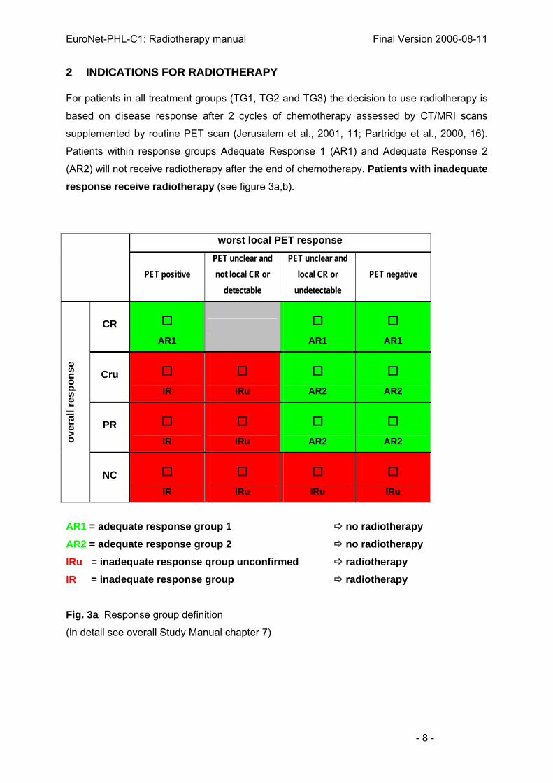
For patients in all treatment groups (TG1, TG2 and TG3) the decision to use radiotherapy is
based on disease response after 2 cycles of chemotherapy assessed by CT/MRI scans
supplemented by routine PET scan (Jerusalem et al., 2001, 11; Partridge et al., 2000, 16).
Patients within response groups Adequate Response 1 (AR1) and Adequate Response 2
(AR2) will not receive radiotherapy after the end of chemotherapy. Patients with inadequate response receive radiotherapy (see figure 3a,b).
worst local PET response
PET positive PET unclear and not local CR or
detectable
PET unclear and local CR or
undetectable PET negative
CR AR1
AR1
AR1
Cru IR
IRu
AR2
AR2
PR IR
IRu
AR2
AR2 ov
eral
l res
pons
e
NC IR
IRu
IRu
IRu
AR1 = adequate response group 1 no radiotherapy AR2 = adequate response group 2 no radiotherapy IRu = inadequate response qroup unconfirmed radiotherapy IR = inadequate response group radiotherapy Fig. 3a Response group definition
(in detail see overall Study Manual chapter 7)
- 8 -
EuroNet-PHL-C1: Radiotherapy manual Final Version 2006-08-11
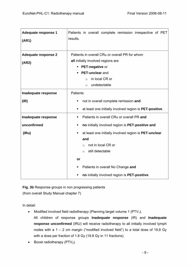
Adequate response 1
(AR1)
Patients in overall complete remission irrespective of PET
results.
Adequate response 2
(AR2)
Patients in overall CRu or overall PR for whom
all initially involved regions are
PET-negative or
PET-unclear and
o in local CR or
o undetectable
Inadequate response
(IR)
Patients
not in overall complete remission and
at least one initially involved region is PET-positive.
Inadequate response
unconfirmed
(IRu)
Patients in overall CRu or overall PR and
no initially involved region is PET-positive and
at least one initially involved region is PET-unclear and
o not in local CR or
o still detectable
or
Patients in overall No Change and
no initially involved region is PET-positive.
Fig. 3b Response groups in non progressing patients
(from overall Study Manual chapter 7)
In detail:
• Modified involved field radiotherapy (Planning target volume 1 (PTV1).
All children of response groups Inadequate response (IR) and Inadequate response unconfirmed (IRU) will receive radiotherapy to all initially involved lymph
nodes with a 1 – 2 cm margin (“modified involved field”) to a total dose of 19,8 Gy
with a dose per fraction of 1.8 Gy (19.8 Gy in 11 fractions).
• Boost radiotherapy (PTV2).
- 9 -
EuroNet-PHL-C1: Radiotherapy manual Final Version 2006-08-11
Children with a poor response and/or bulky residual masses after 2 courses of
chemotherapy will be treated with a boost to residual disease (as defined in Tab 1 on
page 23) with a 1-2 cm margin to a total dose of 10 Gy with 2 Gy per fraction (10 Gy
in 5 fractions).
33 TTIIMMIINNGG OOFF RRAADDIIOOTTHHEERRAAPPYY
Radiotherapy should be started within 3 - 4 weeks after completion of chemotherapy.
For TG-1 patients radiotherapy should follow restaging examinations at the end of
chemotherapy (for those belonging to Inadequate Response-groups IR-groups).
Radiotherapy should begin latest by day 35 after the last dose of chemotherapy.
For TG-2 and TG-3 patients the decision to treat with radiotherapy is taken after restaging
following two courses of OEPA and treatment should be started immediately after recovery of
blood counts following chemotherapy (up to day 25 after the last dose of chemotherapy).
Radiotherapy planning should be scheduled as soon as possible after the decision to treat
and it might be started already during chemotherapy to minimize delays.
Time scale for data documentation see figure 32 in the appendix.
Supra- and infradiaphragmatic fields should be treated concurrently. For larger volumes the
radiation oncologist can decide individually if supra- and infradiaphragmatic irradiation should
be performed separately with a 2 weeks gap.
44 VVOOLLUUMMEE AANNDD DDOOSSEE OOFF RRAADDIIOOTTHHEERRAAPPYY
4.1 Modified Involved Field Radiotherapy (PTV1)
Modified involved field radiotherapy (MIF) contains the involved lymph node(s) as recorded
before chemotherapy plus a safety margin of 1-2 cm taking into account the area of
involvement.
This applies to all patients belonging to Inadequate Response groups. The standard dose is
19,8 Gy. The daily fraction size is 1,8 Gy at the ICRU reference point. Exceptions are the - 10 -
EuroNet-PHL-C1: Radiotherapy manual Final Version 2006-08-11
mediastinum, the para-aortic region, and pelvic areas: chemotherapy response is taken into
account (in particular for lateral borders) to minimise irradiation of sensitive normal tissue
(see figures below). The weekly dose should be distributed on five consecutive days (9 Gy).
All fields should be treated daily. In individual cases, however, 1.5 Gy per day may be used
(large radiation volumes or very young children). This decision is taken by the local radiation
oncologist.
4.2 Boost radiotherapy (PTV2)
Patients with a poor response and/or bulky residual disease after two cycles of OEPA (see
Tab. 1 on page 23) should be treated with a 10 Gy boost delivered in 5 x 2 Gy fractions
In summary:
a) any site of residual disease showing <75% response and >5 cm³ (equal to a sphere of
2.2 cm³).
b) any residual site of disease >100 cm³ (equal to a sphere of 5.8 cm³).
The boost planning target volume (PTV2) is defined by the tumour extent, the lymph
nodes, with poor response after two cycles of chemotherapy with a 1-2cm safety margin. The
boost volume (PTV2) can not exceed the size of the modified involved field (PTV1). Adjacent
residual masses requiring a boost are taken simultaneously in one field (PTV2).
55 MMEEDDIICCAALL AASSPPEECCTTSS OOFF TTRREEAATTMMEENNTT PPLLAANNNNIINNGG
Treatment Planning within the GPOH experience has been based on precise assessment of
disease using CT, MRI, Ultrasound and clinical examination dependent on the specific site.
The integral information on lymph node involvement is drawn onto a pictogram as precisely
as possible (compare figure 4-27). Taking into account the classical field margins for involved
lymph node regions and safety margins for the PTV of 1-2 cm, an individual treatment field is
designed on this pictogram. In the GPOH-experience this treatment planning has been
performed as central review process before radiotherapy had been started (Dieckmann et al.
2002, 6). The individual patient at the hospital is then planned using the treatment plan as
indicated on the pictogram which had been designed centrally. The (imaging) information on
tumour extent and topography is checked and controlled by the local radiation oncologist.
The basic process of treatment planning has been so far fluoroscopic simulation. When using
pre-treatment sectional images for planning it must be noted that diagnostic procedures are
usually performed under circumstances (before chemotherapy, diagnostic CT) that are
- 11 -
EuroNet-PHL-C1: Radiotherapy manual Final Version 2006-08-11
different from those at the time of treatment planning of radiotherapy (after chemotherapy,
planning CT).
The patient is positioned in supine position, the arms beside the body. As the technique of
treatment planning is evolving, the technique to generate standard radiation fields will be in
principle left to the decision of the participating center: either real fluoroscopic simulation with
individual shielding drawn on the simulation film or virtual simulation based on sequential CT
scans, where the individual shielding is drawn onto the Digital Reconstructed Radiograph
(DRR). Standard radiation fields follow the pictograms as given in this chapter (figures 4-27)
and/or the treatment proposal as designed by central review. These standard radiation fields
are named “modified involved fields” (Pötter et al., 2004, 19). They have been introduced in
the HD 90 trial and have been continuously successfully used in HD 95 and HD 2002,
altogether in more than 2000 paediatric Hodgkin patients in these three trials. The local
failure rate as reported was minimal (< 5%, HD 90 Schellong et al., 1999, 29; HD 95 Rühl et
al., 2001, 20).
When using conformal techniques the volume of normal tissues exposed to radiation,
including areas of low radiation doses, should be minimized, because of the risk of stochastic
side-effects (tumour induction). Therefore opposed radiation field techniques, with modulations if necessary, are preferred (e.g. field-in-field techniques, compensators).
Standard principles of pediatric radiotherapy should be applied such as avoiding treatment to
epiphyses or asymmetric irradiation of the spine to minimize growth effects especially in very
young children.
66 TTAARRGGEETT VVOOLLUUMMEE
6.A Modified Involved Field Radiotherapy (PTV1) Modified involved fields should be designed based on the primary lymph node extent on the
topography as it presents at the time of radiotherapy with a 1-2 cm margin.
Specific consideration has to be taken in the following:
• Thoracic disease: lateral margins towards the lung (including mediastinal and hilar
lymph node regions) are determined based on lateral mediastinal borders and hilar
structures as presented on radiographs supported by CT/MRI after two courses of
chemotherapy (figures 11-16). The treated width should cover appropriately the
- 12 -

EuroNet-PHL-C1: Radiotherapy manual Final Version 2006-08-11
standard mediastinal structures including the relevant mediastinal and/or hilar lymph
node areas. The cranio-caudal extent of this modified involved radiation field is
defined by the pre chemotherapy tumour extent with a margin of at least 1-2 cm
(figures 11-16). In case of initial involvement of non-lymphatic organs such as chest
wall or pericardium, extent of initial involvement is used with 1-2 cm safety margin.
• Pelvic disease: chemotherapy response may be taken into account, in particular to
minimise irradiation of sensitive normal tissues such as ovaries (rare).
• Bulky paraaortic involvement: lateral paraaortic field margins should be determined
based on disease extent (spine edge as anatomical landmark) as presented on
CT/MRI after two courses of chemotherapy to minimize renal irradiation (figures
19a/b). The craniocaudal extent of this modified involved field is defined by the pre-
chemotherapy tumour extent with a margin of 1-2 cm.
• Spleen involvement: the whole spleen has to be irradiated. Spleen radiotherapy
includes the spleen (planning CT) with a safety margin of at least 1-2 cm according to
the organ movement due to breathing. The cranial part of the left kidney and lower
parts of the left lung will be included in the treated volume.
• Lung irradiation: If there is initial lung involvement which resolves completely after 2
cycles of chemotherapy, lung irradiation is omitted. In case of residual disease, the
respective lung is irradiated.
• For radiotherapy of the whole lung and liver tolerance dose is 12 – 15 Gy in 1 –
1.2 Gy fractions.
6.A.1 Specific Sites for Modified Involved Field radiotherapy (PTV1)
6.A.1.1 Cervical and supraclavicular region
Notwithstanding the classical definitions of the lymph node regions (Rye conference 1965, in:
Kaplan 1980, 12) the neck and supraclavicular regions and the right and left side are defined
as separate areas. The neck is further separated into an upper and lower neck area (border:
upper edge of larynx). For the individual target definition CT or MRI examination of the pre-
chemotherapy clinical findings is required.
For the inclusion of the whole unilateral neck and supraclavicular region the standard field
borders are defined as follows (standard neck-supraclavicular field, figure 4) :
cranial: mastoid tip and mandibular edge (neck extended)
caudal: 1 cm below clavicle and sternal notch
medial: contralateral spine edge
lateral: acromioclavicular joint
- 13 -

EuroNet-PHL-C1: Radiotherapy manual Final Version 2006-08-11
Fig. 4
Fig. 5 Fig. 6 Fig. 7 Fig. 8
For the individual target, the intial lymph node extension with a safety margin of 1-2 cm is
irradiated. Typical modified involved fields for unilateral neck and supraclavicular involvement
are presented in figures 5-8. Protection of the spine is usually possible for isolated lateral
high neck disease. To avoid severe long term side effects, the whole vertebras have to be
included into the modified involved field, especially in children younger than 14 years.
Any blocking which takes into accout the body surface is not drawn in the pictogram (see
figures 5-8). If appropriate, this may be done for individual cases. (see chapter 10. Technical
and physics aspects)
6.A.1.2 Axilla region
The modified involved field includes the pre-chemotherapy disease with a 1-2 cm margin and
shielding of epiphyses and the humerus.
Standard axilla field borders are defined as follows (see figure 9):
cranial: lower edge of clavicle
caudal: lower edge: to be defined clinically
medial: 1-2 cm medial to the outer lung edge
lateral: inner edge of humerus (slight abduction, epiphyses not to be irradiated)
- 14 -

EuroNet-PHL-C1: Radiotherapy manual Final Version 2006-08-11
Fig. 9 Fig. 10
Modified involved field technique includes the initially involved lymph nodes with a
safety margin of 1-2 cm in all directions taking in consideration growing structures
like epiphyses. If there are radiologically detectable involved lymph nodes in the apex
of the axilla, the lateral supraclavicular region has to be included in the target volume.
Another typical example of modified involved field technique is shown in figure 10.
During radiotherapy planning it is important to keep in mind that diagnostic CT and
MRI scans are often carried out with the patients´ arms elevated, resulting in
significant changes in anatomy when compared to the planning scan.
6.A.1.3. Intrathoracic lymph node involvement
The mediastinum is divided into:
Upper mediastinum - extends to the carina and includes paratracheal and paraoesophageal
LN, pre-aortic LN, LN in the aorto-pulmonary window.
Middle mediastinum - the lymph node areas at the level of the carina: tracheobronchial LN,
bronchopulmonary LN (lung hila), LN adjacent to base of heart, subcarinal LN.
Lower mediastinum - the lymph node areas clearly below the carina along the esophagus,
the lower descending thoracic aorta and the spine and also between heart and sternum
(retrosternal)
To define the field borders of the thoracic radiation fields, CT/MRI scans from status pre
chemotherapy and after the second chemotherapy cycle need to be used. Field limits of the
lateral mediastinum refer to the mediastinal anatomy after the second chemotherapy cycle
including all relevant mediastinal and hilar lymph nodes, whereas for the cranial and caudal
field limits the pre chemotherapy tumour extent is considered with a safety margin of at least
1- 2 cm. (see fig. 11)
- 15 -

EuroNet-PHL-C1: Radiotherapy manual Final Version 2006-08-11
Fig. 11a Fig. 11b
Fig. 11: intrathoracic lymph node involvement: whole mediastinum
a: pre-chemotherapy tumour extent
b: tumour extent after 2 cycles of chemotherapy and, radiation field
If lymph nodes in the region of upper mediastinum are enlarged only the upper
mediastinum is irradiated. Typical field limits are:
cranial: 2 cm above initial involvement (at least 2 cm above sternal notch)
caudal: level of carina
lateral: lateral border of residual lymphoma after second chemotherapy cycle +
safety margin of 1-2 cm
Fig. 12
Fig. 12: Typical lymph node involvement in the upper mediastinum and left supraclavicular
region and radiation field
If lymph nodes in the region of upper and middle mediastinum are involved, the upper and
middle mediastinum and both hilar regions of the lung are included in the radiation field
(figure 13a,b). Typical field limits are:
cranial: 2 cm above initial involvement (e.g. 2 cm above sternal notch)
caudal: 1-2 cm below initial involvement
lateral: mediastinal border and lung hilar regions with safety margin of
1-2 cm
- 16 -
EuroNet-PHL-C1: Radiotherapy manual Final Version 2006-08-11
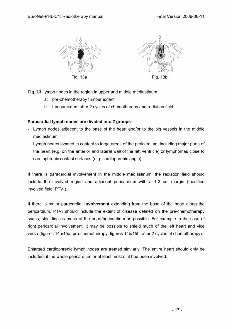
Fig. 13a Fig. 13b
Fig. 13: lymph nodes in the region in upper and middle mediastinum
a: pre-chemotherapy tumour extent
b: tumour extent after 2 cycles of chemotherapy and radiation field
Paracardial lymph nodes are divided into 2 groups:
- Lymph nodes adjacent to the base of the heart and/or to the big vessels in the middle
mediastinum;
- Lymph nodes located in contact to large areas of the pericardium, including major parts of
the heart (e.g. on the anterior and lateral wall of the left ventricle) or lymphomas close to
cardiophrenic contact surfaces (e.g. cardiophrenic angle).
If there is paracardial involvement in the middle mediastinum, the radiation field should
include the involved region and adjacent pericardium with a 1-2 cm margin (modified
involved field, PTV1).
If there is major paracardial involvement extending from the base of the heart along the
pericardium, PTV1 should include the extent of disease defined on the pre-chemotherapy
scans, shielding as much of the heart/pericardium as possible. For example in the case of
right pericardial involvement, it may be possible to shield much of the left heart and vice
versa (figures 14a/15a: pre-chemotherapy, figures 14b/15b: after 2 cycles of chemotherapy).
Enlarged cardiophrenic lymph nodes are treated similarly. The entire heart should only be
included, if the whole pericardium or at least most of it had been involved.
- 17 -
EuroNet-PHL-C1: Radiotherapy manual Final Version 2006-08-11
Fig. 14a Fig. 14b Fig. 15a Fig. 15b
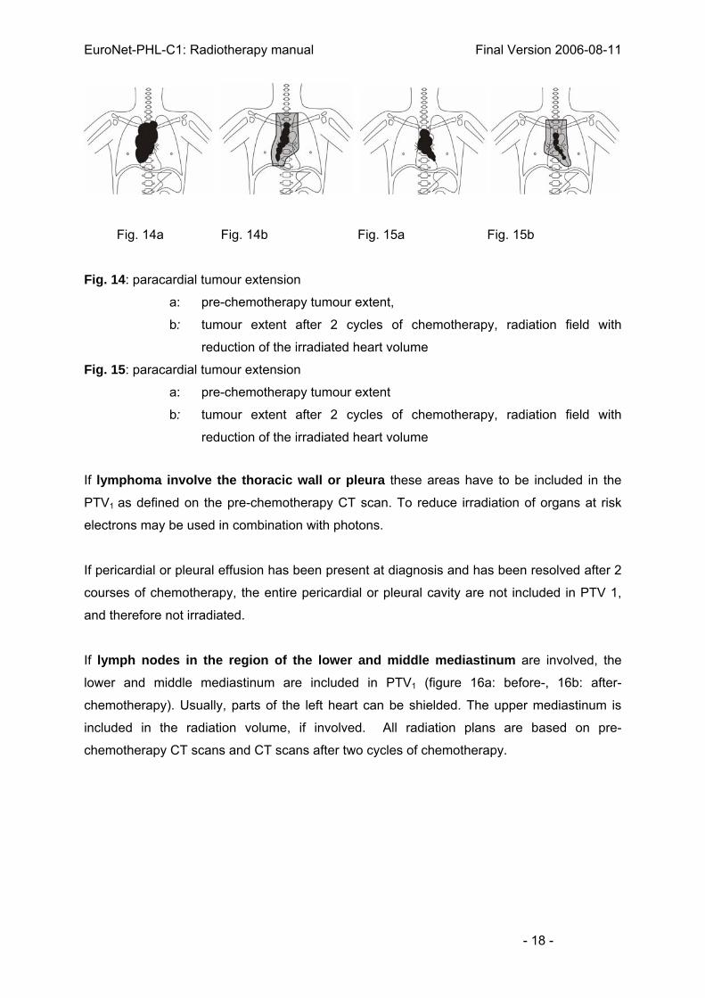
Fig. 14: paracardial tumour extension
a: pre-chemotherapy tumour extent,
b: tumour extent after 2 cycles of chemotherapy, radiation field with
reduction of the irradiated heart volume
Fig. 15: paracardial tumour extension
a: pre-chemotherapy tumour extent
b: tumour extent after 2 cycles of chemotherapy, radiation field with
reduction of the irradiated heart volume
If lymphoma involve the thoracic wall or pleura these areas have to be included in the
PTV1 as defined on the pre-chemotherapy CT scan. To reduce irradiation of organs at risk
electrons may be used in combination with photons.
If pericardial or pleural effusion has been present at diagnosis and has been resolved after 2
courses of chemotherapy, the entire pericardial or pleural cavity are not included in PTV 1,
and therefore not irradiated.
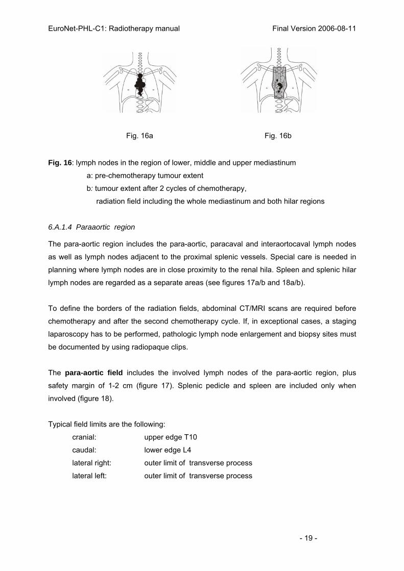
If lymph nodes in the region of the lower and middle mediastinum are involved, the
lower and middle mediastinum are included in PTV1 (figure 16a: before-, 16b: after-
chemotherapy). Usually, parts of the left heart can be shielded. The upper mediastinum is
included in the radiation volume, if involved. All radiation plans are based on pre-
chemotherapy CT scans and CT scans after two cycles of chemotherapy.
- 18 -
EuroNet-PHL-C1: Radiotherapy manual Final Version 2006-08-11
Fig. 16a Fig. 16b
Fig. 16: lymph nodes in the region of lower, middle and upper mediastinum
a: pre-chemotherapy tumour extent
b: tumour extent after 2 cycles of chemotherapy,
radiation field including the whole mediastinum and both hilar regions
6.A.1.4 Paraaortic region
The para-aortic region includes the para-aortic, paracaval and interaortocaval lymph nodes
as well as lymph nodes adjacent to the proximal splenic vessels. Special care is needed in
planning where lymph nodes are in close proximity to the renal hila. Spleen and splenic hilar
lymph nodes are regarded as a separate areas (see figures 17a/b and 18a/b).
To define the borders of the radiation fields, abdominal CT/MRI scans are required before
chemotherapy and after the second chemotherapy cycle. If, in exceptional cases, a staging
laparoscopy has to be performed, pathologic lymph node enlargement and biopsy sites must
be documented by using radiopaque clips.
The para-aortic field includes the involved lymph nodes of the para-aortic region, plus
safety margin of 1-2 cm (figure 17). Splenic pedicle and spleen are included only when
involved (figure 18).
Typical field limits are the following:
cranial: upper edge T10
caudal: lower edge L4
lateral right: outer limit of transverse process
lateral left: outer limit of transverse process
- 19 -
EuroNet-PHL-C1: Radiotherapy manual Final Version 2006-08-11
Fig. 17 Fig. 18
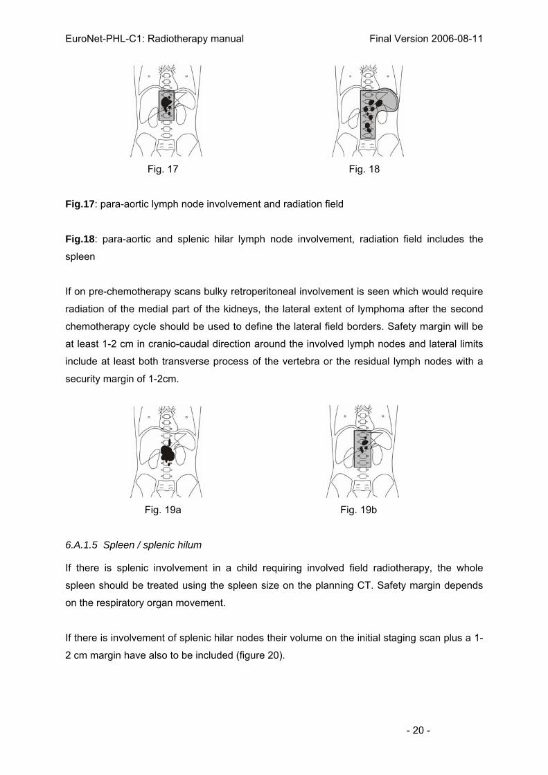
Fig.17: para-aortic lymph node involvement and radiation field
Fig.18: para-aortic and splenic hilar lymph node involvement, radiation field includes the
spleen
If on pre-chemotherapy scans bulky retroperitoneal involvement is seen which would require
radiation of the medial part of the kidneys, the lateral extent of lymphoma after the second
chemotherapy cycle should be used to define the lateral field borders. Safety margin will be
at least 1-2 cm in cranio-caudal direction around the involved lymph nodes and lateral limits
include at least both transverse process of the vertebra or the residual lymph nodes with a
security margin of 1-2cm.
Fig. 19a Fig. 19b
6.A.1.5 Spleen / splenic hilum
If there is splenic involvement in a child requiring involved field radiotherapy, the whole
spleen should be treated using the spleen size on the planning CT. Safety margin depends
on the respiratory organ movement.
If there is involvement of splenic hilar nodes their volume on the initial staging scan plus a 1-
2 cm margin have also to be included (figure 20).
- 20 -

EuroNet-PHL-C1: Radiotherapy manual Final Version 2006-08-11
Fig. 20
Fig. 20: Spleen and splenic hilum involvement and radiation field
If para-aortic lymph nodes are also to be treated, a single volume should be used to
encompass spleen, splenic hilum and paraaortic nodes (figure 18).
Special attention is required in every case for the adjacent upper part of the left kidney.
6.A.1.6 Porta hepatis lymph nodes
If porta hepatis lymph nodes (T12/L 1) are involved, the upper para-aortic region is
included in the target volume regardless of evidence of lymph node enlargement. If, in
addition, lower paraaortic lymph nodes are involved the field is expanded accordingly (figure
21 a,b).
Fig. 21a Fig. 21b
Fig. 21a: Involvement of lymph nodes at the liver hilus and in the para-aortic region:
radiation field does include the para-aortic lymph nodes plus liver hilus
Fig. 21b: Involvement of lymph nodes at the liver hilus, the spleen hilus and in the para-
aortic region: radiation field including also the spleen
Special attention is required for the adjacent upper part of the right kidney.
- 21 -
EuroNet-PHL-C1: Radiotherapy manual Final Version 2006-08-11
6.A.1.7. Iliac region
To define the iliac modified involved radiation fields abdominal CT/MRI scans before
chemotherapy and after 2 cycles are required. Pretherapeutic lymph node extension is
defined as the pre–chemotherapy tumor extension in cranio caudal direction and post-
chemotherapy tumor extension (i.e. after two cycles) in the lateral direction with a safety
margin of 1-2 cm.
Standard fields limits are the following (Fig.22 and 23):
cranial: upper edge of L5
caudal: following the course of the inguinal ligament
lateral: line 1 cm lateral to transverse process of L5 to 1-2 cm medial to the outer
acetabulum border
medial: 1-2 cm medial to the iliac vessels and lymph nodes
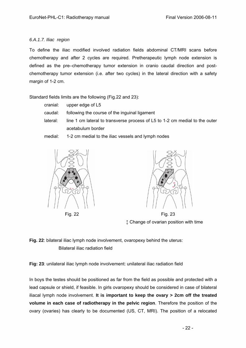
Fig. 22 Fig. 23
↕ Change of ovarian position with time
Fig. 22: bilateral iliac lymph node involvement, ovaropexy behind the uterus:
Bilateral iliac radiation field
Fig: 23: unilateral iliac lymph node involvement: unilateral iliac radiation field
In boys the testes should be positioned as far from the field as possible and protected with a
lead capsule or shield, if feasible. In girls ovaropexy should be considered in case of bilateral
iliacal lymph node involvement. It is important to keep the ovary > 2cm off the treated volume in each case of radiotherapy in the pelvic region. Therefore the position of the
ovary (ovaries) has clearly to be documented (US, CT, MRI). The position of a relocated
- 22 -
EuroNet-PHL-C1: Radiotherapy manual Final Version 2006-08-11
ovary should be marked with a clip to enable a reproducible location of the ovary and a
calculation of the ovarian radiation dose.
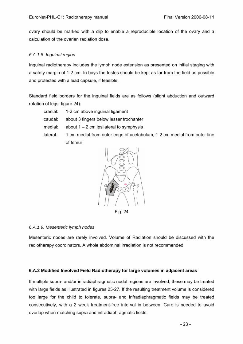
6.A.1.8. Inguinal region
Inguinal radiotherapy includes the lymph node extension as presented on initial staging with
a safety margin of 1-2 cm. In boys the testes should be kept as far from the field as possible
and protected with a lead capsule, if feasible.
Standard field borders for the inguinal fields are as follows (slight abduction and outward
rotation of legs, figure 24):
cranial: 1-2 cm above inguinal ligament
caudal: about 3 fingers below lesser trochanter
medial: about 1 – 2 cm ipsilateral to symphysis
lateral: 1 cm medial from outer edge of acetabulum, 1-2 cm medial from outer line
of femur
Fig. 24
6.A.1.9. Mesenteric lymph nodes
Mesenteric nodes are rarely involved. Volume of Radiation should be discussed with the
radiotherapy coordinators. A whole abdominal irradiation is not recommended.
6.A.2 Modified Involved Field Radiotherapy for large volumes in adjacent areas
If multiple supra- and/or infradiaphragmatic nodal regions are involved, these may be treated
with large fields as illustrated in figures 25-27. If the resulting treatment volume is considered
too large for the child to tolerate, supra- and infradiaphragmatic fields may be treated
consecutively, with a 2 week treatment-free interval in between. Care is needed to avoid
overlap when matching supra and infradiaphragmatic fields.
- 23 -

EuroNet-PHL-C1: Radiotherapy manual Final Version 2006-08-11
Fig. 25 Fig. 26 Fig.27
Fig. 25: bilateral axillary, supraclavicular, cervical and mediastinal involvement:
“mantle field”
Fig. 26: unilateral cervical and supraclavicular involvement; mediastinal, para-aortic
and spleen involvement:
combined supra- and infradiaphragmatic radiation field
Fig. 27: bilateral iliac, para-aortic and spleen involvement:
modified inverted “Y” radiation field
6.B Boost radiotherapy (PTV2)
Areas of residual disease requiring a boost (indications in table 1) should be identified on
restaging scans after two cycles of chemotherapy. These sectional images are used directly
for 3D treatment planning. 3D treatment planning is recommended for the boost volume.
Table 1: Indication for Boost (PTV2)
Indication for Boost (PTV2)
>25 % of initial volume
(≤ 75 % reduction in volume)
and
≤ 25 % of initial volume
(>75 % regression)
and
Residual
volume*
≤ 5 cm³ > 5 cm³ ≤ 100 cm³ > 100 cm³
Dose in PTV2 no boost 10 Gy boost no boost 10 Gy Boost
* Estimate of volume: Diameter (D)1 x D2 x D3 x 0.5
examples: volume of 5 cm³ corresponds to a sphere with a diameter of 2.2 cm
volume of 100 cm³ corresponds to a sphere with a diameter of 5.8 cm
- 24 -
EuroNet-PHL-C1: Radiotherapy manual Final Version 2006-08-11
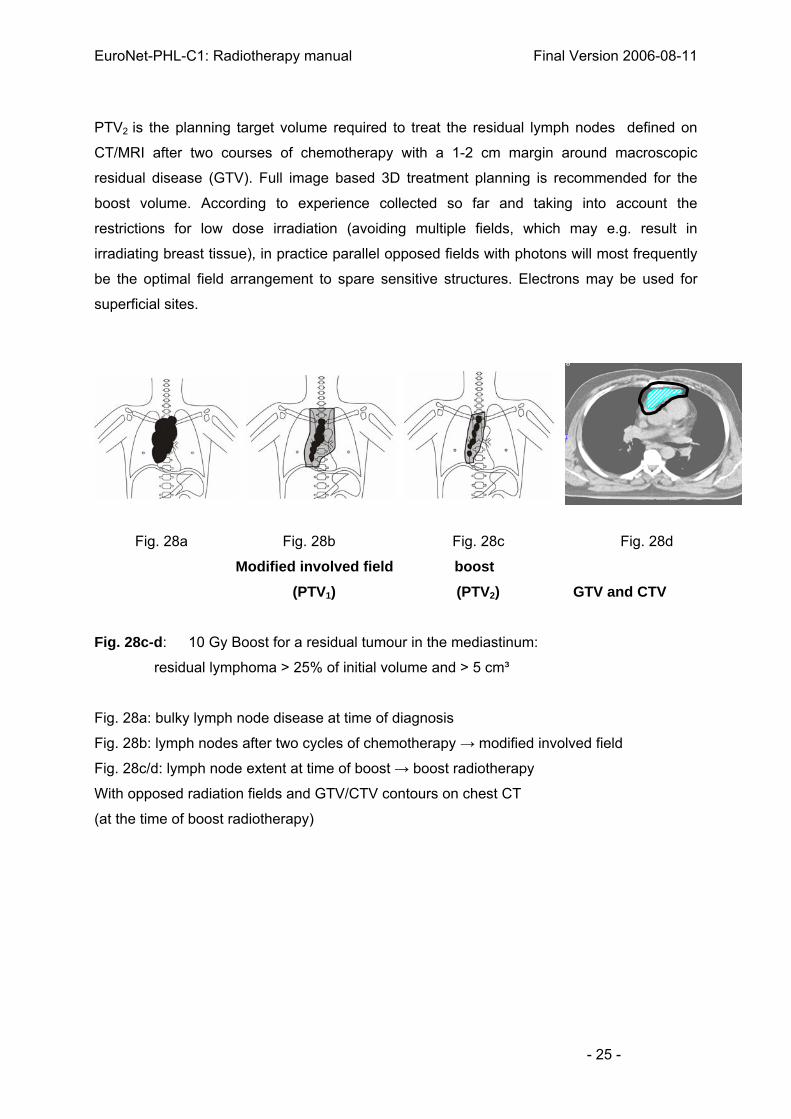
PTV2 is the planning target volume required to treat the residual lymph nodes defined on
CT/MRI after two courses of chemotherapy with a 1-2 cm margin around macroscopic
residual disease (GTV). Full image based 3D treatment planning is recommended for the
boost volume. According to experience collected so far and taking into account the
restrictions for low dose irradiation (avoiding multiple fields, which may e.g. result in
irradiating breast tissue), in practice parallel opposed fields with photons will most frequently
be the optimal field arrangement to spare sensitive structures. Electrons may be used for
superficial sites.
Fig. 28a Fig. 28b Fig. 28c Fig. 28d
Modified involved field boost (PTV1) (PTV2) GTV and CTV
Fig. 28c-d: 10 Gy Boost for a residual tumour in the mediastinum:
residual lymphoma > 25% of initial volume and > 5 cm³
Fig. 28a: bulky lymph node disease at time of diagnosis
Fig. 28b: lymph nodes after two cycles of chemotherapy → modified involved field
Fig. 28c/d: lymph node extent at time of boost → boost radiotherapy
With opposed radiation fields and GTV/CTV contours on chest CT
(at the time of boost radiotherapy)
- 25 -

EuroNet-PHL-C1: Radiotherapy manual Final Version 2006-08-11
Fig. 29a Fig. 29b Fig. 29c Fig. 29d
Modified Involved field boost (PTV1) (PTV2) GTV and CTV
Fig. 29: 10 Gy boost for a residual tumour in the paraaortic region (residual lymphoma
>25% of initial volume and >5 cm³) after 20 Gy with involved field radiotherapy
Fig. 29a: bulky para-aortic lymph node disease at time of diagnosis
Fig. 29b: lymph nodes after two cycles of chemotherapy → radiotherapy field
Fig. 29c/d: lymph node extent at time of boost → boost radiotherapy
with opposed radiation fields and GTV/CTV contours on abdominal CT (at the
time of boost radiotherapy)
If 2 or more lymph node areas require boost treatment, they should be treated independently
with 3D planned volumes. If they are adjacent, they are treated within one volume.
6.C Radiotherapy for non-lymphatic organs
6.C.1 Lung
6.C.1.1 Disseminated involvement of one or both lungs
Disseminated involvement of one or both lungs, which is not completely resolved after two
courses of OEPA, should be treated as in figure 30a/b. Radiotherapy should be 3D planned
(with lung correction). The spine must be irradiated symmetrically, i.e. either fully included
(when both lungs are treated) or excluded (when one lung is treated). In general, the lung is
irradiated (uni- or bilaterally) simultaneously with the
- 26 -

EuroNet-PHL-C1: Radiotherapy manual Final Version 2006-08-11
regional lymph nodes, i.e. the mediastinum and the hilar lung nodes, so that there is no risk
of asymmetric unilateral radiation of the spine. The cumulative dose to a large volume of lung
or to a whole lung should not exceed 12 to 15 Gy, the fraction size should not exceed 1-1.2
Gy. This is achieved by using transmission blocks or field-in-field techniques which permit a
fractional dose of 1.8-2 Gy in the region of mediastinal LN and simultaneously 1-1.2 Gy to
the lung.
Fig. 30a Fig. 30b
Fig. 30: uni and bilateral lung involvement and mediastinal lymphoma
a: unilateral lung and mediastinal radiotherapy with transmission blocks for the right lung to
reduce the dose per fraction and the total dose compared to the mediastinal radiotherapy.
b: bilateral lung and mediastinal radiotherapy with transmission block for both lungs.
6.C.1.2. Localized lung involvement (extranodal involvement)
Only the residual disease after two cycles of OEPA should be treated with an individual
safety margin including the movement of the tumor.
6.C.1.3 Pleura or chest wall
Pleura or chest wall involvement is treated according to the initial pre-chemotherapy extent,
with 1-2 cm safety margins.
6.C.2 Pericardial and or pleural effusion
If pericardial or pleural effusion has been present at diagnosis and has not been resolved
after 2 courses of chemotherapy, pericardial or pleural cavity has to be included in PTV1.
The cumulative dose to a pleural cavity should not exceed 12 to 15 Gy, the fraction size
should not exceed 1-1.2 Gy (see 6.C.1.1). The cumulative dose to the whole pericardial
cavity should not exceed 20 Gy and should be irradiated simultaneously with regional lymph
nodes.
- 27 -
EuroNet-PHL-C1: Radiotherapy manual Final Version 2006-08-11
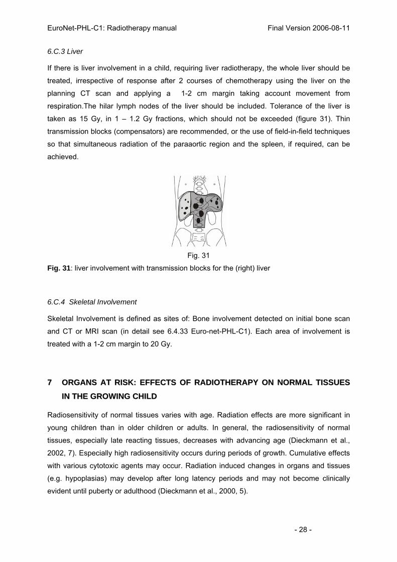
6.C.3 Liver
If there is liver involvement in a child, requiring liver radiotherapy, the whole liver should be
treated, irrespective of response after 2 courses of chemotherapy using the liver on the
planning CT scan and applying a 1-2 cm margin taking account movement from
respiration.The hilar lymph nodes of the liver should be included. Tolerance of the liver is
taken as 15 Gy, in 1 – 1.2 Gy fractions, which should not be exceeded (figure 31). Thin
transmission blocks (compensators) are recommended, or the use of field-in-field techniques
so that simultaneous radiation of the paraaortic region and the spleen, if required, can be
achieved.
Fig. 31
Fig. 31: liver involvement with transmission blocks for the (right) liver
6.C.4 Skeletal Involvement
Skeletal Involvement is defined as sites of: Bone involvement detected on initial bone scan
and CT or MRI scan (in detail see 6.4.33 Euro-net-PHL-C1). Each area of involvement is
treated with a 1-2 cm margin to 20 Gy.
77 OORRGGAANNSS AATT RRIISSKK:: EEFFFFEECCTTSS OOFF RRAADDIIOOTTHHEERRAAPPYY OONN NNOORRMMAALL TTIISSSSUUEESS
IINN TTHHEE GGRROOWWIINNGG CCHHIILLDD
Radiosensitivity of normal tissues varies with age. Radiation effects are more significant in
young children than in older children or adults. In general, the radiosensitivity of normal
tissues, especially late reacting tissues, decreases with advancing age (Dieckmann et al.,
2002, 7). Especially high radiosensitivity occurs during periods of growth. Cumulative effects
with various cytotoxic agents may occur. Radiation induced changes in organs and tissues
(e.g. hypoplasias) may develop after long latency periods and may not become clinically
evident until puberty or adulthood (Dieckmann et al., 2000, 5).
- 28 -
EuroNet-PHL-C1: Radiotherapy manual Final Version 2006-08-11
7.1 Deterministic radiation effects
7.1.1 Skeletal system
Radiosensitivity is greatest in young children (0 to 6 years) and during the pre- pubertal
growth spurt (11-13 years). The extent of the expected impairment depends on the period of
growth of the involved part of the skeleton. Epiphyseal chondroblasts show particularly high
radiosensitivity which is age dependant and dose dependant from a dose of 10 Gy and
increasing with increasing dose (≥ 30 Gy). Perichondral and desmal bone growth are less
affected. Asymmetric radiation of the axial skeleton can result in kyphosis or scoliosis,
whereas symmetric radiation leads to reduced body height. To avoid the development of
scolioses the definition of the radiation field on the spine should be symmetrical. Radiation of
extremities can lead to differences in limb length. After completion of skeletal growth no
undesirable effects on the skeleton should occur at dose levels as recommended in this
protocol (Dieckmann et al., 2000, 5, 7).
7.1.2 Soft tissue (Muscle)
Myoblasts in growing muscles are highly radiosensitive so that, in young children, the growth
of muscles is reduced by doses >25-30 Gy and a muscular hypoplasia can develop within
years where muscle is replaced by fibrotic tissue. It is possible that radiation effects on the
muscles are aggravated by Adriamycin. Only slight radiation effects on muscle growth are
expected from radiotherapy at doses recommended in the protocol after the age of 6.
7.1.3 Spinal cord
Until the age of six years the CNS undergoes a maturation process and is therefore
especially radiosensitive. The total dose to the cervical cord should therefore not exceed 20
Gy for a child. A total dose of 30 Gy is recommended as tolerance dose for the spinal cord.
Nevertheless, individual treatment planning should aim to minimize cord dose without
affecting the planned dose to the involved lymph node regions. No long-term sequelae are
known from radiation of infants and school age children at the dose level recommended in
this protocol.
7.1.4 Lung
Lung tolerance seems not to differ significantly between adults and children at school age.
For whole-lung radiotherapy the dose as recommended in this protocol (12-15 Gy) is unlikely
to lead to any significant impairment in lung function. However, for young children (<6 years), - 29 -
EuroNet-PHL-C1: Radiotherapy manual Final Version 2006-08-11
there is evidence (from nephroblastoma) that whole-lung radiation with a dose of 15 Gy can
lead to permanent restrictive impairment in pulmonary function with a reduced vital capacity
(50 – 75 %).
For partial radiation of the lung at the recommended dose level (20 Gy (30 Gy)) there is no
evidence for significant long term adverse side effects.
Cumulative effects are observed with some cytotoxic agents (e.g. bleomycin, procarbazine).
7.1.5 Heart
Impairment of myocardial and arterial function can occur after a long latency period (~20
years and more) as a long term consequence of radiation induced endothelial proliferation.
There is no clear information about the impact of age at the time of radiation. Radiation of the
heart should be reduced as much as possible, in particular as cardiotoxic anthracyclines
(adriamycin in OEPA) are applied that may lower the overall heart tolerance. Therefore, if
there is paracardial involvement and/or residual paracardial disease, irradiated volume of the
heart and radiation doses to the heart should be minimized as much as possible. A total dose
of 30 Gy should not be exceeded, if significant parts of the heart have to be included.
7.1.6 Breast
Long term adverse deterministic effects on the fully developed breast are not to be expected.
However, the non-developed juvenile breast is highly radiosensitive. After radiation exposure
of about 5 to 10 Gy to the non-developed breast, hypoplasia or aplasia of the whole or partial
breast may occur during adolescence. Therefore, in girls the non-developed breast regions
should be protected as much as possible, which applies for mediastinal, axillary, lung, liver
and spleen radiotherapy (see also stochastic risk).
7.1.7 Kidney
The kidney is radiosensitive. No clear information about the impact of age at the time of
radiation is available. A tolerance dose of 12 Gy for whole kidney irradiation is recommended
and no significant long terms adverse effects are then to be expected.
For partial volumes, e.g. upper left kidney pole in splenic and spleen pedicle radiation, the
dose will be 20 Gy or more which may lead to fibrosis of the upper kidney pole and
consecutive regional functional impairment. However, the overall function of the kidney is
usually not affected by small irradiated volumes (Pötter et al., 1993, 17, 18).
Cumulative toxicity may occur with some cytotoxic agents, as there is e.g. cisplatin or
ifosfamide.
- 30 -
EuroNet-PHL-C1: Radiotherapy manual Final Version 2006-08-11
7.1.8 Liver
For the whole organ a tolerance dose of 15 Gy is recommended. This applies for
radiotherapy of the liver in case of disseminated liver disease. Irradiation of parts of the liver,
e.g. during radiotherapy of the upper paraaortic region, with doses of 20 Gy or more will not
lead to a clinically detectable effect in whole liver function. Cumulative toxic effects have
been described for combined therapy with adriamycin.
7.1.9 Thyroid
Patients may develop sub-clinical hypothyroidism after a latency of 3-4 years or more after a
dose of 20 Gy or more to the thyroid gland. Sub-clinical hypothyroidism can normalize
spontaneously and the need for therapy is controversial. The risk of impairment of thyroid
function increases with higher radiation doses (e.g. 30 Gy), that may lead to clinical
hypothyroidism which requires endocrine replacement. Hyperthyroidism, autoimmune
thyroidopathy and benign cysts due to radiation have been described (see also stochastic
risk).
7.1.10 Testis
The testes are exquisitely radiosensitive. A dose as low as 1-2 Gy may lead to irreversible
impairment of germ cells (spermatogenesis). Therefore testes must be protected during iliac
or inguinal radiation with 20 Gy by direct shielding, appropriate distance and lead capsules, if
feasible. The maximal acceptable dose is < 1 Gy (<5% of 20 Gy). An individual assessment
of the dose to the testes is recommended (Wallace et al., 1997, 33).
7.1.11 Ovary
After a cumulative dose of 5 – 10 Gy, cell death of oocytes is induced resulting in permanent
infertility. Hormone insufficiency occurs after slightly higher doses (e.g. 10 – 15 Gy).
Replacement endocrine therapy, depending on the hormone status, may become necessary
at puberty. In order to avoid ovarian impairment, at least one ovary should be protected by
distance from scattered radiation arising from the treated volume e.g. by double thick blocks
in the direct beam. Usually a distance of more than 2 cm of one ovary from the border of the
treated volume (opposed fields, 6 MV photons, block edges) leads to an acceptable dose of
about 10-20% of the prescribed dose (20 Gy) (Wallace et al., 2005, 34; Sy Ortin et al., 1990,
32; Brämswig et al., 1990, 2; Dieckmann et al., 1996, 4).
- 31 -
EuroNet-PHL-C1: Radiotherapy manual Final Version 2006-08-11
7.1.12 Stochastic radiation effects
The incidence of secondary malignancies increases with radiotherapy volume and, in the
low-dose range, probably also with increasing dose. The risk is increased by combined
therapy with carcinogenic cytotoxic agents. While secondary haematologic malignancies
occur with a latency of median 6.2 years (2.5 – 16.2 years (GPOH-DAL HD studies)) solid
tumors occur, mainly thyroid and breast carcinomas, with increasing frequency much later.
The incidence can therefore not yet be defined exactly in respect of too little long term follow-
up (≥ 20 years) (Bhatia et al., 2003, 1). However, in the GOPH-experience the rate has so far
been rather low with actuarial 5% (Schellong et al., 2004, 30) compared to other groups
(Meadows et al., 1989, 15). Radiation exposure to the developing breast between the ages
of 10 and 16 (- 30) bears an especially high risk of inducing breast cancer.
88 RRAADDIIOOTTHHEERRAAPPYY IINN RREELLAAPPSSEEDD PPAATTIIEENNTTSS
8.1 Indications for radiotherapy
All patients after conventional relapse therapy (chemotherapy not including
autologous transplantation) are irradiated (Schellong et al., 2005, 31).
After autologous transplantation radiotherapy is only used for patients who have a
residual lymph node at CT/MRI in at least one of the initially involved regions (at
the time of restaging on day 50-54 after transplantation), which is PET positive.
8.2 Radiation volume and radiation dose
After conventional relapse therapy: The planning target volume is defined by the extent of
the disease at the time of relapse applying the same principles as defined for modified
involved field radiotherapy (PTV1) in primary treatment (see above).
The radiation dose depends on initial treatment, in particular the extent and dose of
radiotherapy used in primary treatment and the site of relapse and the resulting target for
- 32 -

EuroNet-PHL-C1: Radiotherapy manual Final Version 2006-08-11
radiotherapy. A relevant dose should be prescribed (20-30 Gy). However, a significant risk of
inducing serious adverse late effects (e.g. heart) should not be taken.
The following principles apply:
Patients who have not had previous radiotherapy at the site of relapse will
receive (modified) involved field radiotherapy of 20-30 Gy.
If the relapsed lymph nodes requiring radiotherapy were previously irradiated
with 20 Gy, a further 20-25 Gy should be given.
If the relapsed lymph node was previously irradiated with a dose of greater
than 20 Gy, re-irradiation should be limited to up to 20 Gy taking into account
the dose limits for the respective organs at risk.
Individual decisions are necessary according to the site of the relapse and the
proximity of organs at risk. Therefore, these issues should be discussed in detail with
one of the reference radiotherapists and the study coordinator.
99 CCEENNTTRRAALL RREEVVIIEEWW
For countries with central upfront review (GPOH-associated countries) the treatment plan for
radiotherapy will be provided by the GPOH-study centre before radiotherapy starts. This
treatment plan indicates macroscopic disease and the treated volume on a pictogram and
the dose per fraction and the total dose to be applied. This refers to modified involved field
radiotherapy (PTV1) and also to boost treatment (PTV2), if indicated.
For certain countries treatment plans are established by the local hospitals. They should be
reviewed retrospectively in defined time intervals during the trial.
1100 TTEECCHHNNIICCAALL AANNDD PPHHYYSSIICCSS AASSPPEECCTTSS OOFF TTRREEAATTMMEENNTT PPLLAANNNNIINNGG AANNDD
DDEELLIIVVEERRYY
Prerequisite and technical requirements
For participation in the study the following infrastructure is considered as a prerequisite:
linear accelerator, conventional simulator or virtual simulator, CT, treatment planning system,
possibilities to manufacture individual blocking or beam shaping by multileaf collimators as
- 33 -

EuroNet-PHL-C1: Radiotherapy manual Final Version 2006-08-11
well as portal imaging. For blocking individually moulded blocks, which must be divergent,
multileaf collimators or a combination can be used.
For treatment delivery in paediatric oncology high energy photon beams in the range
between 4 – 10MeV are recommended. High energy photon beams in the above mentioned
energy range, provided by linear accelerators, allow adequate treatment of superficial or
deep situated lymph nodes. High energy electron beams can be used for single beam
techniques to treat superficial lymph nodes (e.g. inguinal) or for treatments of clinically
relevant superficial residual tumour mass (e.g. anterior thoracic wall).
A computerised treatment planning system is mandatory for full three dimensional dose
calculation based on CT information. For large field techniques a dose calculation in multiple
points, i.e. a dose calculation for all target areas, are required. This is to ensure that all lymph
nodes to be irradiated with a large field technique receive an adequate dose (e.g. axillary,
upper cervical region and lower mediastinum within a mantle field treatment).
Multiple neighbouring lymph node regions are to be treated with opposed AP-PA fields using
individual shielding. Lateral opposed fields are to be used for treatment of the epipharynx.
Treatment planning
During simulation, the treatment fields need to be defined with the child in a reproducible
treatment position with adequate immobilisation. This can be done on a conventional
simulator or on a CT simulator. The treatment position needs to be recorded with an X-ray
image or with a DRR. The final treatment parameters used on the treatment unit must be
identical with the ones defined during simulation / treatment planning.
Based on the existing diagnostic information, all targets and organs at risk need to be
outlined on the X-ray or the DRR acquired during treatment planning. Additionally, the
individual shielding needs to be drawn on the simulation X-ray images or DRR. These
images and drawings represent the basic information for focused shielding, which will be
adapted according to the individual anatomical and pathological situation. In case moulded
blocks are used, the block thickness needs to be at least equivalent to 5 half value layers
(HVL) of the block material. Depending on beam energy and other factors of influence the
dose below shielding material must not be higher than 15% of the dose in the dose
specification point. Note that increasing the block thickness (e.g. to 15cm) will limit the
transmitted dose contribution, but will not influence the scattered dose. Nevertheless, in
individual situations (e.g. to shield ovaries) thick blocks might be considered. In case
multileaf collimators are used for individual shielding, leaves and - depending on MLC design
- leaves combined with secondary collimator elements, provide superior shielding in terms of
transmission compared to individually moulded blocks.
- 34 -

EuroNet-PHL-C1: Radiotherapy manual Final Version 2006-08-11
In case of liver and lung irradiation so called “field in field techniques” with a total thickness of
less than 6 cm can be used to limit dose. They need to be designed to reduce the dose in
well defined parts of the field to a predefined value. By doing so different dose levels are
delivered within a single field. For example, using such a transmission block the mediastinum
can be treated to 20Gy while at the same time the dose to the lung can be limited to 15Gy
(Kaplan 1980, 12).
For assessment of target coverage and doses to organs at risk more comprehensively, it is
recommended to perform a CT based post planning with a three dimensional treatment
planning system (TPS). Therefore, a CT must be acquired with the patient in treatment
position, slice thickness 4-8mm. The AP/PA radiation fields as defined during simulation
need to be transferred to the TPS. The treated volumes should not be significantly adapted
or even enlarged during this procedure but rather taken as defined during the classical
simulator based planning, as there has been by now no systematic experience collected with
a full 3 D based treatment planning approach in Hodgkin´s Disease when treating lymph
node regions or modified involved fields. The full 3 D approach is, however, recommended
for treatment planning of the boost volume as here the residual macroscopic disease is
clearly the region of interest: definition of residual GTV, CTV and PTV according to a full 3D
approach in solid tumours is encouraged.
Dose volume calculation
For all patients with 3D planning, relevant organs at risk in the respective region of interest
are to be delineated and Dose volume histograms (DVH) need to be calculated for each
structure. Relevant organs at risk are e.g. lungs (delineated separately), heart, thyroid,
breast, spinal cord, teeth, salivary glands (parotids, submandibular), kidneys, liver, ovaries,
testis. By now, there is no clear notion, if and how soft tissue (e.g. muscle), bone and bone
marrow, although relevant organs at risk in the growing child, should be delineated.
Dose-Volume reporting for these organs at risk (OAR) should be performed according to
parameters for the different organs as established in 3D radiotherapy for solid tumours. It is
not recommended to report only minimum and maximum doses, but also e.g. mean doses,
doses in absolute volumes and/or relative volumes. As there is no agreement on reporting at
present, it is encouraged to develop guidelines for dose volume reporting for relevant organs
in treatment of paediatric Hodgkin´s Disease during the trial, e.g. heart, thyroid.
Dose calculation and dose prescription
For parallel opposed beams the dose prescription point is located midline along the central
beam axis. Dose homogeneity throughout the whole target volume should be within ± 10%,
- 35 -
EuroNet-PHL-C1: Radiotherapy manual Final Version 2006-08-11
which is in line with the recommendations of the International Commission on Radiation Units
and Measurements (ICRU 50, 35).
Dose calculation in more than one point is necessary, because for large fields the dose will
vary due to changing patient dimensions and diameters with respect to the ray line,
especially in cranial caudal direction, and because of differences in depth of lymph node
regions (e.g. neck, supraclavicular region, axilla, mediastinum, etc). Dose variations can be
estimated with simple manual calculations taking into account patient diameter and depth,
but full three dimensional dose calculations based on CT data are preferable.
The definition of dose points for recording and reporting is the responsibility of the radiation
oncologist. Consultation with medical (clinical) physicist is recommended. Irrespective of the
dose calculation method, i.e. manual calculation or calculations performed with a treatment
planning system, all dose points need to be representative for the different lymph node
regions and should represent regions with potential over- or underdosage due to the
respective anatomical conditions.
Relevant dose variations, i.e. dose variations larger than ± 10 %, can be compensated
applying different techniques. In case of overdosage, the overexposed areas can be avoided
by reducing the field size after a certain dose has been reached (“stop dose principle”). In
case of underdosage, an additional dose or fraction can be given to a limited region. Another
alternative to achieve a homogenous dose distribution is the application of missing tissue
compensators, which can be manufactured individually or based on a compensator library
(standardized compensator) (Khung et al 1993) or the use of field in field technique.
Note that due to the small body diameters in paediatric patients, the dose maxima of high
energy photon beams can be located deeper than the supraclavicular lymph nodes despite
the use of parallel opposed beams. In such cases a bolus material (e.g. of 0.5cm) need to be
used in order to shift the dose maximum towards shallower depths.
For highly irregular fields with open field apertures being significantly different from a
regular rectangular field, a sophisticated dose calculation algorithm should be used, which
takes into account the shielding effect of blocks for both head scatter and phantom (volume
scatter) contributions to the total dose.
Field portions outside the body contour can be taken into account in dose calculation by
defining blocks for portion outside the body in the open field aperture. Dose calculation
algorithms, such as Clarkson Integration can then be used to account for this effect of
missing tissue. Full three dimensional dose calculations based on sectional imaging will take
this effect into account as well.
The dose to organs at risk (e.g. testicles) need to be determined with either manual
calculation or a full three dose calculation algorithm, or it can be measured using various in-
vivo dosimetry methods (e.g. for testis).
- 36 -
EuroNet-PHL-C1: Radiotherapy manual Final Version 2006-08-11
In case of lung irradiations inhomogeneity corrections need to be applied, i.e. corrections
which account for the density difference in lung compared to water. If the reduced density of
lung is neglected, significant and unacceptable dose variations (10-20%) will be introduced.
For electron beam applications, the energy needs to be selected such that the 80% isodose
lines fully encompass the target volume at most distal part.
Even if primary treatment planning procedure is based on a simulator technique, during the
course of the treatment a CT should be acquired and based on the treatment geometry, a
post planning should be performed with a three dimensional treatment planning system.
During the post planning procedure dose volume histogram information should be
determined, which will be used for recording and reporting.
Field junction lines and gaps
For supra- and infradiaphragmal radiation (stage III) it is necessary to check whether it is
technically feasible to irradiate the whole target with a large single field, e.g. mediastinum
plus para-aortic region. If possible, a split field technique should be avoided.
In case a split field technique cannot be avoided, the junction line between supra- and
infradiaphragmal field needs to be defined at depth in the body and not only at the surface.
The junction line must not be located in one of the primary target regions. Due to the
divergence of the beam, a certain gap at the patient’s surface needs to be respected. The
size of the gap, usually a few centimetres in length, can be calculated taking into account the
patient and treatment geometry (treatment depth, treatment distance, field size). In order to
avoid over- and underdosage in the spinal cord, the junction line can be moved in regular
intervals (“moving gap technique”). Alternatively, a 2 cm shielding block can be placed at the
backprojection of the spinal cord at the height of the block tray.
1111 TTRREEAATTMMEENNTT DDEELLIIVVEERRYY
Treatment is usually performed with the patient in supine position. Special fixation and
positioning systems are not prerequisite for the study. Nevertheless, the positioning of the
child has to be adequate (e.g. neck cushion) to avoid unnecessary irradiation of organs at
risk (e.g. brain tissue, oral cavity). Positioning has to be identical during treatment planning
and delivery.
Based on the skin markers, radiotherapy fields have to be set up in the same way at the
simulator and at the accelerator. Individual shields or leafs have to be positioned as
described on the simulation films or on the “beams eye view”.
- 37 -

EuroNet-PHL-C1: Radiotherapy manual Final Version 2006-08-11
Positioning of the patient, set up of the radiation field and of the individual shields has to be
controlled during the first irradiation by taking a verification film (portal image). A verification
film is taken then at weekly intervals. Simulation and verification film have to be compared in
regard to field geometry, positioning of the patient and individual shielding and - if necessary
- corrected. Additionally, a photo-documentation of the patient with individual shielding
delineated on the skin of the patient has to be done.
Simulation and verification films as well as photo-documentation are necessary prerequisites
for the radiotherapy quality assurance process as performed during the trial.
The weekly dose should be applied on five consecutive days (9 Gy/10 Gy). All fields for one
target have to be treated daily. In case of large radiation fields (e.g. mantle plus complete
inverted Y) it is advantageous to treat the different targets sequentially.
1122 AACCUUTTEE SSIIDDEE EEFFFFEECCTTSS
Acute side effects during radiotherapy with low doses are rare and mostly temporary. Time
and intensity of side effects may depend additionally on chemotherapy applied.
Moderate mucositis (pharygitis, oesophgitis, or gastroenteritis) may occur.
Skin reactions are rare. Additional thermic and mechanical irritations should be avoided.
Hairloss or delayed hair growing in the occipital region is rare but possible.
Dry mouth (xerostomia) can be observed after irradiation of both parotid glands with a dose
larger than 20 Gy.
Leucocytopenia and thrombocyctopenia are rare and transient. Nevertheless, weekly
blood counts should be taken, in particular in large field radiotherapy (platelets, leucocytes,
red blood count).
- 38 -