radiosensitization of human pancreatic cancer cells by
TRANSCRIPT

Therapeutics, Targets, and Chemical Biology
Radiosensitization of Human Pancreatic Cancer Cells byMLN4924, an Investigational NEDD8-Activating EnzymeInhibitor
Dongping Wei1, Hua Li1, Jie Yu2, Jonathan T. Sebolt1, Lili Zhao1, Theodore S. Lawrence1,Peter G. Smith2, Meredith A. Morgan1, and Yi Sun1
AbstractRadiotherapy is used in locally advanced pancreatic cancers in which it can improve survival in combination
with gemcitabine. However, prognosis is still poor in this setting inwhichmore effective therapies remain needed.MLN4924 is an investigational small molecule currently in phase I clinical trials. MLN4924 inhibits NAE (NEDD8Activating Enzyme), a pivotal regulator of the E3 ubiquitin ligase SCF (SKP1, Cullins, and F-box protein), that hasbeen implicated recently in DNAdamage and repair. In this study, we provide evidence thatMLN4924 can be usedas an effective radiosensitizer in pancreatic cancer. Specifically, MLN4924 (20–100 nmol/L) effectively inhibitedcullin neddylation and sensitized pancreatic cancer cells to ionizing radiation in vitro with a sensitivityenhancement ratio of approximately 1.5. Mechanistically, MLN4924 treatment stimulated an accumulation ofseveral SCF substrates, including CDT1, WEE1, and NOXA, in parallel with an enhancement of radiation-inducedDNA damage, aneuploidy, G2/M phase cell-cycle arrest, and apoptosis. RNAi-mediated knockdown of CDT1 andWEE1 partially abrogatedMLN4924-induced aneuploidy, G2/M arrest, and radiosensitization, indicating a causaleffect. Furthermore, MLN4924 was an effective radiosensitizer in a mouse xenograft model of human pancreaticcancer. Ourfindings offer proof-of-concept for use ofMLN4924 as a novel class of radiosensitizer for the treatmentof pancreatic cancer. Cancer Res; 72(1); 282–93. �2011 AACR.
Introduction
Pancreatic ductal adenocarcinoma is the fourth leadingcause of cancer-related deaths in the United States and oneof the deadliest human malignancies, with an overall 5-yearsurvival rate of approximately 5% (1). Although gemcitabine isthe standard systemic therapy for pancreatic cancer (2), thefinding that as many as 1/3 of pancreatic cancer patients die oflocally destructive disease underscores the importance ofradiotherapy for local disease control (3). Indeed, the combi-nation of radiation with gemcitabine has been shown tosignificantly prolong survival compared with gemcitabinealone (4), and intensifying radiation also seems to improvesurvival (5). Despite these improvements, the prognosis forpatients with pancreatic cancer is still very poor, and newcombinational therapies are urgently needed. Strategies
utilizing small molecule inhibitors to sensitize pancreatictumor cells to radiation (and gemcitabine) are well underway.We have recently shown that small molecule inhibitors ofCHK1 (checkpoint kinase 1), sensitized pancreatic tumor cellsand xenografts to radiation by G2 checkpoint abrogation andhomologous recombination repair inhibition (6). Of the smallmolecule inhibitors tested in phase III clinical trials in pan-creatic cancer, erlotinib, an inhibitor of epidermal growthfactor receptor with only very modest activity in pancreaticcancer, has been shown to improve survival (7, 8). Thus, there isa demand for novel and more effective therapies. Our labora-tory has recently shown that SCF E3 ubiquitin ligases arepromising radiosensitizing targets in cancer cells (9, 10).Whether inhibition of SCF E3 ubiquitin ligases might also bea useful strategy for sensitizing pancreatic cancers to radiationhas not previously been tested.
The SCF (SKP1-Cullin-F-box proteins) E3 ubiquitin ligases,consisting of an adaptor protein SKP1, a scafford protein cullin,a substrate receptor F-box protein, and aRINGprotein RBX1 orRBX2, are the largest multiunit ubiquitin ligases (11, 12)responsible for the ubiquitination of approximately 20% of allubiquitinated proteins for targeted degradation by proteasome(13). The SCF E3s, therefore, regulate numerous biologicalprocesses, including cell-cycle progression, signal transduc-tion, and DNA replication, among others (11, 12). It is wellestablished that the core of SCFE3 ligase is a cullin-RINGfingerprotein complex (14). In human and mouse, there are 7 cullins(cullins 1–3, 4A, 4B, 5, and 7) and 2RING familymembers, RBX1
Authors' Affiliations: 1Division of Radiation and Cancer Biology, Depart-ment of Radiation Oncology, University of Michigan, Ann Arbor, Michigan;and 2Millennium Pharmaceuticals, Inc., Cambridge, Massachusetts
Note: Supplementary data for this article are available at Cancer ResearchOnline (http://cancerres.aacrjournals.org/).
Corresponding Author: Yi Sun, Division of Radiation and Cancer Biology,Department of Radiation Oncology, University of Michigan, 4424BMedicalScience Building 1, 1301 Catherine Street, Ann Arbor, MI 48109. Phone:734-615-1989; Fax: 734-647-9654; E-mail: [email protected]
doi: 10.1158/0008-5472.CAN-11-2866
�2011 American Association for Cancer Research.
CancerResearch
Cancer Res; 72(1) January 1, 2012282
on January 23, 2019. © 2012 American Association for Cancer Research. cancerres.aacrjournals.org Downloaded from
Published OnlineFirst November 9, 2011; DOI: 10.1158/0008-5472.CAN-11-2866

(RING box protein-1) and RBX2, also known as SAG (SensitivetoApoptosis Gene; refs. 11, 12, 15). The ligase activity of the SCFrequires (i) RBX1/RBX2, which binds to ubiquitin-loaded E2,and (ii) cullin neddylation (16–19), the addition of a ubiquitin-like protein NEDD8 to cullin, catalyzed by NEDD8-activatingenzyme E1 (NAE), NEDD8-conjugating enzyme E2 (UBC12),and NEDD8-E3 ligase (20).We have recently shown that inactivation of SCF E3 ligase by
siRNA knockdown of either RBX1 or RBX2/SAG inducedapoptosis and senescence, and sensitized human cancer cellsto radiation (9, 10, 21), and that Sag knockout also sensitizedmouse embryonic stem cells to radiation via inducing apo-ptosis (22). Because MLN4924 effectively inactivates SCF E3 byblocking cullin neddylation (13), we tested our workinghypothesis that MLN4924 could sensitize pancreatic cancercells to radiation. We reported here that MLN4924 indeed actsas a radiosensitizing agent by enhancing radiation-inducedDNA damage, aneuploidy, G2/M arrest, and apoptosis viamechanism including accumulation of CDT1 and WEE1, 2well-known substrates of SCF E3 ubiquitin ligase.
Materials and Methods
Cell cultureTwo humanMiaPaCa-2 and BxPC-3 pancreatic cancer lines,
and human H1299 lung cancer cell line were purchased fromAmerican Type Culture Collection. MiaPaCa-2 and H1299 cellswere grown in DMEM with 10% FBS. BxPC-3 and human lungfibroblast MRC5 cells (a gift from Dr. A. Rehemtulla) werecultured in RPMI1640 with 10% FBS. All lines were tested andwere free of Mycoplasma contamination.
ATPlite growth assay and IC50 determinationCells were seeded in 96-well plates in triplicate and treated
with MLN4924 (13), in various doses for 7 days. Cell viabilitywas measured with ATPlite Kit (Perkin Elmer; ref. 23).
MLN4924 preparation for in vitro and in vivo assaysMLN4924 was synthesized by Millennium Pharmaceuticals,
Inc. (13) and was dissolved in dimethyl sulfoxide to make a 10mmol/L stock solution and kept in �20�C before use. For invivo experiment, MLN4924 was freshly made every week asfollows: To make an 8.32 mg/mL solution, the compound wasdissolved in 10% 2-hydroxypropyl-b-cyclodextrin (HPBCD) insterile water. The pH value was adjusted to 5.0 with 1 mol/LKOH. The solution of MLN4924 was stored at room temper-ature before use.
Radiation exposure and clonogenic assayCells were seeded in 60-mm dishes in duplicate and
exposed to different doses of radiation (Philips RT250,Kimtron Medical) after pretreatment with MLN4924 for 6hours (at 100 nmol/L) or 24 hours (at 20–30 nmol/L).MLN4924 was either washed away (at 100 nmol/L) or stayedin the medium (at 20–30 nmol/L) afterwards, followed byincubation at 37�C for 7 to 9 days. Survival curves were fittedusing the linear-quadratic equation, and the mean inacti-vation dose was calculated (24).
siRNA knockdown of CDT1 and WEE1Two independent sets of siRNA oligonucleotides were
used to target CDT1 or WEE1, respectively. Their sequencesare as follows: siCDT1-1 (50-CGUGGAUGAAGUACCCGACUU-30) and siCDT1-2 (50-GCAAUGUUGGCCAGAUCAAUU-30),and siWEE1-1 (50-GAGGCUGGAUGGAUGCAUUUU-30) andsiWEE1-2 (50-CUCCGGGGUAGUUCUCUCUUU-30), and scram-bled control siRNA (Si-Ctrl: 50-ATTGTATGCGATCGCA-GACTT-30). All oligos were purchased from Dharmacon. Cellswere transfected with siRNA using X-tremeGENE (Roche) andsplit 48 hours later. One portion was used for clonogenic assayand the other portions for immunoblotting (IB) or fluores-cence-activated cell sorting (FACS) profile (23).
ImmunoblottingCells were exposed to various treatments and harvested for
IB as described (23) using antibodies against CDT1, WEE1,CUL1, total CDC2 (Santa Cruz Biotechnology), ORC1, p21, p27(BD Biosciences), NOXA, BIM1, phospho-CHK1 (S345), phos-pho-CHK2 (T68), g-H2AX (Ser139), phospho-CDC2 (Y15),phospho-IkB, Cyclin B1 (Cell Signaling), and b-actin (Sigma).
Fluorescence-activated cell sortingCells were treated with MLN4924, or exposed to radiation
alone or in combination. Cells were harvested 24 or 48 hourspostradiation and analyzed by flow cytometry (23).
Immunofluorescence stainingCells after exposure to various treatments were fixed with
4% paraformaldehyde, permeabilized with 0.5% Triton X-100,blocked with 0.5% bovine serum albumin, and incubated withprimary antibody against g-H2AX (Millipore) at 1:1,000, fol-lowed by incubation with Alexa Fluor 594 goat anti-mouse IgGat 1:2,000. Cellular nuclei were stained with 40,6-diamidino-2-phenylindole, as described (9). The stained cells were observedunder fluorescent microscope (Olympus 1 � 71), using Olym-pus LCP lan F1 lens and Olympus DP70 cameras. The acqui-sition software used was Olympus DP Controller 2002 (Olym-pus Optical Co. Ltd).
In vivo antitumor studyAll animal studies were conducted in accordance with the
guidelines established by theUniversity Committee onUse andCare of Animals. Five million MiaPaCa-2 cells were inoculatedsubcutaneously in both flanks of nude mice. The mice wererandomized and the treatment started when the tumor sizereached 100 mm3 at 14 days after inoculation. MLN4924 (30mg/kg, s.c.) and radiation (1 Gy) were given once a day, 5 days aweek for 3 weeks. Radiationwas delivered directly to the tumorwith the rest of the animal shielded. For combination treat-ment, MLN4924 was given 2 hours prior to radiation exposurewith the same schedule as for individual treatments. Thegrowth of tumors (8 for control group, 12–16 for the othergroups) was measured twice a week and average tumorvolumes were calculated, as estimated from the formula(L � W2)/2. Radiation was conducted in the University ofMichigan Comprehensive Cancer Center Experimental Radi-ation Core.
Radiosensitization of Pancreatic Cancer Cells by MLN4924
www.aacrjournals.org Cancer Res; 72(1) January 1, 2012 283
on January 23, 2019. © 2012 American Association for Cancer Research. cancerres.aacrjournals.org Downloaded from
Published OnlineFirst November 9, 2011; DOI: 10.1158/0008-5472.CAN-11-2866

Statistical analysisANOVAmodels were used with SPSS software for statistical
comparisons involving multiple groups, followed by an SNKpost hoc test to determine significance of each of 2 groups (P <0.05). Tumor volume doubling (tripling) was determined foreach xenograft by identifying the earliest day onwhich it was atleast twice (3 times) as large as on the first day of treatment. Acubic smoothing spline was used to obtain the exact time ofdoubling (tripling). The Kaplan–Meier method was used toanalyze the doubling (tripling) times derived from thesmoothed growth curves, and the cox regression model wasused for comparisons between any 2 treatment groups afteradjusting the initial tumor sizes. Bayesian hierarchical chan-gepoint model was conducted to estimate the tumor growthprofile characterized by a prenadir regression rate, a regressionperiod, a nadir volume, and a postnadir regrowth rate. The 90%highest probability density (HPD) CIs were calculated tocompare these features between different treatments. If theHPD interval of the difference in any feature between twotreatments covers zero, the two treatments are not signifi-cantly different on that feature at the significant level of 0.1(25).
Results
Sensitivity of pancreatic cancer lines to MLN4924 as asingle agent
Our recent studies showed that inactivation of SCF E3ubiquitin ligase by siRNA knockdown of its RING component,RBX1 or RBX2/SAG, sensitized human cancer cells to radiation(9, 10), whereas Sag knockout also sensitizedmouse embryonicstem cells to radiation by inducing apoptosis (22), suggestingthat SCF E3 is an attractive radiosensitizing target. Here, wetested our working hypothesis thatMLN4924, a small moleculeinhibitor of NEDD8 activating enzyme (NAE), which sup-presses cancer cell growth by inhibiting activity of SCF E3ubiquitin ligase via cullin deneddylation (13), could be a novelradiosensitizing agent in pancreatic cancer cells.
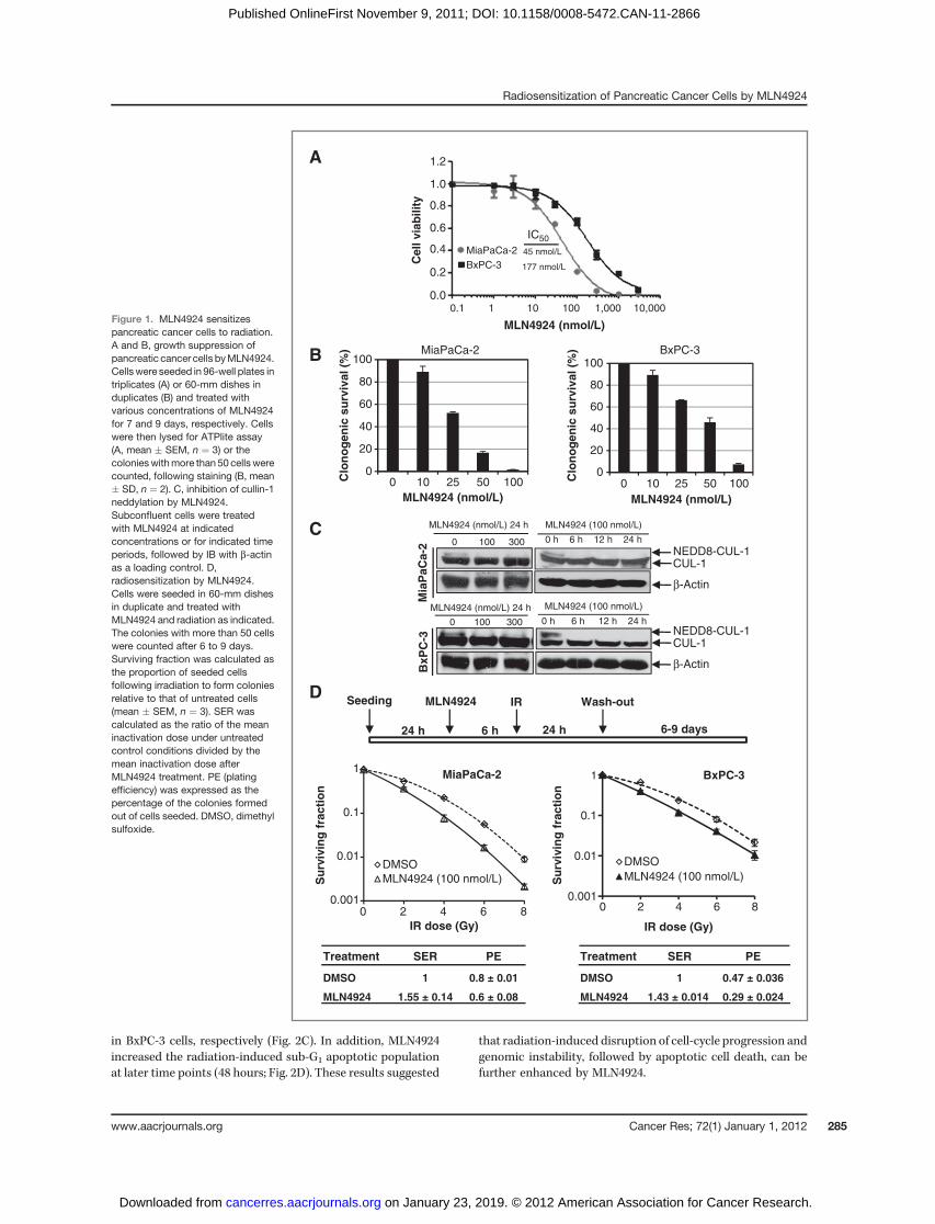
We first determined the sensitivity of 2 pancreatic cancerlines, MiaPaCa-2 and BxPC-3 to MLN4924 as a single agent. Inan ATPlite-based 7-day cell proliferation assay, MLN4924effectively suppressed growth of MiaPaCa-2 and BxPC-3 cellswith an IC50 of 45 or 177 nmol/L, respectively (Fig. 1A). In astandard clonogenic survival assay with MLN4924 in theculture medium for 7 to 9 days, MLN4924 caused a dose-dependent inhibition of colony formation with an IC50 ofapproximately 25 to 50 nmol/L (Fig. 1B). Thus, MLN4924 isa potent inhibitor of cell proliferation and survival in pancre-atic cancer cell lines, with a higher efficacy against MiaPaCa-2cells.
MLN4924 sensitized cancer cells but not normal cells toradiation
We then determined the potential radiosensitizing effect ofMLN4924 in pancreatic cancer cells. We first confirmed thatMLN4924 treatment indeed caused deneddylation of cullin 1;MLN4924 at 100 nmol/L for a 6-hour exposure was sufficient toblock cullin neddylation (Fig. 1C). We next used 2 different
dosing regimens to test MLN4924 radiosensitization. In thefirst regimen (higher MLN4924 dose with shorter exposuretime), cells were pretreated with MLN4924 (100 nmol/L) for 6hours, which effectively caused the accumulation of CDT1 andWEE1 (two critical substrates of SCF E3 ligase, see below;Supplementary Fig. S1), followed by radiation at different dosesup to 8 Gy. MLN4924 was washed out 24 hours postradiationand cells were cultured in MLN4924-free medium for addi-tional 6 to 9 days, allowing colony formation. Under thiscondition, MLN4924 effectively sensitized both pancreaticcancer cell lines to radiation with a sensitivity enhancementratio (SER) of 1.43 to 1.55, respectively (Fig. 1D). In the secondregimen (lower MLN4924 dose with longer exposure time), wepretreated cells with MLN4924 at a lower concentration (20nmol/L forMiaPaCa-2 and 25 nmol/L for BxPC-3) for 24 hours,followed by radiation at different doses up to 8 Gy. Cellswere then cultured for additional 7 to 9 days in the presenceof MLN4924 at these low doses (without medium change).Under this condition, we confirmed that cullin-1 was dened-dylated throughout the experimental periods (SupplementaryFig. S2A) and observed MLN4924 radiosensitization with SERof approximately 1.4 (Supplementary Fig. S2B). Thus, weconclude that MLN4924 is a potent radiosensitizer in pancre-atic cancer cells.
To determine whether radiosensitization by MLN4924 istumor cell selective, we assessed the ability of MLN4924 toradiosensitize H1299 lung cancer cells, pairedwith human lungfibroblast MRC5 cells, because there are no "normal" pancre-atic cells capable of forming colonies in standard clonogenicassays. We found that in H1299 cancer cells, but not in MRC5normal cells, that cullin-1 is highly neddylated, which iscompletely inhibited by MLN4924 at 100 nmol/L (Supplemen-tary Fig. S3A). Consistent with targeting cullin neddylation,MLN4924 was 5-fold more potent in growth suppression ofH1299 cancer cells (IC50¼ 83 nmol/L) thanMRC5 normal cells(IC50 ¼ 429 nmol/L; Supplementary Fig. S3B) and was 4-foldmore potent in inhibition of clonogenic survival of H1299cells (IC50 ¼ 50 nmol/L) than MRC5 normal cells (IC50 ¼200 nmol/L; Supplementary Fig. S3C). Likewise, MLN4924 at aslow as 15 nmol/L sensitized radioresistant H1299 cells (23) toradiation with a SER of 1.39 but had little effect on MRC5 cells(SER ¼ 1.13; Supplementary Fig. S3D). Thus, MLN4924 seemsto selectively target cancer cells with high levels of cullinneddylation and SCF E3 ligase activity.
Radiosensitization by MLN4924 is attributable toenhanced G2/M arrest, aneuploidy, and induction ofapoptosis
To determine the nature of MLN4924 radiosensitization, wecarried out cell-cycle profile of 2 pancreatic cancer cell linestreated with MLN4924, radiation, or MLN4924 in combinationwith radiation (Fig. 2A), and found that MLN4924 remarkablyenhanced the radiation-induced G2/M arrest in MiaPaCa-2 (IRat 34% vs. IR plusMLN4924 at 68%) and BxPC-3 cells (IR at 52%vs. IR plus MLN4924 at 65%; Fig. 2B). MLN4924 also increasedthe aneuploid cell population (DNA content greater than 4N)from 10% in response to radiation alone to 40% in response toradiation plusMLN4924 inMiaPaCa-2 cells and from4% to 24%
Wei et al.
Cancer Res; 72(1) January 1, 2012 Cancer Research284
on January 23, 2019. © 2012 American Association for Cancer Research. cancerres.aacrjournals.org Downloaded from
Published OnlineFirst November 9, 2011; DOI: 10.1158/0008-5472.CAN-11-2866
in BxPC-3 cells, respectively (Fig. 2C). In addition, MLN4924increased the radiation-induced sub-G1 apoptotic populationat later time points (48 hours; Fig. 2D). These results suggested
that radiation-induced disruption of cell-cycle progression andgenomic instability, followed by apoptotic cell death, can befurther enhanced by MLN4924.
Figure 1. MLN4924 sensitizespancreatic cancer cells to radiation.A and B, growth suppression ofpancreatic cancer cells byMLN4924.Cellswere seeded in 96-well plates intriplicates (A) or 60-mm dishes induplicates (B) and treated withvarious concentrations of MLN4924for 7 and 9 days, respectively. Cellswere then lysed for ATPlite assay(A, mean � SEM, n ¼ 3) or thecolonieswithmore than 50 cells werecounted, following staining (B, mean� SD, n ¼ 2). C, inhibition of cullin-1neddylation by MLN4924.Subconfluent cells were treatedwith MLN4924 at indicatedconcentrations or for indicated timeperiods, followed by IB with b-actinas a loading control. D,radiosensitization by MLN4924.Cells were seeded in 60-mm dishesin duplicate and treated withMLN4924 and radiation as indicated.The colonies with more than 50 cellswere counted after 6 to 9 days.Surviving fraction was calculated asthe proportion of seeded cellsfollowing irradiation to form coloniesrelative to that of untreated cells(mean � SEM, n ¼ 3). SER wascalculated as the ratio of the meaninactivation dose under untreatedcontrol conditions divided by themean inactivation dose afterMLN4924 treatment. PE (platingefficiency) was expressed as thepercentage of the colonies formedout of cells seeded. DMSO, dimethylsulfoxide.
B
A
0
20
40
60
80
100
1005025100Clo
no
gen
ic s
urv
ival (%
)
MLN4924 (nmol/L)
MiaPaCa-2
0
20
40
60
80
100
1005025100
Clo
no
gen
ic s
urv
ival (%
)
MLN4924 (nmol/L)
BxPC-3
0.1 1 10 100 1,000 10,0000.0
0.2
0.4
0.6
0.8
1.0
1.2
MiaPaCa-2
BxPC-3
45 nmol/L
177 nmol/L
IC50
Cell v
iab
ilit
y
MLN4924 (nmol/L)
0.001
0.01
0.1
1
86420
Su
rviv
ing
fra
cti
on
IR dose (Gy)
DMSO
MLN4924 (100 nmol/L)
0.001
0.01
0.1
1
86420
Su
rviv
ing
fra
cti
on
IR dose (Gy)
DMSO
MLN4924 (100 nmol/L)
MiaPaCa-2
24 h
Seeding MLN4924 IR Wash-out
6 h 24 h 6-9 days
C
BxPC-3
Mia
PaC
a-2
0 h 6 h 12 h 24 h
MLN4924 (100 nmol/L)MLN4924 (nmol/L) 24 h
0 100 300
β-Actin
NEDD8-CUL-1CUL-1
MLN4924 (nmol/L) 24 h
0 100 300
β-Actin
MLN4924 (100 nmol/L)
NEDD8-CUL-1CUL-1
0 h 6 h 12 h 24 h
BxP
C-3
D
PESERTreatment
0.8 ± 0.011DMSO
0.6 ± 0.081.55 ± 0.14MLN4924
PESERTreatment
0.47 ± 0.0361DMSO
0.29 ± 0.0241.43 ± 0.014 MLN4924
Radiosensitization of Pancreatic Cancer Cells by MLN4924
www.aacrjournals.org Cancer Res; 72(1) January 1, 2012 285
on January 23, 2019. © 2012 American Association for Cancer Research. cancerres.aacrjournals.org Downloaded from
Published OnlineFirst November 9, 2011; DOI: 10.1158/0008-5472.CAN-11-2866

MLN4924 enhanced radiation-induced DNA damageBecause the major cellular effect of ionizing radiation is to
cause DNAdamage and trigger the DNAdamage response (26),we, therefore, examined whether MLN4924 treatment wouldenhance radiation-induced DNA damage and interfere withthe DNA damage repair process. We determined DNA double-strand breaks (DSB) by measuring the overall levels of g-H2AXprotein and formation of nuclear foci upon radiation andMLN4924 treatment, alone or in combination. We found thelevels of g-H2AX increased regardless of single or combination
treatment at an early time point (4 hours). However, a higherprotein level of g-H2AXwas seen in the combination treatmentgroup at 24 hours for both pancreatic cancer cell lines (Fig. 3A).Consistently, we observed a significant increase in the popu-lation of g-H2AX foci positive cells in combination treatmentgroup (Fig. 3B and C). We also determined the DNA damageresponse upon MLN4924 radiation treatment by measuringphosphorylation of CHK1 and CHK2 and found that althoughMLN4924 did not enhance CHK2 phosphorylation, MLN4924did enhance CHK1 phosphorylation in response to radiation,
A
B
C
0 200 400 600 0 200 400 600 0 200 400 600 0 200 400 600
0 200 400 600 0 200 400 600 0 200 400 600 0 200 400 600
PI staining
Ce
ll c
ou
nt
Mia
PaC
a-2
BxP
C-3
G2/M
S
G1
0
20
40
60
80
100
120
Ce
ll-c
yc
le p
rofi
le (
%) MiaPaCa-2 BxPC-3
**
**
0
10
20
30
40
50
Cell
s w
ith
>4N
DN
A (
%)
DMSO
IR
MLN4924
MLN4924 + IR
MiaPaCa-2 BxPC-3
>4N >4N >4N >4N
>4N >4N >4N >4N
DMSO IR MLN4924 MLN4924 + IR
DMSO IRMLN4924 MLN4924 + IR
2152
34
65
1734 32
68
**
**
DMSO
IR
MLN4924
MLN4924 + IR
D
**
0
1
2
3
4
5
6
48 h24 h
Su
b-G
1 c
ells
(Rela
tive f
old
ch
an
ge)
MiaPaCa-2
0
1
2
3
4
5
6
7
8
48 h24 h
DMSO
IR
MLN4924
MLN4924 + IR
BxPC-3 **
Figure 2. MLN4924 alters cell-cycleprogression and inducesapoptosis. Cells were treated withMLN4924 at 100 nmol/L for 24hours (A–C)or up to 48 hours (D), alone or incombination with radiation (6 Gy),followed by FACS analysis forcell-cycle profiling (A), cell-cycledistribution (B), cell population withmore than 4NDNA content (C), andsub-G1 population for apoptosis(D). Shown is mean � SEM (n ¼ 3).�, P < 0.05. One representativeresult is shown for A. DMSO,dimethyl sulfoxide; PI, propidiumiodide.
Wei et al.
Cancer Res; 72(1) January 1, 2012 Cancer Research286
on January 23, 2019. © 2012 American Association for Cancer Research. cancerres.aacrjournals.org Downloaded from
Published OnlineFirst November 9, 2011; DOI: 10.1158/0008-5472.CAN-11-2866

Figure 3. MLN4924 induces DNAdamage and prolongs DNA damageresponse. Cells were treated withMLN4924 (100 nmol/L) alone or incombination with radiation (6 Gy) forindicatedperiodsof time, followedbyIB analysis (A and D), or byimmunofluorescence staining forg-H2AX foci (B and C, left panels).Cells with more than 10 foci werecounted and quantified data isplotted (B and C, right panel). Shownis mean � SEM (n ¼ 5);�, P < 0.05. DAPI, 40,6-diamidino-2-phenylindole; DMSO, dimethylsulfoxide.
A
D
B
0
20
40
60
80
100
% o
f γ-
H2A
X-
po
sit
ive c
ells
(≥10 f
oci)
**
p-CHK1(S345)
Total-CHK1
β-Actin
Total-CHK2
p-CHK2 (T68)
IR
MLN4924
- + - +
- - + +
- + - +
- - + +
- + - +
- - + +
- + - +
- - + +
4 h 24 h 4 h 24 h
MiaPaCa-2 BxPC-3
C
MagnifiedX 60
DAPI
γ-H2AX
IR
MLN4924
MLN4924 + IR
DMSO
Mia
Pa
Ca-2
DMSO
IR
MLN4924
MLN4924 + IR
MagnifiedX 60
Bx
PC
-3
0
20
40
60
80
100
% o
f γ-
H2A
X-
po
sit
ive c
ells
(≥10 f
oci)
**
γ-H2AX
β-Actin
IR
MLN4924
4 h post IR 24 h post IR
- + - +
- - + +
BxPC-3
- + - +
- - + +
- + - +
- - + +
MiaPaCa-2
Radiosensitization of Pancreatic Cancer Cells by MLN4924
www.aacrjournals.org Cancer Res; 72(1) January 1, 2012 287
on January 23, 2019. © 2012 American Association for Cancer Research. cancerres.aacrjournals.org Downloaded from
Published OnlineFirst November 9, 2011; DOI: 10.1158/0008-5472.CAN-11-2866

particularly at the later 24-hour time point (Fig. 3D). Similarenhancement of radiation-induced g-H2AX levels and CHK1phosphorylation byMLN4924 was also observed in H1299 lungcancer cells, which are sensitive to MLN4924 radiosensitiza-tion, but not in MRC5 lung fibroblast, which are resistant toMLN4924 radiosensitization (Supplementary Fig. S4). Takentogether, these results suggested that MLN4924 enhancesradiation-induced DNA damage and prolongs the process ofDNA repair, which likely contributes to its radiosensitizingeffects.
MLN4924 caused accumulation of SCF E3 ligasesubstrates, which is responsible for G2/M arrest andDNA damage response
To identify the mediators of MLN4924 which lead to theenhanced radiation effects, we measured the levels of pro-teins known to be (i) the substrates of SCF E3 ligases and (ii)involved in the regulation of cell-cycle progression, DNAdamage response, and apoptosis (10, 12, 27). We found thatas expected, the levels of cell-cycle regulators, including p21,p27, WEE1, DNA licensing proteins CDT1 and ORC1, andapoptosis regulators NOXA, BIM EL, and pIkBa, increasedsubstantially upon treatment with MLN4924, but notradiation (Fig. 4). The MLN4924–radiation combinationfurther increased the levels of CDT1 and NOXA (Fig. 4), aswell as WEE1 activity, as reflected by enhanced phosphor-ylation of its substrate, CDC2 (Y15; Fig. 4, bottom panels),suggesting these 3 proteins may contribute to MLN4924radiosensitization.
Partial rescue of aneuploidy, G2/M arrest, andradiosensitization by siRNA knockdown of CDT1 andWEE1
Because apoptosis seems to be a late and secondary effect ofMLN4924 radiation (Fig. 2D), likely resulting from enhancedDNA damage and prolonged G2/M arrest, we focused ourattention on CDT1, a DNA licensing protein whose overexpres-sion is known to cause DSB and trigger DNA damage response(9, 28), andWEE1, a protein tyrosine kinase that phosphorylatesCDC2 on Tyr15 to arrest cells at the G2 phase of cell cycle(29, 30). We reasoned that if CDT1 and/or WEE1 were criticalto MLN4924-induced enhancement of radiation effects, weshould be able to abrogate these effects, at least in part, bytheir simultaneous knockdown. Indeed, in MLN4924–radia-tion–treated cells, CDT1 knockdown, using 2 independentsiRNA oligos significantly reduced CHK1 phosphorylation (Fig.5A and Supplementary Fig. S5A), with a consequent reductionof the aneuploid population from approximately 40% to 30%(Fig. 5B) or from34%to19% (SupplementaryFig. S5B). Likewise,WEE1 knockdown using 2 independent siRNA oligos abrogatedCDC2 phosphorylation at Y15 (Fig. 5A, Supplementary Fig. S5A)and reduced the G2/M population from 74% to 55% (Fig. 5C)or 72% to 56% (Supplementary Fig. S5C). Importantly,MLN4924 radiosensitization was largely abrogated uponknockdown of CDT1 or WEE1, as assayed by the first regimen(Fig. 5D and Supplementary Fig. S5D), as well as the secondregimen (Supplementary Fig. S2C). These results clearly suggesta causal effect of CDT1 and/or WEE1 accumulation (upon SCFE3 inactivation by MLN4924) on MLN4924 radiosensitizationin MiaPaCa-2 cells. On the other hand, the incomplete rescueof MLN4924 radiosensitization upon CDT1 and/or WEE1knockdown suggests that other downstream targets (such asp21 or p27, Fig. 4) may also mediate the efficacy of MLN4924.
MLN4924 sensitizedMiaPaCa-2 cells to radiation in an invivo xenograft tumor model
Finally, we assessed MLN4924-mediated radiosensitizationin vivo using the MiaPaCa-2 xenograft model. To ensure thatMLN4924 "hits" its target cullin-1 in in vivo tumors and todetermine when radiation should be delivered post MLN4924administration, we measured the levels of MLN4924-conjugat-ed NEDD8, neddylated cullin-1, SCF substrates, WEE1, andNRF2 at the indicated times after a single administration ofvehicle, MLN4924, radiation, or MLN4924 plus radiation(2 representative tumors per group). As shown in Fig. 6A,MLN4924–NEDD8 adduct was detectable only in groups withMLN4924 treatment, alone (lanes 5 and 6) or in combination(lanes 13 and 14) at the early time point (4 hours), which nearlydisappeared at 24 hours posttreatment (lanes 7 and 8 and 15and 16). Similarly, cullin-1 neddylation was inhibited with acorresponding increase in the SCF E3 substrates WEE1 andNRF2 in MLN4924-treated groups at the early 4-hour timepoint (lanes 5 and 6 and 13 and 14). At later time point (24hours), both neddylated cullin-1 and NRF2 levels returned tothe control levels with WEE1 level remaining slightly higher(lanes 7 and 8 and 15 and 16; Fig. 6A). As expected, radiationalone had no effect on cullin-neddylation or the levels ofselected SCF E3 substrates (lanes 9–12). Our results clearly
CDT1
WEE1
NOXA
BIM EL
p-IkB
β-Actin
- + - +
- - + +
- + - + IR (6 Gy)
MLN4924 (100 nmol/L) + +--
24-h post-IR24-h post-IR
MiaPaCa-2 BxPC-3
p-CDC2 (Y15)
β-Actin
ORC1
p21
p27
Figure 4. MLN4924 induces accumulation of SCF E3 ligase substrates.Subconfluent cells were treated with MLN4924 (100 nmol/L) or radiation(6 Gy) alone or in combination for 24 hours, followed by IB analysis usingindicated antibodies.
Wei et al.
Cancer Res; 72(1) January 1, 2012 Cancer Research288
on January 23, 2019. © 2012 American Association for Cancer Research. cancerres.aacrjournals.org Downloaded from
Published OnlineFirst November 9, 2011; DOI: 10.1158/0008-5472.CAN-11-2866

show that MLN4924 indeed "hits" its target and suggest thatradiation should be delivered 2 hours postadministration ofMLN4924, when SCF E3 is inactivated due to cullindeneddylation.We next determined the in vivo radiosensitizing activity of
MLN4924. As shown in Fig. 6B and C, administration ofMLN4924 alone at a dose of 30 mg/kg s.c./d, 5 d/wk for 3weeks had a moderate inhibitory effect on tumor growth innudemice. Radiation treatment at a clinically relevant dose of 1Gy/d, 5 d/wk for 3 weeks also hadmoderate antitumor activity.
In response to treatment with the combination of MLN4924and radiation, tumor growth was significantly inhibited com-pared with either treatment alone (at day 60; Fig 6B).Similarly, the time required for tumor volume doubling ortripling was significantly increased in response to MLN4924radiation treatment compared with vehicle or radiationtreatment (Fig. 6C). Consistent with this, we also observeda significantly reduced tumor volume nadir correspondingwith a marginally significant increase in the tumor regres-sion rate in response to MLN4924 radiation (compared with
Figure 5. Radiation-enhancingactivity of MLN4924 is inhibited atleast in part by siRNA knockdown ofCDT1 or WEE1. MiaPaCa-2 cellswere transfected with siRNAoligonucleotides targeting CDT1 orWEE1. Forty-eight hours later, oneportion of cells was subjected for IBanalysis (A), the other portion was forFACS analysis (B and C), and stillanother portion was plated forclonogenic assay (D). Shown (B–D) ismean � SD (n ¼ 2).
CDT1
p-CHK1 (S345)
β-Actin
WEE1
p-CDC2 (Y15) SE
p-CDC2 (Y15) LE
Total-CDC2
β-Actin
si-Ctrlsi-CDT1-1 MLN4924 IR
+ + + + - - - -- - - - + + + + - - + + - - + +- + - + - + - +
si-Ctrlsi-WEE1-1 MLN4924 IR
A
B
C
D
+ + + + - - - -
- - + + - - + +- + - + - + - +
- - - - + + + +
0
10
20
30
40
50
Ce
lls
wit
h >
4N
DN
A (
%)
si-ctrl si-CDT1-1
** P = 0.008
* P = 0.026 P = 0.071
* P = 0.035
P = 0.073
γ-H2AX
24-h post-IR24-h post-IR
0.001
0.01
0.1
1
86420
Su
rviv
ing
fra
cti
on
IR dose (Gy)
si-ctrl
si-ctrl + MLN4924
si-CDT1-1 + MLN4924
si-WEE1-1 + MLN4924
PESERTreatment
0.6 ± 0.008 1si-ctrl
0.4 ± 0.007 1.50 ± 0.03 si-ctrl + MLN4924
0.24 ± 0.008 1.22 ± 0.06 si-CDT1-1 + MLN4924
0.26 ± 0.0121.20 ± 0.007 si-WEE1-1 + MLN4924
18
4939
74
0%
20%
40%
60%
80%
100%
120%
Cell c
ycle
pro
file
si-ctrl si-WEE1-1
2946
3755
G2/M
S
G1
Radiosensitization of Pancreatic Cancer Cells by MLN4924
www.aacrjournals.org Cancer Res; 72(1) January 1, 2012 289
on January 23, 2019. © 2012 American Association for Cancer Research. cancerres.aacrjournals.org Downloaded from
Published OnlineFirst November 9, 2011; DOI: 10.1158/0008-5472.CAN-11-2866

radiation; Supplementary Table S1). Importantly, the com-bination treatment was well tolerated by the animals with aminimal loss of body weight (Fig 6D). Taken together, ourresults showed that MLN4924 is a radiosensitizer in pan-creatic cancer, as assessed in both in vitro cell culture and invivo tumor xenograft models, likely by causing accumulationof CDT1 and WEE1, among with other substrates of SCF E3ubiquitin ligase.
Discussion
Our previous work has shown that SCF E3 ubiquitin ligaseis an attractive anticancer target (for review, see refs. 15,31–33). However, because of the complexity of establishing ahigh-throughput (HTS) screen for multi-components SCF E3ubiquitin ligase, there is no single small molecule discoveredthat directly inhibits its ligase activity, although a number of
A
Time required for tumor volume doubling/tripling
TriplingDoublingTreatment
19 (12,23)12.5 (8,19)HPBCD38.5 (22,60)*29.5 (11,49)*MLN492444 (38,47)*37 (34,42)*IR
43 (36,nd)*MLN4924 +IR π 51.5 (42,nd)*π
Data are the median time (days) required for tumor volume doubling/tripling. Upper and lower
limits are indicated in parentheses. Statistical differences were determined by Cox regression
analysis and are indicated versus control* or IR . nd, the upper limit could not be determined.π
B
C
D
IR + MLN4924HPBCD
4h 24 h 4 h 24 h
MLN4924
2 h 22 h
IR
4 h 24 h
MLN4924-NEDD8
NEDD8-CUL1
Tubulin
25R 27R 31R 23R 17R 21R 18R 19R 11R 5R 22R 1R 4R 2R 9R 24R Tumor #
1 2 3 4 5 6 7 8 9 10 11 12 13 14 15 16 Lane #
WEE1
NRF2
0
200
400
600
800
1,000
1,200
706050403020100
Tu
mo
r v
olu
me (
mm
3)
Time (d)
10% HPBCD1 Gy/dMLN4924 30 mg/kg/d1 Gy/d + MLN4924 30 mg/kg/d
**
End
MLN4924 IR Harvest 1 Harvest 2
2 h 2 h 20 h
10
15
20
25
20151050
Bo
dy w
eig
ht
(g)
Time (d)
10% HPBCD
1 Gy/d
MLN4924 30 mg/kg/d
1 Gy/d + MLN4924 30 mg/kg/d
Figure 6. MLN4924radiosensitization in MiaPaCa-2 invivo xenograft tumor model. A,MLN4924 inhibits cullin-1neddylation and causesaccumulationof SCFE3substratesin tumor tissues. In this biomarkerexperiment, 5 � 106 MiaPaCa-2cells were inoculatedsubcutaneously in both flank sides(R, right; L, left) of nude mice. Themice were randomized when thetumor size reached 100 mm3 at 14days after inoculation and weretreated with single dose of HPBCD(vehicle control) or with MLN4924,radiation, or MLN4924-radiationcombination, as indicated. Tumortissues (n ¼ 2 per group) wereharvested at indicated time pointsfor IB. B and C, in vivoradiosensitizing activity ofMLN4924 in the MiaPaCa-2xenograft model. The tumorinoculation and animalrandomization were as describedabove. The number of tumors foreach group are as follows: Control(HPBCD), n ¼ 12, radiation (1 Gy),n ¼ 12, MLN4924 (30 mg/kg, s.c,once a day for 5 d/wk for 3 weeks),n ¼ 16, or combination (MLN4924was given 2–3 hours prior toradiation), n ¼ 16. The tumorgrowth was monitored up to 65days and growth curve plotted. Thearrow indicates the treatment endtime. Student t test was used tocompare each treatment groupwith the control group. Shown aremean � SEM, � indicates P < 0.05(B), and average time periodsrequired for tumors to double ortriple in size (C). D,MLN4924 iswelltolerated by mice. Body weightwas measured during thetreatment and plotted (mean �SEM).
Wei et al.
Cancer Res; 72(1) January 1, 2012 Cancer Research290
on January 23, 2019. © 2012 American Association for Cancer Research. cancerres.aacrjournals.org Downloaded from
Published OnlineFirst November 9, 2011; DOI: 10.1158/0008-5472.CAN-11-2866

HTS methods have been established and optimized to screenfor small molecule inhibitors of single peptide E3 ligases(31, 34). MLN4924 is a newly discovered small moleculeinhibitor of NAE (13). MLN4924 binds to NAE at its activesite to create a covalent NEDD8–MLN4924 adduct, whichresembles NEDD8 adenylate, but cannot be further utilizedin subsequent intraenzyme reactions, thus inhibiting NAEactivity and blocking cullin neddylation (13, 35). Becausecullin neddylation is required for SCF E3 activity (16, 18, 19),MLN4924 becomes the first of a class of "indirect" inhibitorsof SCF E3. By inhibiting SCF E3 ligase activity, MLN4924causes the accumulation of a number of SCF E3 substrates tosuppress the growth of acute myeloid leukemia (36), diffuselarge B-cell lymphoma (37), and many cancer cell linesderived from solid tumors both in vitro and in vivo byinducing apoptosis (13, 36, 37) and senescence (38–40).Importantly, in vivo xenograft assays showed that MLN4924was well tolerated in mice at various doses and treatmentregimens (13), showing a selective killing of cancer cells.With all these preclinical efficacies, MLN4924 has beenadvanced to several phase I clinical trials for solid tumorsand hematologic malignancies (41).In this study, we tested our hypothesis that MLN4924 is a
potent radiosensitizing agent for pancreatic cancer cells,based upon our target validation work using both siRNAknockdown and gene knockout approaches for RBX1 andSAG, the RING components of SCF E3, required for its ligaseactivity (9, 10, 21, 22). For the first time, we showed here thatin in vitro cell culture models, MLN4924 was a potent growthinhibitor as a single agent, as well as a radiosensitizer in 2pancreatic cancer cell lines and in 1 lung cancer cell line, butnot normal lung fibroblast cells. The resistance of normalfibroblasts to MLN4924 radiosensitization is likely due to thelack of cullin neddylation. We also showed in vivo, using aMiaPaCa-2 xenograft model, that MLN4924 inhibited tumorgrowth and conferred radiosensitization with minimal tox-icity to the mice. Mechanistically, we showed that MLN4924enhanced radiation-induced G2/M arrest, aneuploidy, and,eventually, apoptosis by inhibiting cullin neddylation totrigger the accumulation of a number of SCF E3 substrates,related to DNA damage response and cell-cycle regulation.Among these accumulated substrates, CDT1 and WEE1 werefurther increased when combined with radiation and werecritical for enhanced DNA damage and aneuploidy or G2/Marrest, respectively, and eventually for MLN4924 radiosensi-tization, as evidenced by siRNA knockdown-based rescuingexperiments.Precise duplication of the genome at the S phase of each
cell cycle requires initiation of DNA replication from thou-sands of origins. Initiation of too few origins would causecollapse of replication forks, leading to DNA damage andincomplete replication of the genome, whereas more thanone initiation of DNA replication per cell cycle would causeDNA "hyperreplication" or "rereplication" and subsequentDNA damage (42), leading to genomic instability and cancer(43). CDT1 is a key licensing factor which, along with theprotein CDC6, functions to form the prereplicative complex(pre-RC) to initiate DNA replication (44). It was recently
reported that overexpression of Double-arked (Dup), theDrosophila ortholog of CDT1 caused DNA rereplication andDNA damage, followed by caspase activation and apoptoticcell death (45). In human U2-OS cancer cells, CDT1 over-expression caused double strand breaks to trigger DDR,followed by induction of senescence and apoptosis (28).Induction of CDT1 accumulation by MLN4924 was foundto be attributable to DNA rereplication, aneuploidy, andapoptotic death in HCT116 cells (13, 40, 46). Our datapresented here indicated that CDT1 accumulation is alsofound in 2 pancreatic cancer cell lines and is critical forenhanced aneuploidy and MLN4924 radiosensitization.
The WEE1 kinase negatively regulates G2/M transition bydirectly phosphorylating Tyr15 of CDC2, which inhibits theCDC2 activity (30). We have previously shown that WEE1 canbe stabilized and activated by binding to 14-3-3b (47) and is avalid radiosensitizing target, whose inhibition abrogatesthe radiation-induced G2 checkpoint (48, 49). In this study,we found that the level of WEE1, a substrate of SCF E3 ligase(50), increased expectedly upon MLN4924 treatment. TheWEE1 activity was further enhanced when MLN4924 wascombined with radiation. Our rescue experiment showed thataccumulated WEE1 is responsible, at least in part, for aremarkable increase in the G2/M population in response toMLN4924 and radiation, which seems causally related toMLN4924 radiosensitization.
In summary, our study revealed radiosensitizing activityof MLN4924, a small molecule inhibitor of SCF E3, inpancreatic cancer as well as in lung cancer cells, andelucidated its mechanisms of action which include accumu-lation of CDT1 and WEE1, among other cell cycle andapoptosis regulators, which trigger DNA damage, aneuploidyand G2/M arrest, followed by apoptotic death. Our study,therefore, provides the first preclinical proof-of-conceptevidence for future development of MLN4924 as a novelradiosensitizing agent against pancreatic cancer and, pos-sibly, other types of human cancers.
Disclosure of Potential Conflicts of Interest
No potential conflicts of interest were disclosed.
Acknowledgments
The authors thank Millennium Pharmaceuticals, Inc. for providing us withMLN4924.
Grant Support
This work was supported by the NCI grants CA111554, CA118762, andCA156744 to Y. Sun and CA78554 and CA78554-10S231071204 to T.S. Lawr-ence. This work was also partially supported by Millennium Pharmaceuticals,Inc.
The costs of publication of this article were defrayed in part by thepayment of page charges. This article must therefore be hereby markedadvertisement in accordance with 18 U.S.C. Section 1734 solely to indicate thisfact.
Received August 23, 2011; revised October 20, 2011; accepted November 1,2011; published OnlineFirst November 9, 2011.
Radiosensitization of Pancreatic Cancer Cells by MLN4924
www.aacrjournals.org Cancer Res; 72(1) January 1, 2012 291
on January 23, 2019. © 2012 American Association for Cancer Research. cancerres.aacrjournals.org Downloaded from
Published OnlineFirst November 9, 2011; DOI: 10.1158/0008-5472.CAN-11-2866

References1. JemalA,BrayF,CenterMM, Ferlay J,WardE, FormanD.Global cancer
statistics. CA Cancer J Clin 2011;61:69–90.2. SharmaC, Eltawil KM,RenfrewPD,WalshMJ,MolinariM. Advances in
diagnosis, treatment and palliation of pancreatic carcinoma: 1990-2010. World J Gastroenterol 2011;17:867–97.
3. Iacobuzio-Donahue CA, Fu B, Yachida S, Luo M, Abe H, HendersonCM, et al. DPC4 gene status of the primary carcinoma correlates withpatterns of failure in patients with pancreatic cancer. J Clin Oncol2009;27:1806–13.
4. Loehrer PJ, Powell ME, Cardenes HR, Wagner L, Brell JM, Rama-nathan RK, et al. A randomized phase III study of gemcitabine incombination with radiation therapy versus gemcitabine alone inpatients with localized, unresectable pancreatic cancer: E4201. J ClinOncol 2008; ASCO Meeting Abstracts May 20 4506.
5. Ben-Josef E, Griffith K, Francis IR, Khan G, Lawrence T, Abrams R,et al. Phase I radiation dose-escalation trial of intensity-modulatedradiotherapy (IMRT) with concurrent fixed dose-rate gemcitabine(FDR-G) for unresectable pancreatic cancer. J Clin Oncol 2009;27:15s (suppl; abstr 4602).
6. Morgan MA, Parsels LA, Zhao L, Parsels JD, Davis MA, Hassan MC,et al. Mechanism of radiosensitization by the Chk1/2 inhibitorAZD7762 involves abrogation of the G2 checkpoint and inhibitionof homologous recombinational DNA repair. Cancer Res 2010;70:4972–81.
7. Moore MJ, Goldstein D, Hamm J, Figer A, Hecht JR, Gallinger S, et al.Erlotinib plus gemcitabine compared with gemcitabine alone inpatients with advanced pancreatic cancer: a phase III trial of theNational Cancer Institute of Canada Clinical Trials Group. J Clin Oncol2007;25:1960–6.
8. Morgan MA, Parsels LA, Kollar LE, Normolle DP, Maybaum J, Lawr-ence TS. The combination of epidermal growth factor receptor inhi-bitorswith gemcitabine and radiation in pancreatic cancer. ClinCancerRes 2008;14:5142–9.
9. Jia L, Bickel JS,Wu J,MorganMA, Li H, Yang J, et al. RBX1 (RING-boxprotein 1) E3 ubiquitin ligase is required for genomic integrity bymodulating DNA replication licensing proteins. J Biol Chem 2011;286:3379–86.
10. Jia L, Yang J, Hao X, Zheng M, He H, Xiong X, et al. Validation of SAG/RBX2/ROC2 E3 ubiquitin ligase as an anticancer and radiosensitizingtarget. Clin Cancer Res 2010;16:814–24.
11. Deshaies RJ, Joazeiro CA. RING domain E3 ubiquitin ligases. AnnuRev Biochem 2009;78:399–434.
12. Nakayama KI, Nakayama K. Ubiquitin ligases: cell-cycle control andcancer. Nat Rev Cancer 2006;6:369–81.
13. Soucy TA, Smith PG, Milhollen MA, Berger AJ, Gavin JM, Adhikari S,et al. An inhibitor of NEDD8-activating enzyme as a new approach totreat cancer. Nature 2009;458:732–6.
14. Wu K, Fuchs SY, Chen A, Tan P, Gomez C, Ronai Z, et al. The SCF(HOS/beta-TRCP)-ROC1 E3 ubiquitin ligase utilizes two distinctdomains within CUL1 for substrate targeting and ubiquitin ligation.Mol Cell Biol 2000;20:1382–93.
15. Sun Y. E3 ubiquitin ligases as cancer targets and biomarkers. Neo-plasia 2006;8:645–54.
16. Duda DM, Borg LA, Scott DC, Hunt HW, Hammel M, Schulman BA.Structural insights into NEDD8 activation of cullin-RING ligases:conformational control of conjugation. Cell 2008;134:995–1006.
17. Saha A, Deshaies RJ.Multimodal activation of the ubiquitin ligase SCFby Nedd8 conjugation. Mol Cell 2008;32:21–31.
18. Goldenberg SJ, Cascio TC, Shumway SD, Garbutt KC, Liu J, Xiong Y,et al. Structure of the Cand1-Cul1-Roc1 complex reveals regulatorymechanisms for the assembly of the multisubunit cullin-dependentubiquitin ligases. Cell 2004;119:517–28.
19. Kamura T, Conrad MN, Yan Q, Conaway RC, Conaway JW. TheRbx1 subunit of SCF and VHL E3 ubiquitin ligase activates Rub1modification of cullins Cdc53 and Cul2. Genes Dev 1999;13:2928–33.
20. Xirodimas DP. Novel substrates and functions for the ubiquitin-likemolecule NEDD8. Biochem Soc Trans 2008;36:802–6.
21. Jia L, Soengas MS, Sun Y. ROC1/RBX1 E3 ubiquitin ligase silencingsuppresses tumor cell growth via sequential induction of G2-M arrest,apoptosis, and senescence. Cancer Res 2009;69:4974–82.
22. TanM, ZhuY, Kovacev J, Zhao Y, Pan ZQ, Spitz DR, et al. Disruption ofSag/Rbx2/Roc2 induces radiosensitization by increasing ROS levelsand blocking NF-kB activation in mouse embryonic stem cells. FreeRadic Biol Med 2010;49:976–83.
23. Zheng M, Morgan-Lappe SE, Yang J, Bockbrader KM, Pamarthy D,Thomas D, et al. Growth inhibition and radiosensitization of glioblas-toma and lung cancer cells by small interfering RNA silencing of tumornecrosis factor receptor-associated factor 2. Cancer Res 2008;68:7570–8.
24. Fertil B, Dertinger H, Courdi A, Malaise EP. Mean inactivation dose: auseful concept for intercomparison of human cell survival curves.Radiat Res 1984;99:73–84.
25. Zhao L, Morgan MA, Parsels LA, Maybaum J, Lawrence TS, NormolleD. Bayesian hierarchical changepoint methods in modeling the tumorgrowth profiles in xenograft experiments. Clin Cancer Res 2011;17:1057–64.
26. Begg AC, Stewart FA, Vens C. Strategies to improve radiotherapy withtargeted drugs. Nat Rev Cancer 2011;11:239–53.
27. Skaar JR, D'Angiolella V, Pagan JK, Pagano M. SnapShot: F BoxProteins II. Cell 2009;137:1358, e1.
28. Liontos M, Koutsami M, Sideridou M, Evangelou K, Kletsas D, Levy B,et al. Deregulated overexpression of hCdt1 and hCdc6 promotesmalignant behavior. Cancer Res 2007;67:10899–909.
29. Heald R, McLoughlin M, McKeon F. Human wee1 maintains mitotictiming by protecting the nucleus from cytoplasmically activated Cdc2kinase. Cell 1993;74:463–74.
30. Parker LL, Piwnica Worms H. Inactivation of the p34cdc2-cyclin Bcomplex by the human WEE1 tyrosine kinase. Science 1992;257:1955–7.
31. Jia L, Sun Y. SCF E3 ubiquitin ligases as anticancer targets. CurrentCancer Drug Targets 2011;11:347–56.
32. Sun Y. Targeting E3 ubiquitin ligases for cancer therapy. Cancer BiolTherapy 2003;2:623–9.
33. Wei D, Sun Y. Small RING finger proteins RBX1 and RBX2 of SCF E3ubiquitin ligases: the role in cancer and as cancer targets. Genes &Cancer 2010;1:700–7.
34. Sun Y. Overview of approaches for screening for ubiquitin ligaseinhibitors. Methods Enzymol 2005;399:654–63.
35. Brownell JE, Sintchak MD, Gavin JM, Liao H, Bruzzese FJ, Bump NJ,et al. Substrate-assisted inhibition of ubiquitin-like protein-activatingenzymes: the NEDD8 E1 inhibitor MLN4924 forms a NEDD8-AMPmimetic in situ. Mol Cell 2010;37:102–11.
36. Swords RT, Kelly KR, Smith PG,Garnsey JJ,MahalingamD,Medina E,et al. Inhibition of NEDD8-activating enzyme: a novel approach for thetreatment of acute myeloid leukemia. Blood 2010;115:3796–800.
37. Milhollen MA, Traore T, Adams-Duffy J, Thomas MP, Berger AJ, DangL, et al. MLN4924, a NEDD8-activating enzyme inhibitor, is active indiffuse large B-cell lymphoma models: rationale for treatment of NF-{kappa}B-dependent lymphoma. Blood 2010;116:1515–23.
38. Lin HK, Chen Z, Wang G, Nardella C, Lee SW, Chan CH, et al. Skp2targeting suppresses tumorigenesis by Arf-p53-independent cellularsenescence. Nature 2010;464:374–9.
39. Jia L, Li H, Sun Y. Induction of p21-dependent senescence by an NAEinhibitor, MLN4924, as a mechanism of growth suppression. Neopla-sia 2011;13:561–9.
40. Lin JJ, Milhollen MA, Smith PG, Narayanan U, Dutta A. NEDD8-targeting drug MLN4924 elicits DNA rereplication by stabilizing Cdt1in S phase, triggering checkpoint activation, apoptosis, and senes-cence in cancer cells. Cancer Res 2010;70:10310–20.
41. Soucy TA, Smith PG, RolfeM. TargetingNEDD8-activated cullin-RINGligases for the treatment of cancer. Clin Cancer Res 2009;15:3912–6.
42. Arias EE, Walter JC. Strength in numbers: preventing rereplication viamultiplemechanisms in eukaryotic cells. GenesDev2007;21:497–518.
43. Dutta A. Chaotic license for genetic instability and cancer. Nat Genet2007;39:10–1.
Wei et al.
Cancer Res; 72(1) January 1, 2012 Cancer Research292
on January 23, 2019. © 2012 American Association for Cancer Research. cancerres.aacrjournals.org Downloaded from
Published OnlineFirst November 9, 2011; DOI: 10.1158/0008-5472.CAN-11-2866

44. Rialland M, Sola F, Santocanale C. Essential role of human CDT1 inDNA replication and chromatin licensing. JCell Sci 2002;115:1435–40.
45. Mehrotra S, Maqbool SB, Kolpakas A, Murnen K, Calvi BR. Endocy-cling cells do not apoptose in response to DNA rereplication genotoxicstress. Genes Dev 2008;22:3158–71.
46. MilhollenMA,NarayananU, Soucy TA, Veiby PO, Smith PG, AmidonB.Inhibition of NEDD8-activating enzyme induces rereplication and apo-ptosis in human tumor cells consistent with deregulating CDT1 turn-over. Cancer Res 2011;71:3042–51.
47. Wang Y, Jacobs C, Hook KE, Duan H, Booher RN, Sun Y. Binding of14-3-3beta to the carboxyl terminus ofWee1 increasesWee1 stability,
kinase activity, and G2-M cell population. Cell Growth Differ2000;11:211–9.
48. Li J, Wang Y, Sun Y, Lawrence TS.Wild-type TP53 inhibits G(2)-phasecheckpoint abrogation and radiosensitization induced by PD0166285,a WEE1 kinase inhibitor. Radiat Res 2002;157:322–30.
49. Wang Y, Li J, Booher RN, Kraker A, Lawrence T, Leopold WR, et al.Radiosensitization of p53 mutant cells by PD0166285, a novel G(2)checkpoint abrogator. Cancer Res 2001;61:8211–7.
50. Watanabe N, Arai H, Nishihara Y, Taniguchi M, Hunter T, Osada H. M-phase kinases induce phospho-dependent ubiquitination of somaticWee1 by SCFbeta-TrCP. Proc Natl Acad Sci U SA 2004;101:4419–24.
Radiosensitization of Pancreatic Cancer Cells by MLN4924
www.aacrjournals.org Cancer Res; 72(1) January 1, 2012 293
on January 23, 2019. © 2012 American Association for Cancer Research. cancerres.aacrjournals.org Downloaded from
Published OnlineFirst November 9, 2011; DOI: 10.1158/0008-5472.CAN-11-2866

2012;72:282-293. Published OnlineFirst November 9, 2011.Cancer Res Dongping Wei, Hua Li, Jie Yu, et al. an Investigational NEDD8-Activating Enzyme InhibitorRadiosensitization of Human Pancreatic Cancer Cells by MLN4924,
Updated version
10.1158/0008-5472.CAN-11-2866doi:
Access the most recent version of this article at:
Material
Supplementary
http://cancerres.aacrjournals.org/content/suppl/2011/11/09/0008-5472.CAN-11-2866.DC1
Access the most recent supplemental material at:
Cited articles
http://cancerres.aacrjournals.org/content/72/1/282.full#ref-list-1
This article cites 48 articles, 24 of which you can access for free at:
Citing articles
http://cancerres.aacrjournals.org/content/72/1/282.full#related-urls
This article has been cited by 19 HighWire-hosted articles. Access the articles at:
E-mail alerts related to this article or journal.Sign up to receive free email-alerts
Subscriptions
Reprints and
To order reprints of this article or to subscribe to the journal, contact the AACR Publications Department at
Permissions
Rightslink site. Click on "Request Permissions" which will take you to the Copyright Clearance Center's (CCC)
.http://cancerres.aacrjournals.org/content/72/1/282To request permission to re-use all or part of this article, use this link
on January 23, 2019. © 2012 American Association for Cancer Research. cancerres.aacrjournals.org Downloaded from
Published OnlineFirst November 9, 2011; DOI: 10.1158/0008-5472.CAN-11-2866