radiology week one chapters one and two. key terms please know key terms for quiz on friday
TRANSCRIPT

Radiology
Week one
Chapters one and two

Key Terms
• Please know key terms for quiz on Friday

Properties of x-rays
• Produced by the conversion of electric energy into radiation
• Invisible• Travel in straight lines• Can penetrate opaque tissues and
structures• Can affect a photographic emulsion or
digital sensor • Can adversely affect human tissue

History of Dental Radiography1895 Discovery of x-rays W.C. Roentgen
1896 First dental radiograph O.Walkhoff
1896 First dental radiograph in US (Skull)
W.J. Morton
1896 First dental radiograph in US (live patient)
C.E. Kells
1913 First prewrapped dental films
Eastman Kodak Company
1913 First x-ray tube W.D. Coolidge
1923 First dental x-ray machine Victor X-Ray Corp, Chicago

Atom – a fundamental unit of matter
Consists ofTwo Parts:
1.Nucleus
2.OrbitingElectrons

Made of one atom

Dental x-ray production

Ionization=converting atoms into ions
Ion pair

Electromagnetic Radiation
An x-ray is one type of electromagnetic radiation. Electromagnetic radiation represents the movement of energy through space as a combination of electric and magnetic fields. All types of electromagnetic radiation, which also includes radiowaves, tv waves, visible light, microwaves and gamma rays, travel at the speed of light (186,000 miles per second). They travel through space in wave form.


D
W
W
The waves of electromagnetic radiation have two basic properties: wavelength and frequency. The wavelength (W) is the distance from the crest of one wave to the crest of the next wave. The frequency (F) is the number of waves in a given distance (D). If the distance between waves decreases (W becomes shorter), the frequency will increase. The top wave above has a shorter wavelength and a higher frequency than the wave below it.
F = 3
F = 2

radiowaves
tv waves
visible light
x-rays gamma rays
cosmic rays
Which of the above examples of electromagnetic radiation has the shortest wavelength?
Which of the above has the lowest frequency?
Cosmic rays
Radio waves

Bremsstrahlung/General/BrakingRadiation
• Braking = sudden stopping of high speed electrons when they hit the tungsten target in the anode that causes radiation.
• 70% (most) x-rays are produced in this manner.

Characteristic
• Very small amount of x-ray production.
• Can only be produced at 70 kVp and above.
• Causes not only radiation but ionization.

Defining terms
• Primary Radiation – penetrating or useful beam
• Secondary Radiation – created when the primary beam interacts with matter.
• Scatter Radiation – a form of secondary radiation that is deflected in all directions.– Harmful to patient and radiographer

Interactions of X-radiation
• What happens after an x-ray exits the tubehead?– 1. Can pass through the patient without any
interaction– 2. X-ray photons can be completely absorbed
by the patient– 3. X-ray photons can be scattered
• Compton scatter• Coherent scatter
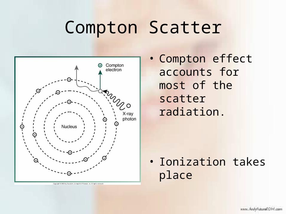
Compton Scatter
• Compton effect accounts for most of the scatter radiation.
• Ionization takes place

Coherent Scatter
• No change in the atom occurs
• X-ray photon simply undergoes a change in direction.

X-ray Equipment
X-ray equipment has three basic components: (1) the x-ray tubehead, which produces the x-rays, (2) support arms, which allow you to move the tubehead around the patient’s head and (3) the control panel, which allows you to alter the duration of the x-ray beam (exposure time) and, on some x-ray machines, the intensity (energy) of the x-ray beam.
1
3
2

PID(cone)
X-ray Tubehead
degrees
The x-ray tubehead is attached to the support arms so that it can rotate up and down (vertically;measured in degrees) and sideways (horizontally) to facilitate proper alignment of the x-ray beam. The PID (Position Indicating Device) is attached to the x-ray tubehead where the x-ray beam exits and it identifies the location of the x-ray beam. Some people refer to the PID as a “cone”; the PID’s on very old x-ray machines used to be coneshaped.

The control panel, like the one above left, allows you to change exposure time but nothing else. Some machines, like the one above right, have controls for changing the mA and kVp settings in addition to exposure time. The individual controls will be discussed more later.
exposure time kVp control
mA control

X-ray Tube
X-rays are produced in the x-ray tube, which is located in the x-ray tubehead. X-rays are generated when electrons from the filament cross the tube and interact with the target. The two main components of the x-ray tube are the cathode and the anode.

(tungsten)
CathodeFocusing cup
Filament
The cathode is composed of a tungsten filament which is centered in a focusing cup. Electrons are produced by the filament (see next slide) and are focused on the target of the anode where the x-rays are produced. The focusing cup has a negative charge, like the electrons, and this helps direct the electrons to the target (“focuses” them; electrons can be focused, x-rays cannot).
side view(cross-section)
front view(facing target)

Thermionic Emission
x-section offilament
hotfilament
When you depress the exposure button, electricity flows through the filament in the cathode, causing it to get hot. The hot filament then releases electrons which surround the filament (thermionic emission). The hotter the filament gets, the greater the number of electrons that are released. (Click to depress exposure button and heat filament).
electrons

Electron Cloud

Anode
Copper stem
Target
The anode in the x-ray tube is composed of a tungsten target embedded in a copper stem. When electrons from the filament enter the target and generate x-rays, a lot of heat is produced. The copper helps to take some of the heat away from the target so that it doesn’t get too hot.
side view front view
Target

X-ray Tube Components
1
2
435
8
6
7
9
1. focusing cup 6. copper stem2. filament 7. leaded glass3. electron stream 8. x-rays4. vacuum 9. beryllium window5. target
(for description, see next slide)

1. Focusing cup: focuses electrons on target2. Filament: releases electrons when heated3. Electron stream: electrons cross from filament to target during length of exposure4. Vacuum: no air or gases inside x-ray tube that might interact with electrons crossing tube5. Target: x-rays produced when electrons strike target6. Copper stem: helps remove heat from target7. Leaded glass: Keeps x-rays from exiting tube in wrong direction8. X-rays produced in target are emitted in all directions9. Beryllium window: this non-leaded glass allows x-rays to pass through. The PID would be located directly in line with this window.
X-ray Tube Components (continued)

Target
Beryllium Window
Focusing cup(filament located inside)
Photo of an X-ray Tube
Leaded glass

The energy of a wave of electromagnetic radiation represents the ability to penetrate an object. The higher the energy, the more easily the wave will pass through the object. The shorter the wavelength, the greater the energy will be and the higher the frequency, the greater the energy will be.
X-ray Energy

A
B
CWhich of the above x-rays has the highest energy?
A: It has the shortest wavelength, highest frequency

Quality Quantity
(primarily)kVp
mA
Time
Filtration
no change
no change
Collimation does not change the energy or number of x-rays in the x-ray beam that reach the film; it just limits the size and shape of the beam.
The quality, or average energy, of the x-ray beam is increased with an increase in kVp or an increase in filtration. The quantity, or number of x-rays, is increased with an increase in time, mA setting and kVp setting.

Filtration
Low-energy x-rays do not contribute to the formation of an x-ray image; all they do is expose the body to radiation. Therefore, we need to get rid of them. The process of removing these low-energy x-rays from the x-ray beam is known as filtration. Filtration increases the average energy (quality) of the x-ray beam.There are two components to x-ray filtration. The first of these, called inherent filtration, results from the materials present in the x-ray machine that the x-rays have to pass through. These include the beryllium window of the x-ray tube, the oil in the tubehead and the barrier material that keeps the oil from leaking out of the tubehead. This removes very weak x-rays.

Filtration (continued)The second component is the addition of aluminum disks placed in the path of the x-ray beam (added filtration). These disks remove the x-rays that had enough energy to get through the inherent filtration but are still not energetic enough to contribute to image formation. Disks of varying thicknesses, when combined with the inherent filtration, produce the total filtration for the x-ray machine. Federal regulations require that an x-ray machine capable of operating at 70 kVp or higher must have total filtration of 2.5 mm aluminum equivalent. (The inherent filtration is “equivalent” to a certain thickness of aluminum). X-ray machines operating below 70 kVp need to have a total filtration of 1.5 mm aluminum equivalent.

Filtration
Inherent
beryllium window of x-ray tube
Added
Aluminum filter (s)
TotalOil/Metal barrier
filter
PID
collimator
barriermaterial
berylliumwindow
oil

filter
PID
The filter is usually located in the end of the PID which attaches to the tubehead.

primary x-ray
scattered x-ray
CollimationCollimation is used to restrict the area of the head that the x-rays will contact. We want to cover the entire film with the x-ray beam, but don’t want to overexpose the patient. Also, when x-rays from the tubehead interact with the tissues of the face, scatter radiation is produced (see below). This scatter radiation creates additional exposure of the patient and also decreases the quality of the x-ray image. (Scatter will be discussed in greater detail in the section on biological effects of x-rays).
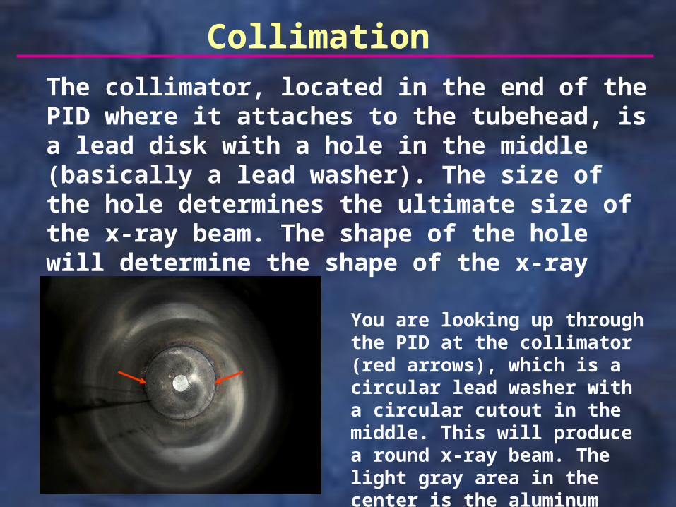
CollimationThe collimator, located in the end of the PID where it attaches to the tubehead, is a lead disk with a hole in the middle (basically a lead washer). The size of the hole determines the ultimate size of the x-ray beam. The shape of the hole will determine the shape of the x-ray beam.
You are looking up through the PID at the collimator (red arrows), which is a circular lead washer with a circular cutout in the middle. This will produce a round x-ray beam. The light gray area in the center is the aluminum filter

The shape of the opening in the collimator determines the shape of the x-ray beam. The size of the opening determines the size of the beam at the end of the PID. PID’s come in varying lengths; longer PID’s have a smaller opening in the collimator.
round
rectangular
Collimation

The x-ray beam continues to spread out as you get further from the x-ray source (target). More surface is exposed on the exit side of the patient than the entrance side. By collimating the beam, less overall surface is exposed and as a result, less scatter radiation is produced. Both of these things reduce patient exposure. 2.75 inches (7 cm) is the maximum diameter of a circular beam or the maximum length of the long side of a rectangular beam at the end of the PID.
collimated beamcollimator
target(x-ray source)
Collimation

If you switch from a 7 cm round PID to a 6 cm round PID, the patient receives 25% less radiation because the area covered by the beam is reduced by 25%.
Rectangular collimation (dotted line at left) results in the patient receiving 55 % less radiation when compared to what they would receive with a 7 cm round PID.
6 cm round
film(4.5 cm long)
entrance
entrance
exit
exit
6 cm7 cm
area covered at skin surface (6 cm round PID)
area covered as beam exits (6 cm round PID)
area covered at skin surface (7 cm round PID)
area covered as beam exits (7 cm round PID)
Collimation

X-ray Beam Modifiers
The following slides identify the various ways of changing the energy of the x-ray beam and the number of x-rays produced during an x-ray exposure.

Exposure Factors
The energy of the x-ray beam and the number of x-rays are primarily regulated by the kVp control, the mA setting and the exposure time. One, two or all three of these exposure factors may need to be adjusted, depending on the size of the patient’s head, the likelihood of patient movement due to tremors or the inability to hold still, etc.. If the exposure factors are not set properly for the current patient, the resultant film may be too light or too dark (see next slide).

Exposure factors too high (too dark)
Correct exposure factors
Exposure factors too low (too light)

1. Recommended kVp, mA, exposure time (e.t.)
2. Increase mA; no change in kVp, e.t.
3. Decrease e.t.; no change in kVp, mA
4. Increase kVp; no change in mA, e.t.
5. Double mA, halve e.t.; no change in kVp
A CB
B
A
C
A
B
overexposed correct exposure underexposed
In the following situations, would you expect the x-ray film to be (A), overexposed, (B) correctly exposed or (C) underexposed? (No change in patient size).