radiology packet 10 pulmonary pattern – bronchial asthma
TRANSCRIPT
Radiology Packet 10
Pulmonary Pattern – Bronchial Asthma
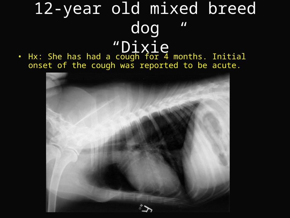
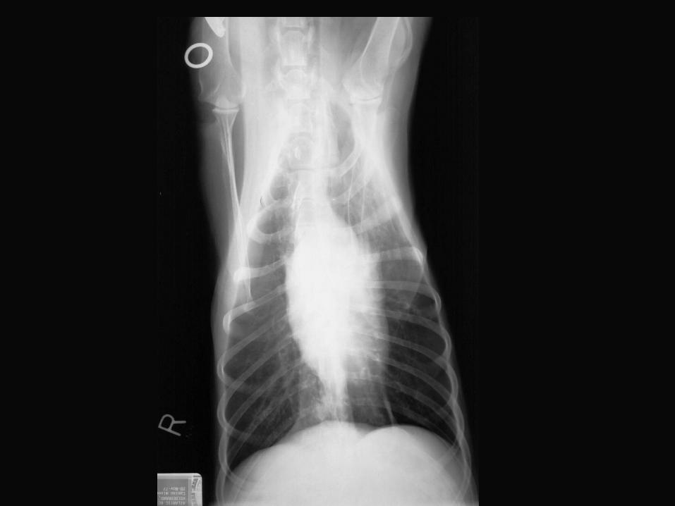
12-year old mixed breed dog“Dixie”
• Hx: She has had a cough for 4 months. Initial onset of the cough was reported to be acute.
12-year old mixed breed dog“Dixie”
• RF– The bronchi are noted to be calcified.– The mineralized bronchi are seen as linear structures overlying the heart.– Mineralized end-on bronchi are also visible.– There is an area of increased interstitial opacity beneath the caudal vena cava.– Multiple small round calcified structures are noted in the periphery of the lungs.– These are pulmonary osteomas or pneumoliths – islands of mineralization within the lung
parenchyma of unknown etiology. They are an incidental finding and should not be mistaken for metastatic disease.
– There is an incidental finding of a small amount of gas in the esophagus cranial to the heart.
• RD– Bronchial lung pattern– Interstitial opacity in the accessory lung lobe
• R/O– Chronic bronchitis
• Cause may be allergic or inflammatory or possibly an inhaled toxin– Parasitic
• Next– Trans-tracheal aspirate or broncho-alveolar lavage
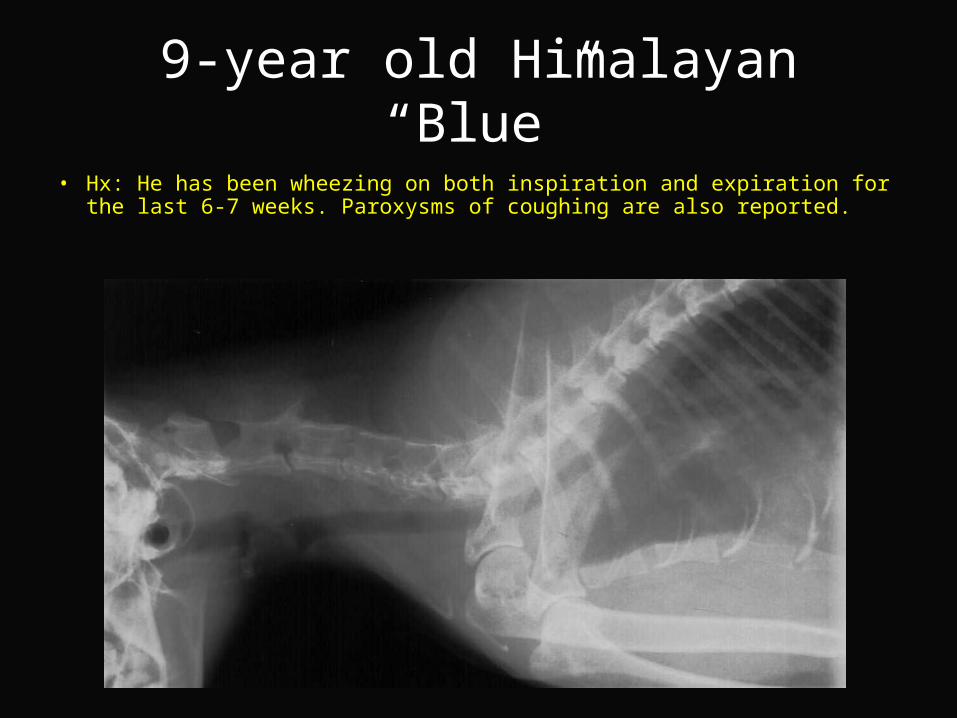
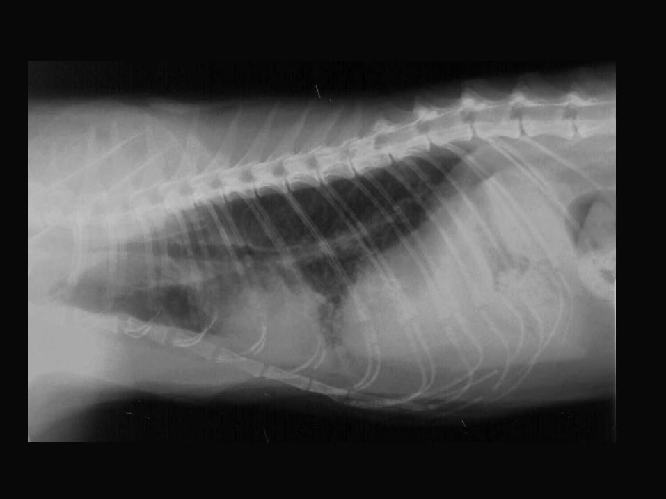
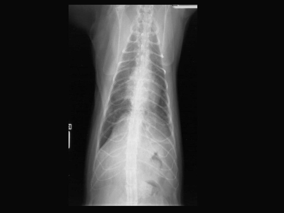
9-year old Himalayan“Blue”
• Hx: He has been wheezing on both inspiration and expiration for the last 6-7 weeks. Paroxysms of coughing are also reported.
9-year old Himalayan“Blue”
• RF– Incidental finding of generalized skeletal osteopenia.– There is a diffuse increase in pulmonary opacity with a faint nodular pattern
visible. – In the lateral view there are patchy pulmonary infiltrates partially obscuring the
cardiac border.– In the VD biew there is a focus of increased opacity in the region of the right
middle lung lobe. An air bronchogram is visible in this area.– The lateral radiograph of the cervical region shows a marked stenosis of the
trachea in the region of the tracheal cartilages.
• RD– Diffuse broncho-interstitial pattern with focal alveolar changes– Tracheal stenosis
• R/O– Feline asthma– Pneumonia
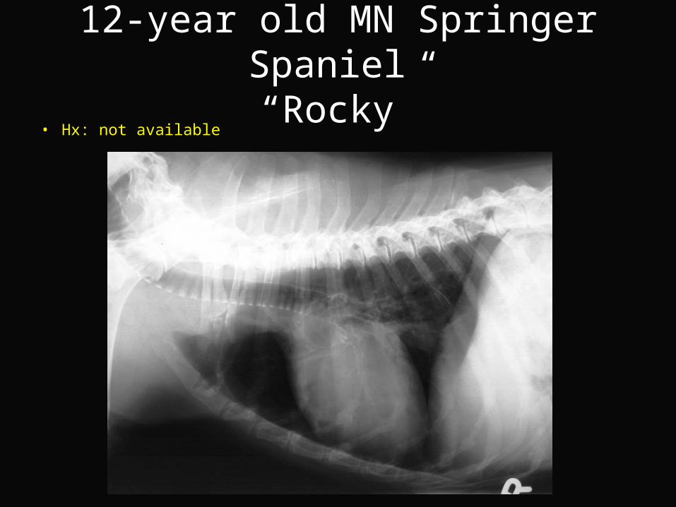
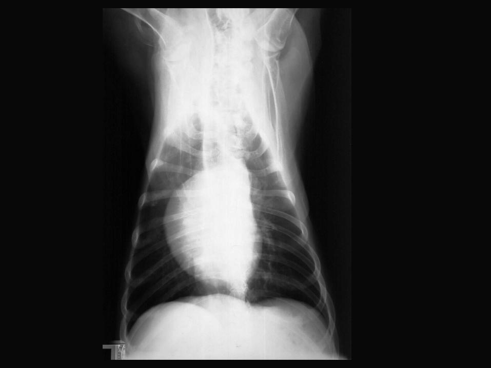
12-year old MN Springer Spaniel “Rocky”
• Hx: not available
12-year old MN Springer Spaniel “Rocky”
• RF– In the lateral view the cardiac silhouette appears round and there is no visible
cardiac apex. This appearance of the heart is typical of that seen in a left lateral recumbent view.
– In the VD view the left margin of the heart is unusual in shape. This is the result of rotation of the patient to the right and projection of the aorta beyond the margin of the heart.
– There is a significant bronchial pattern present.– Bronchial calcification is present extending over the heart and into the
caudodorsal lung fields.– The tracheal cartilages are mineralized
• RD– Bronchial and tracheal calcification– Bronchial pattern
• R/O– Incidental feeding related to normal aging changes– Bronchopneumonia in the past and bronchial mineralization is a permanent
residual change
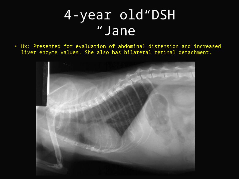
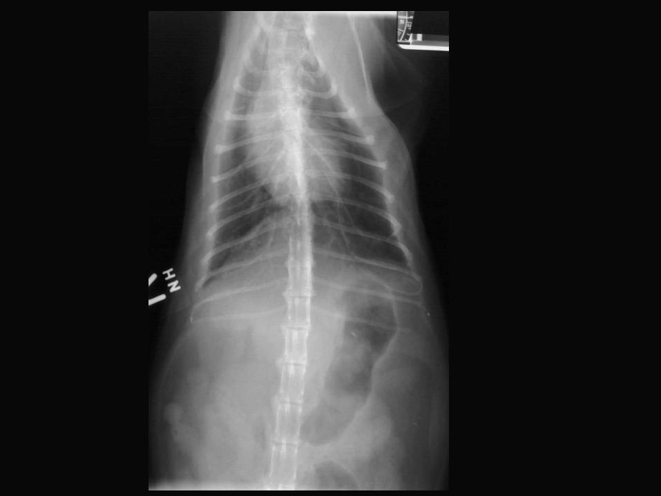
4-year old DSH“Jane”
• Hx: Presented for evaluation of abdominal distension and increased liver enzyme values. She also has bilateral retinal detachment.
4-year old DSH“Jane”
• RF– There is a diffuse interstitial lung pattern present. – There is areas of peribronchial distribution of the interstitial infiltrate are
appreciated.– In the VD view there is tenting of the diaphragm and in the lateral view the
diaphragm is flatter than normal. – Liver extends beyond the costal arch
• RD– Diffuse pulmonary pattern– Thoracic hyperexpansion– Hepatomegaly or could be due to pulmonary hyperinflation
• R/O– Feline airway disease (Asthma)
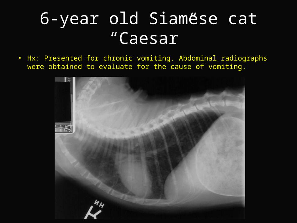
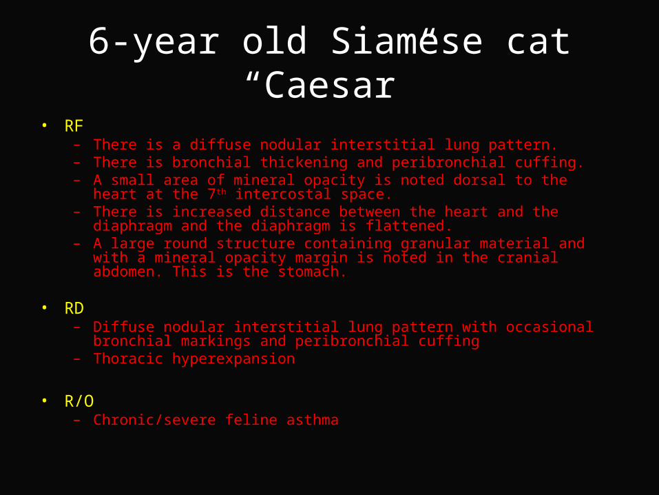
6-year old Siamese cat“Caesar”
• Hx: Presented for chronic vomiting. Abdominal radiographs were obtained to evaluate for the cause of vomiting.
6-year old Siamese cat“Caesar”
• RF– There is a diffuse nodular interstitial lung pattern.– There is bronchial thickening and peribronchial cuffing.– A small area of mineral opacity is noted dorsal to the heart at the 7th intercostal
space.– There is increased distance between the heart and the diaphragm and the
diaphragm is flattened.– A large round structure containing granular material and with a mineral opacity
margin is noted in the cranial abdomen. This is the stomach.
• RD– Diffuse nodular interstitial lung pattern with occasional bronchial markings and
peribronchial cuffing– Thoracic hyperexpansion
• R/O– Chronic/severe feline asthma
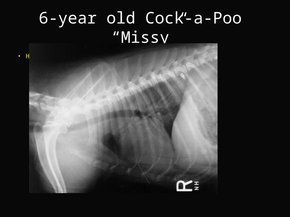
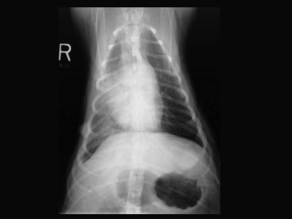
6-year old Cock-a-Poo “Missy”
• Hx: Was hit by a car.
6-year old Cock-a-Poo “Missy”
• RF– Increased opacity and air-bronchograms are present in the region of the right cranial, right
middle lung lobe and the cranial portion of the right caudal lung lobe.– There is slight retraction of the right cranial lung lobe and two faint pleural fissure lines are
seen.– The heart is shifted slightly to the right.– In the Vd view there is increased thickness of the soft tissues along the right lateral thoracic
wall.– A rounded soft tissue opacity structure is seen on the right side at the 6 th intercostal space
and cranial to the stomach. The clear margins of this structure indicate that it is surrounded by air and therefore on the outer surface of the animal, may be enlarged mammary glands.
– In the lateral view there is a mineral opacity structure in the dorsal thorax at the 8 th intercostal space.
– There are fractures of the 8th to 13th ribs on the left side.
• RD– Alveolar pattern of the right cranial, right middle and right caudal lung lobes due to
pulmonary contusions– Mild cardiac shift– Fractured left 8th through 13th ribs– Swelling of the right lateral thoracic wall
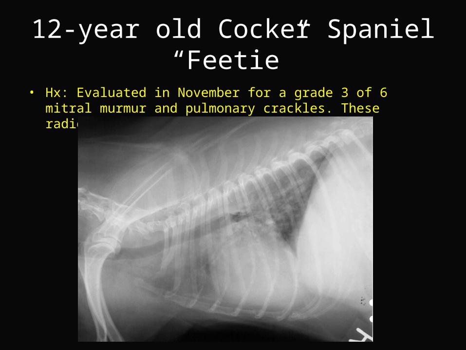
12-year old Cocker Spaniel“Feetie”
• Hx: Evaluated in November for a grade 3 of 6 mitral murmur and pulmonary crackles. These radiographs were obtained in January.
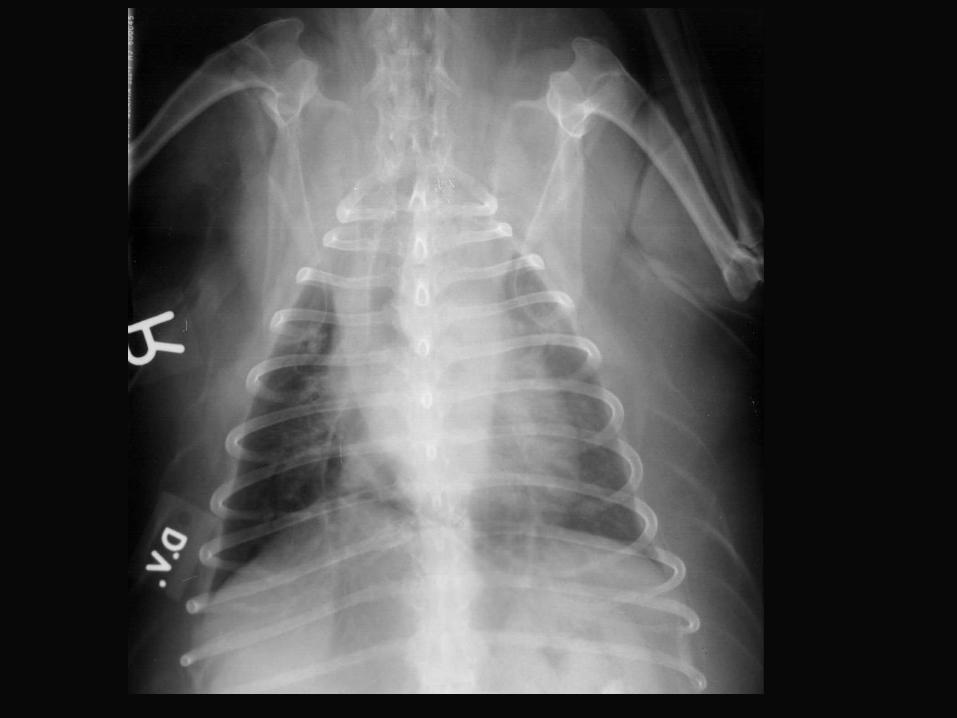
12-year old Cocker Spaniel“Feetie”
• RF– There is increased length of the caudal border of the heart and elevation of the mainstem
bronchi.– There is mild widening of the heart in the lateral view and an increase in sternal contact.– The pulmonary vessels are slightly small (the dog is currently on Lasix)– There is a moderate diffuse increase in pulmonary opacity and bronchial markings are
prominent.– The cranial mediastinum is 2x the width of the vertebral bodies on the DV view. This is the
result of fat deposition.
• RD– Moderate generalized cardiomegaly
• Left-sided enlargement predominates– Diffuse bronchointerstitial lung pattern
• R/O– Mitral valve endocardiosis and insufficiency – Incidental pulmonary changes due to normal aging process– Cardiogenic interstitial pulmonary edema
• Next– Echocardiography