radiographic (x-ray) films - xraykamarul · pdf filehdr 104 chapter 3 radiographic equipment...
TRANSCRIPT
HDR 104
CHAPTER 3
RADIOGRAPHIC EQUIPMENT AND IMAGE RECORDING 1
RADIOGRAPHIC (X-RAY) FILMS
PREPARED BY:MR KAMARUL AMIN BIN ABDULLAH
SCHOOL OF MEDICAL IMAGINGFACULTY OF HEALTH SCIENCE

Slide 2 of 52
TOPIC
CHAPTER 3: Radiographic (X-ray) Films
LEARNING OUTCOMES
At the end of the lesson, the student should be able to:-
Describe types of medical imaging films
Describe the usage and difference of films
Define of monochromatic, orthochromatic and panchromatic
Explain the process of identifying emulsion side for a single emulsion film
Explain the emulsion formation process and film packing
Describe the type of grains in emulsion
Explain problems associated with films and methods of overcoming them
Explain Quality Assurance (QA) test
Compare the relative speed of two films
Slide 3 of 52
TOPIC
CHAPTER 3: Radiographic (X-ray) Films
3
What is X-ray Film?
X-ray film is a photographic receptor consisting of photographically active or
radiation sensitive emulsion coated on a thin sheet like material.
It is responsible to record the physical impression of an object by which we
can get detail about the object.
Slide 4 of 52
TOPIC
CHAPTER 3: Radiographic (X-ray) Films
Classification of the Films
4
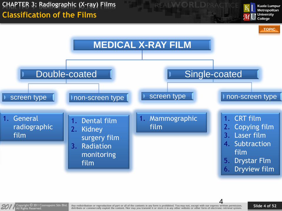
MEDICAL X-RAY FILM
Double-coated
screen type non-screen type
Single-coated
screen type non-screen type
1. General
radiographic
film
1. Dental film
2. Kidney
surgery film
3. Radiation
monitoring
film
1. Mammographic
film
1. CRT film
2. Copying film
3. Laser film
4. Subtraction
film
5. Drystar Flm
6. Dryview film
Slide 5 of 52
TOPIC
CHAPTER 3: Radiographic (X-ray) Films
Direct Exposure Film
Used without intensifying screens.
Used mainly for extremities, previously for mammography.
Requires 10 – 100 times more the exposure dose.
The emulsion is thicker than screen film.
Renders excellent detail.
5
Slide 6 of 52
TOPIC
CHAPTER 3: Radiographic (X-ray) Films
Indirect Exposure Film
These films are used in conjunction with pairs of I.S.
The latent image being produced mainly by light emission from screen
phosphors.
A wide range of different films are available both the blue- sensitive and
green - sensitive.
6
Slide 7 of 52
TOPIC
CHAPTER 3: Radiographic (X-ray) Films
0
2
4
6
8
10
12
14
Series 3
Series 2
Series 1
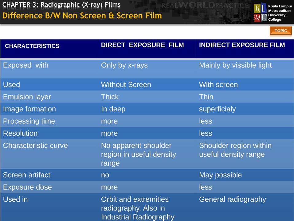
CHARACTERISTICS DIRECT EXPOSURE FILM INDIRECT EXPOSURE FILM
Exposed with Only by x-rays Mainly by vissible light
Used Without Screen With screen
Emulsion layer Thick Thin
Image formation In deep superficialy
Processing time more less
Resolution more less
Characteristic curve No apparent shoulder
region in useful density
range
Shoulder region within
useful density range
Screen artifact no May possible
Exposure dose more less
Used in Orbit and extremities
radiography. Also in
Industrial Radiography
General radiography
Difference B/W Non Screen & Screen Film
Slide 8 of 52
TOPIC
CHAPTER 3: Radiographic (X-ray) Films
Type Of Direct Exposure Film
1. Dental Film
2. Kidney Surgery Film
3. Radiation Monitoring Film
4. Industrial Film
8
Slide 9 of 52
TOPIC
CHAPTER 3: Radiographic (X-ray) Films
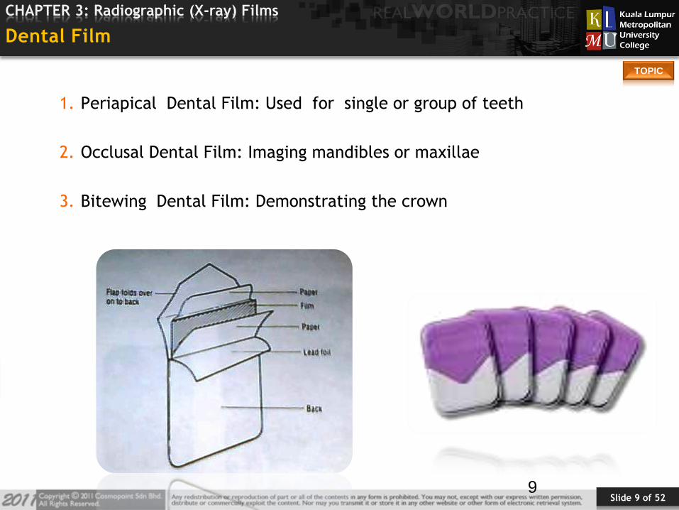
Dental Film
1. Periapical Dental Film: Used for single or group of teeth
2. Occlusal Dental Film: Imaging mandibles or maxillae
3. Bitewing Dental Film: Demonstrating the crown
9
Slide 10 of 52
TOPIC
CHAPTER 3: Radiographic (X-ray) Films
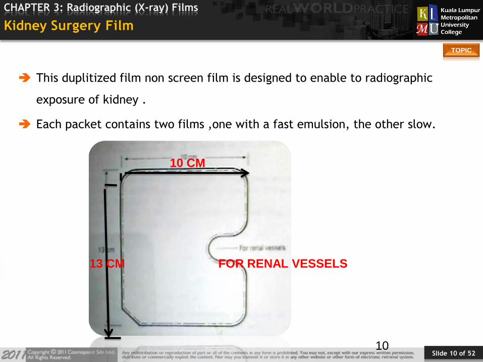
Kidney Surgery Film
This duplitized film non screen film is designed to enable to radiographic
exposure of kidney .
Each packet contains two films ,one with a fast emulsion, the other slow.
10
10 CM
13 CM FOR RENAL VESSELS
Slide 11 of 52
TOPIC
CHAPTER 3: Radiographic (X-ray) Films
Laser Film
A laser printer uses digital electronic signal from an imaging device.
It is high-contrast single-emulsion film with extremely fine grain, also known
as IR film.
Laser film is a silver halide film sensitized red light (Panchromatic) or laser
light, e.g., HN Laser Film, IR Laser Film.
11

Slide 12 of 52
TOPIC
CHAPTER 3: Radiographic (X-ray) Films
Films Used With Cathode Ray Tube OR TV Monitor
These films are used with cathode ray tube camera and multi-formatter.
The emulsions are orthochromatic of medium to high contrast and made
to match a wide variety of CRT phosphor.
The film sizes commonly used are 8” x 10”, 11”x14” and 14”x17”.
Used in following modalities:
1. Ultrasound
2. Computerized tomography
3. Magnetic resonance imaging
4. Nuclear medicine
5. Digital subtraction imaging
12
Slide 13 of 52
TOPIC
CHAPTER 3: Radiographic (X-ray) Films
• A type of single emulsion film used with angiography.
• One type prepares a positive copy of the image.
• The other type enhances subject contrast and detail.
13
• It is used to duplicate the pre-existing film.
• Duplicating film is a single emulsion film that is exposed to ultraviolet.
light or Visible light through existing radiograph to produce a copy.
Duplicating Film
Substraction film
Slide 14 of 52
TOPIC
CHAPTER 3: Radiographic (X-ray) Films
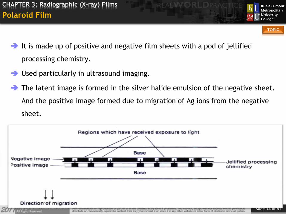
Polaroid Film
It is made up of positive and negative film sheets with a pod of jellified
processing chemistry.
Used particularly in ultrasound imaging.
The latent image is formed in the silver halide emulsion of the negative sheet.
And the positive image formed due to migration of Ag ions from the negative
sheet.
14

Slide 15 of 52
TOPIC
CHAPTER 3: Radiographic (X-ray) Films
The Dry View Film
High quality silver based material coated.
The heat /laser light sensitive layer contains silver halide /silver behnate
crystal.
DRYVIEW Film also a type of laser film having high-resolution,
It is infra red sensitive photothermographic film that needs no wet film
processor.
15
Slide 16 of 52
TOPIC
CHAPTER 3: Radiographic (X-ray) Films
The Drystar Film
Direct thermal printing Drystar dry imaging films are designed to produce the
highest diagnostic grayscale hardcopies. These images can represent the same
"look and feel" as conventional x-ray film.
Blue base
Maximum optical density > 3.5
Daylight film loading (films are insensitive to light)
Shelf life: to be used min. 18 months from packaging date
Storage temperature: 5 - 25 °C
Relative humidity: 30 - 60%
Extended term storage: minimum 20 years
16
Slide 17 of 52
TOPIC
CHAPTER 3: Radiographic (X-ray) Films
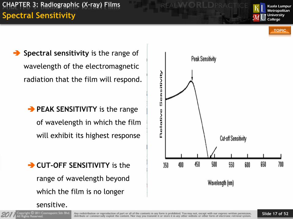
Spectral Sensitivity
Spectral sensitivity is the range of
wavelength of the electromagnetic
radiation that the film will respond.
PEAK SENSITIVITY is the range
of wavelength in which the film
will exhibit its highest response
CUT-OFF SENSITIVITY is the
range of wavelength beyond
which the film is no longer
sensitive.
Slide 18 of 52
TOPIC
CHAPTER 3: Radiographic (X-ray) Films
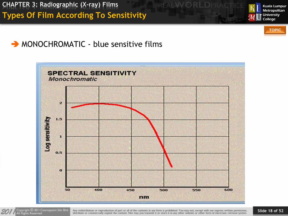
Types Of Film According To Sensitivity
MONOCHROMATIC - blue sensitive films

Slide 19 of 52
TOPIC
CHAPTER 3: Radiographic (X-ray) Films
ORTHOCHROMATIC - green sensitive film
Slide 20 of 52
TOPIC
CHAPTER 3: Radiographic (X-ray) Films
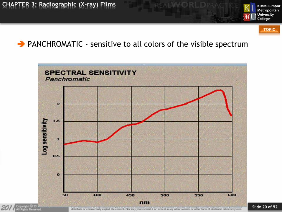
PANCHROMATIC - sensitive to all colors of the visible spectrum
Slide 21 of 52
TOPIC
CHAPTER 3: Radiographic (X-ray) Films
Layers of Radiographic Film
1. Base
2. Subbing layer (Adhesive)
3. Emulsion layer
4. Supercoat
21
Slide 22 of 52
TOPIC
CHAPTER 3: Radiographic (X-ray) Films
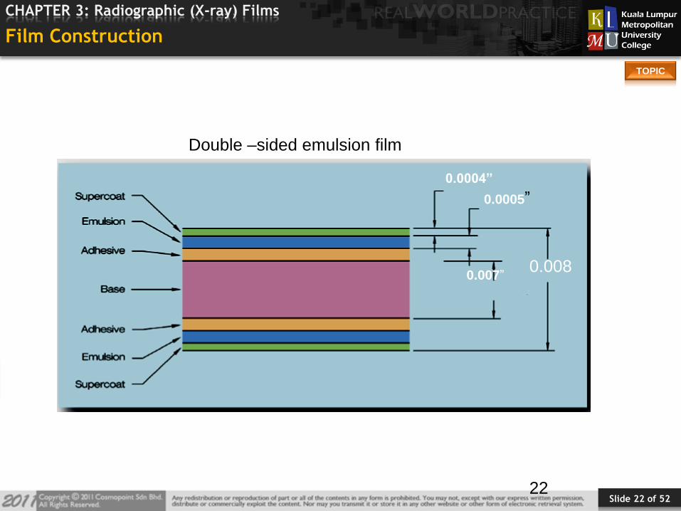
Film Construction
22
0.0004”
0.0005”
0.007”0.008
TOTAL FILM THICKNESS=0.008 INCH
Double –sided emulsion film

Slide 23 of 52
TOPIC
CHAPTER 3: Radiographic (X-ray) Films
2323
0.0004”
0.0005”
0.007”0.008
Anti –Halation /non curl backing
Single –sided emulsion film

Slide 24 of 52
TOPIC
CHAPTER 3: Radiographic (X-ray) Films
0
2
4
6
8
10
12
14
Series 3
Series 2
Series 1
Characteristic Single coated Double coated
Emulsion layer One side Both side
Patient Radiation dose More Less
Noncurl back layer Present Absent
Radiographic detail More Less
Average gradient (G) Very less more
Parallax effect No yes
Contrast Less more
Difference b/w Single Coated And Double Coated X-ray Film
Slide 25 of 52
TOPIC
CHAPTER 3: Radiographic (X-ray) Films
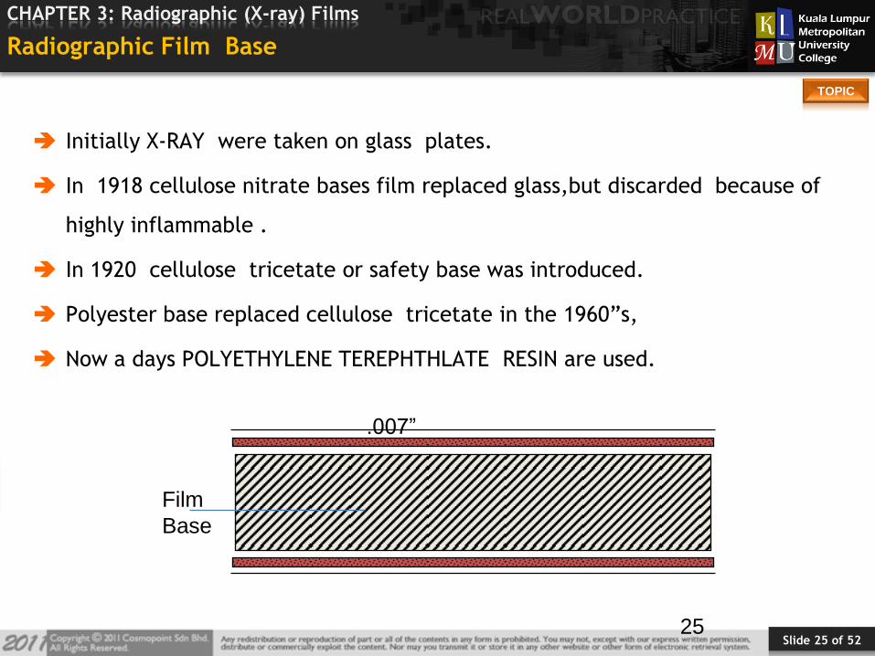
Radiographic Film Base
Initially X-RAY were taken on glass plates.
In 1918 cellulose nitrate bases film replaced glass,but discarded because of
highly inflammable .
In 1920 cellulose tricetate or safety base was introduced.
Polyester base replaced cellulose tricetate in the 1960”s,
Now a days POLYETHYLENE TEREPHTHLATE RESIN are used.
25
Film
Base
.007”

Slide 26 of 52
TOPIC
CHAPTER 3: Radiographic (X-ray) Films
Character of Good Base Material
structural support for fragile emulsion
low light absorption
flexible, thick, & strong
processing
handling
viewbox insertion / removal abuse
dimensional stability
in processing
For archival
varying humidity
NON -FLAMMABLE
26
Slide 27 of 52
TOPIC
CHAPTER 3: Radiographic (X-ray) Films
Function of Base
Provide support for emulsion layer.
To transmit light.
27
Slide 28 of 52
TOPIC
CHAPTER 3: Radiographic (X-ray) Films
Subbing Layer (Adhesive Layer)
Also called Adhesive layer or Substratum layer.
Made of mixture of gelatin solution and solvent of film base.
It keeps emulsion layer and base adhered to each other during coating stage
and processing.
When dye is added, it counteracts cross over effect.
Provides uniform surface over which the emulsion can be coated uniformly.
28
Slide 29 of 52
TOPIC
CHAPTER 3: Radiographic (X-ray) Films
Emulsion Layer
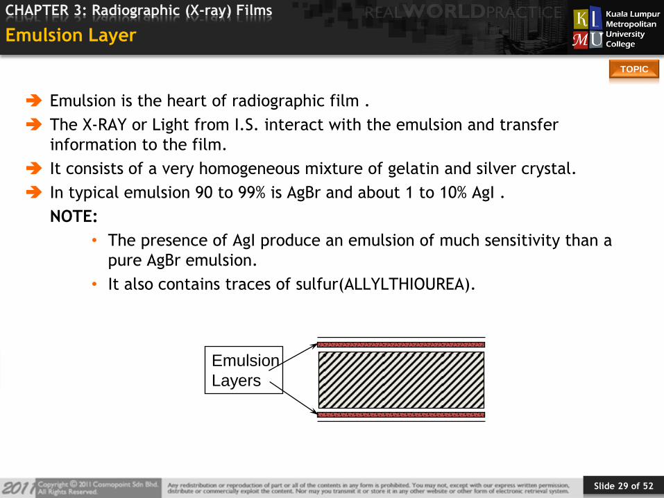
Emulsion is the heart of radiographic film .
The X-RAY or Light from I.S. interact with the emulsion and transfer
information to the film.
It consists of a very homogeneous mixture of gelatin and silver crystal.
In typical emulsion 90 to 99% is AgBr and about 1 to 10% AgI .
NOTE:
• The presence of AgI produce an emulsion of much sensitivity than a
pure AgBr emulsion.
• It also contains traces of sulfur(ALLYLTHIOUREA).
Emulsion
Layers
Slide 30 of 52
TOPIC
CHAPTER 3: Radiographic (X-ray) Films
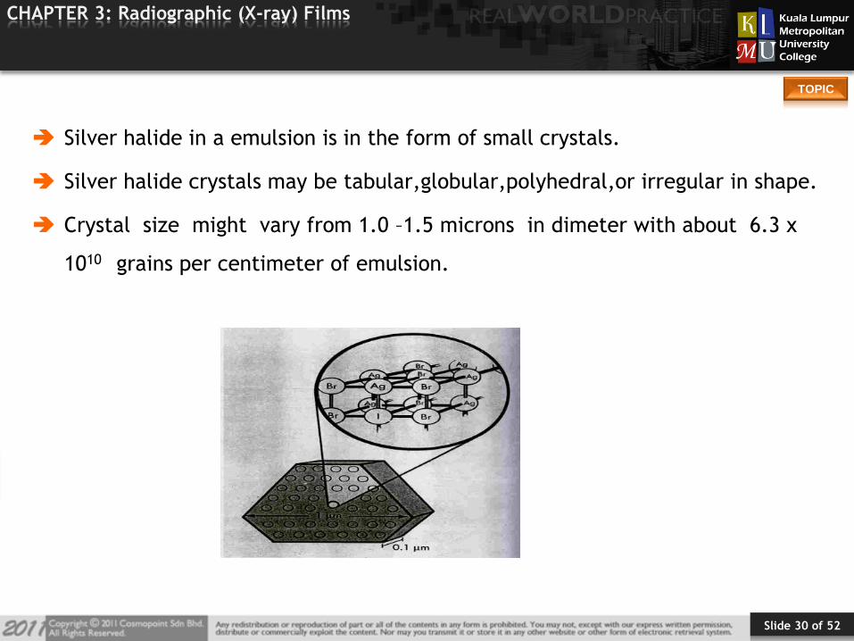
Silver halide in a emulsion is in the form of small crystals.
Silver halide crystals may be tabular,globular,polyhedral,or irregular in shape.
Crystal size might vary from 1.0 –1.5 microns in dimeter with about 6.3 x
1010 grains per centimeter of emulsion.
Slide 31 of 52
TOPIC
CHAPTER 3: Radiographic (X-ray) Films
Grain Technology
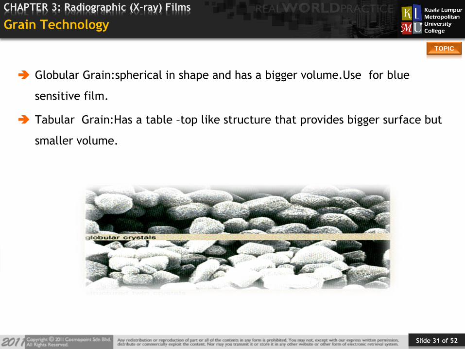
Globular Grain:spherical in shape and has a bigger volume.Use for blue
sensitive film.
Tabular Grain:Has a table –top like structure that provides bigger surface but
smaller volume.
Slide 32 of 52
TOPIC
CHAPTER 3: Radiographic (X-ray) Films
Advantages of Tabular Grain
Increased RESOLUTION due to reduction in cross- over.
Reduction in silver coating weight.
Suitable for 45 s processing.
Slide 33 of 52
TOPIC
CHAPTER 3: Radiographic (X-ray) Films
Grain Size And Distribution
GRAIN SIZE and DISTRIBUTION affects the following:
SPEED: The bigger the average grain size, the higher the speed of the film.
CONTRAST: Affected by size distribution. The more available in the film, the
lower the contrast.
GRAININESS: Graininess is the apparent clumping of the crystal as seen on the
radiograph. The bigger the crystal,the higher the graininess o f the film.
Slide 34 of 52
TOPIC
CHAPTER 3: Radiographic (X-ray) Films
Binder
A binder is an ingredient used to bind together two or more other materials in
mixtures.
The common type of a binder which we can use is Gelatin.
Slide 35 of 52
TOPIC
CHAPTER 3: Radiographic (X-ray) Films
Gelatin
Gelatin is used as the suspending medium and binding agent for the silver
halide particles.
It comes collagen fiber in which primary source are the cartilage, skin and the
protein matrix (ossein) of bone of animals.

Slide 36 of 52
TOPIC
CHAPTER 3: Radiographic (X-ray) Films
Why We Use Gelatin As Binder?
It is a medium in which SILVER NITRATE and SODIUM BROMIDE can react and
the resulting AgBr get finely and evenly dispersed and remain suspended.
In warm state it can be easily spread on the film base.
On cooling, it sets firmly on the base as gel.
Slide 37 of 52
TOPIC
CHAPTER 3: Radiographic (X-ray) Films
Why We Use Gelatin As Binder?
It is flexible and does not crack easily on bending.
It is optically transparent .
Gelatin does not react chemically with the silver halide .
It is porous so the processing chemicals can penetrate to the silver halide
crystals.
Some of the constituents in gelatin enhances the activity of Silver bromide
and some act as antifoggant.
Slide 38 of 52
TOPIC
CHAPTER 3: Radiographic (X-ray) Films
Why We Use Gelatin As Binder?
It is the great advantage of the gelatin in which it can set its intermolecular
space a/c to the condition of the environment, While processing, gelatin
swells up in contact with water, allows processing chemicals to enter the
layer and react with the grains of emulsion, & On drying it regains its former
state.
It is believed that gelatin reduces the tendency of reversal of reaction of
Silver bromide after exposure
Slide 39 of 52
TOPIC
CHAPTER 3: Radiographic (X-ray) Films
Making of The Film Emulsion
The light sensitive layer of a film is termed the Emulsion.
The preparation of emulsion is carried out in four stages:
1. Emulsification
2. Ripening
3. Washing
4. Digestion

Slide 40 of 52
TOPIC
CHAPTER 3: Radiographic (X-ray) Films
Emulsification
Aqueous solution of Silver nitrate and Potassium bromide is mixed with warm
solution of gelatin.
AgNO3 + KBr AgBr + KNO3
Insoluble Silver bromide (AgBr) remains suspended in viscous gelatin.
More rapid process of mixing results small grain size, that results narrow grain
size distribution hence there is low graininess & better resolution.
Note:
More bromide is used to increase the negative charge barrier that helps in
development process.
40

Slide 41 of 52
TOPIC
CHAPTER 3: Radiographic (X-ray) Films
Ripening
Emulsion is placed in certain temperature and more gelatin is mixed. Size of
the grains and their even distribution is determined at this stage
Slow mixing with long ripening at high temp.
=> Fast emulsion (with large grains)
Rapid mixing with short ripening at low temp.
=> Slow emulsion (with fine grains)
Slow mixing with NH3 at low temp.
=> Fast emulsion (with large grains)
41
Slide 42 of 52
TOPIC
CHAPTER 3: Radiographic (X-ray) Films
Washing
After ripening, emulsion is chilled to form thick gel.
This gel is shredded.
It is washed with water that remove KNO3 and excess KBr by diffusion process.
42

Slide 43 of 52
TOPIC
CHAPTER 3: Radiographic (X-ray) Films
Digestion
Shredded and washed emulsion is re-heated to further increase its sensitivity.
Re-heating also make the emulsion liquid and suitable to spread on the film
base.
43

Slide 44 of 52
TOPIC
CHAPTER 3: Radiographic (X-ray) Films
Supercoat (Overcoat)
Protective layer of gelatin
Provides sturdiness to unexposed radiographic film.
Antistatic
Reduces damage from scratches, pressure, or contamination during
storage, handling and processing.
44
Supercoating
Slide 45 of 52
TOPIC
CHAPTER 3: Radiographic (X-ray) Films
Few Additives
Preservative – Phenol as bacteriocide
Silver iodide – To extend sensitivity towards blue range.
Some dyes may extend Colour sensitivity further
Glycerin to make the emulsion pliable
Saponin – To make the emulsion receptive to the processing chemicals
Alcohol – To prevent frothing during coating
45
Slide 46 of 52
TOPIC
CHAPTER 3: Radiographic (X-ray) Films
Coating The Film
Different layers of film are coated on the base material with rollers and
squeezers.
The film lengths are then passed over chilled rollers so that liquefied
gelatinous layers settle and harden.
Then The film lengths are hung like festoons in an air conditioned room to
dry.
Mechanical cutters cut The film lengths in sheets of desirable sizes.
46
Slide 47 of 52
TOPIC
CHAPTER 3: Radiographic (X-ray) Films
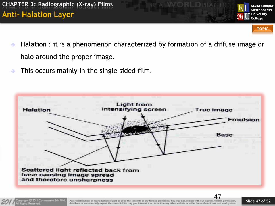
Anti- Halation Layer
Halation : it is a phenomenon characterized by formation of a diffuse image or
halo around the proper image.
This occurs mainly in the single sided film.
47
Slide 48 of 52
TOPIC
CHAPTER 3: Radiographic (X-ray) Films
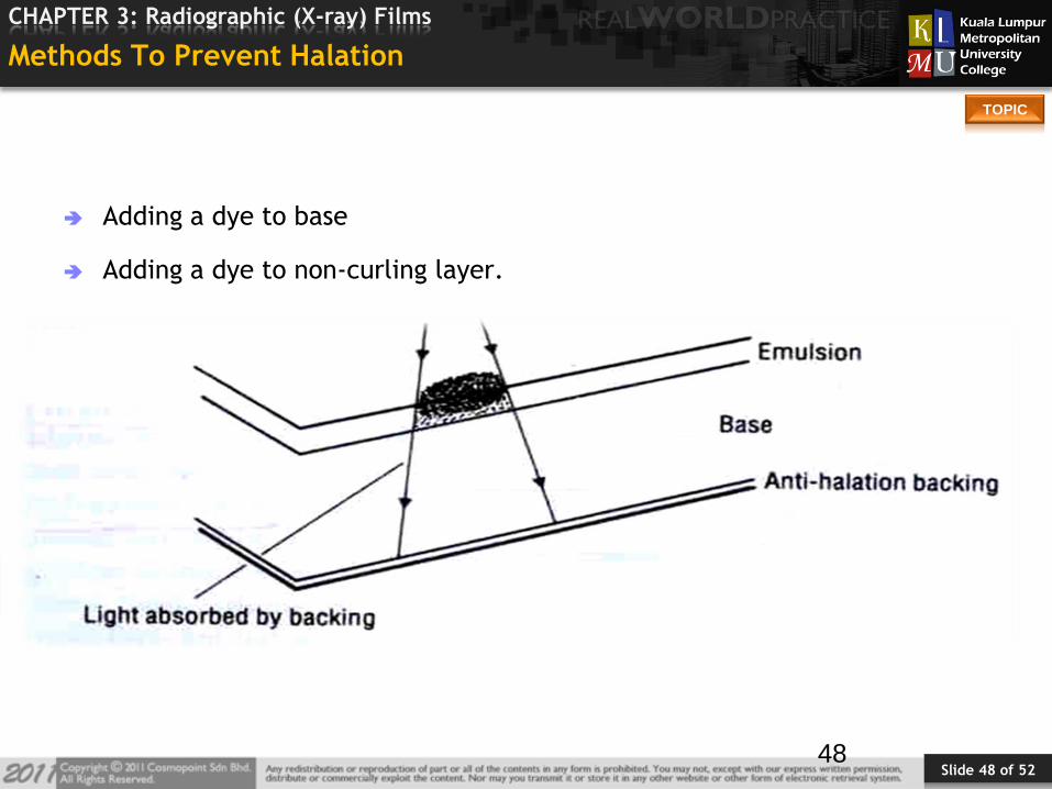
Methods To Prevent Halation
Adding a dye to base
Adding a dye to non-curling layer.
48
Slide 49 of 52
TOPIC
CHAPTER 3: Radiographic (X-ray) Films
Non-curling Layer
Preferred for single sided emulsion film.
This layer is not removed during development.
49

Slide 50 of 52
TOPIC
CHAPTER 3: Radiographic (X-ray) Films
Adding A Dye To Base
These dyes cannot be removed during development.
Dye introduced in the base is carefully controlled because it increase the
density and may interrupt the transparency of the film.
Note-dye used in this should be complementary to the exposing light. e.g.,
red dye is used for green sensitive film, yellow dye is used for blue sensitive
film .
50
Slide 51 of 52
TOPIC
CHAPTER 3: Radiographic (X-ray) Films
Cross Over Effect
It is a type of halation which occurs when film is used with intensifying screen.
Occurs only with double emulsion films and two screens.
Light from one screen expands in the form of a cone as it passes through the
screen and emulsion where a slightly enlarged, less sharp image is formed.
51

Slide 52 of 52
TOPIC
CHAPTER 3: Radiographic (X-ray) Films
Cross Over Effect (Cont’d)
Special dyes incorporated in the emulsion
Colored subbing layer is used.
Addition of magenta dye also reduces cross over effect.
52
Slide 53 of 52
TOPIC
CHAPTER 3: Radiographic (X-ray) Films
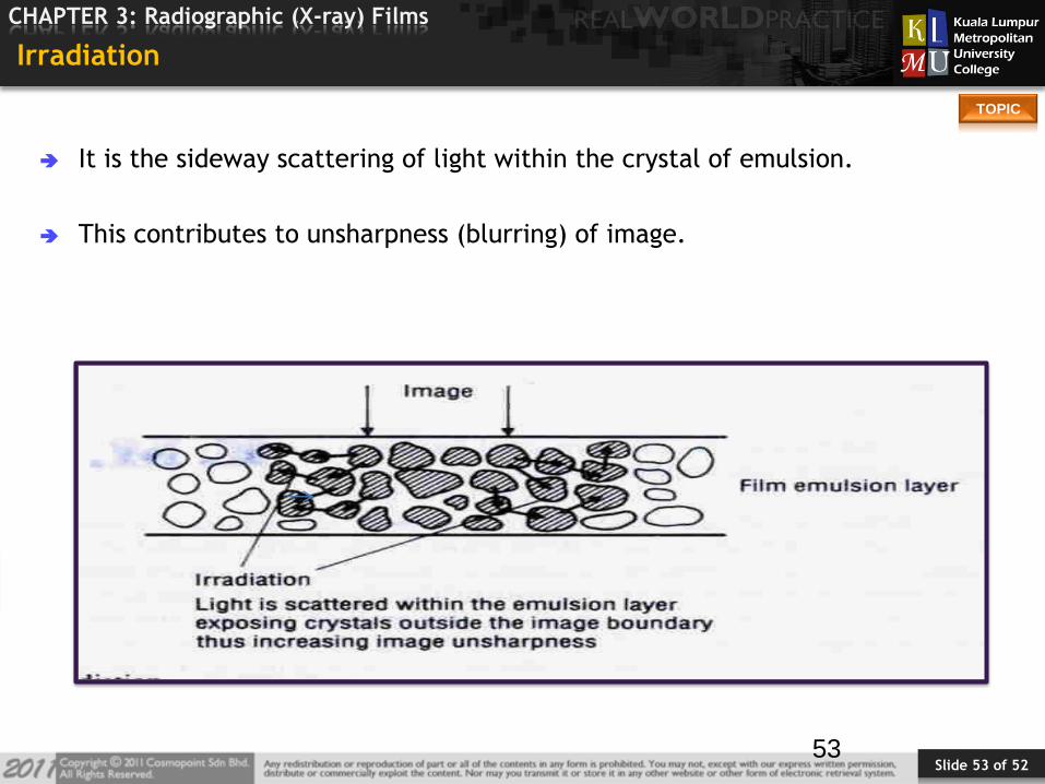
Irradiation
It is the sideway scattering of light within the crystal of emulsion.
This contributes to unsharpness (blurring) of image.
53
Slide 54 of 52
TOPIC
CHAPTER 3: Radiographic (X-ray) Films
How Film Records An Image
There are three steps:-
1. Formation of subject contrast (Optical image)
2. Recording of latent image
3. Conversion of latent image into permanent image (processing)
54

Slide 55 of 52
TOPIC
CHAPTER 3: Radiographic (X-ray) Films
The Latent Image
The latent image is the invisible change in the silver halide crystals.
The interaction between the photons and the silver halide crystals produces
the latent image or manifest image on the emulsion layer.
This interaction is sometimes referred to as the photographic effect.
55

Slide 56 of 52
TOPIC
CHAPTER 3: Radiographic (X-ray) Films
Formation Of Subject Contrast
Subject contrast:- the variation in intensity of x-ray beam after passing the
absorber.
Subject contrast depends upon atomic No., density, thickness of absorber and
the energy of the x-ray beam.
Different intensity of beam react differently with the photographic material
of the film.
56

Slide 57 of 52
TOPIC
CHAPTER 3: Radiographic (X-ray) Films
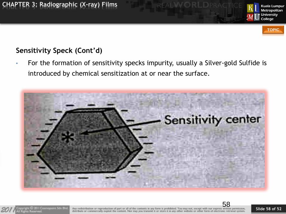
Sensitivity Speck
The shape and lattice structure of silver halide are not perfect.
It causes some imperfection which results in imaging property of crystals.
So the sensitivity specks is that low energy centre of the crystal which acts as
rest house for the 1º electron and development centre for the 2º electron.
57
Slide 58 of 52
TOPIC
CHAPTER 3: Radiographic (X-ray) Films
Sensitivity Speck (Cont’d)
• For the formation of sensitivity specks impurity, usually a Silver-gold Sulfide is
introduced by chemical sensitization at or near the surface.
58
Slide 59 of 52
TOPIC
CHAPTER 3: Radiographic (X-ray) Films
Sensitivity Speck (Cont’d)
The image forming x-rays deposit energy by photoelectric interaction with
atoms of silver halide crystals.
Formation of latent image is given by Gurney-Mott theory
59
Slide 60 of 52
TOPIC
CHAPTER 3: Radiographic (X-ray) Films
-
+
SENSITIVITY SPECK
SILVER HALIDE CRYSTAL
INTERSTITIAL Ag ION
1. Photon Absorption
6. Ag Ion Migration
3. Ag Ion Migration
2. Electron Trapping
+
-
4. Photon Absorption
-+
+
5. Electron Trapping
-

Slide 61 of 52
TOPIC
CHAPTER 3: Radiographic (X-ray) Films
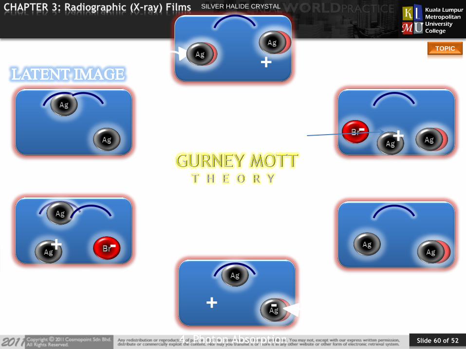
Producing the Latent Image
Radiation interaction releases electrons.
Electrons migrate to the sensitivity center.
At the sensitivity center, atomic silver is formed by attracting an interstitial
silver ion.
The process is repeated many times resulting in the build up of silver atoms.
The remaining silver halide is converted to silver during processing.
The resulting silver grain is formed.
Silver halide that is not irradiated remain inactive. The irradiated and non-
irradiated silver halide produces the latent image.
61
Slide 62 of 52
TOPIC
CHAPTER 3: Radiographic (X-ray) Films
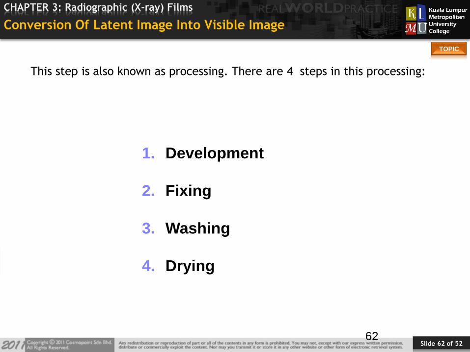
Conversion Of Latent Image Into Visible Image
This step is also known as processing. There are 4 steps in this processing:
62
1. Development
2. Fixing
3. Washing
4. Drying
Slide 63 of 52
TOPIC
CHAPTER 3: Radiographic (X-ray) Films
Characteristics To Be Considered While Selecting Film:
Contrast
Speed
Crossover
Spectral matching
Bulk of purchase
Time of purchase
63
Slide 64 of 52
TOPIC
CHAPTER 3: Radiographic (X-ray) Films
Care And Protection Of Film
Films should be protected from:-
1. Physical damage
2. Light
3. High temperature
4. High relative humidity
5. Harmful gases and fumes
6. X-rays and radioactive source
7. Fire and theft
64
Slide 65 of 52
TOPIC
CHAPTER 3: Radiographic (X-ray) Films
Resolving Power of Films
Ability of a photographic emulsion to record fine details
It is expressed as the number of line pairs per millimeter which can be
distinguished in the image as separate entities
Factors affecting the resolution of an image are – Grain
size, Processing, Diffusion of light inside the emulsion layer and Modular
transfer function
65

Slide 66 of 52
TOPIC
CHAPTER 3: Radiographic (X-ray) Films
Line Pairs Per Millimeter
A black and a white line make a line pair
A test pattern of slits cut on a metal plate with gradual fineness is
exposed, processed and evaluated under magnification.
Radiographic emulsions show 8 – 20 LP/mm
Photographic Fast emulsions show 40 – 50 LP/mm
• Medium emulsions show 70 – 100 LP/mm
• Slow emulsions show over 1000 LP/mm
66
Slide 67 of 52
TOPIC
CHAPTER 3: Radiographic (X-ray) Films
STORAGE AREAS :-
The hospital or x-ray department
The dark room
The imaging room
Storage Of Film
67
Slide 68 of 52
TOPIC
CHAPTER 3: Radiographic (X-ray) Films
Handling and Storage Of Radiographic Film
X-ray film is a sensitive radiation detector and it must be handled in an area
free of radiation.
Film storage must be shielded.
The darkroom adjacent to the x-ray room must be shielded.
If film use is low more shielding may be required.
68

Slide 69 of 52
TOPIC
CHAPTER 3: Radiographic (X-ray) Films
Handling and Storage of Radiographic Film
Improper handling of the film will result in poor image quality due to
artifacts.
Avoid bending, creasing or otherwise rough handling the film. Avoid sharp
objects contacting the film.
Hands must be clean and dry.
Avoid hand creams, lotions or water free hand cleaners.
Static electricity or a dirty processor can cause artifacts.
Artifacts must be avoided.
69
Slide 70 of 52
TOPIC
CHAPTER 3: Radiographic (X-ray) Films
Handling and Storage Of Radiographic Film
Heat and Humidity must be controlled. Film is sensitive to heat and humidity
from the time it is manufactured until the time it is viewed.
Heat and humidity causes fog or a loss of contrast. Film should be stored at
20º C (68º F).
Humidity should be between 40% and 60%.
70
Slide 71 of 52
TOPIC
CHAPTER 3: Radiographic (X-ray) Films
Handling and Storage Of Radiographic Film
Light will expose the film. Film must be handled and stored in dark.
If low level diffuse light exposes the film, fog is increased.
Luminous watches, cell phone and darkroom light leaks should be avoided.
Bright light causes gross exposure.
71

Slide 72 of 52
TOPIC
CHAPTER 3: Radiographic (X-ray) Films
Handling and Storage Of Radiographic Film
Shelf life. All film is supplied in boxes with an expiration date.
Most film is supplied in boxes of 100 sheets.
The oldest film in stock should always be used first. Rotation is important.
Expired will loose speed and contrast and have increased fog.
72
Slide 73 of 52
TOPIC
CHAPTER 3: Radiographic (X-ray) Films
References
No. REFERENCES
1 Richard R. Carlton, Arlene McKenna Adler (2005) Principles of
Radiographic Imaging, Delmar
2 Bushong, S. C. (2008). Radiologic science for technologists. Canada:
Elsevier.