radiation protection by mnsod-plasmid liposome … radiation protection by mnsod-plasmid liposome...
TRANSCRIPT

387D.R. Spitz et al. (eds.), Oxidative Stress in Cancer Biology and Therapy, Oxidative Stress in Applied Basic Research and Clinical Practice, DOI 10.1007/978-1-61779-397-4_19, © Springer Science+Business Media, LLC 2012
Abstract Understanding the molecular mechanism of ionizing irradiation killing of normal tissues compared to tumor cells has uncovered potential therapeutic strate-gies for radiotherapeutic dose escalation in the treatment of solid tumors. One strategy exploits the difference in redox balance between normal tissues and solid tumors with organ-specifi c and systemic (intravenous) administration of plasmid liposomes containing the human manganese superoxide dismutase (MnSOD) transgene. When delivered, this transgene product produced signifi cant protection against single fraction and fractionated radiation in organ-specifi c radioprotective gene therapy by each of several routes of administration (inhalation, swallowed, intravesicle, intra-intestinal), and this strategy was shown to avoid tumor tissue. Furthermore, systemic adminis-tration of MnSOD-PL also provides radioprotection with selective normal tissue reduction of apoptosis compared to tumor. The complex mechanism of selective normal tissue protection involves specifi c differences in redox balance, constitutive MnSOD expression, and compensatory metabolic changes, many of which can enhance the therapeutic effect.
J. S. Greenberger, MD (*) • M. W. Epperly, PhD Department of Radiation Oncology , University of Pittsburgh School of Medicine , 200 Lothrop Street , Pittsburgh , PA 15213 , USA e-mail: [email protected]
V.E. Cagan, PhD Center for Free Radical and Antioxidant Health, Department of Environmental and Occupational Health , University of Pittsburgh , Pittsburgh , PA , USA
J. Peterson, PhD Department of Environmental and Occupational Health , University of Pittsburgh , Pittsburgh , PA, USA
Chapter 19 Radiation Protection by MnSOD-Plasmid Liposome Gene Therapy
Joel S. Greenberger , Valerian E. Cagan , James Peterson , and Michael W. Epperly

388 J.S. Greenberger et al.
19.1 Introduction
Improvement in clinical radiotherapy over the past several decades has relied heavily upon treatment planning techniques and radiotherapy delivery techniques that enhance tumor-specifi c killing and minimize normal tissue dose [ 1– 4 ] . These advances include development of three-dimensional conformal radiotherapy techniques, based upon sophisticated CAT, MRI, PET, and combined imaging tech-niques [ 5 ] ; Intensity Modulated Radiotherapy Treatment (IMRT) beams in which moving multileaf collimator components are used during treatment delivery [ 6 ] ; alternative fractionation techniques; alterations in dose rate and beam energy [ 7 ] ; the development of the Gamma Knife as well as advanced frameless stereotactic treatment devices, and brachytherapy techniques including radioactive seed and radioactive mesh implantation; and high dose rate brachytherapy [ 8– 12 ] . Despite tremendous improvements in radiotherapy treatment planning and beam delivery, normal tissue toxicity remains dose limiting to most tumor targets [ 2– 4, 13 ] .
The therapeutic ratio (ratio of tumor killing over normal tissue toxicity) remains a key factor limiting the development of radiotherapy treatment plans [ 1 ] . Combining ionizing irradiation with new chemotherapeutic agents, many of which are normal tissue radiosensitizers, further limits radiotherapy dose escalation [ 14 ] . The develop-ment of tumor-specifi c radiosensitizers has been one approach toward increasing the therapeutic ratio [ 1 ] . This approach has been limited by the incomplete targeting through intra-arterial or intra-tumor injection of specifi c radiosensitizing agents such as bromadeoxyuridine (BUDR) [ 1 ] or in the case of p53-defi cient tumors, p53-transgene therapy [ 1 ] . One successful approach has been the development of hypoxic cell cytotoxins. Tirapazamine relies upon the hypoxic environment in large epithelial tumors to provide activation of the pro-drug into a cytotoxic moiety [ 14 ] . The use of hypoxic cell cytotoxins has been particularly attractive in head and neck cancer and lung cancer where large hypoxic areas exist in rapidly growing tumors [ 15 ] .
We have developed an alternative approach for improving the therapeutic ratio by decreasing normal tissue irradiation damage through a technique of antioxidant gene therapy [ 16– 19 ] . Tissue culture laboratory and animal preclinical trials dem-onstrated the importance of mitochondrial targeting of the antioxidant therapeutic agent in optimizing therapeutic effect [ 20– 24 ] . This chapter will review molecular biologic, biochemical, cell biologic, and the tissue- and organ-specifi c mechanisms of radioprotection by MnSOD transgene therapy and highlights areas for potential additional improvement of this clinical therapeutic strategy.
19.1.1 Antioxidant Gene Therapy Compared to Direct Delivery of Antioxidants
Initial discoveries in the fi elds of radiation chemistry identifi ed cellular oxygen and water as primary targets for the creation of free radicals also called radical oxygen species (ROS) [ 1, 25– 30 ] . Hydroxyl radical and superoxide were identifi ed as active

38919 Radiation Protection by MnSOD-Plasmid Liposome Gene Therapy
moieties that combine to purine and pyrimidine bases in nuclear DNA. Alkaline sucrose gradient biochemistry techniques analyzing removed nuclei from irradiated cells identifi ed single- and double-strand breaks in DNA and fi rst linked the irradia-tion chemistry events of ionizing irradiation with the molecular biologic changes in irradiated cells [ 1 ] . The kinetics of restoration of DNA integrity after these single- and double-strand breaks led the way toward identifi cation of DNA repair enzymes [ 31– 35 ] and repair enzyme defi cient conditions that were associated with increased radiosensitivity of cells in culture [ 36– 44 ] . These initial studies pointed the way toward development of radioprotective agents by suggesting the possibility that immediate capture, neutralization, or inactivation of irradiation-induced free radicals might result in signifi cant radioprotection [ 1, 45 ] . The development of WR2721 (ethyol, Amifostine), a normal tissue radioprotector, and N-acetylcystine (a sulfhy-dryl compound) both of which act though reactive oxygen species (ROS) or free radical scavenging [ 46 ] serve as two examples of the applications of free radical scavenging [ 47 ] .
During the development of radioprotective free radical scavengers, and identifying the mechanism of action of these compounds, it was quickly appreciated that levels of total cellular antioxidant capacity dropped rapidly after exposure of cells to ionizing irradiation [ 1, 48– 50 ] . It was learned that preservation of the total antioxi-dant cellular levels could be radioprotective [ 49, 50 ] . Paramount in the list of early identifi ed antioxidant compounds was glutathione itself, a free radical scavenger [ 1, 51 ] . Under conditions of lowered glutathione levels, cells were more radiosensitive [ 1, 51 ] . It followed that by adding new free radical scavengers, the consumption of glutathione from the antioxidant pool would be reduced and this would render cells more capable of neutralizing additional irradiation-produced free radicals [ 48 ] .
Radiation biologists studied the kinetics of production of free radicals following irradiation of cells in culture and defi ned several parameters of cellular, tissue, and organ response, which further complicated the strategy of using antioxidant drugs as radioprotectors [ 1 ] . First, irradiation induction of free radicals was found to continue long after irradiation [ 1, 16– 18, 52, 53 ] , and secondly ROS production could be detected outside the irradiated target volume [ 52, 53 ] . These data uncovered the phenomena of irradiation-induced production of cytokines [ 54, 55 ] , which contributed to the delayed toxicity in adjacent or distant sites. The data also defi ned the phenomenon of the “bystander effect,” which included both cell contact and humoral transmission of irradiation damage and free radical induction (ROS) to other cells [ 56 ] . Finally, the mechanism of action of irradiation induced ROS was not limited to nuclear DNA strand breaks and included the oxidation of lipids in cell membranes, most prominently those in the mitochondria [ 49, 50 ] .
Basic radiation chemistry also revealed other natural cellular antioxidant defense mechanisms separate from the antioxidant pool of free radical scavenging small molecules. These studies identifi ed natural radioprotective enzymes including gluta-thione peroxidase, gamma glutamyltranspeptidase, catalase, and the three forms of superoxide dismutases (SODs) [ 26, 27 ] . One form of SOD (MnSOD or SOD2) contains a specifi c 23-amino acid leader sequence that facilitated mitochondrial localization [ 21, 49 ] . In radiation chemistry reactions, superoxide is dismutated by SOD to hydrogen peroxide [ 25 ] , another potent oxidative chemical which itself is

390 J.S. Greenberger et al.
acted upon by catalase to produce benign substances oxygen and water [ 25 ] . The combination of superoxide with nitric oxide, another active free radical, induced in mammalian cells by each of three nitric oxide synthase isoforms, produces the potent free radical peroxynitrite [ 49, 50 ] . Peroxynitrite is one of the most potent inducers of lipid oxidation associated with the formation of the mitochondrial specifi c oxidized lipids [ 48 ] . Conclusions from these observations led to methods by which to reduce the production of peroxynitrite [ 49 ] . The consensus was that reducing levels of peroxynitrite might also decrease ionizing irradiation-induced normal tissue damage [ 48– 50, 57 ] .
Initial translational research experiments by which to adapt these radiation chemistry and cell biology experiments to normal tissue radioprotection in vivo, were largely ineffective. Attempts to deliver MnSOD protein or nitric oxide synthase inhibitor drugs systemically required administration of very high levels to achieve therapeutic effect at the level of the mitochondria [ 58 ] . The therapeutic antioxidant, Tempol, which counteracts both hydroxyl radical and superoxide was only marginally effective in experimental animal studies due to the very high drug dose requirements to gain thera-peutic effect [ 59, 60 ] . This data led to development of techniques to mitochondrially target Tempol and such research not only overcame some of these drug dose problems but further substantiated the importance of the mitochondrial membrane as a key target in prevention of ionizing irradiation-induced apoptosis of normal cells [ 61, 62 ] .
Mitochondrial targeting of MnSOD, through its naturally occurring 23-amino acid mitochondrial localization sequence, combined with the knowledge that very high levels of protein were required to obtain mitochondrial specifi c expression posed the question of whether transgene delivery of the MnSOD cDNA sequence directly to cell nuclei, with a strong promoter to facilitate gene transcription and translation, might be a way to provide adequate mitochondrial targeted levels of protein delivered as such from the “inside out” [ 58 ] . Initial experiments with MnSOD-plasmid liposomes, using a radiosensitive murine hematopoietic progenitor cell line, 32D cl 3, fi rst demonstrated the effectiveness of this approach and also confi rmed the importance of mitochondrial targeting [ 20, 21 ] . Attaching the 23AA mitochondrial localization signal to CU/ZnSOD, a cytoplasmic form of SOD, which itself is non-radioprotective [ 21 ] , had the same effect as MnSOD in radioprotection in vitro [ 21 ] . Conversely, removing the mitochondrial localization sequence from MnSOD produced a non-radioprotective cytoplasmic moiety [ 21 ] . In these experi-ments, mitochondrial targeting of SOD provided signifi cant radioprotection. These experiments were translated in vivo and showed organ-specifi c radioprotection against single fraction and fractionated irradiation [ 21, 63 ] .
19.1.2 The Biochemistry of MnSOD-PL Radioprotective Gene Therapy
Basic biochemistry and radiation chemistry experiments carried out on cells in culture fi rst revealed that 32D cl 3 murine, Interleukin-3 dependent hematopoi-etic progenitor cells, transfected with the MnSOD transgene were signifi cantly

39119 Radiation Protection by MnSOD-Plasmid Liposome Gene Therapy
radioresistant [ 20, 21 ] . The mechanism of this radioresistance was linked to the stabilization of a high level of antioxidant capacity in the cells compared to non-transfected, non-MnSOD-overexpressing cells [ 48 ] . In particular, intracellular levels of glutathione and other radical scavenging antioxidants were shown to persist at higher levels and for longer duration after the cells were exposed to radia-tion compared to parent control cells [ 48 ] .
Cell lines overexpressing MnSOD were compared to parent cell lines with respect to radiation-induced oxidized lipids [ 22, 23 ] . It was discovered that specifi c oxidized lipids, notably phosphatidyl serine and cardiolipin were localized to the mitochondrial membrane in irradiated cells [ 22, 23 ] and the magnitude of lipid oxi-dation was decreased by MnSOD overexpression of clonal cell lines [ 22, 23 ] .
The data on oxidized lipids that demonstrated an increased antioxidant pool size, and decreased susceptibility to specifi c PS and CL lipid oxidation was also detected in experiments in irradiated mouse esophagus [ 64 ] . Esophagus was removed at serial time points after irradiation from mice that received intraesophageal adminis-tration of MnSOD-PL, control liposomes, or no gene therapy. Tissues from animals that received intraesophageal injection of MnSOD-PL showed higher levels of anti-oxidant pool size and decreased lipid oxidation [ 64 ] .
A separate series of experiments demonstrated that treatments that decreased phospholipid peroxidation of the mitochondria also stabilized cytochrome C binding to cardiolipin [ 49, 50 ] .
Cytochrome C, which is a natural and intrinsic component of the mitochondrial respiratory cascade, is nearly 70% bound to cardiolipin in the inner mitochondrial mem-brane [ 49 ] . Thirty percent of cytochrome C is free, but is maintained inside the mito-chondria by an intact mitochondrial membrane. During ionizing irradiation exposure of cells, mitochondrial membrane depolarization is detected, and cytochrome C leaks from the mitochondria into the cytoplasm where it activates caspase-3 leading to cleavage of PARP and subsequent nuclear DNA fragmentation resulting in apoptosis [ 20, 21 ] .
Initial experiments demonstrated that MnSOD-PL treatment prevented cytochrome C leakage out of the mitochondria, but the mechanism was unknown. A separate series of experiments demonstrated that maintaining the antioxidant pool within the mitochondria stabilized cardiolipin binding to cytochrome C and contributed sig-nifi cantly to a decrease in cytochrome C leakage [ 48 ] . Furthermore, it was demon-strated that consumption of antioxidants in the mitochondria led to deleterious effects on one of the most potent enzyme antioxidants, namely MnSOD [ 48 ] . Following ionizing irradiation, MnSOD was demonstrated to be both oxidized and nitrated [ 65 ] . Oxidized MnSOD still retained superoxide dismutation activity, while nitrated MnSOD became less capable of functioning as a superoxide dismutase. Rather, it became a peroxidase causing lipid damage in the mitochondria and lead-ing to cytochrome C leakage and apoptosis [ 65 ] .
In experiments, both in cell lines in vitro and in tissues in vivo including intestine [ 66– 68 ] , ionizing irradiation caused specifi c mitochondrial phospholipid oxidation that was a signature for the oxidative stress induced by ionizing irradiation. MnSOD-PL overexpression in cells and tissues prior to irradiation preserved the antioxidant pool and in some experiments led to a decreased apoptotic response [ 64 ] .

392 J.S. Greenberger et al.
19.2 The Cell Biology of MnSOD-Radioprotective Gene Therapy
Initial experiments were carried out in several preclinical rodent models of irradiation-induced toxicity: esophagitis [ 69 ] , pneumonitis [ 70 ] , cystitis [ 71 ] , intestinal damage [ 72 ] , oral cavity [ 73 ] , and bone marrow failure [ 74 ] . In these models ionizing irradiation was observed to reduce cell numbers, produce apoptosis and ulceration in tissues (in the case of the esophagus), and lead to functional organ inactivation. Irradiation effects on tissues were both direct on cycling cell populations [ 75 ] and indirect via cytokine production [ 76, 77 ] . MnSOD-PL therapy reduced the magnitude of the direct effect by stabilizing the cycling cells. However, it also became evident that irradiation in vivo affected the microenvironment in which proliferating cell populations resided and cytokines were released.
Apoptosis induced by irradiation is known to be more dominant in rapidly cycling cells [ 1 ] . In experiments comparing 32D cl 3 hematopoietic progenitor cells in liquid culture with those in cell pellets, (simulating the hypoxic, contact-inhibited microen-vironment in tissues), irradiation-induced apoptosis was decreased in pelleted cell populations [ 78 ] . Other studies demonstrated that hematopoietic cells in contact with cells of the hematopoietic microenvironment (bone marrow stromal cells) showed reduced ionizing irradiation-induced toxicity both when hematopoietic cells and stromal cells were irradiated together and in experiments in which unirradiated hematopoietic cells were engrafted onto irradiated bone marrow stromal cells [ 79 ] . In both the model of irradiation exposure of both populations and the model of co-cultivation of cell populations with only the stromal cells irradiated, there was decreased toxicity to cycling hematopoietic cell population if MnSOD-PL was added and the total antioxidant pool size was increased [ 48, 80 ] . Therefore, MnSOD-PL treatment of the microenvironment into which restorative and repopulating cells migrate facilitates engraftment [ 80, 81 ] . In recent experiments with chimeric mice having bone marrow replaced with specifi cally marked donor cell populations, it was demonstrated that MnSOD-PL treatment of an epithelial organ (esophagus or lung) facilitated better engraftment of cells of bone marrow origin coming in from outside the irradiation fi eld compared to non-MnSOD-PL treated mice [ 80– 82 ] .
In vitro experiments demonstrated that production of ROS by plateau phase stromal cells increased with irradiation dose [ 79 ] . Antioxidant treatment of irradiated stromal cells decreases ROS production and facilitates improved attachment of engrafted cells in vivo [ 80, 81 ] .
Apoptosis of cells exposed to ionizing irradiation has been shown to be similar to that which occurs in cells deprived of specifi c required growth factors such as IL-3 [ 83 ] . MnSOD-PL overexpression by transgene introduction into IL-3 depen-dent cell facilitated longer duration survival in the absence of IL-3 compared to non-MnSOD overexpressing cells [ 83 ] . These data provide evidence that growth supporting cytokine withdrawal as well as ionizing irradiation induce oxidative stress and depletion of the antioxidant pool in both non-cycling and cycling cells [ 48 ] . Among the non-cycling cells are those of the tissue-specifi c microenvironment.

39319 Radiation Protection by MnSOD-Plasmid Liposome Gene Therapy
The data also demonstrate that non-cycling cells, exposed to irradiation, produce increased levels of ROS and infl ammatory cytokines, such that these cells can induce a toxic indirect effect on homing and engrafting cells. The overexpression of MnSOD in stromal cells prior to irradiation facilitates a reduced depletion of anti-oxidant stores and reduced toxicity of the microenvironment. The data suggest that MnSOD-PL therapy both in vitro and in vivo with respect to stromal cells reduces the irradiated cellular capacity to induce the bystander effect.
19.2.1 MnSOD Radioprotective Gene Therapy Tissue Effects
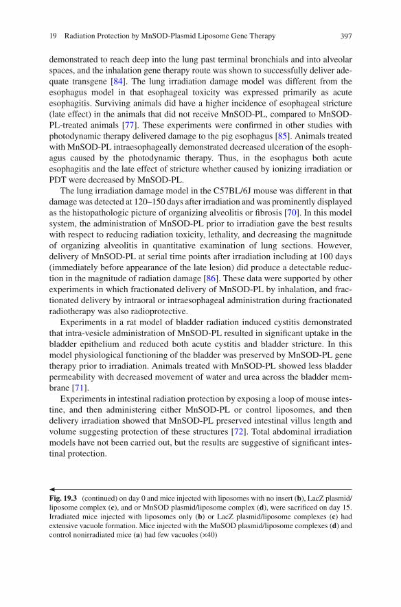
Initial studies in irradiated esophagus quantitated the number of apoptotic bodies and ulcerated areas in the esophagus at serial time points after exposure to single fraction 29 Gy [ 69 ] . In C57BL/6J mice, by 12–14 days after irradiation, signifi cant esophagitis prevented adequate nutrition and animals were observed to be dehy-drated and signifi cantly losing weight. Histopathologic evaluation showed increased apoptotic bodies, microulceration, and signifi cant tissue damage (Figs. 19.1 and 19.2 ). Mice given a single intraesophageal administration of MnSOD-PL, but not empty liposomes, or control LAC-Z transgene-PL showed signifi cant reduction in irradiation-induced apoptosis, improved survival, and the animals showed less dehydration and better weight gain (Fig. 19.3 ; Photographs from [ 69 ] reproduced with permission from Wiley, Inc. Radiation Oncology Investigations ). In addition, apoptotic bodies and microulcerations induced by irradiation were signifi cantly decreased in MnSOD-PL pretreated versus control irradiated tissues [ 80, 81 ] .
These studies in the esophagus demonstrated that MnSOD-PL administration in a single dose or multiple doses during fractionated irradiation resulted in stabilization of the esophageal tissue response, namely expression of cytokine mRNA and elabo-ration of humoral cytokines [ 77 ] . The secondary effects of ionizing irradiation on tissues (cytokine release) were also decreased by MnSOD-PL mediated overexpres-sion of this antioxidant transgene [ 77 ] .
Other tissue-specifi c experiments were carried out with animal models of oral cavity irradiation. In both single fraction and fractionated irradiation to the head and neck region, mice demonstrated signifi cant tongue ulceration, apoptotic bodies in the oral cavity, signifi cant dehydration, and weight loss [ 73 ] . Intraoral administra-tion of MnSOD-PL was demonstrated to introduce transgene deep within the oral cavity tissues [ 73 ] . An epitope-tagged hemagglutinin-tagged MnSOD was utilized to facilitate histochemistry. Histochemical staining showed that cells deep in the tissue going down to the stem cell or basal cell layer were reached by intraoral MnSOD-PL gene therapy [ 75 ] .
In a murine lung irradiation model, intratracheal injection of MnSOD-PL in single and multiple fraction irradiation experiments demonstrated reduction of irradiation-induced acute lung damage [ 70, 76 ] . Inhalation delivery of MnSOD-PL made it easier to facilitate multifraction irradiation experiments since the animals did not suffer trauma of multiple surgeries to the trachea for injection [ 84 ] . MnSOD-PL was

394 J.S. Greenberger et al.
Fig. 19.1 Pathogenesis of irradiation-induced esophagitis. C3H/HeNsd mice were irradiated to 3,500 cGy and sacrifi ced at day 0, 5, or 7 after irradiation. The esophagus was excised, frozen in optimum cutting temperature (OCT) compound, sectioned, and hematoxylin and eosin (H&E) stained. Representative appearances of the esophagus from a mouse sacrifi ced on day 0 (before irradiation) ( a ), day 5 ( b ), and day 7 ( c ) are shown. Five days after irradiation, individual vesicles

39519 Radiation Protection by MnSOD-Plasmid Liposome Gene Therapy
Fig. 19.2 Vacuole formation refl ects esophageal lining cell degeneration. At higher magnifi cation, ( a ) ×400 and ( b ) ×1,000, the vacuoles observed were identifi ed as pyknotic and karyorexic nuclei with expansion of the cells caused by fl uid or fat accumulation
Fig. 19.1 (continued) in squamous cells were detected (see arrow in b ). The vesicles appeared fused to form vacuoles, leading to a separation of the squamous epithelial layer and the submu-cosal layer accompanied by ulceration ( c ). The arrow in C shows an early ulceration. Closer exami-nation of the ulcer also revealed a clear infl ammatory response within the muscle layer, as evidenced by the small dark blue cells that were identifi ed as neutrophils and other infl ammatory cells ( arrow , c ) (×40). Figures 19.1 – 19.3 reproduced with permission from Wiley, Publishers. Reprinted from [ 69 ]
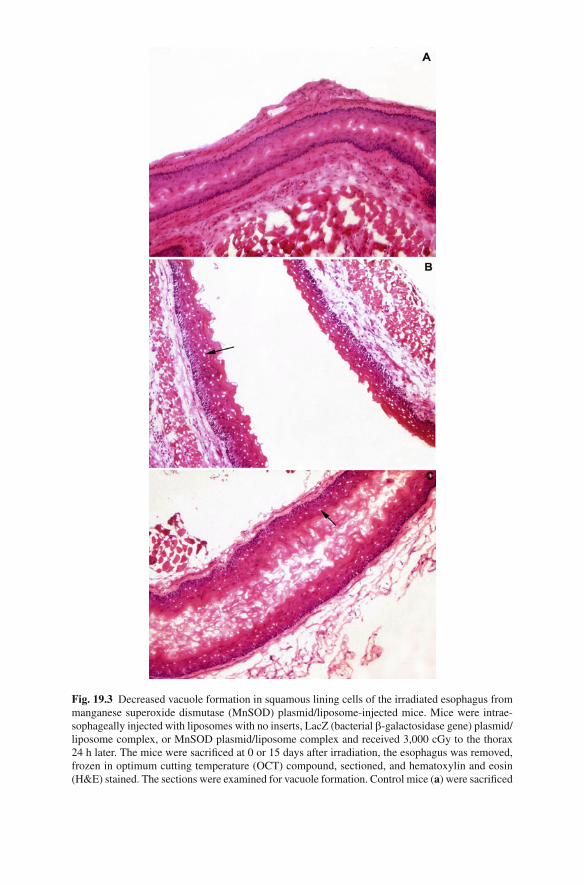
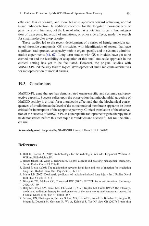
Fig. 19.3 Decreased vacuole formation in squamous lining cells of the irradiated esophagus from manganese superoxide dismutase (MnSOD) plasmid/liposome-injected mice. Mice were intrae-sophageally injected with liposomes with no inserts, LacZ (bacterial b -galactosidase gene) plasmid/liposome complex, or MnSOD plasmid/liposome complex and received 3,000 cGy to the thorax 24 h later. The mice were sacrifi ced at 0 or 15 days after irradiation, the esophagus was removed, frozen in optimum cutting temperature (OCT) compound, sectioned, and hematoxylin and eosin (H&E) stained. The sections were examined for vacuole formation. Control mice ( a ) were sacrifi ced
39719 Radiation Protection by MnSOD-Plasmid Liposome Gene Therapy
demonstrated to reach deep into the lung past terminal bronchials and into alveolar spaces, and the inhalation gene therapy route was shown to successfully deliver ade-quate transgene [ 84 ] . The lung irradiation damage model was different from the esophagus model in that esophageal toxicity was expressed primarily as acute esophagitis. Surviving animals did have a higher incidence of esophageal stricture (late effect) in the animals that did not receive MnSOD-PL, compared to MnSOD-PL-treated animals [ 77 ] . These experiments were confi rmed in other studies with photodynamic therapy delivered damage to the pig esophagus [ 85 ] . Animals treated with MnSOD-PL intraesophageally demonstrated decreased ulceration of the esoph-agus caused by the photodynamic therapy. Thus, in the esophagus both acute esophagitis and the late effect of stricture whether caused by ionizing irradiation or PDT were decreased by MnSOD-PL.
The lung irradiation damage model in the C57BL/6J mouse was different in that damage was detected at 120–150 days after irradiation and was prominently displayed as the histopathologic picture of organizing alveolitis or fi brosis [ 70 ] . In this model system, the administration of MnSOD-PL prior to irradiation gave the best results with respect to reducing radiation toxicity, lethality, and decreasing the magnitude of organizing alveolitis in quantitative examination of lung sections. However, delivery of MnSOD-PL at serial time points after irradiation including at 100 days (immediately before appearance of the late lesion) did produce a detectable reduc-tion in the magnitude of radiation damage [ 86 ] . These data were supported by other experiments in which fractionated delivery of MnSOD-PL by inhalation, and frac-tionated delivery by intraoral or intraesophageal administration during fractionated radiotherapy was also radioprotective.
Experiments in a rat model of bladder radiation induced cystitis demonstrated that intra-vesicle administration of MnSOD-PL resulted in signifi cant uptake in the bladder epithelium and reduced both acute cystitis and bladder stricture. In this model physiological functioning of the bladder was preserved by MnSOD-PL gene therapy prior to irradiation. Animals treated with MnSOD-PL showed less bladder permeability with decreased movement of water and urea across the bladder mem-brane [ 71 ] .
Experiments in intestinal radiation protection by exposing a loop of mouse intes-tine, and then administering either MnSOD-PL or control liposomes, and then delivery irradiation showed that MnSOD-PL preserved intestinal villus length and volume suggesting protection of these structures [ 72 ] . Total abdominal irradiation models have not been carried out, but the results are suggestive of signifi cant intes-tinal protection.
Fig. 19.3 (continued) on day 0 and mice injected with liposomes with no insert ( b ), LacZ plasmid/liposome complex ( c ), and or MnSOD plasmid/liposome complex ( d ), were sacrifi ced on day 15. Irradiated mice injected with liposomes only ( b ) or LacZ plasmid/liposome complexes ( c ) had extensive vacuole formation. Mice injected with the MnSOD plasmid/liposome complexes ( d ) and control nonirradiated mice ( a ) had few vacuoles (×40)

398 J.S. Greenberger et al.
19.2.2 Systemic Radioprotection by MnSOD-PL
Intravenous administration of MnSOD-PL prior to total body irradiation provided protection against both the 9.5- and 10.0-Gy doses. Systemic radioprotection did not induce increased tumorogenesis, and no increase in late effects or new tumors was detected [ 74 ] . MnSOD-PL was detected in tissues for 48–72 h, and expression was documented by RTPCR studies in both the lung and esophagus [ 63, 70, 77 ] .
19.2.3 Combining MnSOD-PL with Other Transgene Therapies or Other Drugs
The radiation chemistry experiment that defi nes the effects of MnSOD suggested that its action would be to enhance and preserve dismutation of superoxide to hydrogen peroxide [ 25– 27 ] . However, hydrogen peroxide itself is a toxic moiety and can cause cell lethality [ 25, 87 ] . Therefore, experiments were carried out to determine whether addition of another transgene for neutralization of hydrogen peroxide might add to radioprotection [ 88, 89 ] . The fi rst experiments involved adding a transgene for glutathione peroxidase [ 63 ] . One publication demonstrated that glutathione peroxidase itself was radioprotective [ 90 ] . Other experimental data failed to confi rm this observation [ 63 ] .
To determine whether overexpression of another enzyme that neutralizes H 2 O
2
catalase increased protection of cells already overexpressing MnSOD-PL, a mito-chondrial targeted catalase transgene was compared to non-mitochondrial targeted catalase for radioprotective capacity in vitro and in vivo. 32D cl 3 hematopoietic progenitor cell lines or a subclone overexpressing MnSOD (2C6) were transfected with the transgene for either catalase or mitochondrial targeted catalase [ 88, 89 ] . These experiments demonstrated that mitochondrial targeting of catalase in cells transfected with mt-CAT were radioprotected [ 88 ] . The signifi cant additional radioprotection observed in 2C6 cells overexpressing mt-CAT as well as MnSOD in this in vitro experiment showed that combining two transgenes in a clonal cell line appeared to be advantageous.
A potential problem with using two different transgenes on separate plasmids delivered by plasmid liposome is that of not adequately facilitating expression of both transgenes in the same cell. Previous studies have demonstrated that under optimal conditions, 20-50% of cells are transduced in the esophagus or lung by MnSOD-PL administration [ 91 ] . Therefore, further experiments will depend upon the ability to construct a single plasmid containing both MnSOD and catalase trans-genes. Experiments with both transgenes on a single plasmid are being carried out to confi rm whether this is additive or potentially synergistic in vivo.
Adding other drugs to MnSOD-PL gene therapy has been a subject of intense interest particularly with respect to orthotopic tumor models and hopeful translation of such results into clinical radiotherapy protocols.

39919 Radiation Protection by MnSOD-Plasmid Liposome Gene Therapy
In previous studies with C57BL/6J mice bearing orthotopic tumors, MnSOD-PL protected normal tissues while not protecting tumors from radiation injury [ 92– 94 ] . In a recent series of experiments, nude mice bearing human orthotopic oral cavity tumors from the CAL33 cell line were treated with intraoral MnSOD-PL prior to a single fraction irradiation [ 95 ] . In the nu/nu model, CAL-33 cells of human origin were treated with Cetuximab, an antibody to the human epidermal growth factor receptor. Mice receiving both Cetuximab and MnSOD-PL demonstrated improved radiation responsiveness. In these studies, animals were examined by micro-PET scanning using a hypoxia imaging technique with F-misonidazole imaging. F-miso targeted hypoxic areas in the CAL-33 tumors and confi rmed the capacity of F-miso to image hypoxic areas. The hypoxic cell cytotoxin, Tirapazamine, was therapeutic when delivered to these mice [ 95 ] . Combining two agents MnSOD-PL plus Cetuximab, MnSOD-PL plus Tirapazamine, or all three agents resulted in signifi cant improvement in local tumor control [ 95 ] . These experiments suggest that MnSOD-PL protection of normal tissues can be supplemented with other therapies to facilitate improved therapeutic ratio and improved outcome.
19.2.4 Tumor Cell Redox Status Differs from Normal Tissues and Facilitates the Use of MnSOD PL Gene Therapy
Studies by St. Clair et al. [ 96 ] and Oberley et al. [ 26– 30 ] as well as others [ 25 ] fi rst demonstrated that many epithelial tumor cell types including those in lung and head and neck cancers have intrinsic reduction of levels of MnSOD activity. These obser-vations suggested that the hypoxic microenvironment in tumors and reversion to anaerobic metabolism may have facilitated down regulation of MnSOD [ 94 ] , which in oxidative metabolism is naturally utilized to counteract ROS production during the electron transport cascade in oxidative metabolism in the mitochondria [ 94 ] . The mechanism of MnSOD reduction in tumors was found to be more complex. Tumor cell lines were demonstrated to have decreased production of MnSOD through point mutations in the promoter of the genes [ 26, 27 ] . Furthermore, some tumor cell systems showed compensatory reduction in glutathione peroxidase such that re-introduction of elevated levels of MnSOD by transgene transfection resulted in sensitivity of those cells to hydrogen peroxide-induced injury. Transfecting into those cells with a transgene for glutathione peroxidase restored the capacity to catabolize hydrogen peroxide [ 90 ] .
One of the prominent concerns in the use of MnSOD-Plasmid Liposome ther-apy for normal tissue protection was the possibility that transgene expression in tumor cells would also provide radioprotection thereby equalizing both sides of the therapeutic ratio at a higher level of protection and potentially compromising the effectiveness of radiotherapy [ 92– 94 ] . While organ-specifi c gene therapy was designed to prevent this by physical means (for example, inhalation of MnSOD-PL resulted in normal bronchiolar and alveolar cell uptake of the transgene while not reaching tumor cells that were in a solid mass and not connected to the airway), the

400 J.S. Greenberger et al.
data showing differences in redox balance between normal tissue and tumors suggested that systemic administration might actually be therapeutic.
The fi rst studies testing this hypothesis involved transfection of epithelial tumor cell lines with MnSOD-PL in vitro [ 93 ] . Epithelial tumor cell lines of both mouse and human origin and of both pulmonary and oral cavity origin demonstrated radio-sensitization by MnSOD-PL [ 93 ] . Cell lines derived from normal tissues of these same tumor sites (normal lung, normal oral cavity) demonstrated MnSOD-PL radio-protection. It appeared that differences in redox balance between normal tissues and tumors might facilitate improvement in therapeutic ratio combining radiosensitization of tumor tissue with radioprotection of normal tissues [ 94 ] . Orthotopic lung tumors at the carina of mice demonstrated that intra-tracheal injection of MnSOD-PL radi-osensitized tumor cells while providing radioprotection of the lung [ 92– 94 ] . In situ histochemistry identifi cation of an epitope-tagged MnSOD transgene failed to identify signifi cant levels of MnSOD in the tumor tissue [ 94 ] , and the radiosensitization of tumor was concluded to be that of indirect effect, of unknown origin. Similar exper-iments with oral cavity, orthotopic tumors in the cheek pouch showed the same result. Intra-oral administration of MnSOD-PL was more tumor radiosensitizing than was intravenous administration while both techniques did provide some tumor cell radiosensitization [ 94 ] .
At the present time, it appears that there are both direct MnSOD transgene-mediated tumor radiosensitizing effects and indirect effects through protection of normal tissue. Such indirect effects could include those of a physiological nature, such as normal tissue radioprotection of blood vessels including limitation of micro-vascular formation in the tumor, or even reduction of blood fl ow to tumor cells. Decrease in production of infl ammatory cytokines by irradiated normal tissues through MnSOD-PL-mediated radioprotection might reduce the availability of such cytokines to growing tumor tissue. Tumor cells have been demonstrated to benefi t from addition of growth factors including TGF b , IL-1, TNF a , and others which are deleterious to normal tissue by the second wave of apoptosis [ 93 ] .
19.2.5 MnSOD-PL Gene Therapy Radioprotection Strategies Lead the Way to Small Molecule Mitochondrial Targeted Radioprotective Agents
Experiments with MnSOD-PL have demonstrated the importance of mitochondrial localization for normal tissue radioprotection. They also identifi ed critical redox differences between normal and tumor tissue, which can facilitate improved radia-tion protection to normal tissue with acceleration of radiation injury in tumors. Long-lived MnSOD-PL radiotherapeutic effects in vivo have demonstrated that sur-vivors of intravenous MnSOD-PL administration prior to total body irradiation are not at increased risk for secondary cancers or additional irradiation-induced life shortening [ 74 ] . All these data suggested that substitution of a small molecule that is mitochondrial targeted and has MnSOD radioprotective capacity might be a more
40119 Radiation Protection by MnSOD-Plasmid Liposome Gene Therapy
effi cient, less expensive, and more feasible approach toward achieving normal tissue radioprotection. In addition, concerns for the long-term consequences of gene therapy in humans, not the least of which is a potential for germ line integra-tion of transgene, induction of mutations, or other side effects, made the search for small molecules a top priority.
These studies led to the recent development of a series of hemigramacidin-tar-geted nitroxide compounds, GS-nitroxides, with identifi cation of several that have signifi cant radioprotective capacity both in organ-specifi c and in systemic adminis-tration experiments [ 61, 62 ] . Long-term studies with GS-nitroxides have yet to be carried out and the feasibility of adaptation of this small molecule approach in the clinical setting has yet to be facilitated. However, the original studies with MnSOD-PL led the way toward logical development of small molecule alternatives for radioprotection of normal tissues.
19.3 Conclusions
MnSOD-PL gene therapy has demonstrated organ-specifi c and systemic radiopro-tective capacity. Success relies upon the observation that mitochondrial targeting of MnSOD activity is critical for a therapeutic effect and that the biochemical conse-quences of irradiation at the level of the mitochondrial membrane appear to be those critical for interruption of the apoptotic pathway. Clinical translation of the observa-tion of the success of MnSOD-PL as a therapeutic radioprotector gene therapy must be demonstrated before this technique is validated and successful for routine clini-cal use.
Acknowledgment Supported by NIAID/NIH Research Grant U19A1068021
References
1. Hall E, Giaccia A (2006) Radiobiology for the radiologist, 6th edn. Lippincott William & Wilkins, Philadelphia, PA
2. Hauer-Jensen M, Wang J, Denham JW (2003) Current and evolving management strategies. Semin Radiat Oncol 13:357–371
3. Gopal R et al (2003) The relationship between local dose and loss of function for irradiation lung. Int J Radiat Oncol Biol Phys 56(1):106–113
4. Marks LB (2002) Dosimetric predictors of radiation-induced lung injury. Int J Radiat Oncol Biol Phys 54(2):313–316
5. Blodgett TM, Meltzer CC, Townsend DW (2007) PET/CT: form and function. Radiology 242(2):50–70
6. Daly ME, Chen AM, Bucci MK, El-Sayed IE, Xia P, Kaplan MJ, Eisele DW (2007) Intensity-modulated radiation therapy for malignancies of the nasal cavity and paranasal sinuses. Int J Radiat Oncol Biol Phys 67(1):151–157
7. Selvaraj RN, Bhatnagar A, Beriwal S, Huq MS, Heron DE, Sonnik D, Brandner E, Surgent R, Mogus R, Deutsch M, Gerszten K, Wu A, Kalnicki S, Yue NJ, Saw CB (2007) Breast skin

402 J.S. Greenberger et al.
doses from brachytherapy using MammoSite HDR, intensity modulated radiation therapy, and tangential fi elds techniques. Technol Cancer Res Treat 6(1):17–22
8. Varlotto JM, Flickinger JC, Niranjan A, Bhatnagar A, Kondziolka D, Lunsford LD (2005) The impact of whole-brain radiation therapy on the long-term control and morbidity of patients surviving more than one year after gamma knife radiosurgery for brain metastases. Int J Radiat Oncol Biol Phys 62(4):1125–1132
9. Tsao MN, Mehta MP, Whelan TJ, Morris DE, Hayman JA, Flickinger JC, Mills M, Rogers CL, Souhami L (2005) The American society for therapeutic radiology and oncology (ASTRO) evidence-based review of the role of radiosurgery for malignant glioma. Int J Radiat Oncol Biol Phys 63(1):47–55
10. Heron DE, Smith RP, Andrade RS (2006) Advances in image-guided radiation therapy – the role of PET-CT. Med Dosim 31(1):3–11
11. Voynov G, Heron DE, Burton S, Grandis J, Quinn A, Ferris R, Ozhasoglu C, Vogel W, Johnson J (2006) Frameless stereotactic radiosurgery for recurrent head and neck carcinoma. Technol Cancer Res Treat 5(5):529–536
12. Beriwal S, Bhatnagar A, Heron DE, Selvaraj R, Mogus R, Kim H, Gerszten K, Kelley J, Edwards RP (2006) High-dose-rate interstitial brachytherapy for gynecologic malignancies. Brachytherapy 5(4):218–222
13. Rubin P, Casarett GW (1968) Clinical radiation pathology. W. B, Saunders, Philadelphia, PA 14. Brown JM, Wilson WR (2004) Exploiting tumour hypoxia in cancer treatment. Nat Rev Cancer
4:437–447 15. Le Q-T (2007) Identifying and targeting hypoxia in head and neck cancer: a brief overview of
current approaches. Int J Radiat Oncol Biol Phys 69(2 Suppl):S56–S58 16. Greenberger JS, Epperly MW, Gretton J, Jefferson M, Nie S, Bernarding M, Kagan V, Guo
H-L (2003) Radioprotective gene therapy. Curr Gene Ther 3:183–195 17. Greenberger JS, Epperly MW (2004) Radioprotective antioxidant gene therapy: potential
mechanisms of action. Gene Ther Mol Biol 8:31–44 18. Greenberger JS, Epperly MW (2005) Pleiotraophic stem cell and tissue effects of ionizing
irradiation protection by MnSOD-plasmid liposome gene therapy. In: Redberry GW (ed) Gene Therapy in Cancer, Chapter VI. Nova Science Publishers, Inc., Hauppage, NY, pp. 191–215
19. Greenberger JS, Epperly MW (2005) Radioprotective gene therapy: current status and future goals. In Vile RG (ed) Viral Therapy of Cancer. Wiley Publications, Hoboken, NJ
20. Epperly MW, Sikora C, Deilippi S, Gretton J, Zhan Q, Kufe DW, Greenberger JS (2002) MnSOD inhibits irradiation-induced apoptosis by stabilization of the mitochondrial membrane against the effects of SAP kinases p38 and Jnk1 translocation. Radiat Res 157:568–577
21. Epperly MW, Gretton JE, Bernarding M, Nie S, Rasul B, Greenberger JS (2003) Mitochondrial localization of copper/zinc superoxide dismutase (Cu/ZnSOD) confers radioprotective func-tions in vitro and in vivo. Radiat Res 160:568–578
22. Belikova NA, Jiang J, Tyurina YY, Zhao Q, Epperly MW, Greenberger J, Kagan VE (2007) Cardiolipin specifi c peroxidase reactions of cytochrome c in mitochondria during irradiation induced apoptosis. Int J Radiat Oncol Biol Phys 69(1):176–185
23. Jiang J, Kurnikov I, Belikova NA, Xiao J, Zhao Q, Vlasova IL, Amoscato AA, Braslau R, Studer A, Fink MP, Greenberger JS, Wipf P, Kagan VE (2007) Structural requirements for optimized delivery, inhibition of oxidative stress and anti-apoptotic activity of targeted nitrox-ides. J Pharmacol Exp Therapeut 320(5):1050–1060
24. Greenberger JS (2008) Gene therapy approaches for stem cell protection. Gene Ther 15:100–108 25. Spitz DR et al (1990) Oxygen toxicity in control and H
2 O
2 -resistant Chinese hamster fi bro-
blasts. Arch Biochem Biophys 279:249–260 26. Oberley LW, Buettner GR (1979) Role of superoxide dismutase in cancer: a review. Cancer
Res 39:1141–1149 27. Oberley LW (1982) Superoxide dismutase and cancer. In: Oberley LW (ed) Superoxide
dismutase. CRC Press, Boca Raton, FL, Vol. II, Chapter 6 28. Zhong W, Oberley LW, Oberley TD, St. Clair DK (1997) Suppression of the malignant
phenotype of human glioma cells by overexpression of manganese superoxide dismutase. Oncogene 14:481–490

40319 Radiation Protection by MnSOD-Plasmid Liposome Gene Therapy
29. Zhong W et al (1996) Inhibition of cell growth and sensitization to oxidative damage by overexpression of manganese superoxide dismutase in rat glioma cells. Cell Growth Differ 7:1175–1186
30. Yan T, Oberley LW, Zhong W, St. Clair DK (1996) Manganese-containing superoxide dismutase overexpression causes phenotypic reversion in SV40-transformed human lung fi broblasts. Cancer Res 56:2864–2871
31. Bennett CB et al (2001) Genes required for ionizing radiation resistance in yeast. Nat Genet 29:426–434
32. Lin Z, Nei M, Ma H (2007) The origins and early evolution of DNA mismatch repair genes-multiple horizontal gene transfers and co-evolution. Nucleic Acids Res 35:7591–7603
33. Cox MC, Battista JR (2005) Deinococcus radiodurans the consummate survivor. Nat Rev Microbiol 3:882–892
34. Daly MJ et al (2004) Accumulation of Mn(II) in deinococcus radiodurans facilitates gamma-radiation resistance. Science 306:1025–1030
35. White O et al (1999) Genome sequence of the radioresistant bacterium deinococcus radio-durans R1. Science 286:1571–1576
36. Morgan JL, Holcomb TM, Morrissey RW (1968) Radiation reaction in ataxia telangiectasia. Am J Dis Child 116:557–558
37. Ho AY et al (2006) Genetic predictors of adverse radiotherapy effects: the gene-PARE project. Int J Radiat Oncol Biol Phys 65:646–655
38. Shiloh Y (2003) ATM and related protein kinases: safeguarding genome integrity. Nat Rev Cancer 3:155–168
39. Abraham RT (2001) Cell cycle checkpoint signaling through the ATM and ATR kinases. Genes Dev 15:2177–2196
40. Casado JA, Nunez MI, Segovia JC, Ruiz de Almodovar JM, Bueren JA (2005) Non-homologous end-joining defect in fanconi anemia hematopoietic cells exposed to ionizing radiation. Radiat Res 164:635–641
41. Taniguchi T et al (2002) Convergence of the fanconi anemia and ataxia telangiectasia signaling pathways. Cell 109:459–472
42. Marcou Y, D’Andrea A, Jeggo PA, Plowman PN (2001) Normal cellular radiosensitivity in an adult fanconi anemia patient with marked clinical radiosensitivity. Radiother Oncol 60:75–79
43. Imamura O et al (2002) Werner and Bloom helicases are involved in DNA repair in a compli-mentary fashion. Oncogene 21:954–963
44. Imamura O, Campbell JL (2003) The human Bloom syndrome gene suppresses the DNA replication and repair defects of yeast dna2 mutants. PNAS 100:8193–8198
45. Stone HB et al (2004) Models for evaluating agents intended for the prophylaxis, mitigation, and treatment of radiation injuries. Report of an NCI Workshop, Dec. 3–4, 2003. Radiat Res 162:711–718
46. Rix-Montel MA (1988) Biophysical aspects of radioprotectors by aminothiols. Eng Med Biol Soc 2:1032–1033
47. Herceg Z, Milosvic Z, Kljajic R, Radnic Z (1990) The effects of use of radioprotective com-pound WR-2721 on haematological parameters in lethally irradiated swines. YRPA, Proceedings of the 3rd Italian-Yugoslav Symposium on Radiation Protection, Plitvice, Yugoslavia pp. 30–35
48. Epperly MW et al (2004) Ascorbate as a “redox-sensor” and protector against irradiation-induced oxidative stress in 32D cl 3 hematopoietic cells and subclones overexpressing human manganese Superoxide Dismutase. Int J Radiat Oncol Biol Phys 58(3):851–861
49. Bayir H et al (2006) Apoptotic interactions of cytochrome c: redox fl irting with anionic phos-pholipids within and outside of mitochondria. Biochim Biophys Acta 1757(5–6):648–659
50. Kagan VE et al (2006) The “pro-apoptotic genies” get out of mitochondria: oxidative lipidom-ics and redox activity of cytochrome c/cardiolipin complexes. Chem Biol Interact 163:15–28
51. Turella P et al (2005) Proapoptotic activity of new glutathione S-transferase inhibitors. Cancer Res 65:3751–3761
52. Vujaskovic Z (2003) Overexpression of extracellular superoxide dismutase protects mice from radiation-induced lung injury. Int J Radiat Oncol Biol Phys 57(4):1056–1066

404 J.S. Greenberger et al.
53. Kang SK et al (2003) Overexpression of extracellular superoxide dismutase protects mice from radiation-induced lung injury. Int J Radiat Oncol Biol Phys 57:1056–1066
54. Anscher MS et al (1998) Plasma TFG,1 as a predictor of radiation pneumonitis. Int J Radiat Oncol Biol Phys 41:1029–1035
55. Rubin P, Johnston CJ, Williams JP, McDonald S, Finkelstein JN (1995) A perpetual cascade of cytokines postirradiation leads to pulmonary fi brosis. Int J Radiat Oncol Bio Phys 33:99–109
56. Mothersill C, Seymour C (2001) Review: radiation-induced bystander effects: past history and future directions. Radiat Res 155:759–767
57. Gorbunov NV et al (2000) Activation of the nitric oxide synthase 2 pathway in the response of bone marrow stromal cells to high doses of ionizing radiation. Radiat Res 154:73–86
58. Greenberger JS, Epperly MW (2007) Antioxidant gene therapeutic approaches to normal tissue radioprotection and tumor radiosensitization. In Vivo 21:141–146
59. Mitchell JB, Russo A, Kuppusamy P, Krishna MC (2000) Radiation, radicals, and images. Ann NY Acad Sci 899:28–43
60. Krishna MC, Grahame DA, Samuni A, Mitchell JB, Russo A (1992) Oxoammonium cation intermediate in the nitroxide-catalyzed dismutation of superoxide. Proc Natl Acad Sci USA 89:5537–5541
61. Fink M et al (2007) Hemigramicidin-TEMPO conjugates: novel mitochondria-targeted anti-oxidants. Crit Care Med 35:5461–5470
62. Jiang J et al (2008) A mitochondria-targeted nitroxide/hemi-gramicidin S conjugate protects Mouse embryonic cells against g -irradiation. Int J Radiat Oncol Biol Phys 70:816–825
63. Epperly MW, Kagan VE, Sikora CA, Gretton JE, Defi lippi SJ, Bar-Sagi D, Greenberger JS (2001) Manganese superoxide dismutase-plasmid/liposome (MnSOD-PL) administration pro-tects mice from esophagitis associated with fractionated irradiation. Int J Cancer 96(4):221–233
64. Epperly MW, Zhang X, Nie S, Cao S, Kagan V, Tyurin V, Greenberger JS (2005) MnSOD-plasmid liposome gene therapy effects on ionizing irradiation induced lipid peroxidation of the esophagus. In Vivo 19:997–1004
65. Han F, Drabek T, Stezoski J, Janesko-Feldman K, Stezoski SW, Clark RSB, Bayir H, Tisherman SA, Kochanek PM (2008) Protein nitration and poly-ADP-ribosylation in brain after rapid exsanguination cardiac arrest in a rat model of emergency preservation and resuscitation. Resuscitation 79(2):301–310
66. Tyurina YY, Tyurin VA, Epperly MW, Greenberger JS, Kagan VE (2008) Oxidative lipidomics of g -irradiation induced intestinal injury. Free Radic Biol Med 44(3):299–314
67. Tyurin VA, Tyurina YY, Ritov VB, Lystsya A, Amoscato AA, Kochanek PM, Hamilton R, DeKosky ST, Greenberger JS, Bayir H, Kagan VE (2010) Oxidative lipidomics of apoptosis: Quantitative assessment of phospholipid hydroperoxides in cells and tissues. Methods Mol Biol 610:353–374
68. Tyurin VA, Tyurina YY, Kochanek PM, Hamilton R, DeKosky ST, Greenberger JS, Bayir H, Kagan VE (2008) Oxidative lipidomics of programmed cell death. Methods Enzymol 442:375–393
69. Stickle RL, Epperly MW, Klein E, Bray JA, Greenberger JS (1999) Prevention of irradiation-induced esophagitis by plasmid/liposome delivery of the human manganese superoxide dis-mutase (MnSOD) transgene. Radiat Oncol Investig 7(6):204–217
70. Epperly MW, Bray JA, Kraeger S, Zwacka R, Engelhardt J, Travis E, Greenberger JS (1998) Prevention of late effects of irradiation lung damage by manganese superoxide dismutase gene therapy. Gene Ther 5:196–208
71. Kanai AJ, Zeidel ML, Lavelle JP, Greenberger JS, Birder LA, deGroat WC, Apodaca GL, Meyers SA, Ramage R, VanBibber MM, Epperly MW (2002) Manganese superoxide dismutase gene therapy protects against irradiation-induced cystitis. Am J Physiol Renal Physiol 44:1152–1160
72. Guo HL, Wolfe D, Epperly MW, Huang S, Liu K, Glorioso JC, Greenberger J, Blumberg D (2003) Gene transfer of human manganese superoxide dismutase protects small intestinal villi from radiation injury. J Gastrointest Surg 7:229–236
73. Guo HL, Seixas-Silva JA, Epperly MW, Gretton JE, Shin DM, Greenberger JS (2003) Prevention of irradiation-induced oral cavity mucositis by plasmid/liposome delivery of the human manganese superoxide dismutase (MnSOD) transgene. Radiat Res 159:361–370

40519 Radiation Protection by MnSOD-Plasmid Liposome Gene Therapy
74. Epperly MW, Smith T, Wang H, Schlesselman J, Franicola D, Greenberger JS (2008) Modulation of total body irradiation induced life shortening by systemic intravenous MnSOD-plasmid liposome gene therapy. Radiat Res 170(4):437–443
75. Epperly MW, Carpenter M, Agarwal A, Mitra P, Nie S, Greenberger JS (2004) Intra-oral man-ganese superoxide dismutase plasmid/liposome radioprotective gene therapy decreases ioniz-ing irradiation-induced murine mucosal cell cycling and apoptosis. In Vivo 18:401–410
76. Epperly MW, Travis EL, Sikora C, Greenberger JS (1999) Magnesium superoxide dismutase (MnSOD) plasmid/liposome pulmonary radioprotective gene therapy: Modulation of irradiation-induced mRNA for IL-1, TNF- a , and TGF- b correlates with delay of organizing alveolitis/fi brosis. Biol Blood Marrow Transplant 5:204–214
77. Epperly MW, Gretton JA, Defi lippi SJ, Sikora CA, Liggitt D, Koe G, Greenberger JS (2001) Modulation of radiation-induced cytokine elevation associated with esophagitis and esopha-geal stricture by manganese superoxide dismutase-plasmid/liposome (SOD-PL) gene therapy. Radiat Res 155:2–14
78. Rajagopalan MS, Gupta K, Epperly MW, Franicola D, Zhang X, Kagan VE, Wipf P, Greenberger JS (2009) The mitochondrial targeted nitroxide JP4-039 augments potentially lethal irradiation damage repair. Vivo 23(5):717–726
79. Gorbunov NV, Pogue-Geile KL, Epperly MW, Bigbee WL, Draviam R, Day BW, Wald N, Watkins SC, Greenberger JS (2000) Activation of the nitric oxide synthase 2 pathway in the response of bone marrow stromal cells to high doses of ionizing radiation. Radiat Res 154:73–86
80. Niu Y, Epperly MW, Shen H, Smith T, Lewis D, Gollin S, Greenberger JS (2008) Intraesophageal MnSOD-plasmid liposome administration enhances engraftment and self-renewal capacity of bone marrow derived progenitors of esophageal squamous epithelium. Gene Ther 15:347–356
81. Epperly MW, Goff JP, Sikora CA, Shields DS, Greenberger JS (2004) Bone marrow origin of cells with capacity for homing and differentiation to esophageal squamous epithelium. Radiat Res 162:233–240
82. Epperly MW, Sikora CA, Defi lippi S, Gretton JE, Greenberger JS (2003) Bone marrow origin of myofi broblasts in irradiation pulmonary fi brosis. Am J Respir Cell Mol Biol 29:213–224
83. Epperly MW, Bernarding M, Gretton J, Jefferson M, Nie S, Greenberger JS (2003) Overexpression of the transgene for manganese superoxide dismutase (MnSOD) in 32D cl 3 cells prevents apoptosis induction by TNF- a , IL-3 withdrawal and ionizing irradiation. Exp Hematol 31(6):465–474
84. Carpenter M, Epperly MW, Agarwal A, Nie S, Hricisak L, Niu Y, Greenberger JS (2005) Inhalation delivery of manganese superoxide dismutase-plasmid/liposomes (MnSOD-PL) protects the murine lung from irradiation damage. Gene Ther 12:685–690
85. Yaron P, Epperly MW, Fernando HC, Klein E, Finkelstein S, Greenberger JS, Luketich JD (2005) Photodynamic therapy induced esophageal-stricture-An animal model: from mouse to pig. J Surg Res 123:67–74
86. Epperly MW, Guo HL, Bernarding M, Gretton J, Jefferson M, Greenberger JS (2003) Delayed intratracheal injection of manganese superoxide dismutase (MnSOD)-plasmid/liposomes provides suboptimal protection against irradiation-induced pulmonary injury compared to treatment before irradiation. Gene Ther Mol Biol 7:61–68
87. Giorgio M, Trinei M, Migliaccio E, Pelicci PG (2007) Hydrogen peroxide: a metabolic by product or a common mediator of ageing signals? Nat RevMol Cell Biol 8:722–730
88. Epperly MW, Melendez JA, Zhang X, Nie S, Pearce L, Peterson J, Franicola D, Dixon T, Greenberger BA, Komanduri P, Greenberger JS (2009) Mitochondrial targeting of a catalase transgene product by plasmid liposomes increases radioresistance in vitro and in vivo. Radiat Res 171(5):588–595
89. Bai J, Rodriguez AM, Melendez JA, Cederbai AI (1999) Overexpression of catalase in cytosolic or mitochondrial compartment protects HepG2 cells against oxidative injury. J Biol Chem 274:26217–26224
90. Epperly MW, Melendez A, Zhang X, Franicola D, Smith T, Greenberger BA, Komanduri P, Greenberger JS (2009) Mitochondrial targeting of a catalase transgene product by plasmid liposomes increases radioresistance in vitro and in vivo. Radiat Res 171(5):588–595

406 J.S. Greenberger et al.
91. Epperly MW, Guo HL, Jefferson M, Wong S, Gretton J, Bernarding M, Bar-Sagi D, Greenberger JS (2003) Cell phenotype specifi c duration of expression of epitope-tagged HA-MnSOD in cells of the murine lung following intratracheal plasmid liposome gene therapy. Gene Ther 10:163–171
92. Epperly MW, Defi lippi S, Sikora C, Gretton J, Kalend K, Greenberger JS (2000) Intratracheal injection of manganese superoxide dismutase (MnSOD) plasmid/liposomes protects normal lung but not orthotopic tumors from irradiation. Gene Ther 7(12):1011–1018
93. Guo H, Epperly MW, Bernarding M, Nie S, Gretton J, Jefferson M, Greenberger JS (2003) Manganese superoxide dismutase-plasmid/liposome (MnSOD-PL) intratracheal gene therapy reduction of irradiation-induced infl ammatory cytokines does not protect orthotopic lewis lung carcinomas. In Vivo 17:13–22
94. Epperly MW, Wegner R, Kanai AJ, Kagan V, Greenberger EE, Nie S, Greenberger JS (2007) Irradiated murine oral cavity orthotopic tumor antioxidant pool destabilization by MnSOD-plasmid liposome gene therapy mediates tumor radiosensitization. Radiat Res 267:289–297
95. Greenberger JS, Dixon T, Franicola D, Niu Y, Zhang X, Epperly MW (2008) Manganese superoxide dismutase (MnSOD)-plasmid liposomes, C225, and Tirapazamine combination enhances radiotherapy of human orthotopic oral cavity squamous tumors. ASTRO, 2008. Int J Rad Oncol Biol Physics 72(1 (suppl 28), #1119):56
96. St. Clair DK, Wan XS, Oberley TD, Muse KE, St. Clair WH (1992) Suppression of radiation-induced neoplastic transformation by overexpression of mitochondrial superoxide dismutase. Mol Carcinog 6:238–242