radiation- its medicinal uses
TRANSCRIPT


Although scientists have only known about radiation since the 1890s, they have developed a wide variety of uses for this natural force. Today, to benefit humankind, radiation is used
in medicine, academics, and industry, as well as for generating electricity. In addition, radiation has useful applications in such areas as agriculture, archaeology
(carbon dating), space exploration, law enforcement, geology (including mining), and many others.
Banguilan, Castro &
Rojas
BS Physics IV

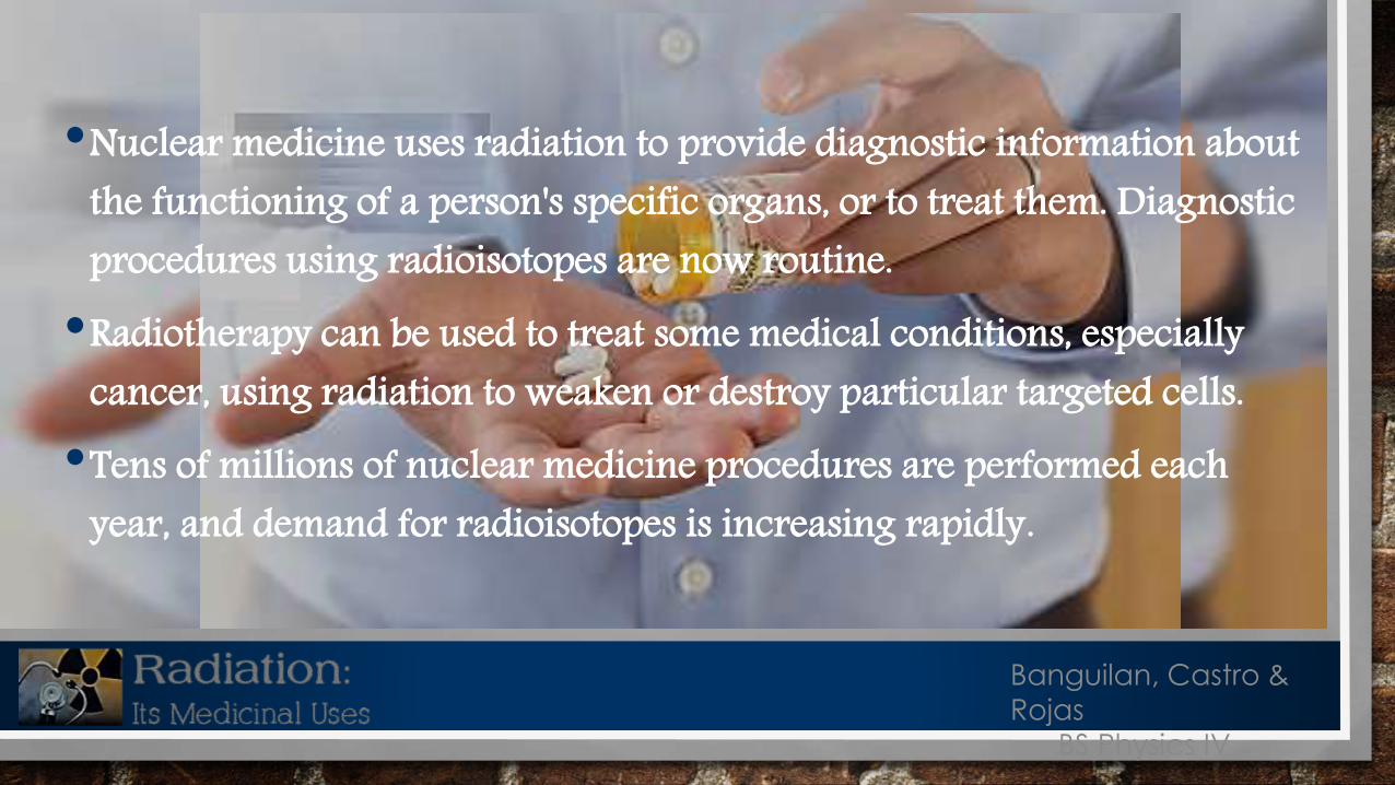
•Nuclear medicine uses radiation to provide diagnostic information about the functioning of a person's specific organs, or to treat them. Diagnostic procedures using radioisotopes are now routine.•Radiotherapy can be used to treat some medical conditions, especially
cancer, using radiation to weaken or destroy particular targeted cells.•Tens of millions of nuclear medicine procedures are performed each
year, and demand for radioisotopes is increasing rapidly.
Banguilan, Castro &
Rojas
BS Physics IV

• Sterilization of medical products• Today, Over half of all medical equipment used in modern hospitals is sterilized using radiation.
•New Drug testing• Over 80% of all new drugs are tested with radioactive tagging before approval
•Medical Imaging• Therapy
• Approximately 10% of medical procedures use radiation to treat a variety of diseases, including many types of cancers, heart disease, gastrointestinal, endocrine, neurological disorders and other abnormalities with in the body.
Areas of Medicine Where Radiation is Used
Banguilan, Castro &
Rojas
BS Physics IV
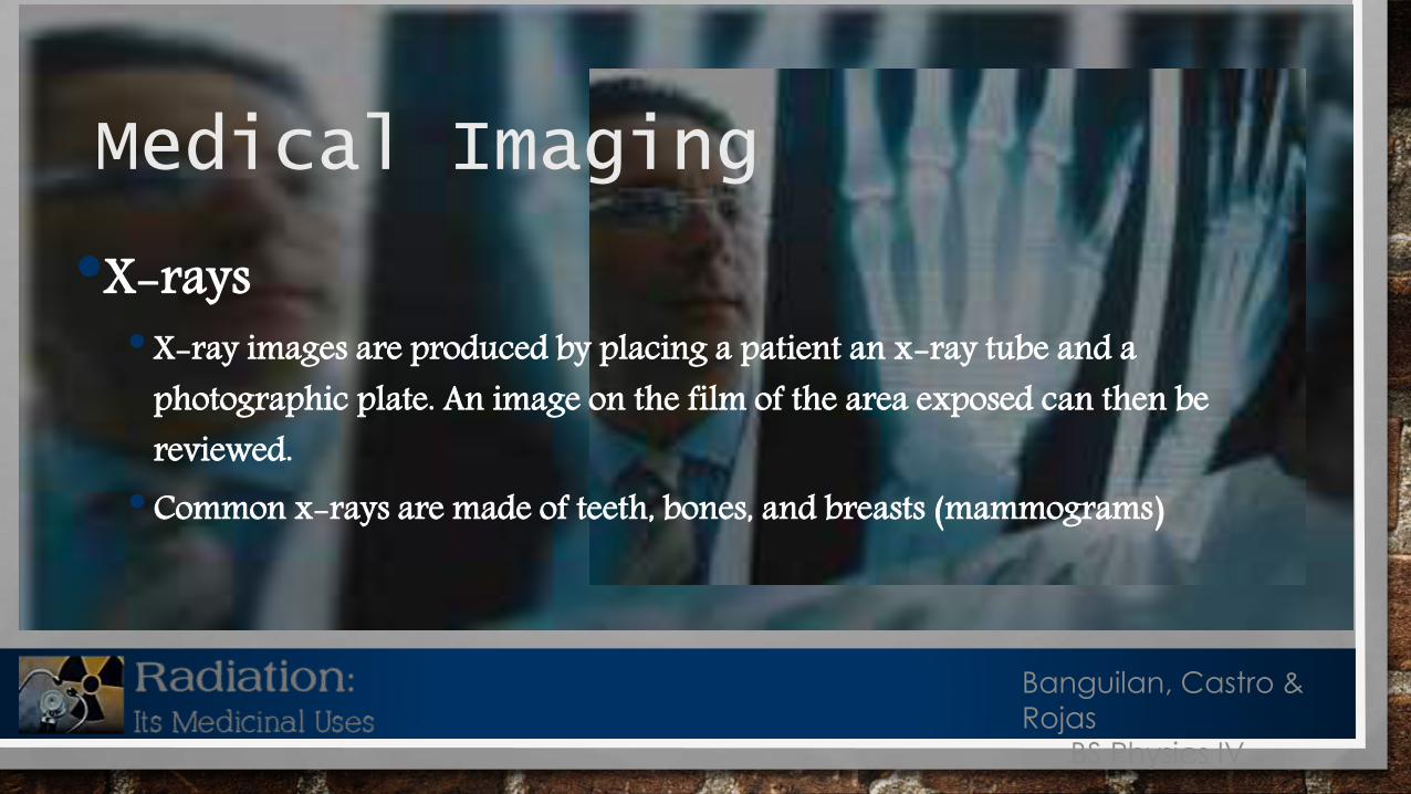
•X-rays• X-ray images are produced by placing a patient an x-ray tube and a
photographic plate. An image on the film of the area exposed can then be reviewed.• Common x-rays are made of teeth, bones, and breasts (mammograms)
Medical Imaging
Banguilan, Castro &
Rojas
BS Physics IV

•Magnetic Resonance Imaging (MRI)•MRI is an imaging technique used to visualize internal structures of the body
in detail. MRI can create more detailed images of the human body than those possible with X-rays.• This procedure uses a magnetic field and pulses or radio waves to make
picture of organs and structures inside the body. The water in our bodies is made up of millions of atoms that are magnetically charged. When placed in a magnetic field these atoms line up with a field much like a compass points to the North Pole.
Medical Imaging
Banguilan, Castro &
Rojas
BS Physics IV
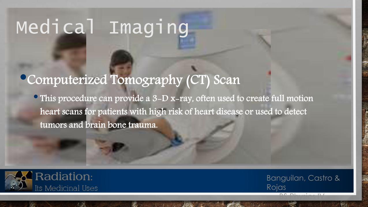
•Computerized Tomography (CT) Scan• This procedure can provide a 3-D x-ray, often used to create full motion
heart scans for patients with high risk of heart disease or used to detect tumors and brain bone trauma.
Medical Imaging
Banguilan, Castro &
Rojas
BS Physics IV
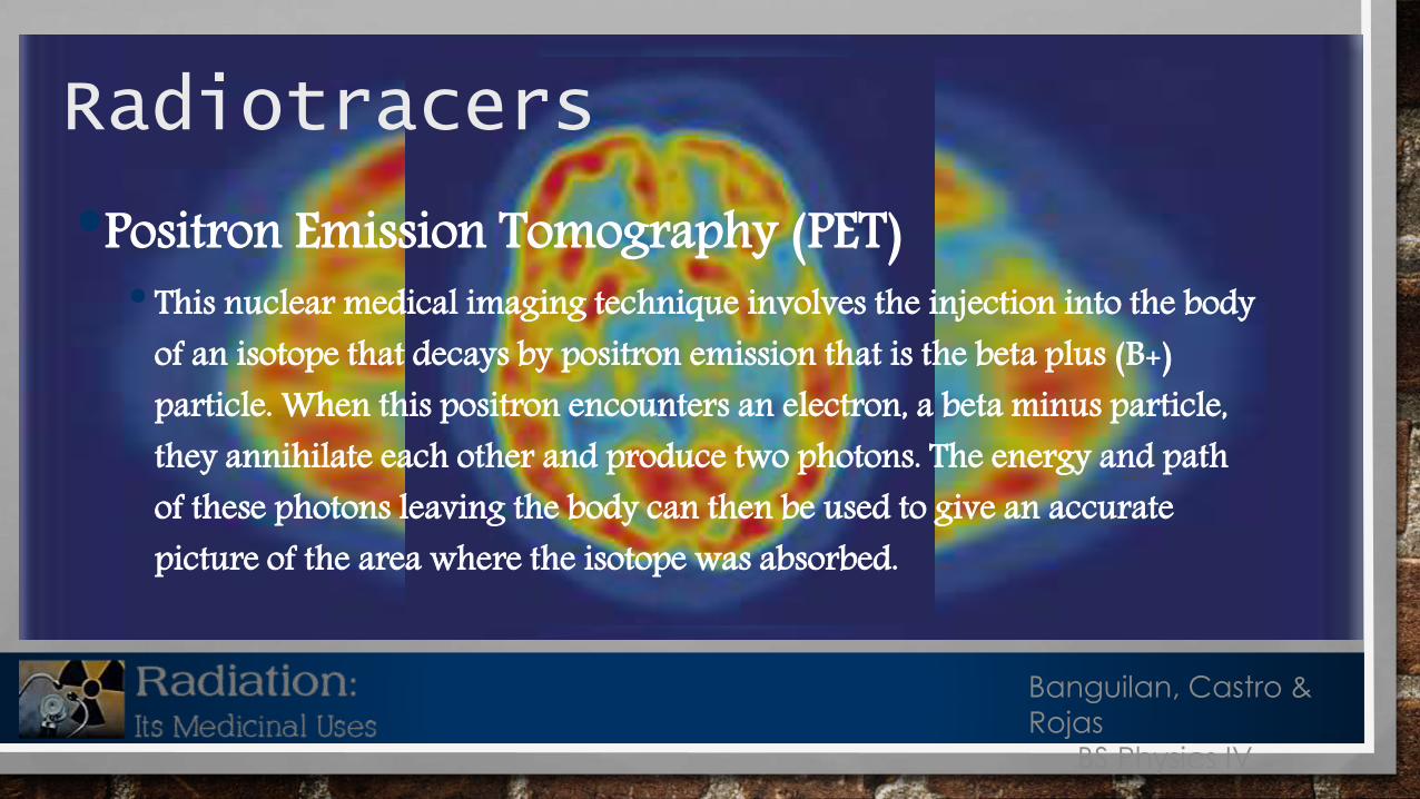
•Positron Emission Tomography (PET)• This nuclear medical imaging technique involves the injection into the body
of an isotope that decays by positron emission that is the beta plus (B+) particle. When this positron encounters an electron, a beta minus particle, they annihilate each other and produce two photons. The energy and path of these photons leaving the body can then be used to give an accurate picture of the area where the isotope was absorbed.
Radiotracers
Banguilan, Castro &
Rojas
BS Physics IV

•Single Photon Emission Computed Tomography (SPECT)• SPECT is similar to PET is its use of radioactive tracer material and detection of
gamma rays. In contrast with PET however, the tracer used in SPECT emits gamma radiation that is measured directly. Where PET tracers emit positrons that annihilate with electrons up to a few millimeters away, causing two gamma photons to be emitted in opposite directions. A PET scanner detects these emissions coincident in time, which provides more local radiation event information and thus higher resolution images than SPECT. SPECT scans, however, are significantly less expensive that PET scans, in part because they are able to use longer lived, more easily obtainable radioisotopes that PET needs.
Radiotracers
Banguilan, Castro &
Rojas
BS Physics IV
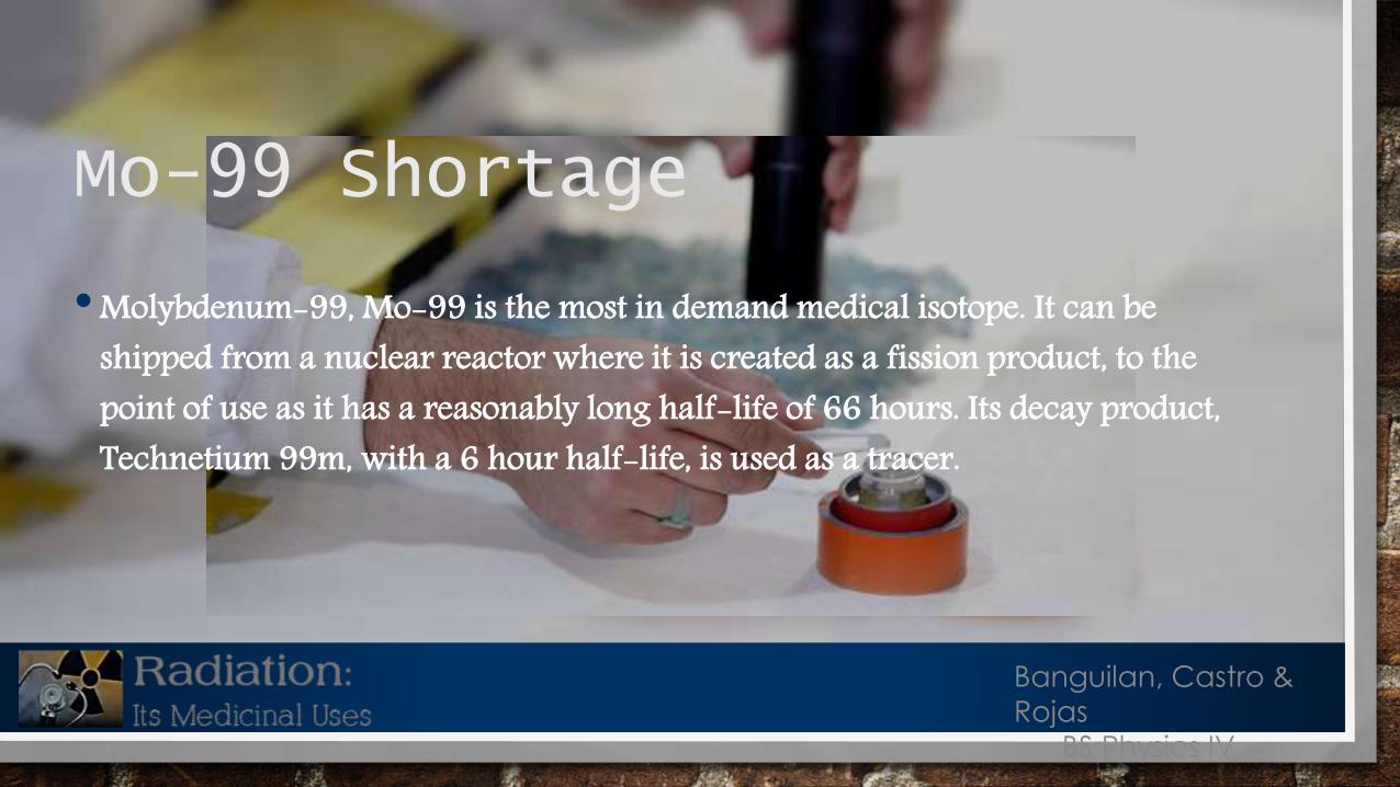
•Molybdenum-99, Mo-99 is the most in demand medical isotope. It can be shipped from a nuclear reactor where it is created as a fission product, to the point of use as it has a reasonably long half-life of 66 hours. Its decay product, Technetium 99m, with a 6 hour half-life, is used as a tracer.
Mo-99 Shortage
Banguilan, Castro &
Rojas
BS Physics IV

Radiation in medical therapy was first applied to the treatment of the thyroid cancer. The patient drinks a determined amount of the solution spiked with radioactive iodine-131. This radioisotope preferentially lodges in the thyroid. The beta emissions of this radioisotope subsequently target and destroy the cancer in the thyroid.
The Use of Radiation in Medical Therapy
Banguilan, Castro &
Rojas
BS Physics IV

• External radiation therapy uses an external beam of radiation to focus on cancerous growths. An incident beam of x-rays or protons is moved around the patient in a precise manner so that the beam remains focused on the tumor minimizing the length of time the penetrating radiation beam doesn’t remain on any of the healthy cell for vey long.• Internal radionuclide therapy can be administered by planting a small radiation source, usually a gamma or beta emitter in the target area(s). Iridium 192 implants are used often in the brain and breast regions. They are produced in wire form and are introduced through a catheter to the target area. After administering the correct dose, the implant wire is removed.
Banguilan, Castro &
Rojas
BS Physics IV

• Boron Neutron Capture Therapy (BNCT)• In this procedure, boron is injected onto the patient to preferentially concentrate at the tumor site. A neutron beam is then focused on the boron. Neutrons react with the boron to produce alpha materials that destroy the malignant cells in the immediate vicinity of the concentrated boron. Since alpha particles are stopped at a very short distance from their point of origin, intense radiation damage is localized.
Banguilan, Castro &
Rojas
BS Physics IV

•Gamma Knife Radiosurgery (Cyber Knife)•Minimizing injury to healthy cells, radiation therapy involves rotating an external
radiation beam around the patient. The radiation from the radioactive source is delivered from many directions, with the beam continually focused on the target abnormality with only small amounts of radiation passing through healthy tissue.
Therapy
Banguilan, Castro &
Rojas
BS Physics IV

•Brachytherapy• It is a from of the internal radio therapy where a radiation source is placed inside or next to the area requiring treatment. Brachytherapy involves the precise placement of short-range radioisotopes directly at the site of the cancerous tumor. These are enclosed in a protective capsule or wire that allows the ionizing radiation to escape. The radiation treats and kills surrounding tissue, but prevents the charge of radioisotopes from moving or dissolving in the body fluids. The capsule may be removed later, or with some isotopes, it may be allowed to remain in place for prolonged treatment. A key feature of brachytherapy is that the radiation affects a localized area around the radiation source. In addition, if the patient moves, of if there’s any movement of the tumor within the body during treatment, the radiation source retains its correct position in relation to the tumor.
Banguilan, Castro &
Rojas
BS Physics IV
• There is no doubt that medical research will find more ways to use radiation and radioisotopes to improve our lives.
Banguilan, Castro &
Rojas
BS Physics IV

Reference
• American Nuclear Society, “Medical Use of Isotopes”
Banguilan, Castro &
Rojas
BS Physics IV
