rachel m. frank, md; mark a. slabaugh, md; hip …...hip pain in active patients jfponline.com vol...
TRANSCRIPT

736 The Journal of family PracTice | December 2012 | Vol 61, no 12
Hip pain in active patients: What you may be missing When a patient who’s physically active complains of hip pain, don’t be too quick to label it a “hip pointer” that requires time and rest to heal. Consider this more nuanced—and effective—approach.
Hip pain is a common complaint, and commonly mis-understood. Although the pain can be associated with a broad spectrum of conditions, the presenta-
tion is often vague and nonspecific. Thus, hip pain and injury are frequently attributed, of-
ten incorrectly, to a “hip pointer”—a contusion of soft tissues against the iliac crest. It’s not unusual for patients who receive this diagnosis to be treated conservatively for prolonged pe-riods, leading some previously active individuals to abandon their favorite sport or self-impose limits on the activities they engage in.1
But it doesn’t have to be this way. Minimally invasive hip arthroscopy and advances in im-
aging, instrumentation, and devices have made it easier to identify and address underlying pathology associated with hip pain, helping patients return to their previous level of ac-tivity more rapidly.2,3 And, while many conditions associated with hip pain can be treated conservatively, family physi-cians—whom patients often go to first—should not hesitate to provide a referral when more aggressive treatment or diag-nostic confirmation is needed.
We created this guide with family physicians in mind. Our focus here is primarily on anterior hip pain—the most common presentation—in active, or athletic, patients.
When did the pain begin? Where does it hurt?Before performing a physical examination, find out as much as possible about the onset of pain and when and under what circumstances it occurs. (A review of hip anatomy is provided at the end of this article at jfponline.com.) Did it begin sud-denly, after an acute injury or a particular physical maneu-ver? Or is the pain insidious, as was the case with one of our patients?
Rachel M. Frank, MD; Mark A. Slabaugh, MD; Robert C. Grumet, MD; Charles A. Bush-Joseph, MD; Walter W. Virkus, MD; Shane J. Nho, MD, MSDivision of Sports Medicine (Drs. Frank, Slabaugh, Grumet, Bush-Joseph, Virkus, and Nho); Hip Preservation Center (Drs. Bush-Joseph, Virkus, and Nho), Department of Orthopedic Surgery, Rush University Medical Center, Chicago, Ill
The authors reported no potential conflict of interest relevant to this article.
CASE c
PRACtiCE RECoMMENDAtioNS
› Consider both musculo-skeletal and nonmusculo-skeletal causes in patients with vague complaints of hip and groin pain. B
› Use imaging studies to con-firm a hip pain diagnosis. B
› Refer patients who fail to respond to nonsurgi-cal treatment to a sports medicine specialist or an orthopedic surgeon. B
Strength of recommendation (SoR)
Good-quality patient-oriented evidence
Inconsistent or limited-quality patient-oriented evidence
Consensus, usual practice, opinion, disease-oriented evidence, case series
A
B
C

737JfPonline.com Vol 61, no 12 | December 2012 | The Journal of family PracTice
ima
ge ©
Joe g
or
ma
n
Minimally invasive hip arthroscopy and advances in imaging and devices have made it easier to get patients back to their previous level of activity more quickly.
Trendelenburg sign—a pelvic tilt when the weight is shifted to the affected extremity.
Although most individuals with hip pain will not have an obvious gait abnormality, any patient who walks with a limp or needs crutches requires an immediate referral to an orthopedic surgeon.
include these elements in the physical examExamine the hip with the patient sitting on the side of the exam table. Assess range of motion (ROM), comparing the range of flex-ion, extension, and internal/external rotation on the affected and unaffected sides. Include the following maneuvers:
z impingement testing. In patients with FAI and osteoarthritis, impingement test-ing—encompassing Flexion, ADDuction, and Internal Rotation (FADDIR)—will elicit pain. The maneuver can be tested starting at 458 of hip flexion, increasing to approximate-ly 1208. Pain with <458 of hip flexion indicates that the impingement is severe.
Such testing can also reveal labral tears, which may be caused by FAI or other struc-tural abnormalities. In a patient with ante-
CASE c mack Q, a 27-year-old man with an 8-month history of right hip pain, sought care at our medical center for an achy pain in his right groin; he also described an occasional “clicking and popping sensation” in his groin but denied any trauma. The pain worsened with prolonged sitting and certain activities, such as squatting, twisting, and putting on shoes and socks. our patient had stopped playing soccer because it hurt too much. he had tried physical therapy, oral anti-inflam-matories, and a corticosteroid injection, with little relief.
Start with a gait assessment The physical examination should begin with a gait assessment. Consider the patient’s abil-ity to bear weight and his or her foot angle.
An individual with a stress fracture will have difficulty bearing weight on the affect-ed side, resulting in a limp, or antalgic gait. A patient with femoral acetabular impinge-ment (FAI) will often exhibit greater exter-nal rotation of the foot on the affected side compared with the other foot. And a patient with weakened abductor muscles, typically be-cause of severe osteoarthritis, will exhibit the

738 The Journal of family PracTice | December 2012 | Vol 61, no 12
rior labral tears, FADDIR will produce groin pain; posterior labral tears will produce pain when the patient is sitting with legs hanging off the exam table and the contralateral leg is brought to the chest and the affected limb fully extended.
In patients with hip pain and bursitis, applying downward pressure will elicit a snapping sound as the iliopsoas snaps over the iliopectineal eminence or femoral head. Flexion, ABduction, and External Rotation (FABER) can also be used to diagnose il-iopsoas tendonitis: The test is positive if it elicits pain in the affected extremity or in the sacroiliac joint on the opposite side.
z Log roll. A painful response to this test, which involves internally and externally ro-tating the affected hip while it is relaxed and the knee fully extended, is an indication of synovitis of the hip caused by intra-articular pathology. To test hip stability, externally ro-tate the leg while it is extended. If the hip is stable, the leg will return to a neutral position; microinstability of the hip is likely if the leg remains in the rotated position.
z Muscular strength testing. To assess for tendinopathy in the hip area, the patient should be in a seated position and contract the internal and external rotators and the ad-ductor muscles while you apply resistance. To test abductor strength, have the patient as-sume a lateral position and hold and abduct the leg on the affected side while you apply resistance.
Hip flexion strength should be tested with the patient in both supine and seated positions. A patient with quadriceps ten-donitis will have much greater pain with re-sisted hip flexion in the supine position vs the seated position; the opposite is true for a pa-tient with iliopsoas tendonitis. (See “Did you know ... ? Hip pain facts and findings” on page 739 for additional diagnostic tips.)
Perform a neurologic evaluation to rule out a back condition that might radiate pain into the anterior hip; ask the patient to do a sit-up while you apply resistance to test for abdominal wall pathology, as well.
z Hip palpation. This aspect of the physi-cal exam is important regardless of the cause of the pain but especially crucial for pediatric and adolescent patients, whose anterior hip
pain may be related to apophyseal injury. Palpate the superior iliac spine (and over the inferior iliac spine in thin patients) to deter-mine if the sartorius or rectus femoris has been injured. The area just lateral to the sym-physis will be tender to palpation in patients with osteitis pubis.
Refer or treat? Here’s what to considerWhile the history and physical should provide ample information for a differential diagno-sis, imaging studies are generally required for confirmation. Clinical assessment— including physical exam, imaging, and intra- articular injection—of patients with hip pain is up to 98% accurate in identifying hip ab-normalities, with arthroscopy as the gold standard.4
CASE c on physical examination, mr. Q had right hip extension to 0º, flexion to 110º, ex-ternal rotation to 50º, and internal rotation to neutral; he also had positive impingement and subspine impingement tests, a painful arc of motion from 12 to 4 o’clock, tenderness over the hip adductor, and pain with resisted hip adduction. he did not walk with a limp.
Diagnostic studies included plain radio-graphs, which demonstrated that the joint space was well preserved. We identified subtle anatomical abnormalities on the femo-ral head-neck junction, known as a cam de-formity. magnetic resonance imaging (mri) revealed an anterior superior labral tear with cartilage delamination.
Stress fractures affect runners, military recruitsIn addition to long-distance runners who have recently increased the frequency, du-ration, or intensity of training,5,6 military recruits have a higher incidence of stress fractures due to the rapid onset of intensive training. Stress fractures can also occur in patients who do not have a history of intense activity but have metabolically weakened bone, in some cases as a result of an eating disorder.7
Patients with femoral neck stress fractures typically present with activity-related anterior
FABER (flexion, abduction, and external rotation) can be used to diagnose iliopsoas tendonitis: the test is positive if it elicits pain in the affected extremity or in the sacroiliac joint on the opposite side.

Hip pAin in ACtive pAtients
739JfPonline.com Vol 61, no 12 | December 2012 | The Journal of family PracTice
in patients with femoral neck stress fractures, radiographs are often negative for up to 4 weeks after the onset of pain, while magnetic resonance imaging can detect an abnormality within a day or 2.
groin pain that is relieved by rest; initially, they may be only mildly affected, but the condi-tion worsens in those who continue to “work through the pain.” By the time such individu-als seek treatment, they almost always have pain with weight bearing and an antalgic gait.
Symptoms consistent with a femoral neck stress fracture can be further evaluated with plain radiographs. However, x-rays are often negative for up to 4 weeks after the onset of pain.8 In cases in which radiographs are nega-tive but the physical exam is suggestive of a stress fracture, MRI—which can detect an ab-normality within a day or 2 of injury8,9—should be used to confirm the diagnosis (FiGURE 1).
z treatment. A complete femoral neck fracture portends impending displacement and requires emergent evaluation by an or-thopedist, and superior neck changes, also known as tension-sided stress fractures, require urgent treatment with percutane-ous screw fixation.9 However, compression-sided, or inferior, stress fractures can be treat-ed with restricted weight bearing and activity modification. Gradual resumption of activ-ity is allowed only after the patient has been asymptomatic for 6 weeks; recurrent pain in-dicates residual stress reaction, and signals that activities should be abated.
osteonecrosis has many causes Necrosis of the femoral head is a debilitating and progressive condition primarily affect-ing patients between the ages of 20 and 50 years.10 It has multiple (and diverse) causes, including trauma, steroids, alcohol, smok-ing, lupus, sickle cell anemia, and coagulopa-thies, as well as scuba diving. But about 20% of cases have no apparent cause.11,12
Patients with osteonecrosis of the hip typically present with groin pain, often de-scribed as a deep, intermittent ache that in-terferes with activities of daily living. Exam findings depend on the stage of presentation. Early on, pain will occur only with extreme ROM; in advanced cases, ROM is restricted and pain occurs even with limited motion.
Femoral head collapse due to loss of the structural integrity of the subchondral bone—which occurs in 80% of cases12—is thought to be caused by decreased blood flow. Plain ra-diography can confirm a diagnosis of osteo-necrosis in patients with advanced disease, but MRI is useful for evaluating patients with earlier clinical presentations.
z treatment of osteonecrosis is dictated by the stage of the disease, but remains con-troversial because no intervention has been shown to prevent progression in all cases.12
Did you know ... ? Hip pain facts and findings• a patient with quadriceps tendonitis will have much greater pain with resisted hip flexion
in a supine position vs a seated position. The opposite is true for a patient with iliopsoas tendonitis.
• Patients with femoral neck stress fractures typically present with activity-related anterior groin pain that is relieved by rest. initially, they may be only mildly affected, but the condi-tion worsens in those who continue to “work through the pain.”
• Plain radiography can confirm a diagnosis of osteonecrosis in patients with advanced dis-ease, but magnetic resonance imaging is useful for evaluating earlier clinical presentations.
• Patients with labral tears often exhibit what has been called the “c-sign”—so named for the shape patients make with their hand as they grip their hip just above the greater tro-chanter to indicate where it hurts.
• athletes who experience adductor strains often play sports in which kicking or frequent changes in direction are required, such as football, hockey, and soccer, and are generally able to tell you exactly what they were doing when the injury occurred.
• unlike other hernias, a sports hernia (athletic pubalgia) does not involve a bulge of tissue protruding through one part of the body into another. instead, it occurs when the oblique abdominal muscles strain or completely tear away from the pubis.
conTinueD

740 The Journal of family PracTice | December 2012 | Vol 61, no 12
All patients should be referred to a specialist. Those without collapse or cartilage damage can be treated surgically with core decom-pression, possibly with additional vascular-ized bone grafting,13,14 while those with more advanced disease typically require a total hip replacement at a relatively young age. Results for total hip replacement in patients with os-teonecrosis are thought to be inferior to hip replacement in patients with osteoarthritis, although comparison is difficult because of the differences in age and activity levels in these 2 groups.15,16
Femoral acetabular impingement can occur on the cam or pincer side FAI pathology can exist on either the femoral (cam) or acetabular (pincer) side,17 or both.18 In pure cam impingement, the anterior fem-oral neck loses its normal concave anatomy and develops a “bump,” which impinges on the anterosuperior labrum during hip flex-ion, causing labral tears and delamination of the adjacent cartilage.
Pure pincer impingement arises from a prominent acetabular rim, causing overcov-erage of the femoral head. Acetabular labral tears result from the repetitive impaction with flexion and internal rotation.
Patients report an insidious onset of groin pain that is exacerbated by flexion-type sports, such as hockey, football, and golf,19 as well as activities of daily living. In patients
Watchful waiting is never indicated for osteonecrosis, which leads to collapse in 80% of cases.
with cartilage damage, even walking can be painful. Physical examination of patients with FAI reveals findings that are similar to those of patients with acetabular labral tears. Abnormally large cam lesions or acetabular overcoverage will result in restriction of hip ROM, especially internal rotation and flexion due to a mechanical block.
Radiographs (FiGURE 2) are essential to diagnose FAI and to distinguish this condi-tion from an isolated labral tear.20 Cam im-pingement will be best demonstrated on a cross-table lateral radiograph, which shows an asphericity of the femoral head/neck junction anteriorly, while pincer impinge-ment will show overcoverage of the femoral head on an AP radiograph. MRI or magnetic resonance arthrography (MRA) is frequently obtained to see whether any cartilage dete-rioration has occurred. Computed tomog-raphy, which can provide a 3-dimensional reproduction of the hip morphology, is often used for preoperative planning when surgical intervention is required.
z treatment. Surgical intervention is often needed to correct or remove the ab-normal anatomy, and both arthroscopic and open surgery are recommended.20 Both methods include osteoplasty at the femoral head/neck junction and/or the acetabular rim to allow the proximal femur to articulate with the acetabulum without injury to the la-brum with flexion and internal rotation.21
Results of both open and arthroscopic osteoplasty of the femur and acetabulum are still preliminary, with only a few studies reporting mid-term results. Open surgery typically has longer recovery and rehabilita-tion, but advocates emphasize the improved ability to contour the femur or acetabulum. Both open and arthroscopic procedures have about an 8% to 13% rate of revision in short-term follow-up.17
Labral tears occur with trauma and certain sports In addition to FAI, causes of labral tears in-clude dysplasia, instability, trauma, and degeneration, as well as sports that require repetitive hip flexion and/or pivoting, such as hockey, soccer, and football.22,23
Patients with labral tears typically pres-
FiGURE 1
MRI reveals a femoral neck stress fracture
ima
ges
co
ur
Tesy
of:
sh
an
e J.
nh
o, m
D, m
s

Hip pAin in ACtive pAtients
ent with anterior hip pain radiating to the groin, worsening with twisting motions, run-ning, walking, and sitting for prolonged peri-ods. Clicking or catching may occur, as well. Patients may exhibit what one researcher called the “c-sign”—so named for the shape patients make with their hand as they grip their hip just above the greater trochanter to indicate where it hurts.4 The work-up for labral tears includes radiographs and, often, MRA, which is nearly 100% specific.24
z treatment. Conservative treatment, which may include activity modification or rest and ice, nonsteroidal anti-inflammatory drugs (NSAIDs), and physical therapy, is often effective for labral tears; when such measures fail, surgical intervention is indicated. A sys-tematic review found a 67% satisfaction rate after 3.5 years in patients who had undergone labral debridement, and complete resolution of mechanical symptoms in nearly 50%.25 An-other study showed similar results for hip ar-throscopy, with symptom relief continuing for 4.8 years after surgery, on average, and 84% of patients able to return to their previous level of activity.26
The long-term results of labral de-bridement are unknown, however, and the possibility of an association between this procedure and the development of arthritis remains. Most specialists prefer anatomic repair to restore normal hip kinematics and, potentially, long-term hip function,27,28 but structural abnormalities must also be ad-dressed to prevent failure of the repair or re-current tears.
iliopsoas tendonitis: You know the snapOften referred to as internal snapping of the hip or internal coxa saltans, iliopsoas ten-donitis/bursitis can be a recalcitrant cause of anterior hip pain. Snapping of the iliopsoas leading to bursitis or tendonitis can occur at the iliopectineal eminence, the femoral head, or the lesser trochanter.29 Runners and ballet dancers are often affected.30,31
Snapping in itself is not an indication of pathology, but chronicity of symptoms is. Patients with relatively acute symptoms typically have only bursitis, while a longer duration of symptoms leads to tendonitis or tendinopathy.32
z treatment. First-linetherapy is nonoperative, and includes activity mod-ification, rest, ice, NSAIDs, and physical therapy. Advise patients to refrain from activities causing pain, and to apply ice to the affected every 20 min-utes (with a 20- to 30-min-ute off period) for one to 2 hours. Physical thera-py focuses on stretching the iliopsoas and rec-tus femoris muscles and strengthening the ham-string muscles to relieve the stress on the anterior pelvis. If such treatment is unsuccessful, ultrasound can be used to guide a therapeutic injection of cortisone.33 If this fails to bring relief, fractional lengthening of the iliopsoas tendon can be performed to eliminate snapping and relieve pain.34
Muscular strains/avulsion fractures: Sports and age play a roleAlthough strains can affect any of the anterior muscles around the hip, in active individuals the adductors are most commonly affected. Skeletally immature patients are an excep-tion: apophyseal fractures at the origin of the sartorius and rectus femoris muscles are more common than muscular strains in this patient population.
Athletes who experience adductor strains often play sports in which kicking or frequent changes in direction are required—eg, football, hockey, and soccer35—and gen-erally are able to tell you exactly what they were doing when the injury occurred. Physi-cal examination can reveal focal findings, with swelling and tenderness confined to the anteromedial aspect of the hip along the ad-ductor muscle group. MRI can help differen-tiate the site of true pathology.36
z treatment of adductor strains is non-operative, with rest, ice, and activity modifi-cation until the tendon heals. In the rare case in which complete tendon avulsion is found, surgical reattachment is needed.
z Apophyseal fracture in skeletally
741JfPonline.com Vol 61, no 12 | December 2012 | The Journal of family PracTice
FiGURE 2
Femoral acetabular impingement with a prominent pincer lesion

742 The Journal of family PracTice | December 2012 | Vol 61, no 12
Labral tears are associated with structural abnormalities, such as femoral acetabular impingement, dysplasia, and degeneration.
immature patients typically occurs during participation in a sport that requires rapid acceleration and deceleration with the hip in an extended position. In such patients, stretching the affected muscle should repro-duce the pain. Radiographs are diagnostic and will often show minimal displacement of the apophysis. Treatment is almost always nonoperative. Surgical intervention is rarely needed, and only indicated with displace-ment >2 cm.37
Athletic pubalgia: A challenging Dx Also referred to as sports hernia, athletic pubalgia is an enigmatic cause of anterior hip pain in athletes. Diagnosis can be espe-cially challenging, and patients may have lingering symptoms for years before the cause is discovered.38 A sports hernia, un-like other hernias, does not involve a bulge of tissue protruding through one body part into another. In contrast, a sports hernia oc-curs when the oblique abdominal muscles strain or completely tear away from the pu-bis. A recent systematic review found that the underlying etiology involves posterior ingui-nal wall weakening, which can be a result of poorly balanced hip adductor and abdomi-nal muscle activation.39
Patients with sports hernia will often present with anterior hip and/or groin pain, especially with hip extension, twisting, and turning. In addition, patients can have pain in the lower abdomen and, in males, in the testicles. Physical examination will usually show pubic point tenderness, which is exac-erbated by resisted hip adduction.40 MRI and ultrasound are extremely helpful in diagnos-ing and forming a treatment plan.39
z the initial treatment of choice for sports hernias is nonoperative, and the first step is always activity modification or tem-porary avoidance of symptom-producing activities. Additional modalities in-clude NSAIDs, ice, and physical therapy to strengthen the surrounding muscles. Surgical intervention, if needed, may be done laparoscopically or via an open ap-proach with direct repair.40,41
Less common causes to considerWhile the conditions detailed here account
for most anterior hip etiologies, there are other less common causes to consider. One such cause is osteitis pubis, an umbrella term for conditions that affect the area surround-ing the symphysis pubis. Patients with oste-itis pubis present with pain over the anterior aspect of the pelvis that is worse with sit-ups, rising from a chair, or any activity where con-traction of the rectus muscles occurs.29 Ten-derness is found directly over and just lateral to the pubic symphysis. Radiographs are fre-quently negative, but occasionally chronic degenerative changes at the symphysis are present in addition to symphyseal narrow-ing. Additional imaging is often necessary for diagnosis.
z Neuropathies. When history, physical examination, and imaging studies have ruled out other causes, neuropathies (ilioinguinal, genitofemoral, and obturator) should be con-sidered, particularly in patients with vague, radiating anterior hip and/or groin pain.42
In pediatric patients, Legg-Calve-Perthes disease and slipped capital femoral epiphy- sis are possibilities, as well.
Getting patients back on trackRehabilitation after hip injury resulting in anterior hip pain will be determined by the site, type, and mechanism of injury, as well as the severity. Restrictions in weight bear-ing and the use of an assistive device may be needed to prevent excessive stress on bone and supporting soft-tissue structures in the early stages of healing. Physical therapy, as needed, should initially focus on early con-trolled ROM of the hip joint to prevent both intra- and extra-articular adhesions and ex-cessive scar tissue formation.2
For patients who undergo surgery, much of the focus will be on strengthening the supporting musculature—the hip ab-ductor group, anterior and posterior thigh musculature, and core stabilizing muscles. Neuromuscular training may be needed to promote normal biomechanics and mini-mize compensatory movement patterns. For athletes, cardiovascular training and a return- to-play program should be implemented, as well.2,43,44

Hip pAin in ACtive pAtients
743JfPonline.com Vol 61, no 12 | December 2012 | The Journal of family PracTice
CASE c mr. Q was diagnosed with right hip pain due to a labral tear secondary to a cam femoral acetabular impingement. given that he had failed nonoperative treatment and had long-standing pain, we recommend-ed surgery for this patient. he underwent right hip arthroscopic labral repair, acetabu-lar rim trimming, acetabular microfracture,
femoral osteochondroplasty with capsular plication. at 12-month follow-up, he was do-ing well, with resolution of the presurgical pain and return to all athletic activities. JFP
CoRRESPoNDENCE rachel m. frank, mD, Department of orthopedic surgery, rush university medical center, 1611 West harrison street, suite 300, chicago, il 60612; [email protected]
references 1. Margo K, Drezner J, Motzkin D. Evaluation and management of
hip pain: an algorithmic approach. J Fam Pract. 2003;52:607-617.
2. Leunig M, Beaule PE, Ganz R. The concept of femoroacetabu-lar impingement: current status and future perspectives. Clin Orthop Relat Res. 2009;467:616-622.
3. Enseki KR, Martin RL, Draovitch P, et al The hip joint: ar-throscopic procedures and postoperative rehabilitation. J Or-thop Sports Phys Ther. 2006;36:516-525.
4. Byrd JW, Jones KS. Diagnostic accuracy of clinical assessment, magnetic resonance imaging, magnetic resonance arthrogra-phy, and intra-articular injection in hip arthroscopy patients. Am J Sports Med. 2004;32:1668-1674.
5. Fredericson M, Jennings F, Beaulieu C, et al. Stress fractures in athletes. Top Magn Reson Imaging. 2006;17:309-325.
6. Matheson GO, Clement DB, McKenzie DC, et al. Stress frac-tures in athletes. A study of 320 cases. Am J Sports Med. 1987;15:46-58.
7. Stanitski CL, McMaster JH, Scranton PE. On the nature of stress fractures. Am J Sports Med. 1978;6:391-396.
8. Sofka CM. Imaging of stress fractures. Clin Sports Med. 2006;25:53-62, viii.
9. Shin AY, Gillingham BL. Fatigue fractures of the femoral neck in athletes. J Am Acad Orthop Surg. 1997;5:293-302.
10. Mont MA, Hungerford DS. Non-traumatic avascular necrosis of the femoral head. J Bone Joint Surg Am. 1995;77:459-474.
11. Lavernia CJ, Sierra RJ, Gomez-Marin O. Smoking and joint re-placement: resource consumption and short-term outcome. Clin Orthop Relat Res. 1999;(367):172-180.
12. Lavernia CJ, Sierra RJ, Grieco FR. Osteonecrosis of the femoral head. J Am Acad Orthop Surg. 1999;7:250-261.
13. Smith SW, Fehring TK, Griffin WL, Beaver WB. Core decom-pression of the osteonecrotic femoral head. J Bone Joint Surg Am. 1995;77:674-680.
14. Fairbank AC, Bhatia D, Jinnah RH, et al. Long-term results of core decompression for ischaemic necrosis of the femoral head. J Bone Joint Surg Br. 1995;77:42-49.
15. Chandler HP, Reineck FT, Wixson RL, et al. Total hip replace-ment in patients younger than thirty years old. A five-year follow-up study. J Bone Joint Surg Am. 1981;63:1426-1434.
16. Wei SY, Klimkiewicz JJ, Lai M, et al. Revision total hip ar-throplasty in patients with avascular necrosis. Orthopedics. 1999;22:747-757.
17. Bedi A, Chen N, Robertson W, et al. The management of labral tears and femoroacetabular impingement of the hip in the young, active patient. Arthroscopy. 2008;24:1135-1145.
18. Guanche CA, Bare AA. Arthroscopic treatment of femoroac-etabular impingement. Arthroscopy. 2006;22:95-106.
19. Philippon M, Schenker M, Briggs K, et al. Femoroacetabular impingement in 45 professional athletes: Knee Surg Sports Traumatol Arthrosc. 2007;15:908-914.
20. Sierra RJ, Trousdale RT, Ganz R, et al. Hip disease in the young, active patient: evaluation and nonarthroplasty surgical op-tions. J Am Acad Orthop Surg. 2008;16:689-703.
21. Byrd JW, Jones KS. Prospective analysis of hip arthroscopy with 10-year followup. Clin Orthop Relat Res. 2009;468:741-746.
22. Burnett RS, Della Rocca GJ, Prather H, et al. Clinical presen-tation of patients with tears of the acetabular labrum. J Bone Joint Surg Am. 2006;88:1448-1457.
23. Bare AA, Guanche CA. Hip impingement: the role of arthros-
copy. Orthopedics. 2005;28:266-273.
24. Toomayan GA, Holman WR, Major NM, et al. Sensitivity of MR arthrography in the evaluation of acetabular labral tears. AJR Am J Roentgenol. 2006;186:449-453.
25. Robertson WJ, Kadrmas WR, Kelly BT. Arthroscopic manage-ment of labral tears in the hip: a systematic review of the litera-ture. Clin Orthop Relat Res. 2007;455:88-92.
26. Kamath AF, Componovo R, Baldwin K, et al. Hip arthroscopy for labral tears: review of clinical outcomes with 4.8-year mean follow-up. Am J Sports Med. 2009;37:1721-1727.
27. Larson CM, Giveans MR. Arthroscopic debridement versus refixation of the acetabular labrum associated with femoroac-etabular impingement. Arthroscopy. 2009;25:369-376.
28. Larson CM, Guanche CA, Kelly BT, et al. Advanced techniques in hip arthroscopy. Instr Course Lect. 2009;58:423-436.
29. Tibor LM, Sekiya JK. Differential diagnosis of pain around the hip joint. Arthroscopy. 2008;24:1407-1421.
30. Holmich P. Long-standing groin pain in sportspeople falls into three primary patterns, a “clinical entity” approach: a prospec-tive study of 207 patients. Br J Sports Med. 2007;41:247-252.
31. Winston P, Awan R, Cassidy JD, et al. Clinical examination and ultrasound of self-reported snapping hip syndrome in elite ballet dancers. Am J Sports Med. 2007;35:118-126.
32. Blankenbaker DG, De Smet AA, Keene JS. Sonography of the iliopsoas tendon and injection of the iliopsoas bursa for diag-nosis and management of the painful snapping hip. Skeletal Radiol. 2006;35:565-571.
33. Adler RS, Buly R, Ambrose R, et al. Diagnostic and therapeutic use of sonography-guided iliopsoas peritendinous injections. AJR Am J Roentgenol. 2005;185:940-943.
34. Anderson SA, Keene JS. Results of arthroscopic iliopsoas ten-don release in competitive and recreational athletes. Am J Sports Med. 2008;36:2363-2371.
35. Maffey L, Emery C. What are the risk factors for groin strain injury in sport? Sports Med. 2007;37:881-894.
36. Verrall GM, Slavotinek JP, Fon GT, et al. Outcome of conserva-tive management of athletic chronic groin injury diagnosed as pubic bone stress injury. Am J Sports Med. 2007;35:467-474.
37. Pointinger H, Munk P, Poeschl GP. Avulsion fracture of the anterior superior iliac spine following apophysitis. Br J Sports Med. 2003;37:361-362.
38. Unverzagt CA, Schuemann T, Mathisen J. Differential diagno-sis of a sports hernia in a high-school athlete. J Orthop Sports Phys Ther. 2008;38:63-70.
39. Caudill P, Nyland J, Smith C, et al. Sports hernias: a systematic literature review. Br J Sports Med. 2008;42:954-964.
40. Ahumada LA, Ashruf S, Espinosa-de-los-Monteros A, et al. Athletic pubalgia: definition and surgical treatment. Ann Plast Surg. 2005;55:393-396.
41. Anderson K, Strickland SM, Warren R. Hip and groin injuries in athletes. Am J Sports Med. 2001;29:521-533.
42. Petchprapa CN, Rosenberg ZS, Sconfienza LM, et al. MR imag-ing of entrapment neuropathies of the lower extremity. Part I. Radiographs. 2010;30:983-1000.
43. Voight M, Robinson K, Gill L, et al. Postoperative guidelines for hip arthroscopy in the active population. Sports Health. 2010;2:222-230.
44. Stalzer S, Wahoff M, Scanlan M. Rehabilitation following hip arthroscopy. Clin Sports Med. 2006;25:337-357.
A sports hernia, unlike other hernias, does not involve a bulge of tissue protruding through one body part into another.
The Journal of family PracTice | December 2012 | Vol 61, no 12743a
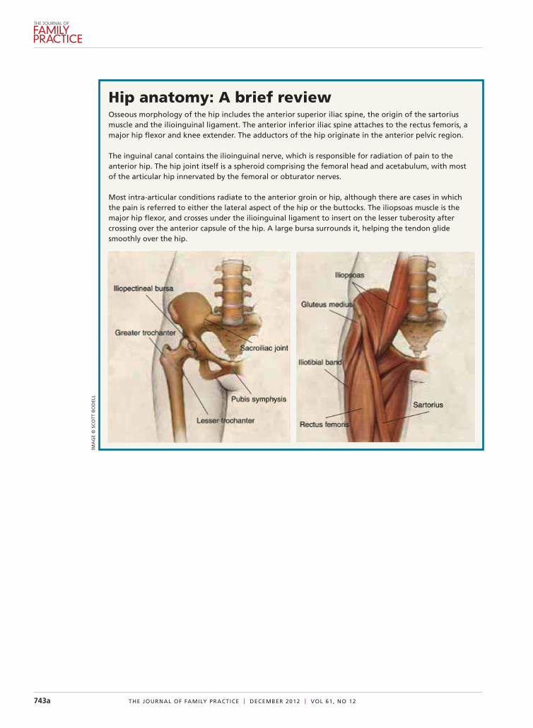
Hip anatomy: A brief review osseous morphology of the hip includes the anterior superior iliac spine, the origin of the sartorius muscle and the ilioinguinal ligament. The anterior inferior iliac spine attaches to the rectus femoris, a major hip flexor and knee extender. The adductors of the hip originate in the anterior pelvic region.
The inguinal canal contains the ilioinguinal nerve, which is responsible for radiation of pain to the anterior hip. The hip joint itself is a spheroid comprising the femoral head and acetabulum, with most of the articular hip innervated by the femoral or obturator nerves.
most intra-articular conditions radiate to the anterior groin or hip, although there are cases in which the pain is referred to either the lateral aspect of the hip or the buttocks. The iliopsoas muscle is the major hip flexor, and crosses under the ilioinguinal ligament to insert on the lesser tuberosity after crossing over the anterior capsule of the hip. a large bursa surrounds it, helping the tendon glide smoothly over the hip.
ima
ge
© s
co
TT b
oD
ell