(r)-mandelonitrile lyase from the fern - plant physiology
TRANSCRIPT

Plant Physiol. (1995) 109: 1231-1238
Purification and Characterization of a Nove1 (R)-Mandelonitrile Lyase from the Fern Phlebodium aureum'
Harald Wajant*, Siegfried Forster, Dirk Selmar, Franz Effenberger, and Klaus Pfizenmaier
lnstitut für Zellbiologie und lmmunologie der Universitat Stuttgart, Allmandring 31 (H.W., K.P.), and lnstitut für Organische Chemie der Universitat Stuttgart, Pfaffenwaldring 55 (S.F., F.E.), 70569 Stuttgart, Germany; and
Botanisches lnstitut der Technischen Universitat Braunschweig, Mendelssohnstrasse 4, 38092 Braunschweig, Germany (D.S.)
Using high-performance liquid chromatography and nuclear magnetic resonance we identified vicianin as the cyanogenic com- pound of Phlebodium aureum. The (R)-hydroxynitrile lyase involved during cyanogenesis in the catabolism of the aglycon ([RI-mande- lonitrile) was purified to apparent homogeneity. The purified ho- loenzyme is a homomultimer with subunits of M, = 20,000. At least three isoforms of the enzyme exist. In contrast to other hydroxyni- trile lyases, mandelonitrile lyase (MDL) from P. aureum was not inhibited by sulfhydryl- or hydroxyl-modifying reagents, suggesting a different catalytic mechanism. The enzyme is active over a broad temperature range, with maximum activity between 35 and 50"C, and a p H optimum at 6.5. In contrast to (RI-MDLs isolated from several species of the Rosaceae family, (R)-MDL from f . dureum is not a flavoprotein. The substrate specificity was investigated using immobilized enzyme and diisopropyl ether as solvent. The addition of cyanide to aromatic and heterocyclic carbonyls is catalyzed by this (R)-MDL, whereas aliphatic carbonyls are poorly converted.
If cyanogenic plants, which are widely distributed among higher plants, are damaged, HCN is released into the environment. This phenomenon, designated as cyano- genesis, is caused by the catabolism of cyanogenic glyco- sides or, in some cases, cyanogenic lipids (Seigler, 1991). Catabolism of cyanogenic glycosides is initiated by p-glu- cosidases, which hydrolyze the cyanogenic glycoside to cyanohydrin (a-hydroxynitrile) and a saccharide. Subse- quently, the unstable cyanohydrin decomposes spontane- ously or enzymatically by the action of an a-HNL to cya- nide and a carbonyl compound (Conn, 1981). Thus, it is suggested that the predominant physiological role of cya- nogenesis is protection from predators (Nahrstedt, 1985). However, for seedlings of Hevea brasiliensis, it was shown that cyanogenic glycosides also serve as N-storage com- pounds (Lieberei et al., 1985; Selmar et al., 1988).
To date, HNLs of various angiosperms have been puri- fied and characterized (Poulton, 1988; Kuroki and Conn, 1989; Wajant and Mundry, 1993; Hughes et al., 1994; White et al., 1994). According to their FAD content, HNLs can be divided into two groups: flavoprotein and nonflavoprotein
' This work was supported by Bundesministerium fiir For- schung und Technologie, Germany, grant No. A03U-ZSP Stuttgart and Deutsche Forschungsgemeinschaft grant No. WA 1025/1-1.
* Corresponding author; fax 49-711-685-7484. 1231
HNLs. Flavoprotein HNLs are exclusively found in two subfamilies of the Rosaceae family (Gerstner et al., 1968), whereas nonflavoprotein HNLs have been described for several families of higher plants (Poulton, 1988; Selmar et al., 1988; Kuroki and Conn, 1989). The flavoprotein HNLs isolated from Rosaceae species are (RI-MDLs. They are a11 glycoproteins of similar size (Poulton, 1988) and are sero- logically related (Gerstner and Pfeil, 1972). The flavopro- tein HNLs do not catalyze a net oxidation or reduction reaction (Jorns, 1979). Therefore, it was proposed that (R)- MDL from Rosaceae evolved from an ancestoral flavopro- tein, which lost its capability to catalyze oxidation/reduc- tion reactions. According to this hypothesis, the FAD of flavoprotein HNL serves to maintain the structural integ- rity of the enzyme (Jorns, 1979).
In contrast to the flavoprotein HNLs, which form a ho- mogenous group of enzymes, the nonflavoprotein HNLs form an extremely heterogenous group of proteins. They differ in size, extent of glycosylation, subunit composition, and substrate specificity (Poulton, 1988; Kuroki and Conn, 1989; Hughes et al., 1994). Even acetone cyanohydrin lyases from Linum usitafissimum and Manihot esculenta, which have the same cyanohydrin as natural substrate, show no common biochemical properties. Whereas the acetone cy- anohydrin lyase from L. usitatissimium is a homodimer of 82 kD catalyzing the synthesis of (X)-cyanohydrins (Xu et al., 1988; Albrecht et al., 1993), MeHNL (EC 4.1.2.37) is a homotrimeric molecule of 90 kD accepting (S)-cyanohy- drins as substrate (Hughes et al., 1994; Wajant et al., 1995). Furthermore, a serological cross-reactivity can be detected neither between MeHNL and HNL from L. usitafissimum (acetone cyanohydrin lyase; EC 4.1.2.37) nor among other nonflavoprotein HNLs (Wajant et al., 1995).
Recently, the genes encoding for the flavoprotein HNL from Prunus serotina (Cheng and Poulton, 1993) and the nonflavoprotein MeHNL (Hughes et al., 1994) and SbHNL (EC 4.1.2.11) (Wajant et al., 1994), respectively, were cloned. There are no obvious sequence homologies be-
Abbreviations: DFP, diisopropyl fluorophosphate; EE, enantio- meric excess; HNL, hydroxynitrile lyase; MDL, mandelonitrile lyase; MeHNL, HNL from Manihot esculenta (acetone cyanohydrin lyase); PaMDL, MDL from Prunus amygdalum; PhaMDL, MDL from Phlebodium aureum; SbHNL, HNL from Sorghum bicolor ([SI- p-hydroxy-MDL).
www.plantphysiol.orgon December 25, 2018 - Published by Downloaded from Copyright © 1995 American Society of Plant Biologists. All rights reserved.
1232 Wajant et al. Plant Physiol. Vol. 109, 1995
tween the deduced amino acid sequences of these HNLs. For the HNL from P. serotina, limited homologies to other flavoproteins were found (Cheng and Poulton, 1993). For MeHNL, no homologies to proteins with known function were described (Hughes et al., 1994). However, analysis of the SbHNL sequence revealed considerable homologies to Ser carboxypeptidases (Wajant et al., 1994). In particular, SbHNL contains a catalytic triad Ser-Asp-His (Wajant et al., 1994), which is described for three groups of independently evolved Ser proteases (Liao and Remington, 1990). There- fore, Ser proteases are regarded as an example of conver- gent molecular evolution (Liao and Remington, 1990). Fac- ing the lack of sequence homologies among the three cloned HNLs and taking into account the biochemical het- erogeneity of HNLs, it was proposed that HNLs also have independently evolved from severa1 ancestral enzymes (Wajant et al., 1994).
Here we describe the purification of a nove1 (R)-MDL from the fern Pklebodium aureum, which contains no FAD. To our knowledge, this MDL is the first HNL isolated from Filitaceae. PhaMDL (EC 4.1.2.10) possesses no common properties with the flavoprotein lyases from Rosaceae, ex- cept that it has the same natural substrate, mandelonitrile, which is released from prunasin in Prunus sp. and from vicianin in P. aureum, respectively. PhaMDL is a multimer of 20-kD subunits and is suitable for synthesis of (R)- cyanohydrins in organic media.
MATERIALS A N D METHODS
Plant Materials
Pklebodium aureum was cultivated at 22°C under natural light conditions. Directly after harvest, the leaves were frozen in liquid nitrogen, crushed using a mortar and pestle, and lyophylized.
Chemicals and Biochemicals
Except where noted, amino acid-modifying agents were purchased from Sigma. Protein standards for gel filtration were obtained from Pharmacia (catalase, aldolase, BSA, and ovalbumin) or Serva (Paramus, NJ) (carbonic anhy- drase and trypsin inhibitor). Protein standards for SDS- PAGE and chromatography resins were also obtained from Pharmacia, and the BCA protein assay kit was obtained from Pierce.
Extraction and Purification of Cyanogenic Glycosides
To remove lipids and other lipophilic substances, the freeze-dried powder was extracted with petrol ether using a soxhlet apparatus. To extract the cyanogenic glycosides, the defatted plant material was suspended in methanol (2 mL/g fresh weight) and homogenized in an Ultra Turrax homogenizer (IKA, Staufen, Germany) (3 X 60 s). After filtration, the methanol extract was evaporated. The dried material was dissolved in methanol/water (30:70), filtered (Spartam 308 [Schleicher & Schiill], 0.45 pm), and applied to an HPLC system, using a preparative RP-18 column (16 mm i.d.; 100 mm long; 15 pm particle size). Separation was
performed isocratically with methano1:water (30:70) at a flow rate of 5 mL/min. Cyanogenic glycosides were de- tected by cyanide liberation after their hydrolysis. For tliis, after evaporation of methanol, aliquots of each fraction were combined with P-glucosidase (emulsin, Serva, 2 mg/mL in phosphate buffer, pH 5.5) and incubated for 1 h at room temperature. To dissociate the hydroxynitriles pro- duced, NaOH (1 N) was added. After neutralization, cya- nide was analyzed using the Spectroquant kit for cyanide (Merck, Darmstadt, Germany). The fraction containing cy- anogenic glycosides was freeze dried and purified by a second HPLC, which was run on an analytical column (RP-18,4 mm i.d.; 250 mm long; 5 pm particle size). Again, separation was performed isocratically with methano1:wa- ter (30:70) at a flow rate of 1 mL/min.
Characterization and ldentification of Vicianin
For mass spectroscopy, the fast atom bombardment tech- nique with xenon and glycerol as the matrix was used. Mass spectroscopy was performed in positive as well. as in negative mode. 'H- and 13C-NMR spectra were recorded at ambient temperature with a Bruker (Bremen, Germany) 300 NMR-spectrometer in DMSO as solvent. For higher resolution, two-dimensional 'H-NMR spectra (correlated spectroscopy) were also obtained.
Assay for MDL Activity
MDL activity was determined by measuring the conver- sion of (R)-mandelonitrile to benzaldehyde and cy anide (Jorns, 1979). The increase of A,,9 caused by the production of benzaldehyde was recorded spectrophotometrically for 1 to 2 min. The slow base-catalyzed, nonenzymatic decom- position of mandelonitrile, determined in a separate con- trol reaction, was subtracted. Assays were carried out in 1 mL of 50 mM sodium acetate (pH 5.3) containing 0.084 mM mandelonitrile. The amount of enzyme that decomp'oses 1 pmol of mandelonitrile in 60 s under these conditions is defined as 1 unit.
Protein Concentration Determinations
Protein concentrations were determined by a commercial assay (BCA assay, Pierce) with BSA as standard.
Enzyme Purification
al., 1994). PaMDL was purified as described elsewhere (Lactble et
Purification of MDL from f . aureum
A11 steps of enzyme purification, except the fast protein liquid chromatography procedures, were carried out on ice.
Step 7 . Anion-Exchange Chromatography on Q-Sepharose FF
Ten to 20 g of freeze-dried powdered plant material of P. aureum were extracted three times with 15 volumes of 50
www.plantphysiol.orgon December 25, 2018 - Published by Downloaded from Copyright © 1995 American Society of Plant Biologists. All rights reserved.

Mandelonitrile Lyase from Phlebodium aureum 1233
mM sodium acetate (pH 5.7). The suspensions were centri- fuged at 40,OOOg for 30 min in a Ti 45 rotor, and the supernatants were collected. The pooled supernatants were loaded at a flow rate of 3 mL/min onto a Q-Sepharose FF filled HR10/10 column (Pharmacia), previously equili- brated with 20 mM sodium acetate, pH 5.7. Subsequently, the column was washed with 20 mM sodium acetate (pH 5.7) at the same flow rate until the AZs0 reached the initial base line. Bound materials were eluted with a 200-mL linear gradient of O to 0.4 M NaCl in 20 mM sodium acetate (pH 5.7) at a flow rate of 2 mL/min. Fractions of 5 mL were collected in an ice-cooled rack and assayed for MDL activ- ity, and positive fractions were pooled.
Step 2. Chromatofocusing on a Mono P HR 5/20 Column
Active pools from step 1 were dialyzed for 24 to 28 h with two changes against deionized H,O and were subse- quently applied at a flow rate of 0.5 mL/min onto a Mono P HR 5/20 column (Pharmacia), equilibrated in polybuffer PB 94 (Pharmacia) diluted 1:10 in 25 mM piperazine-C1 (pH 5.0). Bound proteins were eluted at a flow rate of 0.25 mL/min using a linear gradient (192 mL) of polybuffer PB 94 diluted 1:lO in 25 mM piperazine-C1 (pH 4.0), and active fractions were pooled.
Step 3. Gel-Filtration Chromatography on Superdex 200
Protein samples (5-10 mL) were applied to a HiLoad 26/60 Superdex 200 prep grade column (Pharmacia) and eluted with 20 mM Tris-C1 (pH 7.5) at a flow rate of 1 mL/min.
Step 4. Anion-Exchange Chromatography on Mono Q
Fractions from step 3 with high specific activity were pooled and concentrated by application on Mono Q HR 5/5 (Pharmacia), equilibrated in 20 mM Tris-C1 (pH 7.5). Protein was eluted in a 20-mL linear gradient of O to 0.4 M NaCl in 20 mM Tris-C1 (pH 7.5) a t a flow rate of 1 mL/min.
Step 5. Cation-Exchange Chromatography on Mono S
Fractions from step 4 with high specific activity were pooled, diluted 1 : l O with 30 mM sodium citrate (pH 3.9), and loaded at a flow rate of 1 mL/min onto Mono S HR 5/5 (Pharmacia), equilibrated with 30 mM sodium citrate (pH 3.9). Proteins were eluted using a linear gradient from O to 0.4 M NaCl in 30 mM sodium citrate (pH 3.9) at a flow rate of 0.5 mL/min.
Gel Electrophoresis
SDS-PAGE was performed with a 1-mm-thick polyacryl- amide gel as described by Laemmli (1970). Samples were boiled at 100°C for 5 min in the presence of 1% SDS and 75 mM DTT. Gels were silver stained according to the method of Blum et al. (1987).
Estimation of Apparent Molecular Mass
Apparent molecular masses of two native MDL isofofms were estimated by means of gel-filtration chromatography on a HiLoad 26/60 Superdex 200 prep grade column. Three milliliters of each purified isoform of MDL were loaded onto the column at a flow rate of 0.4 mL/min and eluted at the same flow rate with 20 mM Tris-C1 buffer (pH 7.5) containing 400 mM NaC1. The column was calibrated with the following protein standards: catalase (232 kD), aldolase (158 kD), BSA (67 kD), ovalbumin (43 kD), carbonic anhy- drase (29 kD), and trypsin inhibitor (20.1 kD).
Effects of Temperature and p H on MDL Activity
Enzyme reactions for temperature experiments were car- ried out in 100 mM sodium acetate (pH 6.0) containing 0.84 mM (R,S)-mandelonitrile. For determination of the pH de- pendente of MDL activity, the reaction was measured at 23°C. The buffers used for this purpose were 50 mM so- dium citrate (pH 3.3-5.2), 50 mM sodium acetate (pH 5.3- 6.5), and Tris-C1 (pH 6.5-7.5), each containing 0.84 mM (R,S)-mandelonitrile. Reactions were initiated by addition of 20 to 40 ng of MDL (0.37-0.74 unit). The observed activity was corrected for the base-catalyzed spontaneous decomposition of (R,S)-mandelonitrile at the appropriate temperature or pH.
Data Analysis of Enzyme Kinetics
The initial velocity for a wide range of substrate concen- trations was determined in triplicate in 100 mM sodium acetate (pH 6.0) at 23°C for fixed enzyme concentrations of MDL isoforms. The obtained data were analyzed according to the method of Michaelis and Menten (Palmer, 1981). An accurate estimation of V,,, was obtained from a Line- weaver-Burk plot (Palmer, 1981). Statistical parameters (confidence interval, SD) were determined using the Sigma Plot software from Jandel Scientific (Carle Madera, CA).
Effects of Additives
The effect of various inhibitors on the activity of purified MDL was examined using (R,S)-mandelonitrile as sub- strate (0.84 mM). The enzyme (approximately 100 ng) was incubated with the inhibitors at various concentrations in 100 p L of 20 mM sodium acetate (pH 6.0) at 37°C for 10 min. Subsequently, the enzymatic reaction was started by addition of 20 pL of enzyme solution to 1 mL of sodium acetate (pH 5.3) containing 0.84 mM mandelonitrile.
UV/Visible Spectra
Spectra were measured in a UVIKON 710 spectropho- tometer (Kontron, Neufahrn, Germany), recording 100 nm/min, using a 1-cm path length. The MDLs were ana- lyzed in 5 mM Tris-C1 (pH 7.5).
Synthesis of (R)-Cyanohydrins in Organic Media
PaMDL (EC 4.1.2.10) and PhaMDL were immobilized on avicel-cellulose as described elsewhere (Ziegler et al.,
www.plantphysiol.orgon December 25, 2018 - Published by Downloaded from Copyright © 1995 American Society of Plant Biologists. All rights reserved.
1234 Wajant et al. Plant Physiol. Vol. 109, 1'395
1990). Then, a mixture of immobilized enzyme, appropriate carbonyl (0.5 mmol), and HCN (2.6 mmol) in diisopropyl ether (4 mL) was shaken for 4.5 h at room temperature. To obtain the cyanohydrins, the immobilized enzyme was removed by filtration and the remaining solution was con- centrated. Reaction yields were determined by NMR, and optical purity (in percentage EE) was measured by GC.
RESULTS A N D DlSCUSSlON
ldentification of (R)-Vicianin
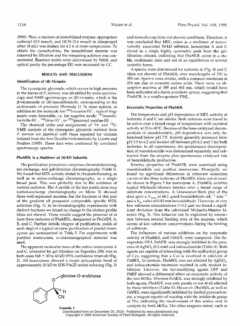
The cyanogenic glycoside, which occurs in high amounts in the leaves of P. aureum, was identified by mass spectros- copy and NMR spectroscopy as (X)-vicianin, which is the P-vicianoside of (X)-mandelonitrile, corresponding to the arabinoside of prunasin (Formula 1). In mass spectra, in addition to the molecule ion 426(vicianin-H)+, typical frag- ments were detectable, i.e. for negative mode: ','(mande- lonitrile-H)+, '49(Ara-H)+, or 293(prunasyl residue-H)+.
The chemical shifts and assignments of 'H- and I3C- NMR analysis of the cyanogenic glycoside isolated from P. aureum are identical with those reported for vicianin isolated from the fern Davallia trickomanoides by Lizotte and Poulton (1985). These data were confirmed by correlated spectroscopy spectra.
PhaMDL 1s a Multimer of 20-kD Subunits
The purification procedure comprises chromatofocusing, ion exchange, and gel-filtration chromatography (Table I). We found that MDL activity eluted in chromatofocusing, as well as in anion-exchange chromatography, as a single broad peak. This was probably due to the existence of various isoforms. The A profile of the last purification step (cation-exchange chromatography on Mono S) showed three well-separated maxima, but the fractions of this part of the gradient a11 possessed comparable specific MDL activities (Fig. 1). In re-chromatography experiments with distinct fractions we found no change in the elution profile (data not shown). These results suggest the presence of at least three isoforms of PhaMDL, designated as PhaMDL A, B, and C. Further details (degree of purification, yields at each step) of a typical enzyme purification of pooled isoen- zymes are summarized in Table I. For experiments with purified isoenzymes, re-chromatographed material was used.
The apparent molecular mass of the native isoenzymes A and C, estimated by gel filtration on Superdex 200, was in both cases 168 t 30 to 40 kD (95% confidence interval) (Fig. 2). All isoenzymes showed a single polypeptide band of approximately 20 kD in SDS-PAGE under reducing (Fig. 3)
,glucose-O-arabi nose O
Formula 1
and nonreducing (data not shown) conditions. Therefore, it was concluded that MDL exists as a multimer of nonco- valently associated 20-kD subunits. Isoenzyme A and C eluted as a single highly symmetric peak from the gel- filtration column, indicating that PhaMDL exists in a sta- ble, multimeric state and not in an equilibrium of several unstable forms.
A spectra were determined for isoforms A (Fig. 4) and C (data not shown) of PhaMDL over wavelengths of :!30 to 450 nm. Spectra were similar, with a common maximum at 278 nm due to aromatic amino acids. There were no ab- sorption maxima at 389 and 463 nm, which would have been indicative of a flavin prosthetic group, suggesting that PhaMDL is a nonflavoprotein HNL.
Enzymatic Properties of PhaMDL
The temperature and pH dependence of MDL activity of isoforms A and C are similar. Both isoforms were found to be active over a broad range of temperature with maximal activity at 35 to 40°C. Because of the base-catalyzed d'ecom- position of mandelonitrile, pH dependence was only de- termined below pH 7.5. Activity generally increased from pH 3.5 to 6.2 and leveled off between pH 6.2 and 7 for both isoforms. In a11 experiments, the spontaneous deconnposi- tion of mandelonitrile was determined separately and sub- tracted from the enzyme plus spontaneous catalyzed rate of benzaldehyde production.
Kinetics properties of PhaMDL were examined using mandelonitrile and purified isoenzymes. Principally we found no significant differences in substrate saturation curves of the three isoforms of PhaMDL (data not shown). As shown in Figure 5 for isoenzyme A, PhaMDL exhibits typical Michaelis-Menten kinetics over a broad range of substrate concentrations. A Lineweaver-Burk plot of the data gave a V,,, of 60.1 pmol benzaldehyde mL-' min-' and a K , value of 0.83 mM benzaldehyde. However, at very low substrate concentrations (<0.2 p ~ ) we found a signif- icant deviation from the calculated Michaelis-Menten ki- netics (Fig. 5). This behavior can be explained by interac- tion between several binding sites of the enzyme, which occurs at low substrate concentrations during the biinding of substrate.
The influences of various additives on the enzymatic activity of PhaMDL and PaMDL were compared. The fla- voprotein HNL PaMDL was strongly inhibited in the pres- ente of AgNO, (0.2 miv) and iodoacetamide (Table 11). Both agents are capable of interacting with the sulfhydryl group of Cys, suggesting that a Cys is involved in catalysis of PaMDL. In contrast, PhaMDL was not affected by AgNO,, and iodoacetamide treatment resulted in only modest in- hibition. Likewise, the Ser-modifying agents DFP and PMSF showed a differential effect on enzymatic activity of the two MDLs. Whereas PaMDL was strongly inhibited by both agents, I'haMDL was only poorly or not at a11 affected by these inhibitors (Table 11). However, PhaMDL as well as PaMDL were significantly inhibited by diethyl pyrocarbon- ate, a reagent capable of reacting with the imidazole group of His, indicating the involvement of this amino acid in catalysis by both MDLs. The other reagents tested, such as
www.plantphysiol.orgon December 25, 2018 - Published by Downloaded from Copyright © 1995 American Society of Plant Biologists. All rights reserved.

Mandelonitrile Lyase f r om Phlebodium aureum
300 -
2 250 - 'E . r" 200 - U
a, o 150-
n v) -
100 - z
I 1 50 -
0 -
1235
Table 1. furification of PhaMDL All values were determined for pools comprising all three isoenzymes.
Step Total Protein Total Activitv Specific Activitv Yield Purification
mg units un its/mg % - fold
Crude extract 190 2,200 11.6 1 O0 - Q-Sepharose FF 20 2,100 105 95 9.1 Chromatofocusing 1.5 720 480 33 41 Gel filtration 0.2 600 3,000 27 260 Mono Q (pH 7.5) 0.1 2 600 5,000 27 430 Mono S (pH 3.9) 0.025 467 19,000 21 1,600
ions and reducing agents, did not affect the activities of the MDLs (Table 11).
Use of MDL for the Synthesis of (R)-Cyanohydrins in Organic Media
In recent years, several groups have developed proce- dures for the synthesis of optically active cyanohydrins (Effenberger et al., 1987, 1990; Smitskamp-Wilms et al., 1991). Such cyanohydrins are important building blocks for the synthesis of a-hydroxy acids, a-hydroxy ketones, or p-ethanolamines. In addition to chemical methods, such as the enantio-selective addition of trimethylsilylcyanide to aldehydes in the presence of chiral catalysts (Dalton et al., 1991; Hayashi et al., 1993), the HNL-catalyzed addition in organic media is of special interest (Effenberger et al., 1987, 1990). The use of HNLs in organic media avoids the base- catalyzed racemic addition, which spontaneously occurs in aqueous systems, and allows the conversion of substrates that are poorly soluble in water (Effenberger et al., 1987, 1990).
MDLs from Rosaceae have been successfully used for the synthesis of a wide range of aromatic and heterocyclic
0.0
g 0.1
4 5 0.1 ::
c
I 0.1
0.1
0.1
1 .o 0.9
0.8
0.7
0.6
0.5 2 2 0.4
0.3
0.2
0.1
0.0
E
O 5 10 15 20 25 30 35
fraction number
Figure 1. Cation-exchange chromatography of MDL isoenzymes. The MDL-containing fraction obtained after anion-exchange chro- matography on Mono S was applied at a flow rate of 1 mL/min on a Mono S HR 5/5 (Pharmacia), equilibrated with 30 mM sodium citrate (pH 3.9). The adsorbed enzyme was eluted using a linear gradient from O to 0.4 M NaCl in 30 mM sodium citrate (pH 3.9) at a flow rate of 0.5 mL/min. Fractions of 0.5 mL were collected and assayed for MDL activitv.
(R)-cyanohydrins (Effenberger et al., 1987; Smitskamp- Wilms et al., 1991), whereas synthesis of aliphatic (R)- cyanohydrins with MDLs from Rosaceae is hampered by the reduced specific activity of these enzymes for the re- spective carbonyls (Smitskamp-Wilms et al., 1991). How- ever, Albrecht et al. (1993) described the use of HNL from L. usitatissimum for these applications. Moreover, SbHNL has been used for the synthesis of (S)-cyanohydrins (Effen- berger et al., 1990; Smitskamp-Wilms et al., 1991). How- ever, this enzyme allows only the effective synthesis of aromatic (S)-cyanohydrins. The usefulness of MeHNL and HNL from H. brusiliensis (EC 4.1.2.37), which are (S)-HNLs, for the production of aliphatic (9-cyanohydrins is under investigation by several groups (Klempier et al., 1993; Wa- jant et al., 1995).
To evaluate the biotechnological potency of PhaMDL, we have used immobilized enzyme to catalyze the formation of aromatic, heterocyclic, and aliphatic (RI-cyanohydrins (Table 111) and compared it with the catalytic properties of PaMDL. Like PaMDL, PhaMDL preferentially accepted ar- omatic and heterocyclic carbonyls, but aliphatic carbonyls
170 180 190 200 210 220 230 240 250
elution volume [ml]
Figure 2. Estimation of native molecular mass of MDL A and C by gel-filtration chromatography on Superdex 200. Three milliliters of purified MDL A and C (10 pg/mL) were loaded onto a HiLoad 26/60 Superdex 200 preparative grade column (Pharmacia), equilibrated in 20 mM Tris buffer (pH 7.5) containing 400 mM NaCl a ta flow rate of 0.4 mL/min. Molecular mass markers (O) used were catalase (232 kD), aldolase (1 58 kD), BSA (67 kD), ovalbumin (43 kD), carbonic anhydrase (29 kD), and trypsin inhibitor (20.1 kD). Both isoenzymes, A and C, of MDL eluted in fractions corresponding to 168 k D (O). Dotted lines show the 95% confidence interval.
www.plantphysiol.orgon December 25, 2018 - Published by Downloaded from Copyright © 1995 American Society of Plant Biologists. All rights reserved.

1236 Wajant et al. Plant Physiol. Vol. 109, 1995
M A B CkD
94 -
67 -
43 -
30 -
20.1 -
14.4 -i
Figure 3. SDS-PAGE of MDL isoenzymes. Twenty microliters (20-60ng) of peaks A, B, and C (Fig. 1) were reduced by treatment with DTTand separated by electrophoresis on a 17% acrylamide gel. Proteinbands were visualized by silver staining. Molecular mass markers(lane M) used were phosphorylase B (94 kD, 64 ng), BSA (67 kD, 83ng), ovalbumin (43 kD, 147 ng), carbonic anhydrase (30 kD, 83 ng),soybean trypsin inhibitor (20.1 kD, 80 ng), and lactalbumin (14.4 kD,121 ng).
were not excluded. In contrast to PaMDL, 2-thiophen alde-hyde was an efficiently converted substrate for PhaMDL,indicating that use of PhaMDL may be superior to PaMDLin selected applications.
Comparison of PhaMDL with Other HNLsMolecular size and polypeptide arrangement are quite dif-
ferent from those of (R)-MDLs isolated from Rosaceae speciesas well as from those of (S)-MDL from Ximenia americana(Kuroki and Conn, 1989). Moreover, various inhibitors af-fected PhaMDL and PaMDL in a totally different manner(Table II). For PhaMDL, lack of inhibition by DFP, PMSF,
0.060
g 0,040 -
0,020 -
0,000 -
200 250 300 350 400 450
wavelength (nm)Figure 4. Absorption spectrum of purified PhaMDL. The spectrumwas determined for PhaMDL isoenzyme A with a protein concentra-tion of 40 /xg/mL in 50 mM sodium acetate, pH 5.7.
i I i i- 2 - 1 0 1 2 3 4
1/ISJ
T
0 1 2 3 4 5 6
(R,S)-mandelonitrile concentration (mM)
Figure 5. Response of MDL to increasing concentrations of (R,S)-mandelonitrile. Original data from triplicates of a typical experimentwith fixed amounts of MDL isoform A. Rate values (/xmol min~'mg~') for the dissociation of (R,5)-mandelonitrile to benzaldehydeand HCN were plotted against substrate concentration. A Lin-eweaver-Burk plot of the data gave a Vmax of 60.1 /xmol benzalde-hyde ml"1 min^1 and a Km value of 0.83 mM benzaldehyde (coef-ficient of determination value 0.99) (inset I). Inset II, The deviation ofexperimentally determined rate values from the calculated Michae-lis-Menten kinetics in the early interval of the curve (0-0.2 mM).
Table II. Effects of various reagents on MDL activityComparison of PaMDL and PhaMDL.
Reagent
lodoacetamide
PMSF
DFP
Diethyl pyrocarbonate
AgN03
DTTEDTAEGTACaCI2MgCI2MnCI2ZnCI2CuSO4
Concentration
10220.520.520.510.255511111
Inhibition
PaMDL PhaMDL
93705025805779509585<5<5<5<5<5<5<5<5
30<5<5<510<56060<5<5<5<5<5<5<5<5<5<5
www.plantphysiol.orgon December 25, 2018 - Published by Downloaded from Copyright © 1995 American Society of Plant Biologists. All rights reserved.

Mandelonitrile Lyase from Phlebodium aureum 1237
Table 111. MDL-catalyzed synthesis of (R)-cyanohydrins from car- bonyls and HCN
Comparison of PaMDL and PhaMDL. Reactions were carried out with immobilized enzyme in diisopropyl ether for 1 to 4.5 h at room temperature. Yields were determined by NMR, and E € of (R)-cyano- hydrins was measured by GC analysis of O-acetyl derivatives. n.d., Not determined.
Yield €E
PhaMDL PaMDL PhaMDL PaMDL Carbonyl
% % Benzaldehyde a2 99 99 99 Butanale 42 1 00 55 90 Thiophen-2-aldehyde 92 48 99 87 2-Pentanone 17 78 n.d. n.d.
iodoacetamide, and AgNO, suggest that there is no Cys or Ser involved in catalysis. In contrast, PaMDL was inhibited by a11 of these compounds, indicating the involvement of Ser and/or Cys in the catalytic mechanism of this enzyme. The inhibition of enzymatic activity by Ser-modifying agents has also been described for SbHNL (Wajant et al., 1994) and MeHNL (Wajant et al., 1995). Therefore, it seems possible that PhaMDL, in contrast to PaMDL, uses a catalytic mechanism different from other HNLs. Furthermore, in opposition to MDLs from Rosaceae, PhaMDL contains no FAD. Differences in the kinetics properties of both enzymes were found as well. We found for PhaMDL a K,,, value of 0.83 mM benzaldehyde and deviations from Michaelis-Menten kinetics a t low sub- strate concentrations, whereas for MDLs from Rosaceae, Michaelis-Menten kinetics have been described with a K,,, value of 94 FM benzaldehyde (Xu et al., 1986). Such striking differences between HNLs possessing the same natural sub- strate are also found for acetone cyanohydrin lyases from L. usitatissimum and M. esculenta. These HNLs differ in subunit composition, molecular size, and stereoselectivity for chiral substrates (Xu et al., 1988; Albrecht et al., 1993; Wajant et al., 1995). Taking into account the lack of sequence homologies among the three cloned HNLs, so far, and the biochemical heterogeneity described above and elsewhere (Wajant e t al., 1994,19951, we believe that convergent evolution of HNL is probable. According to this idea, severa1 species of plants have recruited distinct enzymes (proteases) during evolution to adopt them for the same function: the accelerated release of HCN from cyanohydrins.
ACKNOWLEDCMENTS
We thank Dr. Victor Wray (Gesellschaft fiir Biochemische For- schung, Braunschweig, Germany) for recording NMR spectra and Dr. Hans Martin Schiebel (Organisches Institut, Technische Uni- versitat Braunschweig, Germany) for performing the mass spec- troscopical analysis.
Received June 5, 1995; accepted October 10, 1995. Copyright Clearance Center: 0032-0889/95/109/1231/08.
LITERATURE ClTED
Albrecht J, Jansen I, Kula MR (1993) Improved purification of an (R)-oxynitilase from Linum usitatissimum (flax) and investigation of the substrate range. Biotechnol Appl Biochem 17: 191-203
Blum H, Beier H, Gross HJ (1987) Improved silver staining of plant proteins, RNA and DNA in polyacrylamide gels. Electro- phoresis 8: 93-99
Cheng IP, Poulton JE (1993) Cloning of cDNA of Prunus serotina (R)-( +)-mandelonitrile lyase and identification of a putative FAD-binding site. Plant Cell Physiol 34: 1139-1143
Conn EE (1981) Cyanogenic glycosides. In PK Stumpf, EE Conn, eds, The Biochemistry of Plants: A Comprehensive Treatise, Vol 7: Secondary Plant Products. Academic Press, New York, pp
Dalton DM, Garner CM, Fernández JM, Gladysz JA (1991) A new asymmetric synthesis of cyanohydrins and oxygen-functional- ized derivatives: stereoselective addition of cyanide to chiral rhenium aldehyde and ketone complexes of the formula [$ C,H,)Re(NO)(PPh)(O=CRR')]+BF;. J Org Chem 56: 6823-6829
Effenberger F, Horsch B, Forster S , Ziegler T (1990) Enzyme- catalyzed synthesis of 6)-cyanohydrins and subsequent hydro- lysis to (3-a-hydroxy-carboxylic acids. Tetrahedron Lett 31:
Effenberger F, Ziegler T, Forster S (1987) Enzyme-catalyzed cya- nohydrin synthesis in organic solvents. Angew Chem Int Ed Engl 2 6 458-460
Gerstner E, Matzke V, Pfeil E (1968) Zur chemischen und biolo- gischen Systematik der Rosaceen. Naturwissenschaften 55:
Gerstner E, Pfeil E (1972) Zur Kenntnis des Flavinenzyms Hy- droxynitril-Lyase (D-Oxynitrilase). Hoppe-Seylers Z Physiol Chem 353: 271-286
Hayashi M, Miyamoto Y, Inoue T, Oguni N (1993) Enantioselec- tive trimethylsilylcyanation of some aldehydes catalyzed by chiral Schiff base-titanium alkoide complexes. J Org Chem 58:
Hughes J, Carvalho JP de C, Hughes MA (1994) Purification, characterization, and cloning of a-hydroxynitrile lyase from cas- sava (Manihot esculenta Crantz). Arch Biochem Biophys 311:
Jorns MS (1979) Mechanism of catalysis by the flavoenzyme oxynitrilase. J Biol Chem 254: 12145-12152
Klempier N, Griengl H, Hayn M (1993) Aliphatic (3-Cyanohy- drins by enzyme catalyzed synthesis. Tetrahedron Lett 34:
Kuroki GW, Conn EE (1989) Mandelonitrile lyase from Ximeniu amevicana L.: stereospecificity and lack of flavin prosthetic group. Proc Natl Acad Sci USA 86: 6978-6981
Laemmli UK (1970) Cleavage of structural proteins during the assembly of the head of bacteriophage T4. Nature 227: 680-685
Lauble HP, Miiller K, Schindelin H, Forster S , Effenberger F (1994) Crystallization and preliminary X-ray diffraction studies of mandelonitrile lyase from almonds. Proteins 19: 343-347
Liao DI, Remington SJ (1990) Structure of wheat serine car- boxypeptidase I1 at 3.5-A resolution. J Biol Chem 265: 6528-6531
Lieberei R, Selmar D, Biehl B (1985) Metabolization of cyanogenic glucosides in Hevea brusiliensis. Plant Syst Evol 150 49-63
Lizotte PA, Poulton JE (1985) Identification of (R)-vicianin in Dnvallia trickomanoides Blume. Z Naturforsch 41c: 5-8
Nahrstedt A (1985) Cyanogenic compounds as protecting agents for organisms. Plant Syst Evol 1 5 0 3547
Palmer T (1981) Understanding Enzymes. John Wily & Sons, New York
Poulton JE (1988) Localization and catabolism of cyanogenic gly- cosides. Ciba Found Symp 140: 67-91
Seigler DS (1991) Cyanide and cyanogenic glycosides. In GA Rosenthal, MR Berenbaum, eds, Herbivores: Their Interactions with Secondary Plant Metabolites. Academic Press, New York,
Selmar D, Lieberei R, Biehl B (1988) Mobilization and utilization of cyanogenic glycosides. Plant Physiol 86: 711-716
Smitskamp-Wilms E, Brussee J, van der Gen A, van Scharren- burg GJM, Sloothaak JB (1991) Hydroxynitrile lyases from almond and sorghum as biocatalysts. Recl Trav Chim Pays-Bas Belg 110: 209-215
Wajant H, Forster S , Bottinger H, Effenberger F, Pfizenmaier K (1995) Acetone cyanohydrin lyase from Manihot esculenta (cas-
479-500
1249-1250
561-563
151 5-1 522
496-502
4769-4772
pp 35-77
www.plantphysiol.orgon December 25, 2018 - Published by Downloaded from Copyright © 1995 American Society of Plant Biologists. All rights reserved.

1238 Wa jan t et al. Plant Physiol. Vol. 109, 15195
sava) is serologically distinct from other hydroxynitrile lyases. Plant Sci 108: 1-11
Wajant H, Mundry KW (1993) Hydroxynitrile lyase from Sorghum bicolor: a glycoprotein heterotetramer. Plant Sci 89: 127-133
Wajant H, Mundry KW, Pfizenmaier K (1994) Molecular cloning of hydroxynitrile lyase from Sorghum bicolor (L.). Homologies to serine carboxypeptidases. Plant Mo1 Biol 26: 735-746
White W, McMahon J, Sayre RT (1994) Regulation of cyanogen- esis in cassava. Acta Hortic 375: 69-77
Xu LL, Singh BK, Conn EE (1986) Purification and characteriza- tion of mandelonitrile lyase from Prunus lyonii. Arch Biochem Biophys 250: 322-328
Xu LL, Singh BK, Conn EE (1988) Purification and characteriza- tion of acetone cyanohydrin lyase from Linum usitutiss,+". Arch Biochem Biophys 263: 256-263
Ziegler T, Horsch B, Effenberger F (1990) Ein einfacher Zugang zu (R)-a-Hydroxycarbonsauren und (R)-l-amino-2-alkoholen aus (R)-Cyanhydrinen. Synthesis 7: 575-578
www.plantphysiol.orgon December 25, 2018 - Published by Downloaded from Copyright © 1995 American Society of Plant Biologists. All rights reserved.