r. k. mishra - world laparoscopy hospital · retroperitoneal laparoscopy, using a laparoscope and...
TRANSCRIPT

World Laparoscopy Hospital Advance Laparoscopic Surgery
Retroperitoneoscopy
R. K. Mishra

World Laparoscopy Hospital Advance Laparoscopic Surgery
History
Bartel in 1969 first reported endoscopic visualization of the pelvic retroperitoneum
1974 Wittmoser performed a retroperitoneal endoscopic lumbar sympathectomy
The technique called ‘lumboscopy’ was extensively used and popularized by Sommerkamp (1974) for exposing the kidney for renal biopsy
Retroperitoneal laparoscopy, using a laparoscope and pneumoinsufflation, was only started in 1979 by Wickham, for performing a ureterolithotomy.

World Laparoscopy Hospital Advance Laparoscopic Surgery
Anatomy

World Laparoscopy Hospital Advance Laparoscopic Surgery
Anatomy

World Laparoscopy Hospital Advance Laparoscopic Surgery
Vascular Structures

World Laparoscopy Hospital Advance Laparoscopic Surgery
Patient Position
The patient should
placed in the flank
position, with the
kidney bridge
elevated.
Operating table bent
to widen the space
between the 12th rib
and the iliac crest

World Laparoscopy Hospital Advance Laparoscopic Surgery
Patient Position
World Laparoscopy Hospital Advance Laparoscopic Surgery
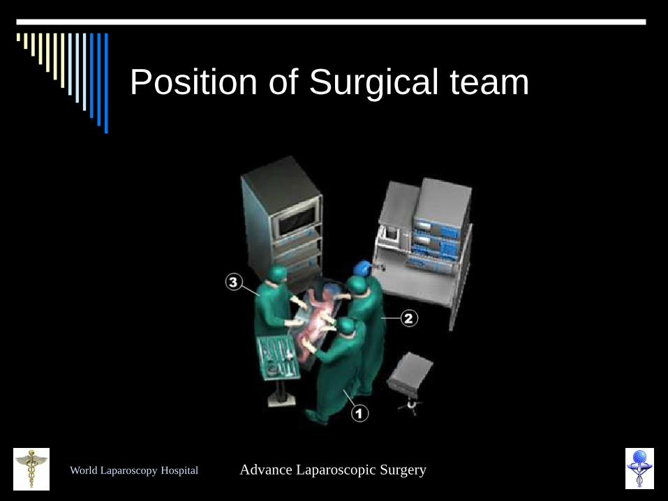
Position of Surgical team

World Laparoscopy Hospital Advance Laparoscopic Surgery
Difficulties
No preformed space in retroperitoneal
area
Pneumoretroperitoneum merely by
insufflation through a Veress needle is
impossible.
Break up the tough fibrous trabeculae &
dense areolar & fibro-fatty tissue is
necessary to allow creation of a
satisfactory pneumoretroperitoneum.

World Laparoscopy Hospital Advance Laparoscopic Surgery
Port Position
10 mm incision made between 12th rib & iliac crest over the posterior axillary line
Two more 5 mm ports inserted anterior and posterior to the camera port under videoscopic guidance.
A fourth port inserted if necessary just beneath the 12th rib and lateral to the paraspinal muscle.

World Laparoscopy Hospital Advance Laparoscopic Surgery
Port Position

World Laparoscopy Hospital Advance Laparoscopic Surgery

World Laparoscopy Hospital Advance Laparoscopic Surgery

World Laparoscopy Hospital Advance Laparoscopic Surgery

World Laparoscopy Hospital Advance Laparoscopic Surgery
Port position for nephrectomy
The flank muscle fibre is bluntly
separated
The Thoracolumbar fascia
gently pierced with a fingertip.
A space is created by gentle
finger dissection of the retro
peritoneum, anterior to the
psoas muscle and posterior to
Gerota's fascia.

World Laparoscopy Hospital Advance Laparoscopic Surgery
Retroperitoneal space
Balloon Canulla
System is necessary to
create retroperitoneal
space
Balloon should be
inflated with 400ml to
800ml of normal saline
Pressure of balloon
should not be more
than 100 cm of water

World Laparoscopy Hospital Advance Laparoscopic Surgery
Access technique
Separation of Garota’s fascia from psoas muscle by balloon should be under
vision
Filling by balloon should be maintained for 5 minutes to achieve adequate
haemostasis

World Laparoscopy Hospital Advance Laparoscopic Surgery
Access Technique
After removing balloon the port wound size is decreased
12 to 20 mm of Hg pressure is required to maintain retroperitoneal
working space.
All the trocar should be ideally fixed with sterile adhesives

World Laparoscopy Hospital Advance Laparoscopic Surgery
Retroperitoneal Laparoscopic
Nephrectomy

World Laparoscopy Hospital Advance Laparoscopic Surgery
Retroperitoneoscopic
Ureterolithotomy

World Laparoscopy Hospital Advance Laparoscopic Surgery
THANK YOU