quiz 3: page 76 fascial fitness: by thomas myers · pdf filelead to new take-aways: ... in...
TRANSCRIPT

CECap
pr
ov
ed
QUIZ 3: PAGE 76
If you are interested in the role of fascia infitness training, the following questionslead to new take-aways:
• Most injuries are connective-tissue (fas-cial) injuries, not muscular injuries—sohow do we best train to prevent and repair damage and build elasticity and resilience into the system?
• There are 10 times more sensory nerveendings in your fascia than in your mus-cles; therefore, how do we aim proprio-ceptive stimulation at the fascia as well asthe muscles?
• Traditional anatomy texts of the musclesand fascia are inaccurate, based on a fun-damental misunderstanding of our move-ment function—so how can we work withfascia as a whole, as the “organ system ofstability”?Consciously or unconsciously, you have
been working with fascia for your wholemovement career—it is unavoidable. Now,however, new research is reinforcing the importance of fascia and other connectivetissue in functional training (Fascia Congress2009). Fascia is much more than “plasticwrap around the muscles.” Fascia is the organ system of stability and mechano-regulation. Understanding this may revolu-tionize our ideas of “fitness” (Varela & Frenk1987). Research into the fascial net upsetsboth our traditional beliefs and some of ournew favorites as well. The evidence all pointsto a new consideration within overall fitnessfor life—hence the term fascial fitness. Thisarticle lays out the emerging picture of thefascial net as a whole and explores three ofthe many aspects of recent research that giveus a better understanding of how best to trainthe fascial net. >>
FASCIAL FITNESS:B y T h o m a s M y e r s
38 Apri l 2011 IDEA Fitness Journal
TRAINING
IN
THE
NEURO

Apri l 2011 IDEA Fitness Journal 39
Research shows why taking a different
approach to exercise and the movement
brain is the wave of the future.
MYOFASCIALWEB
Muscle Isolation vs.
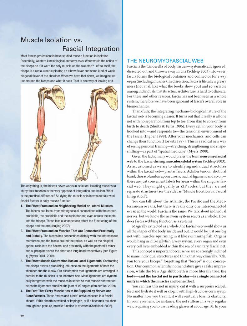
Fascial IntegrationMost fitness professionals have studied muscle function in isolation.Essentially, Western kinesiological anatomy asks: What would the action ofthe biceps be if it were the only muscle on the skeleton? Left to itself, thebiceps is a radio-ulnar supinator, an elbow flexor and some kind of weak diagonal flexor of the shoulder. When we have that down, we imagine weunderstand the biceps and what it does. That is one way of looking at it.
The only thing is, the biceps never works in isolation. Isolating muscles tostudy their function is the very opposite of integration and holism. What is the practical difference? Studying the muscle solo leaves out four vitalfascial factors in daily muscle function:1. The Effect From and on Neighboring Medial or Lateral Muscles.
The biceps has force-transmitting fascial connections with the coraco-brachialis, the brachialis and the supinator and even across the septainto the triceps. These fascial connections affect the functioning of the biceps and the arm (Huijing 2007).
2. The Effect From and on Muscles That Are Connected Proximallyand Distally. The biceps has connections distally with the interosseousmembrane and the fascia around the radius, as well as the bicipitalaponeurosis into the flexors; and proximally with the pectoralis minorand supraspinatus via the short and long head respectively (see Figure1) (Myers 2001, 2009).
3. The Effect Muscle Contraction Has on Local Ligaments. Contractingthe biceps exerts a stabilizing influence on the ligaments of both theshoulder and the elbow. Our assumption that ligaments are arranged inparallel to the muscles is an incorrect one. Most ligaments are dynami-cally integrated with the muscles in series so that muscle contractionhelps the ligaments stabilize the joint at all angles (Van der Wal 2009).
4. The Fact That Every Muscle Has to Be Supplied by Nerves andBlood Vessels. These “wires and tubes” arrive encased in a fascialsheath. If this sheath is twisted or impinged, or if it becomes too shortthrough bad posture, muscle function is affected (Shacklock 2005).
40
THE NEUROMYOFASCIAL WEBFascia is the Cinderella of body tissues—systematically ignored,dissected out and thrown away in bits (Schleip 2003). However,fascia forms the biological container and connector for everyorgan (including muscles). In dissection, fascia is literally a greasymess (not at all like what the books show you) and so variableamong individuals that its actual architecture is hard to delineate.For these and other reasons, fascia has not been seen as a wholesystem; therefore we have been ignorant of fascia’s overall role inbiomechanics.
Thankfully, the integrating mechano-biological nature of thefascial web is becoming clearer. It turns out that it really is all onenet with no separation from top to toe, from skin to core or frombirth to death (Shultz & Feitis 1996). Every cell in your body ishooked into—and responds to—the tensional environment ofthe fascia (Ingber 1998). Alter your mechanics, and cells canchange their function (Horwitz 1997). This is a radical new wayof seeing personal training—stretching, strengthening and shape-shifting—as part of “spatial medicine” (Myers 1998).
Given the facts, many would prefer the term neuromyofascialweb to the fascia-dissing musculoskeletal system (Schleip 2003).As accustomed as we are to identifying individual structureswithin the fascial web—plantar fascia, Achilles tendon, iliotibialband, thoracolumbar aponeurosis, nuchal ligament and so on—these are just convenient labels for areas within the singular fas-cial web. They might qualify as ZIP codes, but they are notseparate structures (see the sidebar “Muscle Isolation vs. FascialIntegration”).
You can talk about the Atlantic, the Pacific and the Medi -terranean oceans, but there is really only one interconnectedocean in the world. Fascia is the same. We talk about individualnerves, but we know the nervous system reacts as a whole. Howdoes fascia webbing function as a system?
Magically extracted as a whole, the fascial web would show usall the shapes of the body, inside and out. It would be just one bignet with muscles squirming in it like swimming fish. Organswould hang in it like jellyfish. Every system, every organ and evenevery cell lives embedded within the sea of a unitary fascial net.
This concept is important because we are so strongly inclinedto name individual structures and think that way clinically: “Oh,you tore your biceps,” forgetting that “biceps” is our concep-tion. Our common scientific nomenclature gives a false impres-sion, while the New Age shibboleth is more literally true: thebody—and the fascial net in particular—is a single connectedunity in which the muscles and bones float.
You can tear this net in injury, cut it with a surgeon’s scalpel,feed and hydrate it well or clog it with high-fructose corn syrup.No matter how you treat it, it will eventually lose its elasticity.In your eye’s lens, for instance, the net stiffens in a very regularway, requiring you to use reading glasses at about age 50. In your

Apri l 2011 IDEA Fitness Journal 41
skin, the net frays to cause wrinkles. Key elements like hip carti-lage may fail you before you die, and need replacement, but whenyou finally breathe your last breath your fascial web will still bethe same single net you started with.
It’s no small wonder that this system, like the nervous and cir-culatory systems, would develop complex signaling and homeo-static mechanisms (Langevin et al. 2006). The larger wonder isthat we have not really seen or explored the connective-tissue sys-tem’s responses until now.
A DEFINITION OF TERMSIn medicine, the term fascia designates tissues with specific topol-ogy and histology, as distinct from tendon, ligament or otherspecified tissues. In this article, however, we are using fascia asan overall name for this systemic net of connective tissue, becausethere is no generalized term (Huijing & Langevin 2009).
Connective tissue includes the blood and blood cells, andother elements not part of the structural net we are examining.Perhaps the closest term would be extra-cellular matrix (ECM),which includes everything in your body that isn’t cellular (seeFigure 3). The ECM has three main elements:• fibers: the strong pliable weave—consisting primarily of col-
lagen (which has 12 types) and its cousins elastin and retic-ulin—that both separates compartments and binds themtogether
• glue: the variable and colloidal gels like heparin, fibronectinand hyaluronic acid that accommodate change and providethe substrate for other cells like nerves and epithelia
• water: the fluid that surrounds and permeates the cells as amedium of exchange; mixes with the glue to make materialsof differing properties; and keeps the fibers wet and pliableThough the ECM will be our topic just below, the term fascia
as we define it also includes fibroblasts and mast cells, which giverise to the fibers and glue and then remodel them in responseto the demands of injury, training and habit.
The principal structural element in the ECM comprises thefibers collagen, elastin and reticulin. Collagen is by far the mostcommon of these, and by far the strongest. This is the white,sinewy stuff in meat. The collagen fiber is a triple helix; if it wasa half-inch thick, it would be about a yard long and look like anold three-strand rope (Snyder 1975). Collagen fibers can bearranged in regular directional rows, as they are in tendons orligaments (dense regular), or in random crisscross ways, like felt(dense or loose irregular).
The collagen fibers cannot actually stick to each other but areglued together by other proteins called glycoaminoglycans(GAGs), which are mucopolysaccharides, both of which are longwords for snot. We are held together by mucous, a colloidal sub-stance, which, by varying its chemistry slightly, can display a sur-prising array of properties, from thick and sticky to fluid and
lubricating. The fernlike molecules of mucous open to absorbwater (they are hydrophilic) or close and bind to themselveswhen water is absent. Depending on their chemistry, they eitherbind layers together or allow them to slide on each other(Grinnell 2008).
The phenomenon we call “stretch” or lengthening (and thatscientists call “creep” or hysteresis) is a function not of the colla-gen fibers lengthening but of the fibers sliding along each otheron the glue of the hydrated GAGs (Sbriccoli et al. 2005). Take thewater out of the GAGs, and the result is tissue that is mightily reluctant to stretch (Schleip 2003).
Most injuries occur when connective tissue is stretched fasterthan it can respond. The less it is hydrated, the less elastic response it has in it.
The Body Electric?
Connective-tissue cells produce the fibers and the GAGs, andthese materials are then altered to form a remarkable variety ofbuilding materials. If you were to try to recreate your structuralbody out of items you could buy at Home Depot®, what wouldyou need? Wood or PVC for the bones, silicon rubber for the car-tilage, lots of string, wire, tubing, plastic sheeting, rubber bands,cotton, nets, grease and oil—the list goes on. Would you try tobuild a body without duct tape?
Your body manufactures all these materials and many moreby mixing together various proportions of the ECM’s fibers andglue and altering the chemistry in different ways (Snyder 1975).In bone, the fiber matrix is there—much like leather—but themucousy ground substance has been systematically replaced withmineral salts. Cartilage has the same leathery substrate, but theglue has been dried into a tough but pliable “plastic” that perme-ates the fibrous leather. In ligament and tendon, almost all theglue has been squeezed out. In blood and joint fluid, the fiber exists only in a liquid form, until it hits the air, when it forms ascab. This manufactory in your body is fascinating: the dentin inyour teeth, your gums, your heart valves, even the clear corneaof your eye—are all formed in this fashion. >>
FIGURE 1. Deep Front Arm Line
Al and the DFAL blue line. The Anatomy TrainsMyofascial Meridians map out the longitudinalfascial connections that link one muscle to thenext as functional wholes.

42 Apri l 2011 IDEA Fitness Journal
Remodeling and Tensegrity
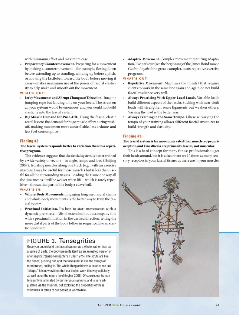
Your muscles may determine your shape in the training sense,but connective tissue determines your shape in the overall sense.It holds the bones together, pulling in on them as they press out(like a tensegrity system; see Figure 3).
The ECM is capable of remodeling itself in a variety of ways(Chen et al. 1997). Just as your muscles remodel themselves inresponse to training, the fascia remodels itself in response todirect signaling from the cells (Langevin et al. 2010); injury(Desmouliere, Chapponnier & Gabbiani 2005); long-held mechanical forces (Iatrides et al. 2003); use patterns (includingemotional ones); gravity; and certain chemistry within your body(Grinnell & Petroll 2010). The complexities of remodeling arejust now being explored in the lab; the details will be revealedover the coming decade.
The idea of tensegrity (tension and integrity) and the phe-nomenon of remodeling are the basis for structural therapy, including yoga and the forms of manual therapy commonlyknown as Rolfing®, or Rolfing Structural Integration, and itsdeep-tissue relatives, including foam rolling. Change the de-mand—as we do in bodywork and personal training—and thefascial system responds to that new demand. This commontheme points to a future where manual therapy and movementtraining combine to form a powerful method for • restoring natural settings for posture and function;• steering small problems away from developing into big ones
later on;• easing the long-term consequences from injury; and • extending functional movement farther and farther up the
age scale.
HOW TO TRAIN THENEUROMYOFASCIAL WEBIf the fascia is a singular space-organizing adjustable tensegritythat traverses the whole body and regulates—both locally and asa whole—the biomechanics of tension and compression, we canthen ask: How can we train this system, in conjunction with ourwork on muscles and neural control, to prevent and repair injuryand build resilience into the system?
The answer to this question is still developing—rapidly—bothin the laboratory and on the training floor. Some research is con-firming our images and practices as they have developed and aretraditionally applied. Here we focus on a few surprising sets of
findings that are (or soon will be) changing our ideas of how theneuromyofascial web really works and what role connective tis-sue plays in developing overall fitness for life. More of these results can be found at www.fascialftness.de or in the fascial fit-ness section of www.anatomytrains.com.
Finding #1
Specific training can enhance the fascial elasticity essential tosystemic resilience.
Fascial elasticity has not been recognized until recently, andthe mechanisms involved are still being studied (Chino et al.2008). Nevertheless, applications to training are already evident.The basic news is that connective tissue—even dense tissues liketendons and aponeuroses—is much more significantly elasticthan previously thought. The second essential part of that newsis that fascial elasticity is stored and returned very quickly. Inother words, it is more like a superball than a Nerf™ ball. Thus,fascial elasticity is a factor only when the motion is cyclic andquickly repeated, as in running, walking or bouncing, but not asin bicycling, in which the repetitive cycle is far too slow to takeadvantage of fascia’s elastic properties.
Measurements of calf lengthening during running haveshown that much of the length required for dorsiflexion is com-ing from an elastic stretch of the fascia, while the muscle is con-tracting isometrically (Kubo et al. 2006). This contradicts ourprevious understanding that the tendon was nonelastic, and thatthe muscles were lengthening and shortening during these cyclicmotions prior to and following footfall.
The runners who train for and employ more of this elastic-ity will be using less muscle power (read: less glucose) duringtheir runs, as they are storing energy in the stretch and then get-ting it back during the release. Thus, they will be able to runlonger with less fatigue.
Building in this elasticity is a matter of putting a demand onthe tissues to act in this way. Doing this slowly (compared withmuscle training) is a definite attribute of fascial training (it maytake 6–24 months to build fascial elasticity).W H AT ’ S I N :
• Bouncing. When you land on the ball of your foot, you decel-erate and accelerate in such a way that you not only make useof but actually build elasticity into the tendons and entire fas-cial system. The best training effect seems to follow the pleas-ure principle: feel for that sense of elegance, an ideal resonance
This article uses the generalized term fascia to denote the interconnected net of fibers and glue. 2A. Two muscles held together by “fuzz”—areolar tissue. 2B.The “strapping tape” nature of the fascia covering the quadriceps. 2C. The very delicate, gluey tissue that allows change and movement beneath our skin, be-tween our muscles, and anywhere anatomical structures have to slide on each other.
2A 2B 2C
FIGURE 2. Title
Apri l 2011 IDEA Fitness Journal 43
with minimum effort and maximum ease.• Preparatory Countermovement. Preparing for a movement
by making a countermovement—for example, flexing downbefore extending up to standing, winding up before a pitch,or moving the kettlebell toward the body before moving itaway—makes maximum use of the power of fascial elastic-ity to help make and smooth out the movement.
W H AT ’ S O U T:
• Jerky Movements and Abrupt Changes of Direction. Imaginejumping rope but landing only on your heels. The stress onall your systems would be enormous, and you would not buildelasticity into the fascial system.
• Big Muscle Demand for Push-Off. Using the fascial elastic recoil lessens the demand for huge muscle effort during push-off, making movement more controllable, less arduous andless fuel-consumptive.
Finding #2
The fascial system responds better to variation than to a repeti-tive program.
The evidence suggests that the fascial system is better trainedby a wide variety of vectors—in angle, tempo and load (Huijing2007). Isolating muscles along one track (e.g., with an exercisemachine) may be useful for those muscles but is less than use-ful for all the surrounding tissues. Loading the tissue one way allthe time means it will be weaker when life—which is rarely repet-itive—throws that part of the body a curve ball.W H AT ’ S I N :
• Whole-Body Movements. Engaging long myofascial chainsand whole-body movements is the better way to train the fas-cial system.
• Proximal Initiation. It’s best to start movements with a dynamic pre-stretch (distal extension) but accompany thiswith a proximal initiation in the desired direction, letting themore distal parts of the body follow in sequence, like an elas-tic pendulum.
• Adaptive Movement. Complex movement requiring adapta-tion, like parkour (see the beginning of the James Bond movieCasino Royale for a great example), beats repetitive exerciseprograms.
W H AT ’ S O U T:
• Repetitive Movement. Machines (or minds) that requireclients to work in the same line again and again do not buildfascial resilience very well.
• Always Practicing With Upper-Level Loads. Variable loadsbuild different aspects of the fascia. Sticking with near-limitloads will strengthen some ligaments but weaken others.Varying the load is the better way.
• Always Training in the Same Tempo. Likewise, varying thetempo of your training allows different fascial structures tobuild strength and elasticity.
Finding #3
The fascial system is far more innervated than muscle, so propri-oception and kinesthesia are primarily fascial, not muscular.
This is a hard concept for many fitness professionals to gettheir heads around, but it is a fact: there are 10 times as many sen-sory receptors in your fascial tissues as there are in your muscles
FIGURE 3. TensegritiesOnce you understand the fascial system as a whole, rather than asa series of parts, the body presents itself as an animated version ofa tensegrity (“tension-integrity”) (Fuller 1975). The struts are likethe bones, pushing out, and the fascial net is like the strings ormembranes, pulling in. The whole thing achieves a balance we call“shape.” It is now evident that our bodies work this way cellularlyas well as on the macro level (Ingber 2008). Of course, our humantensegrity is animated by our nervous systems, and is very ad-justable via the muscles, but exploring the properties of thesestructures in terms of our bodies is worthwhile.

44 Apri l 2011 IDEA Fitness Journal
(Stillwell 1957). The muscles have spindles that measure lengthchange (and over time, rate of length change) in the muscles.Even these spindles can be seen as fascial receptors, but let’s bekind and give them to the muscles (Van der Wal 2009). For eachspindle, there are about 10 receptors in the surrounding fascia—in the surface epimysium, the tendon and attachment fascia, thenearby ligaments and the superficial layers. These receptors include the Golgi tendon organs that measure load (by measur-ing the stretch in the fibers), paciniform endings to measure pres-sure, Ruffini endings to inform the central nervous system ofshear forces in the soft tissues, and ubiquitous small interstitialnerve endings that can report on all these plus, apparently, pain(Stecco et al. 2009; Taguchi et al. 2009).
So when you say you are feeling your muscles move, this is abit of a misnomer. You are “listening” to your fascial tissues muchmore than to your muscles.
Here are three interesting findings that go along with this basic eye-opener: 1. Ligaments are mostly arranged in series with the muscles, not
in parallel (Van der Wal 2009). This means that when youtense a muscle, the ligaments are automatically tensed to sta-bilize the joint, no matter what its position. Our idea thatthe ligaments do not function until the joint is at its full extension or torsion is now outmoded; for example, ligamentsfunction all through a preacher curl, not just at the ends of themovement.
2. Nerve endings arrange themselves according to the forces thatcommonly apply in that location in that individual, not accord-ing to a genetic plan, and definitely not according to the anatom-ical division we call a muscle. There is no representation of a“deltoid” inside your movement brain. That’s just a concept over
in your cortex, not in your biological organization.3. Apparently, sensors in and near the skin are more active in
detecting and regulating movement than the joint ligamentreceptors (Yahia, Pigeon & DesRosiers 1993).
W H AT ’ S I N :
• Skin and Surface Tissue Stimulation to Enhance Pro -prioception. Rubbing and moving the skin and surface tis-sues is important to enhance fascial proprioception. Oneweightlifter is having good results scrubbing himself with a veg-etable brush before going into competition.
• Directing Clients to Feel Their Fascial Tissues. Taking atten-tion—your own and your client’s—away from the muscles andputting it into the surrounding fascial tissues can help preventinjury and make the perception of kinesthesia more accurateand fully informed. Sensuous body activity coupled with ahigh level of kinesthetic acuity (think: cat) may prevent injurybetter than being tough.
W H AT ’ S O U T:
• Isolated Muscle Orientation. Exercising a single muscle ormuscle group is nearly impossible; every exercise is stimulat-ing multiple nerves, involving multiple muscles and employ-ing fascial tissues all around the site of effort, as well as“upstream” and “downstream” from it.
• Joint-Receptor Emphasis. Given that the ligaments are often tensed by the muscles, the emphasis on joint receptors—while important—needs to be replaced with a more generalattention to the whole area, from the skin on down.
This discussion has focused on biomechanical factors; it hasomitted nutritional and humoral considerations, as well as con-stitutional differences in fascia, which have recently come up forstudy. A deeper understanding of the role of fascia in trainingchanges your perspective, your work, your words and your effect. Fascia is not just cling wrap. n
Thomas Myers is the author of the best-selling Anatomy Trains:Myofascial Meridians for Manual and Movement Therapists(Churchill-Livingston 2009) and director of Kinesis Inc., which offers professional development courses worldwide for a variety ofmovement-centered approaches. A student of Ida Rolf, MosheFeldenkrais and R. Buckminster Fuller, Myers has practiced inte-grative bodywork in the USA and Europe since 1974.
ReferencesChen, C.S., et al. 1997. Geometric control of cell life and death. Science, 276 (5317), 1425–28.Chino, K., et al. 2008. In vivo fascicle behaviour of synergistic muscles on concentric
and eccentric plantar flexion in humans. Journal of Electromyography and Kinesiology,18 (1), 79–88.
Desmouliere, A., Chapponier, C., & Gabbiani, G. 2005. Tissue repair, contraction, andthe myofibroblast. Wound Repair Regeneration, 13 (1), 7–12.
Fascia Congress. 2009. www.fasciacongress.org/2009. Fuller, R.B. 1975. Synergetics. New York: Macmillan.Grinnell, F. 2008. Fibroblast mechanics in three-dimensional collagen matrices. Trends
in Cell Biology, 12 (3), 191–93.
CECap
pr
ov
ed
Take Quizwww.ideafit.com/april-2011-courses
or mail the quiz on page tk.
You are “listening” to your fascial tissues much more than to your muscles.
Apri l 2011 IDEA Fitness Journal 45
Grinnell, F., & Petroll, W. 2010. Cell motility and me-chanics in three-dimensional collagen matrices.Annual Review of Cell and Developmental Biology, 26,335–61.
Horwitz, A. 1997. Integrins and health. ScientificAmerican, 276, 68–75.
Huijing, P. 2007. Epimuscular myofascial force transmis-sion between antagonistic and synergistic muscles canexplain movement limitation in spastic paresis.Journal of Biomechanics, 17 (6), 708–24.
Huijing, P.A., & Langevin, H. 2009. Communicatingabout fascia: History, pitfalls and recommendations.In P.A. Huijing et al. (Eds.), Fascia Research II: BasicScience and Implications for Conventional andComplementary Health Care. Munich, Germany:Elsevier GmbH.
Iatrides, J., et al. 2003. Subcutaneous tissue mechanicalbehaviour is linear and viscoelastic under uniaxial ten-sion. Connective Tissue Research, 44 (5), 208–17.
Ingber, D. 1998. The architecture of life. ScientificAmerican, 278, 48–57.
Ingber, D. 2008. Tensegrity and mechanotransduction.Journal of Bodywork and Movement Therapies, 12 (3),198–200.
Kubo, K., et al. 2006. Effects of series elasticity on the hu-man knee extension torque-angle relationship in vivo.Research Quarterly for Exercise & Sport, 77 (4), 408–16.
Langevin, H. 2006. Connective tissue: A body-wide sig-naling network? Medical Hypotheses, 66 (6), 1074–77.
Langevin, H., et al. 2010. Fibroblast cytoskeletal remod-eling contributes to connective tissue tension. Journalof Cellular Physiology. E-pub ahead of publication. Oct.13, 2010.
Myers, T.W. 1998. Kinesthetic dystonia, Journal ofBodywork and Movement Therapies, 2 (2), 101–14.
Myers, T.W. 2009. Anatomy Trains: Myofascial Meridansfor Manual and Movement Therapists. New York:Churchill-Livingston.
Sbriccoli, P., et al. 2005. Neuromuscular response tocyclic loading of the anterior cruciate ligament. TheAmercian Journal of Sports Medicine, 33 (4), 543–51.
Schleip, R. 2003. Fascial plasticity—a new neurobiolog-ical explanation. Journal of Bodywork and MovementTherapies. Part 1: 2003, 7 (1), 11–19; part 2: 2003, (2),104–16.
Shacklock, M. 2005. Clinical Neurodynamics. Burlington,MA: Butterworth-Heinemann.
Shultz, L., & Feitis, R. 1996. The Endless Web. Berkeley,CA: North Atlantic Books.
Snyder, G. 1975. Fasciae: Applied anatomy and physiol-ogy. Kirksville, MO: Kirksville College of Osteopathy.
Stecco, C., et al. 2009. Treatment of phantom-limb painaccording to the fascial manipulation technique: A pi-lot study. In P.A. Huijing et al. (Eds.), Fascia ResearchII: Basic Science and Implications for Conventional andComplementary Health Care. Munich, Germany:Elsevier GmbH.
Stillwell, D.L. 1957. Regional variations in the innerva-tion of deep fasciae and aponeuroses. The AnatomicalRecord, 127 (4), 635–53.
Taguchi, T., et al. 2009. The thoracolumbar fascia as asource of low back pain. In P.A. Huijing et al. (Eds.),Fascia Research II: Basic Science and Implications forConventional and Complementary Health Care.Munich, Germany: Elsevier GmbH.
Van der Wal, J. 2009 The architecture of the connectivetissue in the musculoskeletal system: An often over-looked functional parameter as to proprioception inthe locomotor apparatus. In P.A. Huijing et al. (Eds),Fascia Research II: Basic Science and Implications forConventional and Complementary Health Care.Munich, Germany: Elsevier GmbH.
Varela, F., & Frenk, S. 1987. The organ of form. Journalof Social and Biological Structures, 10 (1), 1073–83.
Yahia, L.H., Pigeon, P., & DesRosiers, E.A. 1993.Viscoelastic properties of the human lumbodorsal fas-cia. Journal of Biomedical Engineering 15, 425–29.