quantitative trait loci affecting the difference in ... · 1 quantitative trait loci affecting the...
TRANSCRIPT

1
Quantitative Trait Loci Affecting The Difference in Pigmentation
Between Drosophila yakuba and D. santomea
MARY ANNA CARBONE,* 1,
ANA LLOPART,†1,2
MATTHEW DEANGELIS,†,3
JERRY A. COYNE† AND TRUDY F. C. MACKAY
*
* Department of Genetics, North Carolina State University, Raleigh, North Carolina 27695
† Department of Ecology and Evolution, University of Chicago, Chicago, Illinois 60637
1 The first two authors contributed equally to this work
2 Present address: Department of Biological Sciences, The University of Iowa, Iowa City, IA
52242
3 Present address: Pediatric Intensive Care Unit, Lucile Packard Children’s Hospital
725 Welch Road, Palo Alto, CA 94304
Genetics: Published Articles Ahead of Print, published on June 21, 2005 as 10.1534/genetics.105.044412

2
Running head: QTL for Drosophila pigmentation
Keywords: Speciation, species differences, composite interval mapping
Corresponding author: Mary Anna Carbone
Department of Genetics
Campus Box 7614
North Carolina State University
Raleigh, NC 27695
Tel: 919-515-5789
Fax: 919-515-3355
E-mail: [email protected]

3
ABSTRACT
Using quantitative trait locus (QTL) mapping, we studied the genetic basis of the difference in
pigmentation between two sister species of Drosophila: D. yakuba, which, like other members of
the D. melanogaster subgroup, shows heavy black pigmentation on the abdomen of males and
females, and D. santomea, an endemic to the African island of São Tomé, which has virtually no
pigmentation. Here we mapped four QTLs with large effects on this interspecific difference in
pigmentation: two on the X chromosome, and one each on the second and third chromosomes.
The same four QTLs were detected in male hybrids in the backcrosses to both D. santomea and
D. yakuba, and in the female D. yakuba backcross hybrids. All four QTLs exhibited strong
epistatic interactions in male backcross hybrids, but only one pair of QTLs interacted in females
from the backcross to D. yabuka. All QTLs from each species affected pigmentation in the same
direction, consistent with adaptive evolution driven by directional natural selection. The regions
delimited by the QTLs included many positional candidate loci in the pigmentation pathway,
including genes affecting catecholamine biosynthesis, melanization of the cuticle, and many
additional pleiotropic effects.

4
INTRODUCTION
During the Modern Synthesis, the dominant view of the genetics of species differences
was that of Ronald FISHER (1930), who believed that such differences were almost invariably
due to the accumulation of many genes, each of small phenotypic effect. Tests of this
proposition, however, were limited by the lack of genetic markers in most crossable but
differentiated species, although some data suggested that species differences could occasionally
be due to genes of large effect (ORR and COYNE 1992).
Recently, however, the advent of molecular techniques has improved our ability to study
the genetics of species differences. Two innovations have been crucial. First, quantitative trait
locus (QTL) mapping enables us to localize genes responsible for species differences by
determining their association with molecular markers at known sites. Second, molecular
techniques such as germline transformation enable us to determine directly whether a candidate
gene affects a species difference. ORR (2001) describes these innovations and the results of
recent genetical studies using them. Although most data derive from a small number of
organisms (ORR’s study describes only 13 analyses, six from Drosophila and four from the
monkeyflower genus Mimulus), the results show that while differences in traits between species
can be polygenic, genes of large effect are involved more frequently than previously suspected.
Here we describe a genetic analysis of a striking character difference — the degree of
abdominal pigmentation — between two sister species of Drosophila: D. yakuba and D.
santomea.
D. yakuba is widely distributed in open habitats across sub-Saharan Africa and the
islands near the continent (including Madagascar). In contrast, D. santomea is endemic to the
860 km2 volcanic island of São Tomé, in the Gulf of Benin 255 km west of the coast of

5
Gabon (LACHAISE et al. 2000). On the mountain Pico do São Tomé, D. yakuba occurs at
elevations below 1450 m, while D. santomea occupies the mist forests at elevations between
1153 and 2024 m. (D. yakuba is also widespread throughout lowland São Tomé, probably as
a result of a secondary invasion after the initial common ancestor evolved into D. santomea.)
Between about 1100 and 1450 m in elevation, the ranges of the two species overlap, with the
ratio of D. yakuba/D. santomea shifting from 2:1 to 1:20 as one moves upward through this
zone. The species show substantial sexual isolation when tested in the laboratory (LACHAISE
et al. 2000), and, using morphological criteria, one finds a low frequency (ca. 1%) of hybrids
in the zone of overlap.
Molecular evidence puts the divergence between D. yakuba and D. santomea at about
400,000 years ago (LLOPART et al. 2002). In interspecific crosses, F1 male hybrids are sterile
but female hybrids are fertile, and thus can be crossed to either parental species (LACHAISE et
al. 2000; CARIOU et al. 2001). This fertility permits genetic analysis using backcross
individuals.
The diagnostic differences between the species include male genital morphology and
sex-comb tooth number (LACHAISE et al. 2000; COYNE et al. 2004), but the most striking
difference involves abdominal pigmentation. Among the nine species in the D. melanogaster
subgroup, eight of them, including D. yakuba, have similar patterns of dark pigmentation:
males possess thin black stripes along the posterior portions of tergites 2, 3, and 4, while
tergites 5-7 are completely black. Females of these species have stripes along the posterior
portions of all tergites, and tergites 5-7 show substantial but not complete black pigmentation.
In contrast, D. santomea males show virtually no pigmentation, and females show only very
light striping on the posterior parts of tergites 2-5, with no pigmentation on other tergites.

6
(Photographs of these differences are given in Figures 1 and 2 of LACHAISE et al. 2000 and
Fig. 1 of LLOPART et al. 2002). Given the dark pigmentation in all but one species in the
subgroup, including the outgroup species D. orena and D. erecta, it is nearly certain that the
absence of dark pigmentation in D. santomea is a novel derived trait.
The adaptive significance of this pigmentation difference, if any, is unknown. Although
these species show strong sexual isolation (COYNE et al. 2002), this does not diminish when
flies are tested in the dark, suggesting that the pigmentation difference is not a cue for mate
discrimination (LLOPART et al. 2002). Moreover, the relationship between pigmentation and
temperature is opposite to that expected from other studies of Drosophila: individuals within a
species or closely related species living in colder conditions are almost invariably darker (e.g.,
DAVID et al. 1985; GIBERT et al. 1998), yet D. santomea, which lives at higher altitudes than
D. yakuba, is lighter.
In previous genetic analyses using three morphological markers and eight molecular
markers, we determined that at least three genes were involved in the pigmentation difference
between D. santomea and D. yakuba females, and five genes between males. In each case,
the genes resided on all three major chromosomes, with the X chromosome having a
particularly strong effect in males (LLOPART et al. 2002).
In this study we extend and refine our previous analysis, using a more accurate method of
measuring pigmentation as well as a QTL analysis employing 32 molecular markers, which
enables us to map “pigmentation genes” more accurately. Our goals are to determine the number
of genes involved in this morphological difference, their chromosomal locations, whether the
same genetic regions affect the pigmentation difference in both males and females, and whether
QTLs from a given species tend to affect the character in the same direction, implying that the

7
species difference evolved by natural selection (ORR 1998). Finally, the mapping of QTLs to
fairly restricted regions of the genome may eventually allow us to identify specific loci involved
in this species difference.
MATERIALS AND METHODS
Drosophila strains: All flies were maintained in 8-dram vials containing standard
cornmeal-agar-Karo media on a 12h:12h light:dark cycle at 24°C. One isofemale strain was used
from each species. The D. yakuba Taï 18 strain was derived from a female collected by D.
LACHAISE in 1983 in the Taï rainforest on the border between Liberia and the Ivory Coast. The
D. santomea STO.4 stock was derived from a female collected in March 1998 in the Obo Natural
Reserve on São Tomé Island. These two strains were used (and further described) in our
previous work on the genetics of pigmentation in these species (LLOPART et al. 2002). The
strains are homosequential in chromosome banding pattern except for the right arm of the second
chromosome: the D. yakuba Taï 18 strain is polymorphic for inversion 2Rn, which covers about
40% of the right arm of chromosome 2 (LEMEUNIER and ASHBURNER 1976).
Crosses: Backcross (BC) hybrids were produced by crossing four-day-old virgin D.
yakuba Taï 18 females to virgin D. santomea STO.4 males, and then backcrossing virgin F1
females to males from both species. Genetically, female BC hybrids to D. yakuba are either
homozygous D. yakuba or heterozygous D. yakuba/santomea, and have mitochondrial DNA
from D. yakuba. Similarly, female BC hybrids to D. santomea are either homozygous D.
santomea or heterozygous D. yakuba/santomea, and have mitochondrial DNA from D. yakuba.
Male BC hybrids have the same autosomal and mitochondrial genotypes as females, but the X-
linked loci are either pure D. santomea or D. yakuba, with the Y chromosome from the parental

8
male used in the backcross. We scored pigmentation in about 50 males and 50 females from
each of the pure species and the reciprocal F1 hybrids, and between 73 and 544 males and
females from each of the two backcrosses (Table 1). To improve the precision of QTL mapping,
we selected backcross individuals with extreme and intermediate pigmentation phenotypes for
subsequent pigmentation scoring and genotyping. For each of the first three BC genotypes listed
in Table 1, we selected flies of each sex from a sample of about 12,500 individuals (about 50
bottles, each containing about 250 flies of each sex). One-third of the total individuals chosen
were judged by eye to have very dark pigmentation, one-third to have very light pigmentation,
and the remaining third were chosen randomly from individuals with intermediate phenotypes.
(Equal numbers of all three classes were chosen from each bottle inspected until we had
accumulated about 500 flies of each sex in each backcross. Thus for each sex we selected about
4% of total individuals inspected; this stringent selection facilitates the precision of mapping).
However, females from the backcross of F1 individuals to D. santomea showed little variation in
pigmentation, and so all of these were chosen randomly.
Pigmentation scores: All scoring of pigmentation was done on 4-day-old virgin flies.
We scored only the three posterior tergites of each fly (segments 5, 6, and 7) by examining the
fly under a dissecting microscope. A pigmentation score was assigned based on both the
percentage of the tergite that was pigmented and the degree of pigmentation. First, the
proportion of the tergite covered by black pigment was estimated to the nearest 0.05 (5%). Then,
the relative degree of pigmentation was measured within the pigmented area. Using color
standards, we assessed the degree of pigmentation within the pigmented area using a five-point
scale ranging from 1 (very light pigmentation, slightly darker than background color) to 5 (dark,
shiny black), with intermediate numbers representing intermediate degrees of pigmentation.

9
Unpigmented areas were given a score of 0. We limited ourselves to assigning only three shades
of black to each tergite. The percentage of the area of each tergite covered by each shade of
pigmentation was then multiplied by the intensity of pigmentation, and these areas were
summed. This gives each tergite a minimum possible pigmentation score of 0 (no area
pigmented) to 5 (tergite completely covered with very dark pigmentation [1.0 × 5]). These areas
were summed for all three tergites, yielding a minimum possible pigmentation score for a given
fly of 0 and a maximum possible score of 15. As shown in Table 1, this procedure discriminates
well between the pigmentation of these species.
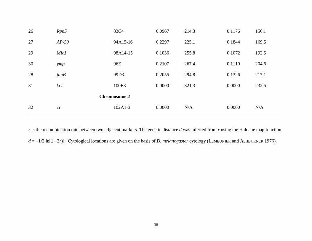
Molecular markers: We identified single nucleotide polymorphisms (SNPs) and
insertion/deletion variants (indels) that discriminated between the D. yakuba Taï18 and the D.
santomea STO.4 strains for 41 nuclear regions: y, per, sog, v, sn, Rux, f, bnb, Hex-A, AnnX,
su(f), l(2)gl, Rad1, RpL27A, Gart, salr, vkg, Rep4, Adh, His3, barr, Sara, Hex-C, Ngp, Kr, Lsp1-
γ, RpL14, dib, sfl, Sod, Est6, Ssl1, hb, Xdh (ry), Rpn5, AP-50, Mlc1, ymp, janB, krz, ci. Newly
reported sequences were deposited in GenBank under accession numbers DQ068949 (Gart_T18)
and DQ068950 (Gart_sto4); otherwise see LLOPART et al. (2005) for details. To determine
nucleotide sites differentially fixed between D. yakuba Taï18 and D. santomea STO.4, we tested
20 individuals (10 per strain) for each region by direct sequencing of PCR products obtained
from single fly DNA extractions (ASHBURNER 1989) using D. melanogaster or D. yakuba
primers. We cleaned PCR products using the Wizard Magnesil PCR clean-up system (Promega
Corp. Madison, WI), and sequenced them directly with an ABI PRISM® 3100 Genetic Analyzer
(Applied Biosystems Inc. Foster City, CA). We edited the sequences with the Sequencher 3.0
software (Gene Codes Corp. Ann Harbor, MI), and aligned them using the ClustalX program
(THOMPSON et al. 1997).
10
In total, we sequenced approximately 17.5 kb in each of the 20 flies tested. We detected
263 nucleotide differences fixed between Taï18 and STO.4, that is, 70% of the total nucleotide
variation. Among these fixed differences we selected 32 that affect a restriction endonuclease
site to be used as markers in the genotyping procedure. Table 2 lists the 32 markers, their
relative order within the D. yakuba chromosomes, and the conditions for genotyping.
We inferred the relative order of markers within each chromosome in D. yakuba/D.
santomea from the D. yakuba genome project (http://www.genome.wustl.edu/projects/yakuba/;
version 040407).
Marker genotypes: All BC individuals from the pigmentation assays were stored at –80oC
in 0.5 ml eppendorf tubes. Genomic DNA was extracted from each BC individual using the
Puregene (Gentra Systems; Minneapolis, MN) single-fly DNA extraction protocol. The sample
consisted of 506 BC D. santomea males, 73 BC D. santomea females, 537 BC D. yakuba males,
and 526 BC D. yakuba females. The genotypes of the 1642 BC hybrids were determined for all
32 markers (i.e. 52,544 genotypes).
The 32 molecular markers were designed using sequence data from the parental strains of
D. santomea and D. yakuba. The aligned sequences were used to develop PCR primers using
Primer3 (ROZEN and SKALETSKY 2000) and restriction enzyme (RE) digestions. Genotyping was
performed using Restriction Fragment Length Polymorphism (RFLP) analysis by PCR
amplification from genomic DNA using RedTaq DNA polymerase (Sigma; St. Louis, MO)
followed by RE digestion (see Table 2 for primers, RE, and conditions). All RE’s were
purchased from New England Biolabs (Beverly, MA) and primers were purchased from MWG
Biotech (High Point, NC). The digested PCR products were run on a 3% agarose gel stained
with ethidium-bromide, imaged with the Bio-Rad ChemiDoc System PC RS-170 using Quantity

11
One (version 4.2.1) software, and manually genotyped by assigning a “0” (homozygous D.
santomea), “1” (D. yakuba/D. santomea heterozygote) or “2” (homozygous D. yakuba) to each
marker genotype. A recombination map based on the 1642 BC hybrids (Table 3) was
constructed using the Haldane mapping function.
QTL mapping: QTLs affecting variation in pigmentation between D. yakuba and D.
santomea were mapped in each BC population using composite interval mapping (CIM; ZENG
1994) and implemented using QTL Cartographer software (BASTEN et al. 1999). CIM tests
whether an interval between two markers contains a QTL affecting the trait while simultaneously
controlling for the effect of QTL located outside the interval using multiple regression on marker
co-factors. Marker co-factors were chosen by forward selection – backward elimination
stepwise regression. The likelihood ratio (LR) test statistic is −2ln(L0/L1), where L0/L1 is the
ratio of the likelihood under the null hypothesis (i.e., there is no QTL in the test interval) to the
alternative hypothesis (there is a QTL in the test interval). LR test statistics were computed every
2 cM with marker co-factors 10 cM or more from the test location. We used permutation analysis
to determine appropriate significance thresholds that take into account the multiple tests
performed and correlations among markers. We permuted trait and marker data 1000 times, and
recorded the maximum LR statistic across all intervals for each permutation. LR statistics
calculated from the original data that exceed the 50th greatest LR statistic from the permuted data
are significant at the experiment-wise 5% level under the null hypothesis (CHURCHILL and
DOERGE, 1994; DOERGE and CHURCHILL, 1996). The approximate boundaries of regions
containing QTLs were determined by taking 2 LOD intervals (9.22 LR) surrounding the point of
greatest significance and interpolating the cytological location of the interval based on the
observed amount of recombination between flanking markers.

12
We estimated the effects of each QTL as the difference between the appropriate
homozygous genotypes and heterozygous D. santomea/D. yakuba genotypes at the peak LR,
scaled by the phenotypic standard deviation. The effects in females and autosomes of both sexes
are thus estimates of a − d in the cross to D. yakuba and −a − d in the cross to D. santomea,
where −a and a are, respectively, the genotypic values in D. santomea and D. yakuba, and d is
the heterozygous effect (FALCONER and MACKAY 1996). The effects of X-linked QTLs in males
are estimates of 2a.
We evaluated pairwise epistatic interactions between all possible marker pairs by running
ANOVA models to account for the main effects of all significant markers and one pairwise
interaction between markers (DILDA and MACKAY 2002). The ANOVAs were performed with
the PROC GLM procedure using SAS 8.02 software (Cary, NC). Interactions with P-values less
than 0.0001 are significant based on a Bonferroni correction for 496 tests per BC population. We
estimated the effects of significant two-locus interactions from the least-squares means of the
four marker locus classes as [(011 + 022) − (012 + 021)], where the first subscript is 1 if the marker
has a homozygous genotype for either parental species and 2 if the marker has a heterozygous
genotype, and the second subscript takes on the same values for the other marker in the
interaction. Standard errors of the interaction effects were estimated as described by DILDA and
MACKAY (2002).
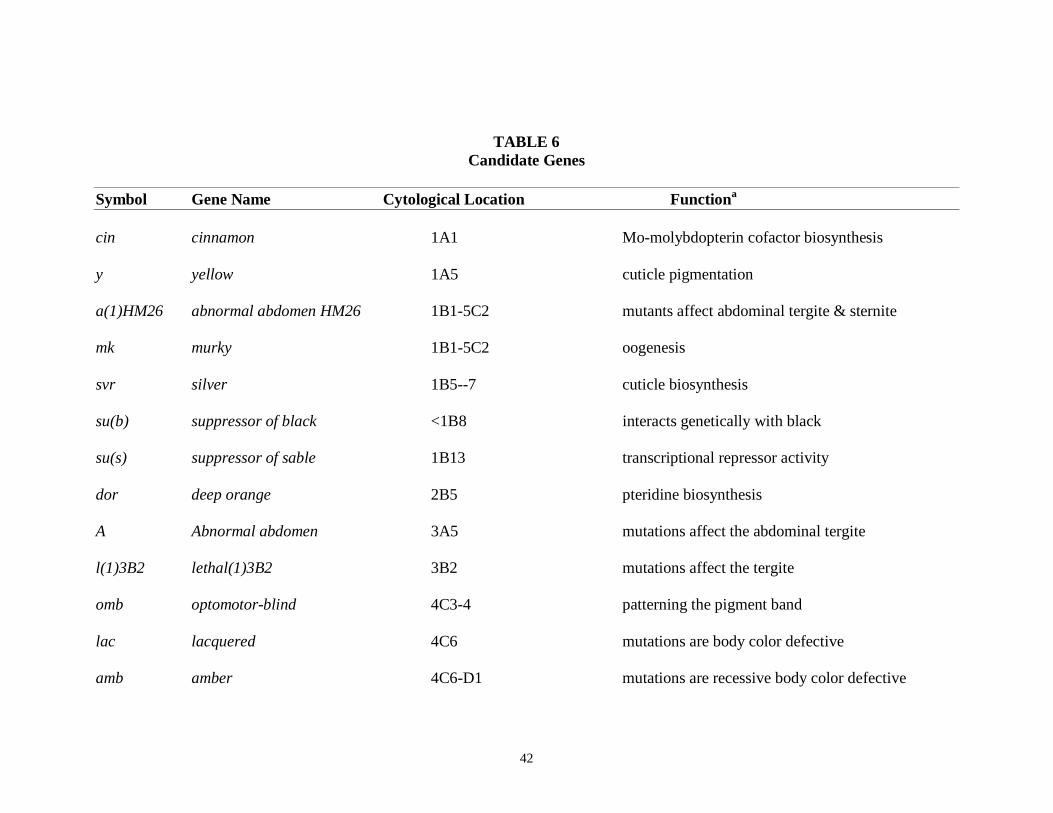
Candidate genes: Cytological bands in D. melanogaster of the markers that define the
interval under the QTL peak were obtained using Flybase (DRYSDALE and CROSBY, 2005).
Using the known cytological positions of these markers in D. melanogaster, we determined the
corresponding positions in D. yakuba (LEMEUNIER and ASHBURNER,1976; ASHBURNER, 1989).
This defined an interval both in D. melanogaster and in D. yakuba that allowed us to search for

13
candidate genes within that interval. We obtained a complete list of candidate genes involved in
pigmentation in D. melanogaster from Flybase (DRYSDALE and CrOSBY, 2005). Candidate genes
were identified based on the markers that delimit each QTL, and the presence of pigmentation
genes between these two markers (Table 6).
RESULTS
Pigmentation of pure species and F1 hybrids: Table 1 gives the mean pigmentation
scores, standard errors, and sample sizes for the pure species, the reciprocal F1 hybrids, and the
backcross hybrids used for genotyping. The difference between the pure species is substantial:
the mean pigmentation score of D. yakuba males and females is 14.22 and 9.85, respectively, and
for D. santomea males and females 0.63 and 1.02, respectively. As seen in our previous
analysis, (LLOPART et al. 2002) reciprocal F1 hybrid males show a large effect of the X
chromosome on pigmentation: these males have pigmentation scores fairly close to those of
males from the species of the maternal parent. The difference in pigmentation scores between
the two classes of F1 males is highly significant (t = 27.1, 100 d.f., P < 0.001). The relative effect
of the X chromosome in male pigmentation can be judged as the percentage of the total
difference between males of the two species explained by the difference between the reciprocal
F1 males; this effect is about 60%. This effect is much larger than the relative size of this
chromosome (constituting roughly 21% of the haploid genome [Table 3]) and suggests either
that the X chromosome carries a disproportionate number of genes affecting pigmentation, or that
individual X-linked genes have disproportionately large effects. (The QTL analysis below shows
that the second explanation is most likely to be correct.)

14
In contrast to males, the F1 females do not differ significantly in pigmentation scores
(Table 1; t = 1.55, 99 d.f., P = 0.12). There is thus no evidence for a maternal or mitochondrial
effect affecting pigmentation of these females, who are identical in nuclear genotype. The mean
score of all F1 females (4.45) is slightly lighter than the average score of females for the two
species (5.43), showing a small amount of dominance for the D. santomea phenotype.
QTLs affecting variation in pigmentation in BC hybrids: We mapped four QTLs with
large effects on pigmentation (Table 4, Figure 1). The same four QTLs were detected in male
hybrids in the backcrosses to both D. santomea and D. yakuba: two QTLs were on the X
chromosome (between markers 1-2 and markers 6-10), one QTL was on the second chromosome
(between markers 15-18) and one QTL was on the third chromosome (between markers 23-26).
The magnitude of the QTL effects ranged from 0.49 – 1.42 phenotypic standard deviations in the
backcross to D. santomea, and accounted for 67% of the total phenotypic variation. Similarly,
the QTL effects ranged from 0.62 – 1.62 phenotypic standard deviation in the backcross to D.
yakuba, and accounted for 58% of the total phenotypic variation. In both backcrosses, the sum of
the estimated QTL effects equaled or exceeded that expected from the difference between the
parental genotypes. In the backcross to D. santomea, the expected difference in pigmentation is
–11.29 (i.e. the difference in pigmentation between D. santomea males (0.63) and (Y X S) F1
males (11.92)); whereas the sum of the QTL effects was –11.64. In the backcross to D. yakuba,
the expected difference in pigmentation is 10.5 (i.e. the difference in pigmentation between D.
yakuba males (14.22) and (S X Y) F1 males (3.72)); whereas the sum of the QTL effects is 16.15.
Thus, it is likely that we have detected all of the QTLs affecting variation in pigmentation in this
hybridization, and that our selective genotyping protocol led to over-estimation of effects

15
(LYNCH and WALSH 1998). It is also possible that estimates of main effects have been biased by
epistatic interactions (see below).
Four QTLs in the same positions affected variation in pigmentation in the female D.
yakuba BC hybrids. The magnitude of the QTL effects ranged from 0.31 – 0.83 phenotypic
standard deviations, and accounted for 63% of the total phenotypic variance. The sum of the
estimated QTL effects slightly exceeded that expected from the difference between the parental
genotypes in this backcross, again suggesting that we have detected all of the QTL affecting
variation in pigmentation in this hybridization. The expected difference in pigmentation is 5.39
(i.e. the difference in pigmentation between D. yakuba females (9.85) and F1 females (4.46 on
average)); whereas the sum of the QTL effects is 5.62. We observed only a single X chromosome
QTL (between markers 6-10) in the D. santomea BC females, accounting for 43% of the total
phenotypic variance. This could be attributable to a lack of power to detect QTLs in this cross,
since only 73 flies were assessed for genotype – phenotype associations, compared to over 500
individuals in each of the other crosses. Indeed, this QTL only accounted for 40% of the
expected difference in pigmentation (–3.44; i.e. the difference in pigmentation between D.
santomea females (1.02) and F1 females (4.46 on average)).
The QTL effects were largely additive within loci. In the BC to D. yakuba, the effects of
the two X-chromosome QTLs in females (a − d) were approximately half that of the effects in
males (2a), consistent with d = 0. In addition, the effects of the chromosome 3 QTL in males
from the backcrosses to D. yakuba and D. santomea were equal and opposite, as expected if d =
0. The second chromosome QTL had a larger effect in the BC to D. yakuba than to D. santomea,
suggesting partial dominance of the D. santomea genotype. Since dominance of D. santomea

16
QTLs reduces the power to detect QTLs in the backcross to D. santomea, this could also account
for our failure to detect this QTL in females from this backcross.
Epistatic interactions: We assessed all possible epistatic interactions between pairs of
markers within each cross and sex (Table 5, Figures 2 – 4). We observed significant epistasis
(after correcting for multiple tests) in males from both backcrosses between markers in regions
encompassed by the QTLs, but not between QTL regions and regions without main effects, or
between two regions with no main effects on pigmentation (Figure 2). The significant
interactions were between the two X-chromosome QTLs; the X-chromosome QTL between
markers 6-10 and the chromosome 2 QTL; the X-chromosome QTL between markers 6-10 and
the chromosome 3 QTL; and the chromosome 2 and chromosome 3 QTLs (Figure 2, Table 5).
The nature of these interactions is illustrated in Figure 3, where the effect of a Y/S substitution at
the second locus is shown in the form of reaction norms, conditional on the genotype of the first
locus (where Y denotes a D. yakuba allele and S denotes a D. santomea allele at the QTL). In the
absence of epistasis, the effect of the substitution at the second locus would be independent of
the genotype of the first, and the reaction norms would be parallel.
In the backcross to D. santomea, we expect the hemizygous Y or heterozygous SY
genotype at the second locus to be more pigmented than the hemizygous S or homozygous SS
genotype at this locus. However, for all the interacting markers in this backcross, this is only true
if the genotype at the first locus is Y (or SY). Either there is no difference between the genotypes
at the second locus if the first is S (SS), or, for the case of the interaction between the second X-
chromosome QTL and the chromosome 2 QTL, the SS genotype at the chromosome 2 QTL is
actually more pigmented than the SY genotype at this QTL when the X-chromosome QTL is S
(Figure 3). In other words, the effect of a Y-S substitution in an otherwise S background is

17
smaller than the effect of an S-Y substitution at each QTL in the Y background. Equivalently, the
effect of Y-S substitutions at two interacting loci in the homozygous S background is greater
than additive, and the effect of S-Y substitutions at two interacting loci in the heterozygous SY
background is less than additive. This is illustrated in Figure 4, where the sum of the effects of
substituting single Y alleles at each QTL in the S background would yield a predicted
pigmentation score of 4.09 for the YYYY haplotype, whereas the observed score is 9.43.
The epistatic interactions are more complicated in the backcross to D. yakuba. Here we
expect the hemizygous Y or homozygous YY genotype at the second locus to be more
pigmented than the hemizygous S or heterozygous SY genotype at this locus. In the interaction
between the two X-chromosome QTLs (marker 1 × marker 9), this is true if marker 1 is Y. On
the other hand, in the interactions with the chromosome 3 QTL (marker 8 × marker 25 and
marker 18 × marker 25), this is true if marker 8 is S and marker 25 is SY (Figure 3). However,
the interaction between the second X-chromosome QTL and the chromosome 2 QTL (marker 8 ×
marker 18) is in the opposite direction to that expected: the SY genotype at marker 18 is actually
more pigmented than the YY genotype at this marker, but only if marker 8 is Y. Overall, the
effect of Y-S substitutions at two interacting loci in the heterozygous SY background is less than
additive, and the effect of S-Y substitutions at two interacting loci in the homozygous YY
background is greater than additive. Figure 4 shows that the sum of the effects of substituting
single Y alleles at each QTL would yield a predicted pigmentation score of 15.15 for YYYY
haplotype, whereas the observed score is 13.43.
A single epistatic interaction was observed in females from the backcross to D. yakuba,
between the second X chromosome QTL (markers 6-10) and the chromosome 2 QTL (data not
shown). The direction of the epistatic effects between these QTLs is the same as in males from

18
this hybridization. The effect of Y-S substitutions at the two interacting loci in the heterozygous
SY background is less than additive, and the effect of S-Y substitutions at two interacting loci in
the homozygous YY background is greater than additive. Figure 4 shows that the sum of the
effects of substituting single Y alleles at each QTL would yield a predicted pigmentation score of
11.17 for YYYY haplotype, whereas the observed score is 9.49.
DISCUSSION
We have mapped at least four QTLs with large effects associated with the variation in
pigmentation between D. yakuba and D. santomea. The QTLs mapped to the same locations in
both males and females in the backcrosses to D. yakuba and in males in the backcross to D.
santomea: two QTLs mapped to the X chromosome, and one each to the second and third
chromosomes. Thus, the loss of pigmentation in D. santomea involved evolutionary changes in
several genes, which probably affected both sexes. (Although only a single QTL was detected in
females in the backcross to D. santomea, it mapped to the same location as one of the X
chromosome QTLs, and the small sample size of this population and narrow range of
pigmentation conspire to reduce the power to detect QTLs with small effects.)
This study not only largely expands and refines the earlier results of LLOPART et al.
(2002), in which pigmentation differences were assessed in backcross hybrids using eight
molecular markers, but also provides the first accurate chromosomal locations of genetic factors
associated with these differences. In LLOPART et al. (2002), the QTL of largest effect was also
associated with AnnX at the base of the X chromosome, with a second QTL with smaller effect at
the tip of the X chromosome associated with y. The locations of the autosomal QTLs are also
concordant between the two studies. The QTL with the smallest effect detected by LLOPART et

19
al. (2002) was associated with the marker at bric-à-brac 1 (bab1) at the tip of 3L, which is only
marginally significant in the backcross to D. yakuba females in this study (LR = 14 between the
Lsp1γ and dib markers). One possible explanation for this small discrepancy is that the bab1
marker is in linkage disequilibrium with the chromosome 3 QTL mapped in this study. This,
however, is not likely because the map distance between the major QTL detected on
chromosome 3 and the bab1 region is greater than 100 cM. It is possible that the discrepancy
could be due to the fact that the methods used to score abdominal pigmentation in both studies,
although correlated, are different.
The results presented here raise the interesting possibility that the genetic basis of
pigmentation differences between D. yakuba and D. santomea is fairly simple. We infer that we
have detected all major QTLs accounting for variation in pigmentation in these backcross
hybrids (with the exception of females in the backcross to D. santomea), since the sum of the
QTL effects equals or exceeds that expected from the difference between parental strain means.
Further high-resolution mapping is required to determine whether single genes or multiple
closely linked loci are responsible for the large QTL effects. Nevertheless, all QTLs from the
same species affected pigmentation in the same direction, suggesting that the species difference
might have arisen by natural selection (ORR 1998). We were not able to formally test this
hypothesis, since a minimum of six QTLs are required to reject the null hypothesis (ORR 1998).
However, sequencing of the relevant loci may show, by the ratio of coding versus non-coding
substitutions, whether selection was involved in their divergence.
Only two other studies have investigated the genetic basis of pigmentation differences
between closely related species of Drosophila. HOLLOCHER et al. (2000b) studied two Caribbean
species in the Drosophila cardini group having extremely different pigmentation patterns: D.

20
arawakana (a light-colored sexually dimorphic species) and D. nigrodunni (the darkest sexually
monomorphic species of the D. dunni subgroup). Using quantitative measures of abdominal
pigmentation in F1 hybrids and backcross flies, the authors conclude that, at least for the
posterior segment of the abdomen (“area 3” in HOLLOCHER et al. 2000a, fig. 2), which is roughly
equivalent to the area scored in our analysis, there are paternal and maternal effects, with no
particular effect of the X chromosome. The second study mapped QTLs for the difference in
pigmentation between D. americana and D. novamexicana (WITTKOPP et al. 2003) using 23
molecular markers. Five genes (y, e, Ddc, omb and bab) previously implicated in the
development and evolution of abdominal pigmentation were used as markers. The authors
indicate that this species difference is polygenic with no significant effect of the X chromosome
but with significant effects of three of the five autosomes. There is little genetic commonality
between the results reported by these two studies and our results. Of course, unless there is a
very limited number of genes that could be potentially responsible for differences in
pigmentation, one does not expect the genetic architecture to be shared among distantly related
species.
Our observations of epistatic interactions between QTLs with main effects on
pigmentation are consistent with genes corresponding to the QTLs that are in the same
pathway(s). In the absence of high resolution mapping, however, we can only speculate about
what candidate genes might correspond to the QTLs. An obvious candidate for the QTL at the tip
of the X chromosome is yellow (y) itself, and complementation tests to D. santomea using a y
mutation in D. yakuba are consistent with a very small contribution of mutations at the y locus in
the pigmentation difference between these species (LLOPART et al. 2002). In addition, two

21
enhancers of y are located in the region embraced by the QTL at the base of the X chromosome;
these could contribute to the interactions between the two X chromosome QTLs.
Several candidate genes affecting body pigmentation have been identified by mutagenesis
in D. melanogaster (DRYSDALE and CROSBY, 2005), and co-localize to the regions containing
QTLs affecting pigmentation differences between D. yakuba and D. santomea (Table 6).
Catecholamines are required for proper melanization and sclerotization of the Drosophila cuticle
(WRIGHT 1987; WALTER et al. 1996). The Ddc gene cluster on chromosome 2 (genetically
defined by Df(2L)TW130; 37B9-C1,2;D1-2) contains at least 18 functionally related genes
involved in the catecholamine pathway, including Catsup, Ddc, Dox-A2, amd, and l(2)37Ca
(STATHAKIS et al. 1995). Mutations in 11 of the loci in this complex (including Ddc and amd)
produce melanotic psueudotumors, indicating abnormal catecholamine metabolism (WRIGHT
1996), and mutations in 14 of the loci affect the formation, sclerotization or melanization of the
cuticle (WRIGHT 1996). The Ddc cluster co-localizes with the QTL on chromosome 2. Pu
encodes GTP cyclohydrolase, the rate limiting step in the synthesis of tetrahydobiopterin, the co-
factor required for the phosporylation of tyrosine hydroxylase, which is in turn the rate limiting
step in the synthesis of dopamine (STATHAKIS et al. 1999). Pu also co-localizes with the QTL on
chromosome 2. The silver (svr) gene, which encodes proteins that are members of the
carboxypeptidase family (SETTLE et al. 1995), co-localizes with the QTL at the tip of the X
chromosome. Mutations in svr affect pigmentation, wing shape and catecholamine pools
(WRIGHT 1987). tan (t) is an excellent candidate gene corresponding to the QTL at the base of
the X chromosome. t is probably the structural gene for beta-alanyldopamine hydrolase activity; t
mutants have reduced dopamine levels (WRIGHT 1987).

22
Additional candidate genes in the pigmentation pathway include optomotor-blind
(omb), black (b), Cysteine proteinase-1 (Cp1), and Black cells (Bc) (WRIGHT 1987; WITTKOPP et
al. 2002; 2003). The developmental gene, omb, co-localizes with the QTL at the tip of the X
chromosome. omb encodes a T-box transcription factor that is necessary for patterning the
pigment band in each adult abdominal segment of Drosophila melanogaster (KOPP and DUNCAN
1997; 2002; KOPP et al. 1997). A recent study by BRISSON et al. (2004) examined patterns of
nucleotide variation at the omb locus in D. polymorpha, a species highly polymorphic for
abdominal pigmentation. Two classes of haplotypes that appear to be under balancing selection
were associated with variation in abdominal pigmentation in this species.
b, Cp1 and Bc all co-localize with the chromosome 2 QTL. b encodes a product
involved in beta-alanine biosynthesis; b mutants are heavily pigmented. Cysteine proteinase 1
(Cp1) encodes a product with cathepsin L activity; deletion studies of Cp1 have shown complete
female sterility and reduced pigmentation in abdominal segments 1-5 (GRAY et al. 1998). Bc
encodes a tyrosinase, which catalyzes the de novo synthesis of melanin from tyrosine (WITTKOPP
et al. 2003).
Conspicuously absent from the list of strong potential candidate genes are bab1 and
bab2, two closely linked genes at the tip of chromosome 3L that are thought to be repress male-
specific abdominal pigmentation in females (KOPP et al. 2000) and contribute significantly to
variation of abdominal pigmentation in females of D. melanogaster (KOPP et al. 2003). The
expression of bab is correlated with pigmentation across a diverse range of Drosophila species,
such that species in which neither sex is pigmented express exhibit similar expression of Bab in
males and females, but species in which abdominal tergites of males are more pigmented than
females have female-specific Bab expression (KOPP et al. 2000). Thus, it was possible a priori

23
that overexpression of Bab in D. santomea could have resulted in loss of pigmentation in both
sexes. This is not the case, however, since none of the QTLs map in the vicinity of bab. Further,
Bab2 protein is expressed in a dimorphic melanogaster-like pattern in D. santomea (GOMPEL and
CARROLL 2003), which is inconsistent with mutations at bab affecting the difference in
pigmentation between D. santomea and D. yakuba.
While it is plausible that the loss of pigmentation in D. santomea was driven by natural
selection, it is also possible that selection acted on pleiotropic effects of genes affecting
pigmentation, and not pigmentation itself. All of the candidate genes listed in Table 3 have
highly pleiotropic effects on traits related to fitness, including reproduction and immune
response. For example, Ddc catalyzes the final step in the biosynthesis of the neurotransmitters
dopamine and serotonin. Dopamine is required in Drosophila for normal development
(NECKAMEYER 1996); ovarian maturation, fecundity and sexual receptivity in females
(NECKAMEYER 1996; 1998a); learning (TEMPEL et al. 1984; NECKAMEYER 1998b); locomotion
(PENDLETON et al. 2002) and aggressive behavior (BAIER et al. 2002). Serotonin also regulates or
modulates a variety of behaviors in many animal species, including aggression, feeding, learning,
locomotion, sleep and mood (BLENAU and BAUMANN 2001). Further speculation about the nature
of the pleiotropic effects (and sex-specific epistatic effects) of genes affecting variation in
pigmentation between these species must await the positional cloning of these genes.
Acknowledgements: We thank BETHUEL MGUMBA and ERIC GROSSMAN for technical help as
well as AMANDA J. MOEHRING and TED J. MORGAN for helpful discussions. This work was
funded by NIH research grants to J. A. C. (GM 58260) and T. F. C. M. (GM45344, GM 58260).

24
LITERATURE CITED
ASHBURNER, M., 1989 Drosophila: A laboratory manual. Cold Spring Harbor Press, Cold
Spring Harbor, N. Y.
BASTEN, C. J., B. S. WEIR and Z-B. ZENG, 1999 QTL Cartographer, Version 1.13. Department
of Statistics, North Carolina State University, Raleigh, NC.
BAIER, A., B. WITTEK and B. BREMBS, 2002 Drosophila as a new model organism for the
neurobiology of aggression? J. Exp. Biol. 205: 1233-1240.
BLENAU, W., and A. BAUMANN, 2001 Molecular and pharmacological properties of insect
bioamine receptors: Lessons from Drosophila melanogaster and Apis mellifera. Arch.
Insect Biochem. Phys. 48: 13-38.
BRISSON, J. A., A. R. TEMPLETON and I. DUNCAN, 2004 Population genetics of the
developmental gene optomotor-blind (omb) in Drosophila polymorpha: evidence for a
role in abdominal pigmentation variation. Genetics 168: 1999-2010.
CARIOU, M. L., J. F. SILVAIN, V. DAUBIN, J. L. DALAGE and D. LACHAISE, 2001 Divergence
between Drosophila santomea and allopatric or sympatric populations of D. yakuba using
paralogous amylase genes and migration scenarios along the Cameroon volcanic line.
Mol. Ecol. 10: 649-660.
CHURCHILL, G. A., and R. W. DOERGE, 1994 Empirical threshold values for quantitative trait
mapping. Genetics 138: 963-971.
COYNE, J. A., S. ELWYN, S. Y. KIM and A. LLOPART, 2004 Genetic studies of two sister species
in the Drosophila melanogaster subgroup, D. yakuba and D. santomea. Genet. Res. 84:
11-26.

25
COYNE, J. A., S. Y. KIM, A. S. CHANG, D. LACHAISE and S. ELWYN, 2002 Sexual isolation
between two species with overlapping ranges: Drosophila santomea and Drosophila
yakuba. Evolution 56: 2424-2434.
DAVID, J. R., P. CAPY, V. PAYANT and S. TSAKAS, 1985 Thoracic trident pigmentation in
Drosophila melanogaster: differentiation of geographical populations. Genet. Sel. Evol.
17: 211-224.
DILDA, C. L., and T. F. C. MACKAY, 2002 The genetic architecture of Drosophila sensory bristle
number. Genetics 162: 1655-1674.
DOERGE, R. W., and G. A. CHURCHILL, 1996 Permutation tests for multiple loci affecting a
quantitative character. Genetics 142: 285-294.
DRYSDALE R.A., CROSBY M.A. and The FlyBase Consortium, 2005 FlyBase: genes and gene
models. Nucleic Acids Research 33:D390-D395. http://flybase.org/
FALCONER, D. S., and T. F. C. MACKAY, 1996 Introduction to Quantitative Genetics, Ed. 4.
Longman, London.
FISHER, R. A., 1930 The Genetical Theory of Natural Selection: A complete variorum edition.
Oxford University Press, Oxford.
GRAY, Y. H. M., J. A. SVED, C. R. PRESTON and W. R. ENGELS, 1998 Structure and associated
mutational effects of the cysteine proteinase (CP1) gene of Drosophila melanogaster.
Ins. Mol. Biol. 7: 291-293.
GIBERT, P., B. MORETEAU, J. C. MORETEAU, R. PARKASH and J. R. DAVID, 1998 Light body
pigmentation in Indian Drosophila melanogaster: a likely adaptation to a hot and arid
climate. J. Genetics 77: 13-20.
GOMPEL, N. and S. B. CARROLL, 2003 Genetic mechanisms and constraints governing the

26
evolution of correlated traits in drosophilid flies. Nature 424: 931-935.
HOLLOCHER, H., J. L. HATCHER, AND E. G. DYRESON, 2000a. Evolution of abdominal
pigmentation differences across species in the Drosophila dunni subgroup. Evolution
54:2046-2056.
HOLLOCHER, H., J. L. HATCHER, AND E. G. DYRESON, 2000b. Genetic and developmental
analysis of abdominal pigmentation differences across species in the Drosophila dunni
subgroup. Evolution 54:2057-2071.
KOPP, A., R. K. BLACKMAN and I. DUNCAN, 1999 Wingless, decapentaplegic and EGF receptor
signaling pathways interact to specify dorso-ventral pattern in the adult abdomen of
Drosophila. Development 126: 3495-507.
KOPP, A., and I. DUNCAN, 1997 Control of cell fate and polarity in the adult abdominal
segments of Drosophila by optomotor-blind. Development 124: 3715-3726.
KOPP, A., and I. DUNCAN, 2002 Anteroposterior patterning in adult abdominal segments of
Drosophila. Dev. Biol. 242: 15-30.
KOPP, A., I. DUNCAN, D. GODT and S. B. CARROLL, 2000 Genetic control and evolution of
sexually dimorphic characters. Nature 408: 553-559.
KOPP, A., R. M. GRAZE, S. XU, S. B. CARROLL and S. V. NUZHDIN 2003 Quantitative trait loci
responsible for variation in sexually dimorphic traits in Drosophila melanogaster.
Genetics 163: 771-787.
LACHAISE, D., M. HARRY, M. SOLIGNAC, F. LEMEUNIER, V. BENASSI, et al., 2000 Evolutionary
novelities in islands: Drosophila santomea, a new melanogaster sister species from Sao
Tome. Proc. Roy. Soc. Lond. B 267: 1487-95.
LEMEUNIER, F., and M. ASHBURNER, 1976 Relationship within the melanogaster subgroup

27
of the genus Drosophila (Sophophora). II. Phylogenetic relationships between six
species based upon polytene chromosome banding sequences. Proc. Roy. Soc. Lond. B
193: 275-294.
LLOPART, A., S. ELWYN, D. LACHAISE and J. A. COYNE, 2002 Genetics of a difference in
pigmentation between Drosophila yakuba and D. santomea. Evolution 56: 2262-2277.
LLOPART, A., S. ELWYN and J. A. COYNE, 2002 Pigmentation and mate choice in Drosophila.
Nature 419: 360.
LLOPART, A., D. LACHAISE and J. A. COYNE, 2005 Multilocus analysis of introgression between
two sympatric sister species of Drosophula: D. yakuba and D. santomea . Genetics, in
press.
LYNCH, M., and B. WALSH, 1998 Genetics and analysis of quantitative traits. Sinauer
Associates, Sunderland, MA.
NECKAMEYER, W., 1996 Multiple roles for dopamine in Drosophila development. Dev. Biol.
176: 209-219.
NECKAMEYER, W., 1998a Dopamine modulates female sexual receptivity in Drosophila
melanogaster. J. Neurogenet. 12: 101-114.
NECKAMEYER, W. 1998b Dopamine and mushroom bodies in Drosophila: experience-dependent
and –independent aspects of sexual behavior. Learn. Mem. 5: 157-165.
ORR, H. A., 1998 Testing natural selection vs. genetic drift in phenotypic evolution using
quantitative trait locus data. Genetics 149: 2099-2014.
ORR, H. A., 2001 The genetics of species differences. Trends Ecol. Evol. 16: 343-350.
ORR, H. A., AND J. A. COYNE, 1992 The genetics of adaptation: a reassessment. Amer. Natur.
140: 725-742.

28
PENDLETON, R. G., A. RASHEED, T. SARDINA, T. TULLY and R. HILLMAN, 2002 Effects of
tyrosine hydroxylase mutants on locomotor activity in Drosophila: a study in functional
genomics. Behav. Genet. 32: 89-94.
ROZEN, S., and H. J. SKALETSKY, 2000 Primer3 on the WWW for general users and for biologist
programmers, pp. 365-386 in Bioinformatics Methods and Protocols: Methods in
Molecular Biology, edited by S. KRAWETZ and S. MISENER. Humana Press, Totowa, N. J.
(http://fokker.wi.mit.edu/primer3/).
SETTLE, S. H. Jr., M. M. GREEN and K. C. BURTIS, 1995 The silver gene of Drosophila
melanogaster encodes multiple carboxypeptidases similar to mammalian prohormone-
processing enzymes. Proc Natl. Acad. Sci. USA 92: 9470-9474.
STATHAKIS, D. G., D. Y. BURTON, W. E. MCIVOR, S. KRISHNAKUMAR, T. R. F. WRIGHT, et al.,
1999 The Catecholamines up (Catsup) protein of Drosophila melanogaster functions as
a negative regulator of tryrosine hydroxlyase activity. Genetics 153: 361-382.
STATHAKIS, D. G., E. S. PENTZ, M. E. FREEMAN, J. KULLMAN, G. R. HANKINS, et al., 1995 The
genetic and molecular organization of the Dopa decarboxylase gene cluster of
Drosophila melanogaster. Genetics 141: 629-655.
TEMPEL, B. L., M. S. LIVINGSTONE and W. G. QUINN, 1984 Mutations in the dopa
decarboxylase gene affect learning in Drosophila. Proc. Natl. Acad. Sci. USA 81: 3577-
3581.
THOMPSON, J. D., T. J. GIBSON, F. PLEWNIAK, F. JEANMOUGIN and D. G. HIGGINS, 1997 The
CLUSTAL_X windows interface: flexible strategies for multiple sequence alignment
aided by quality analysis tools. Nucleic Acids Res 25: 4876-4882.

29
WALTER, M. F., L. L. ZEINEH, B. C. BLACK, W. E. MCIVOR, T. R. F. WRIGHT, et al., 1996
Catecholamine metabolism and in vitro induction of premature cuticle melanization in
wild type and pigmentation mutants of Drosophila melanogaster. Arch. Insect Biochem.
Physiol. 31: 219-233.
WITTKOPP, P. J., S. B. CARROLL and A. KOPP, 2003 Evolution in black and white: genetic
control of pigment patterns in Drosophila. Trends Genet. 19: 495-504.
WITTKOPP, P. J., J. R. TRUE and S. B. CARROLL, 2002 Reciprocal functions of the Drosophila
yellow and ebony proteins in the development and evolution of pigment patterns.
Development 129: 1849-1858.
WRIGHT, T. R. F., 1987 The genetics of biogenic amine metabolism, sclerotization, and
melanization in Drosophila melanogaster. Adv. Genet. 24: 127-222.
WRIGHT, T. R. F., 1996 The Wilhelmine E. Key 1992 Invitational lecture. Phenotypic analysis
of the Dopa decarboxylase gene cluster mutants in Drosophila melanogaster. J. Hered.
87: 175-190.
ZENG, Z.-B., 1994 Precision mapping of quantitative trait loci. Genetics 136: 1457-1468.

30
TABLE 1
Pigmentation scores of pure D. yakuba, D. santomea,
F1 hybrids from the reciprocal crosses, and backcross individuals.
Genotype Sex Mean score (SE) N
D. yakuba Taï 18 M 14.22 (0.30) 51
F 9.85 (0.15) 53
D. santomea STO.4 M 0.63 (0.05) 50
F 1.02 (0.06) 56
F1 (Y X S) M 11.92 (0.21) 51
F 4.62 (0.18) 50
F1 (S X Y) M 3.72 (0.21) 51
F 4.29 (0.12) 51
Backcross (F1 X Y) M 7.26 (0.18) 544
F 7.42 (0.09) 544
Backcross (F1 X S) M 4.50 (0.16) 517
F 2.05 (0.13) 73
“Y” = D. yakuba, “S” = D. santomea. (Taï 18 and STO.4 strains were used in all crosses). In all
crosses the genotype of the female parent is given first. All F1 females were produced by
crossing D. yakuba females to D. santomea males.

31
TABLE 2
Molecular polymorphisms discriminating D. yakuba and D. santomea
Cytological PCR Restriction Marker Location Primer Sequence (5’-3’) Type Ta (oC) Endonuclease
y 1A5 CGCTGCGTGTTTGTTTATTT S 55 AvaII
GCGAATGTTCAAAGAATAATTTC
per 3B1-2 TTCCAGTTCTCCGAATCAGC S 55 BbvI
CCTTAGGGCTGAGCCACTCT
sog 13E1 GCTGGCGTACAACATTGAAA S 57 XhoI
CTCGGTGGCCACATTCAC
v 9F11 AGACTCCCTTCCTGCCTTTC S 55 SspI
TGAGAGCTCCAGTTCCGACT
rux 5D2 CATTTGCTCATCCGTTTCCT S 55 HpyCH4IV
GTGCTTGTAGCGCGTTGTC

32
f 15F4--7 CTCGCCGAATGGCAGCAT S 55 HpaII
AATGTACGTCCGCCTGGAT
bnb 17D6 TTCCTTCTCCTGCTCCTTGA S 55 PvuII
CCGAGAAGAAGTCCATCGAG
Hex-A 8E10 GGTACCCAGCTCTTCGATCA S 57 HhaI
GGCAATGGCATCCTTTAGAA
AnnX 19C1 AAACCAGAGAGCTGCCTTCA S 55 Taq1α
ATTCTCCTTGCGACGTCTTG
su(f) 20E TGGTGGGCAAAAGTCAAAAT ID 57 NcoI
AAAATCTTAGCCGCCTGGAC
l(2)gl 21A5 TGACGTCGCTGAAGTTCTTG S 54 MseI
GATGGGCCAGCTTATATTGC
Rad1 23A1 ATGAATGTGCTGTCCGAGTG S 55 HpyCH4IV
GTTCGTGGAACACCTTCGAT
RpL27A 24F3 ATCAAGCGGAAGAAGACCAG S 55 NcoI
GACCTTGCCGAAGTAACCAG

33
salr 32E4-F1 AGCTGACTGATCCCAACCAG S 60 ScrF1
GATGATGCCGTTGGAGAACT
Rep4 34B4 TCACGGAGTACGAACACCAA S 57 Taq1α
TACGGGTCAGTTCCTCCTTG
His3 39D3-E1 TTTCAGGACCACAAACCACA ID 57 MfeI
CCGTTTGCCCCTTATAAACA
barr 38B1-2 GCAGTGCAGGATGAAGATCA S 53 HaeIII
TTGGAGTCCACCTCCAGAAC
Sara 57E6 CGACCACAAACCCTGAATTT S 55 HphI
CATGTTATCCGGCACCCATA
Kr 60F5 ACCAGCCATGAGTGGAGATT S 56 MlyI
CTACAGAGCTGGCTCCATCC
Lsp1γ 61A6 CAAAACCCACCACAAGCAG S 52 AluI
CCTTGTACTCCTTCTCGTACATGAT
dib 64A5 AGTCCTTTTCTCCCCAGGAA S 52 NruI
ATTGGGCCTGGCTGAGTT

34
sfl 65B3-4 GGGTAATCCCTGTGACGATG S 52 NsiI
TTCCGATGGAAAGAAGTCCA
Est-6 69A1 TCCTGCCTACGCTTTTGTCT ID 52 MseI
AAAAGTAGTCGTCGCCATGC
Ssl1 80B2 GGTGCCCAGTAGTGGTGAGT S 52 BsrI
GACGCACATTTTCGAGATCA
ry 87D9 CGCTTTGAGCAAAAATCCA S 52 SacI
GAAGAACAAGCTCACCACCA
Rpn5 83C4 TACCGAGGGCAAGATTTACG S 57 MseI
TGCTGATCTTCTTGGCAATG
AP-50 94A15-16 AGTGCAAGTTCGGCATCAA S 57 HaeIII
GAATGGCAGCGAAATGTCTT
Mlc1 98A14-15 TGCAAACAGAGTTCGTCCAG S 52 Tsp5091
AACGGGCATTATCAGCATGT
ymp 96E CCTCGAGACCCGCAGTAGT S 53 HaeIII
CACCTCGCACTTCTGATTGA

35
janB 99D3 CATGGCTTCACGAAATACGG S 57 SalI
CTTACCCTGGAGGTGCCATA
krz 100E3 CGCATGTTGTCAAATAAAATCG S 55 MseI
TTTTTGGGATAACCCATTATTCA
ci 102A1-3 AGCCCTTGCAGTGAAGACTC S 50 HpaI
TGGTAGGTCTGCTACGTCC
Cytological locations are given on the basis of D. melanogaster cytology (LEMEUNIER and ASHBURNER 1976). The order of the
markers in the first column reflects their relative positions in D. yakuba/D. santomea chromosomes inferred from the D. yakuba
genome project (http://www.genome.wustl.edu/projects/yakuba/).
The marker type is S, SNP; ID, insertion/deletion. The PCR protocol for all markers is 1 cycle 94 oC, 2 minutes; 35 cycles 94
oC, 30 seconds; TA, 30 seconds; 72 oC, 30 seconds; 1 cycle 72 oC, 4 minutes; where the annealing temperature, TA, is listed. PCR
products were digested with a restriction endonuclease, run on a 3% agarose gel stained with ethidium bromide, imaged with the Bio-
Rad Chemi Doc System PC RS-170 using Quantity One (version 4.2.1) software, and manually genotype

36
TABLE 3
Molecular markers and map positions
D. santomea D. yakuba Marker Marker Genetic Genetic Number Name Cytological Location r distance (cM) r distance (cM) Chromosome X
1 y 1A5 0.0432 0.0 0.0245 0.0
2 per 3B1-2 0.1572 4.5 0.1468 2.5
3 sog 13E1 0.1883 23.4 0.1364 19.9
4 v 9F11 0.3005 47.0 0.2690 35.8
5 rux 5D2 0.0570 93.0 0.0913 74.4
6 f 15F4-7 0.0777 99.0 0.1110 84.5
7 bnb 17D6 0.0363 107.5 0.0329 97.2
8 Hex-A 8E10 0.0760 111.2 0.0593 100.6
9 AnnX 19C1 0.0501 119.5 0.0254 106.9
10 su(f) 20E 0.0000 124.7 0.0000 109.5

37
Chromosome 2
11 l(2)gl 21A5 0.0639 0.0 0.0865 0.0
12 Rad1 23A1 0.1054 6.8 0.0922 9.5
13 RpL27A 24F3 0.1364 18.7 0.1665 19.7
14 salr 32E4-F1 0.1002 34.6 0.1001 39.9
15 Rep4 34B4 0.2073 45.8 0.2023 51.2
16 His3 39D3-E1 0.0345 72.6 0.0132 77.1
17 barr 38B1-2 0.1140 76.1 0.1477 78.4
18 Sara 57E6 0.2694 89.1 0.2653 96.0
19 Kr 60F5 0.0000 127.8 0.0000 133.8
Chromosome 3
20 Lsp1γ 61A6 0.2729 0.0 0.1966 0.0
21 Dib 64A5 0.2712 39.5 0.1345 25.0
22 sfl 65B3-4 0.2314 78.5 0.2700 40.6
23 Est-6 69A1 0.3057 109.6 0.2493 79.5
24 Ssl1 80B2 0.2211 156.9 0.1637 114.0
25 ry 87D9 0.2159 186.1 0.1797 133.8
38
26 Rpn5 83C4 0.0967 214.3 0.1176 156.1
27 AP-50 94A15-16 0.2297 225.1 0.1844 169.5
29 Mlc1 98A14-15 0.1036 255.8 0.1072 192.5
30 ymp 96E 0.2107 267.4 0.1110 204.6
28 janB 99D3 0.2055 294.8 0.1326 217.1
31 krz 100E3 0.0000 321.3 0.0000 232.5
Chromosome 4
32 ci 102A1-3 0.0000 N/A 0.0000 N/A
r is the recombination rate between two adjacent markers. The genetic distance d was inferred from r using the Haldane map function,
d = −1/2 ln[1 −2r)]. Cytological locations are given on the basis of D. melanogaster cytology (LEMEUNIER and ASHBURNER 1976).

39
TABLE 4
QTLs affecting variation in pigmentation between D. yakuba and D. santomea
Backcross Population Sex QTL Peak LRa LRa Effect (SE)b Effect/σpc R2 d
F1 females F 1A5-13E1 1A5 26.71 0.68 (0.18) 0.31 0.0169
× D. yakuba males 15F4-20E 8E10 87.08 1.44 (0.56) 0.66 0.0584
34B4-57E6 34B4 441.74 1.81 (0.76) 0.83 0.4382
69A1-83C4 80B2 130.57 1.69 (0.69) 0.78 0.1135
M 1A5-13E1 3B1-2 65.13 2.60 (0.65) 0.62 0.0327
15F4-20E 17D6 525.94 6.75 (2.24) 1.62 0.3186
34B4-57E6 34B4 193.19 3.29 (0.70) 0.79 0.1060
69A1-83C4 80B2 216.23 3.51 (0.91) 0.84 0.1227

40
F1 females F 15F4-20E 19C1 53.485 −1.39 (0.68) −1.27 0.4280
× D. santomea males
M 1A5-13E1 1A5 67.88 −2.16 (0.45) −0.61 0.0409
15F4-20E 17D6 450.81 −5.03 (1.84) −1.42 0.3964
34B4-57E6 34B4 199.18 −1.73 (0.61) −0.49 0.1397
69A1-83C4 80B2 133.46 −2.72 (0.76) −0.77 0.0966
a. QTL regions are estimated from 2 LOD support intervals (P ≤ 0.05). The peak is the cytological location with the highest
likelihood ratio (LR). Cytological locations are given on the basis of D. melanogaster cytology (LEMEUNIER and
ASHBURNER 1976).
b. Effects were estimated from the least-squares means of the two marker locus classes as: [01−02], where the subscript is 1
if the marker has a homozygous genotype and 2 if the marker has a heterozygous or hemizygous genotype. The standard
error is listed in brackets (SE).
c. Effect divided by the phenotypic standard deviation. See footnote “a” for the calculation of the effect.
d. R2 is the proportion of variance explained by the QTL and is estimated by: R2 = (so2 – s1
2)/s2, where s2 is the variance of the
trait, so2 is the sample variance of the residuals, and s1
2 is the variance of the residuals (BASTEN et al. 1999).

41
TABLE 5
Epistatic effects of QTLs affecting variation in pigmentation between D. yakuba and D. santomea
Backcross Population Sex Markers Effect (SE)a Effect/σpb P-value
F1 females × D. yakuba males M 1A5 × 19C1 1.38 (0.30) 0.33 <0.0001
8E10 × 57E6 −1.54 (0.32) −0.37 <0.0001
8E10 × 87D9 −2.50 (0.33) −0.60 <0.0001
57E6 × 87D9 −1.33 (0.17) −0.32 0.0001
F 8E10 × 39D3-E1 −1.04 (0.25) −0.48 <0.0001
F1 females × D. santomea males M 1A5 × 19C1 1.29 (0.30) 0.36 0.0001
8E10 × 57E6 2.59 (0.58) 0.73 <0.0001
8E10 × 87D9 2.17 (0.47) 0.61 <0.0001
57E6 × 87D9 1.64 (0.36) 0.46 <0.0001
a. See text for details. The standard error (SE) is given in brackets.
b. The QTL effect divided by the phenotypic standard deviation.
42
TABLE 6 Candidate Genes
Symbol Gene Name Cytological Location Functiona cin cinnamon 1A1 Mo-molybdopterin cofactor biosynthesis
y yellow 1A5 cuticle pigmentation
a(1)HM26 abnormal abdomen HM26 1B1-5C2 mutants affect abdominal tergite & sternite
mk murky 1B1-5C2 oogenesis
svr silver 1B5--7 cuticle biosynthesis
su(b) suppressor of black <1B8 interacts genetically with black
su(s) suppressor of sable 1B13 transcriptional repressor activity
dor deep orange 2B5 pteridine biosynthesis
A Abnormal abdomen 3A5 mutations affect the abdominal tergite
l(1)3B2 lethal(1)3B2 3B2 mutations affect the tergite
omb optomotor-blind 4C3-4 patterning the pigment band
lac lacquered 4C6 mutations are body color defective
amb amber 4C6-D1 mutations are recessive body color defective

43
pt platinum 7F1 mutations are body color defective
t tan 8A1-B8 beta-alanyl-dopamine hydrolase activity
s sable 11F1-12A1 encodes a product involved in pigmentation
e(y)1 enhancer of yellow-1 16E1 interacts genetically with y, w, z, ct and sc
e(y)3 enahncer of yellow-3 18C-D interacts genetically with y, w, z, ct and sc
mel melanized 19B3-C3 mutations are recessive body color defective
mal maroon-like 19D1 Mo-molybdopterin cofactor biosynthesis
mel1 melanized-like 19E1+ mutations affect the abdominal tergite
vao varied outspread 19E7 mutations are body color defective
su(f) suppressor of forked 20E encodes a product with putative poly(A) binding
b black 34D5 glutamate decarboxylase activity
yellow-c yellow-c 35B8 cuticle pigmentation
Catsup Catecholamines up 37B11 regulation of catecholamine metabolism
Dox-A2 Diphenol oxidase A2 37B12 endopeptidase activity
Ddc Dopa decarboxylase 37C1 dopamine biosynthesis; pigmentation patterning
amd α methyl dopa-resistant 37C1 involved in cuticle biosynthesis
l(2)37Ca lethal(2)37Ca 37C5 interacts genetically with Ddc

44
tyr1 tyrosine-1 38A6-C1 mutations are body color defective
pr purple 38B3 involved in pteridine biosynthesis
Bkd Blackoid 43E18-52D7 mutations are body color defective
dkb dark bubbly <49D7 mutations are body color defective
Cp1 Cysteine proteinase-1 50C18-20 mutations affect the abdominal segment 1-5
U Upturned 53A mutations are body color defective
Bc Black cells 54F6 involved in melanization defense response
Pu Punch 57C7-8 tetrahydrobiopterin biosynthesis
D Dichaete 70D3 mutations affect the abdominal segment 3-7
db dark body 73C1-D2 mutations are body color defective
Crn Crown <77B3 mutations are dominant body color defective
kkv krotzkopf verkehrt 83A1 chitin synthase activity
a. In cases where the function is unclear, mutant phenotypes are listed. All information, including cytological locations, was
retrieved from the FlyBase website (http://www.flybase.org) (DRYSDALE and CROSBY, 2005).

45
FIGURE CAPTIONS
FIGURE 1. − QTLs affecting variation in pigmentation between D. yakuba and D. santomea.
(A) F1 females [from D. yakuba (males) × D. santomea (females)] backcrossed to D. yakuba
males. (B) F1 females [from D. yakuba (females) × D. santomea (males)] backcrossed to D.
santomea males. Molecular makers are indicated as closed triangles on the X-axis. Plots are
likelihood ratio (LR) test statistics for pigmentation differences between males (teal) and females
(magenta) as determined by composite interval mapping. Significance thresholds for each cross
were determined by permutation and are approximately LR = 10 for each cross, denoted by the
dashed horizontal line.
FIGURE 2.− Pairwise epistasis between markers. The significance of all pairwise interactions
between markers for males from the backcross to D. santomea is indicated above the diagonal,
and for interactions for males from the backcross to D. yakuba below the diagonal. � P <
0.0001 (Bonferroni correction); �0.0001 < P < 0.001; � 0.001 < P < 0.01; � 0.01 < P < 0.05.
FIGURE 3. − Significant epistatic interactions between QTLs, depicted as reaction norms. Values
shown are the mean pigmentation scores (Y-axis) for a particular genotype in the background of
another genotype. (A) Backcrosses to D. santomea males. (B) Backcrosses to D. yakuba males.
Epistatic interactions are shown for the significant markers common to both backcross
populations, as shown in Fig. 2 (M1 × M9, M8 × M18, M8 × M25, M18 × M25). See Table 3 for
marker definitions. Marker genotypes are indicated as D. santomea (A) or D. yakuba (B)
homozygote (●); D. santomea/D. yakuba heterozygote (▲).

46
FIGURE 4. − Mean pigmentation scores (Y-axis) of the eight marker haplotypes derived from the
four QTLs with large effects on pigmentation. The letters denote the genotype of the QTL allele
(S = D. santomea, Y = D. yakuba), and the order of the letters indicates the genotype for the
QTL at the tip of the X chromosome, the QTL at the base of the X chromosome, the second
chromosome QTL, and the third chromosome QTL, respectively. Haplotypes refer to the
markers at the peak LR in the respective QTL analyses. See Table 3 for marker descriptions.
Haplotypes for male backcross hybrids are hemizygous D. yakuba and/or D. santomea for the
two X chromosome QTLs. The X chromosome QTLs in female backcross hybrids and all
autosomal QTLs are heterozygous or homozygous; the marker genotype of the non-recurrent
parent is indicated. (A) Males from the backcross to D. santomea. Haplotypes are for markers
M1, M7, M15 and M24. (B) Males from the backcross to D. yakuba. Haplotypes are for markers
M2, M7, M15 and M24. (C) Females from the backcross to D. yakuba. Haplotypes are for
markers M1, M8, M15 and M24. The bar-graphs are color-coded (increasing ratio of black to
yellow) to indicate increasing number of D. yakuba alleles and pigmentation scores.

47
0
100
200
300
400
500
600
0.01
20.5
41.4 63 85 105
123
8.85
30.7
51.8
72.6
93.1
115 20
41.5
63.5
84.6
107
128
150
171
192
214
233
255
275
297
319
Position (cM)
Lik
elih
oo
d R
atio
0
100
200
300
400
500
600
0.01
18.5
37.8
57.8
76.4
94.5
9.51
27.7 46
65.2
84.5
104
124
0.01 20 39
58.7
78.7
97.5
116
136
156
174
193
211
229
Position (cM)
Lik
elih
oo
d R
atio
A
B

48
1 -
y
2 -
per
3 -
sog
4 -
v
5 -
Ru
x
6 -
f
7 -
bn
b
8 -
Hex
A
9 -
An
nX
10 -
su
(f)
11 -
l(2)
gl
12 -
Rad
1
13 -
Rp
L27
A
14 -
sal
r
15 -
Rep
4
16 -
His
3
17 -
bar
r
18 -
Sar
a
19 -
Kr
20 -
Lsp
1
γ
21 -
Dib
22 -
sfl
23 -
Est
-6
24 -
Ssl
1
25 -
ry
26 -
Rp
n5
27 -
Ap
-50
28 -
Mlc
1
29 -
ym
p
30 -
jan
B
31 -
krz
32 -
ci
1 - y
2 - per
3 - sog
4 - v
5 - Rux
6 - f
7 - bnb
8 - HexA
9 - AnnX
10 - su(f)
11 - l(2)gl
12 - Rad1
13 - RpL27A
14 - salr
15 - Rep4
16 - His3
17 - barr
18 - Sara
19 - Kr
20 - Lsp1 γ
21 - Dib22 - sfl
23 - Est-6
24 - Ssl1
25 - ry
26 - Rpn5
27 - Ap-50
28 - Mlc1
29 - ymp
30 - janB
31 - krz
32 - ci
BC to D. santomea
BC
to
D. y
aku
ba

49
0
1
2
3
4
5
6
Pig
men
tatio
n S
core
S M8 Genotype Y
M8 x M18 0
2
4
6
8
10
12
14
Pig
men
tatio
n S
core
Y M8 Genotype S
M8 x M18
0
1
2
3
4
5
6
7
Pig
men
tatio
n S
core
S M8 Genotype Y
M8 x M25
0
2
4
6
8
10
12
Pig
men
tatio
n S
core
Y M8 Genotype S
M8 x M25
0
0.5
1
1.5
2
2.5
3
3.5
4
4.5
5
Pig
men
tatio
n S
core
SS M18 Genotype SY
M18 x M25 0
2
4
6
8
10
12
Pig
men
tatio
n S
core
YY M18 Genotype SY
M18 x M25
A B
0
2
4
6
8
10
12
Pig
men
tatio
n S
core
0
0.5
1
1.5
2
2.5
3
3.5
4
4.5
5
Pig
men
tatio
n S
core
S M1 Genotype Y Y M1 Genotype S
M1 x M9 M1 x M9
Pig
men
tati
on
Sc o
re
Pig
men
tati
on
Sc o
re
Pig
men
tati
on
Sco
re
Pig
men
tati
on
Sco
re
Pig
men
tati
on
Sco
re
Pig
men
tati
on
Sco
re
Pig
men
tati
on
Sco
re
Pig
men
tati
on
Sc o
re

50
A
C
B