quantitative structure-activity relationship (qsar) studies of the inhibition of xanthine oxidase by...
TRANSCRIPT

Recueil des Travaux Chimiques des Pays-Bas, I10/5, May 1991 139
Recueil Review
Red. Trav. Chim. Pays-Bas 110, 139-150 (1991) 0165-0513/91/05139-12S3.50
Quantitative Structure-Activity Relationship (QSAR) studies of the inhibition of xanthine oxidase by heterocyclic compounds
Han S. D. Naeff, Maurice C. R. Franssen and Henk C. van der Plas
Laboratory of Organic Chemistry, Agricultural University Wageningen, Dreijenplein 8 , 6703 HB Wageningen, The Netherlands (Received October 1st. 1990)
Introduction**
Quantitative Structure-Activity Relationship (QSAR) studies
Researchers have been using physicochemical properties to study the biological activity of organic compounds since the 1880s. Even at that early date, researchers realized that, because of its reliance on structural formulae, the language of synthetic organic chemistry was not the most suitable one for understanding structure-activity relationships (SARs)’. Accordingly, those first studies, which compared only one molecular property with biological activity, were often unsuccessful. Over the last thirty years, it has become clear that the
** Chem. Absfr. names: allopurinol (4) = 1,5-dihydro-4H-pyrazolo[3,4-d]pyrimidin-4- -one dist-benzohypoxanthine (76) = 3,7-dihydro-6H-imidao[4,5-h]- quinazolin-6-one /in-benzohypoxanthine (74) = 3,7-dihydro-8H-imidazo[4,5-g]- quinazolin-%one prox-benzohypoxanthine (75) = 3,8-dihydro-9H-imidazo[4,5-f]- quinazolin-9-one FAD = riboflavin 5’-(trihydrogen diphosphate), 5’-+ 5’-ester with adenosine guanine (Table 11) = 2-amino- 1,7-dihydro-6H-purin-6-one hypoxanthine (la) = 1,7-dihydro-6H-purin-6-one lumazine = 2,4( IH,3H)-pteridinedione /in-napthohypoxanthine (89) = 3,8-dihydro-9H-benzimidazo- [5,6-g]quinazolin-9-one /in-napthoxanthine (90) = 3,6-dihydro-8H-benzimidazo[5,6-g]- quinazoline-7,9-dione pterin = 2-amino-4( 1 H)-pteridinone sepharose = agarose 6-thiouric acid (3b) = 1,6,7,9-tetrahydro-6-thioxo-3H-purine- 2,8-dione uric acid (3a) = 7,9-dihydro- 1 H-purine-2,6,8(3H)-trione xanthine (2) = 3,7-dihydro-lH-purine-2,6-dione
biological activity of a compound may depend on more than one physicochemical property (or other process) of the biological object. These include permeation and transport, drug metabolism, and interaction with the biological organism. This review limits discussion of the SAR to the physical and physicochemical properties of an organic compound; permeation and transport, and drug metabolism in the SAR fall outside this scope. The SAR study came into its own in the 1960s, as com- puters were becoming widely available. For the first time, it was possible to use more than one parameter in an SAR study, and to calculate a quantitative relationship between biological activity and the parameters describing a con- generic group of compounds. Since then, numerous meth- ods for studying quantitative structure-activity relation- ships (QSARs) have been developed. These include the non-parametric methods of Free and Wilson2 and Fujita and Ban3, the parametric method of Hansch and co-workers4, discriminant analysis’, and pattern recognition6. The choice of a method depends on several factors (e.g., quality of biological data, number of compounds tested, degree of variance in the results, and ratio of time required for synthesis and biological testing). The most widely used method is the linear free-energy approach, also known as the Hansch approach4. It attributes variation in biological activity (BA) to the different substituents whose different physicochemical properties modify BA relative to the parent compound. These properties can be translated into parame- ters and then added and combined. The parameters are adopted from physical chemistry and are assumed to be electronic, steric, hydrophobic, and dispersive. An explan- ation of the parameters that are used in the present study is given in Table I. The most widely used substituent parameters are the Hammett constant (ts), the Tafl steric constant ( E s ) , the hydrophobic constant (n), and the molar refractivity (MR). Although Hansch and Leo” have tabulated the values of many substitution constants, the database is far
I40
Table I Description of linear free-energy-related parameters used.
Han S . D . Naeff et al. / Quantitative Structure-Activity Relationship (QSAR) studies
u(u,,,, up)
Parameter I Name
Hammett constant
P
A
Steric properties
Partition coefficient Hydrophobic constant
Taft steric constant
MR Molar refractivity
Description
L , B , , B ,
MTD*
Defined only for meta and para substituents in aromatic rings to represent elec- tronic character; a positive value for u denotes an electron-withdrawing character, a negative value for u denotes an electron-donating character.
Sterimol parameters Minimum topological difference
LogP, taken as a measure of the hydrophobicity of the molecule; to measure P, it is best to use an octanol/water system. A = IogP, - IogP,, where Px is the partition coefficient of the substituted compound, and P, the partition coefficient of the unsubstituted reference compound.
MR = [(q’ - I)/($ + 2)] .MW/p, where q is the refractive index for the sodium D line, MW is the molecular weight, and p is the density of the compound; MR can be used as a steric parameter in the absence of E,; MR also measures the elec- tronic effect and can reflect the dipole-dipole interaction at the active site.
Related to the acid-catalyzed hydrolysis of u-substituted acetates (XCH,COOR); represents the steric effect influencing intramolecular and intermolecular hindrance to the reaction or binding. Directional steric parameters, where L is a measure of the length of the substituent, B , a measure of its smallest width, and B, a measure of its largest width. Deviation from an optimum standard molecule.
from complete. The Hansch approach results in the follow- ing equation:
BA = a + b . n + c . ~ + d . E , + e . M R (1)
where BA is the concentration (C) that is required to pro- duce some standard biological response. BA is often expressed as the inverse of C, or as the logarithm of the inverse of C. The values of coefficients a , b, c, d , and e in Eqn. 1 are calculated by least-squares multiple regression analysis. This method also provides a measure of - t: an indication of the significance of the coefficient of
- r 2 : that fraction of the variance in the BA data that is
- s: the standard deviation of the observed BA values; - F the ratio between the variances of the observed and
the calculated BA; it indicates the significance of the total equation.
After performing multiple regression analysis, one examines the data set for interesting equations (i.e., those that contain statistically significant terms, make mechanistic sense, and do not “overfit” the data). This last quality means that, in general, only one parameter is used for a minimum of three observed BA values.
each parameter;
explained by the equation;
Xanthine oxidase
Xanthine oxidase has three prosthetic groups, namely flavine adenine dinucleotide (FAD), molybdenum, and an iron-sulfur cluster. For the most part, the mechanism of the electron-transfer sequence (from the substrate, through the prosthetic groups, towards oxygen) has been resolved. The molecular weight of xanthine oxidase, however, is high (275 000 to 300000), making X-ray studies difficult16. Extended X-ray absorption fine-structure spectroscopy (EXAFS) studies of the structure of its molybdenum cen- ters have only been partially successful17.
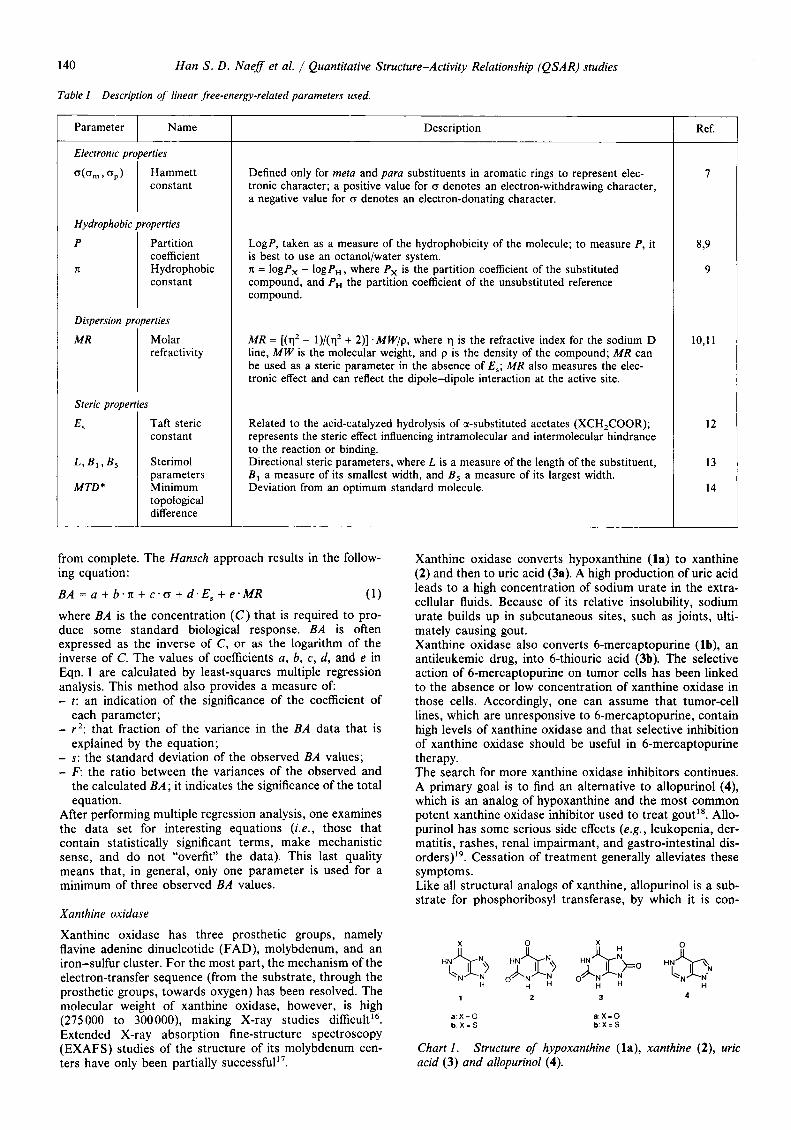
Xanthine oxidase converts hwoxanthine ( la)
Ref.
I
8,9
9
10,Il
12
13
14
o xanthine (2) and then to uric acid (3a). A high production of uric acid leads to a high concentration of sodium urate in the extra- cellular fluids. Because of its relative insolubility, sodium urate builds up in subcutaneous sites, such as joints, ulti- mately causing gout. Xanthine oxidase also converts 6-mercaptopurine (Ib), an antileukemic drug, into 6-thiouric acid (3b). The selective action of 6-mercaptopurine on tumor cells has been linked to the absence or low concentration of xanthine oxidase in those cells. Accordingly, one can assume that tumor-cell lines, which are unresponsive to 6-mercaptopurine, contain high levels of xanthine oxidase and that selective inhibition of xanthine oxidase should be useful in 6-mercaptopurine therapy. The search for more xanthine oxidase inhibitors continues. A primary goal is to find an alternative to allopurinol (4), which is an analog of hypoxanthine and the most common potent xanthine oxidase inhibitor used to treat gout”. Allo- purinol has some serious side effects (e .g . , leukopenia, der- matitis, rashes, renal impairmant, and gastro-intestinal dis- orders)”. Cessation of treatment generally alleviates these symptoms. Like all structural analogs of xanthine, allopurinol is a sub- strate for phosphoribosyl transferase, by which it is con-
1 2 3 4
a X = O b X = S
a ’ X . 0 b ’ X = S
Chart 1 . acid (3) and allopurinol (4).
Structure of hypoxanthine (la), xanthine (2), uric

Recueil des Travaux Chimiques des Pays-Bas, 11015, May 1991
verted to a ribonucleotide with an anti-metabolic poten- tial,’. Briley and Eisenthal” have reported that allopurinol can also be converted to a nucleoside by purine nucleoside phosphorylase. Although it has not yet been proved, medi- cal researchers suspect that prolonged ingestion of these compounds could well be harmful. The aim of this article is to review the QSAR studies that are performed on the inhibition of xanthine oxidase, and combine the results obtained into a model for the active site of the enzyme.
141
QSAR studies of inhibition of xanthine oxidase
Inhibition by phenyl-substituted 9-phenylguanines
Baker and co-workers22 synthesized an extensive series of xanthine oxidase inhibitors and measured their biological activity. Silipo and Hansch2’ compiled Baker’s data and made a QSAR study of derivatives of phenyl-substituted 9-phenylguanines (Table 11). The QSAR study by Silipo and Hansch revealed a correla-
tion between inhibitory activity of the compounds, molar refractivity, and Taft steric parameters:
10g(1/150) = 0.267( f O.06)MR3 - 0.647( f 0.12){MR3 .MR,} + 1.2911 f 0.39)E,, + 0.101( & 0.04)MR4 + 0.252( f 0.1 l )E, , + 4.552( f 0.45) ( 2 )
n = 65 r = 0.910 s = 0.308
The subscript of the parameters shows the position of the substituent (R) . The positive coefficient of MR, indicates a positive interaction for bulky substituents at position 3. The negative coefficient of the cross-product term MR, * MR, shows that bulky substituents at positions 3 and 4 substan- tially reduce the inhibitory activity, suggesting that the sub- stituent space near these positions is limited. This is caused primarily by the substituent at position 4. The coefficient of MR, is small and the positive coefficient of E,, indicates steric hindrance at that position (the bulkier the sub- stituent, the more negative the value of E,) . Moreover, the high coefficient for E,, indicates strong steric hindrance at position 2.
Table I1 Phenyl-substituted 9-phenylguanines and their inhibition activity towards xanthine oxidase.
0
Compound
5 6 7 8 9
10 I1 12 13 14 1s 16 17 18 19 20 21 22 23 24 25 26 27 28 29 30 31 32 33 34 35 36 37
R“
2-c1 2-Br 3-X-3’-SOzF, 4-OCH3 3-Y-3’-S02F, 4-OCH3 3-X-4’-S02F, 4-OCH3 2,3-CH = CHCH = CH 3-Y-4‘-S02F, 4-OCH3 4-Z-4‘-SOzF 4-N(CH3 )z 4-NHCOCH,Br 4-CI 4-C(CH3), 4-CH3 4-CF3 3,4-C12 4-O(CH,),X-4’-SO,F 4-Z-3’-S02F 3,4-(OCH3)2 4-Y-4‘-S02F 4-O( CHz)zY-4 ’-SO,F 4-O(CHz),X-4’-SOzF 4-CzH5 4-O( CHZ 13Y-3 ‘-S O2F 2-F 4-(W),CH3 3-NH2 4-O(CH2)2Y-3’-S02F 4-OCH’ 4-0(CH,)zY’-4’-CH3.3’-SO,F 4-CONHZ 3,4-CH =CHCH = CH H 4-O(CHz),X-3 ’-SOzF
- Logl,,b
5.09 5.1 1 5.25 5.31 5.35 5.38 5.39 5.60 5.68 5.72 5.74 5.74 5.80 5.89 5.96 6.00 6.02 6.14 6.15 6.16 6.16 6.17 6.20 6.21 6.21 6.22 6.28 6.30 6.3 1 6.38 6.39 6.39 6.40
Compound
38 39 40 41 42 43 44 45 46 47 48 49 50 51 52 53 54 55 56 57 58 59 60 61 62 63 64 65 66 67 68 69
R”
4-0(CH,),X-4’-SOzF 4-Y-3’-SOzF 3-CI CH(CH3 12 4-C6H5 3-CH3 3-NHCHO 3-OCH3 4-OH
4-O(CHz),X’-4’-CH,, 3’-SO,F 3-Y -3‘-SOzF 4-OCzH5 3-NHCOCH20C6H4-4‘-S02F 4-O(CHZ),X’-Z‘-Cl, 5’-SOzF 3-Y’-4‘-CH,. 3-SO,F
3-NHCOC,H, 3-NHCOCH,Br 3-Yf-2’-C1, 5’-SO,F 4-O(CH,)z-X’-2’-OCH,, 5’-SO,F 3-X’-2’-Cl, 5’-SO,F 3-Y-4‘-S02F 3-X ’-3 ’KI, 4‘-SOzF 4-NHCO(CH2)2C6H4-4’-SO2F 3-X-4‘-S02F 3-X’-4’-CH3, 3’-SO,F 3-X’-2’-OCH3, 5’-SOzF 3-NHCOCH,C,H4-4’-SOZF 3-NHCO(CH,),C,H,-4‘-SO2F
- Logl,,b
6.48 6.55 6.57 6.60 6.60 6.62 6.64 6.66 6.68 6.74 6.82 6.92 6.96 6.96 7.00 7.04 7.04 7.08 7.09 7.14 7.15 7.15 7.16 7.28 7.29 7.48 7.58 7.62 7.74 7.80 7.82 8.00
a X = NHCONHC,H,, Y = NHCOC6H4, Z = NHSO,C,H,, X’ = NHCONHC,H,, Y‘ = NHCOC,H,. from Ref. 23.
- Logl,, values were taken
142 Han S . D. Naeff et al . / Quantitative Structure-Activity Relationship (QSA R) studies
Allopurinol'(4y
Inhibition by phenyl-substituted 6-phenyl-4(3 H)-pteridinones
530
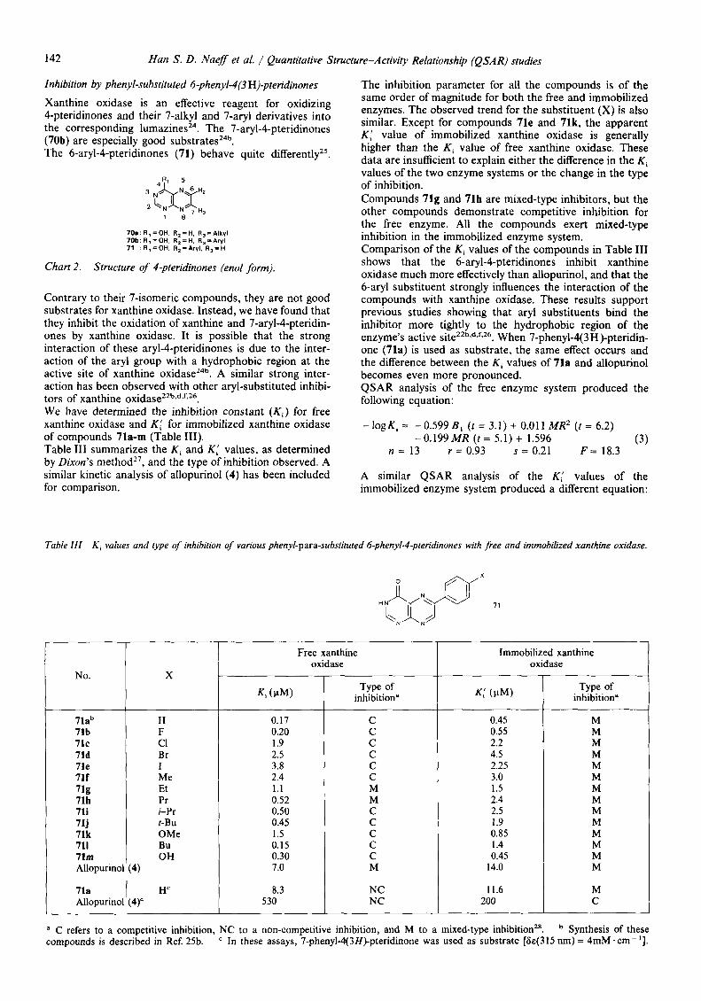
Xanthine oxidase is an effective reagent for oxidizing 4-pteridinones and their 7-alkyl and 7-aryl derivatives into the corresponding l ~ m a z i n e s ~ ~ . The 7-aryl-4-pteridinones (70b) are especially good substrates24b. The 6-aryl-4-pteridinones (71) behave quite d i f fe ren t l~~~.
NC
70a:R,=OH. R,=H. R,=Alkyl 70b: R,=OH. R,=H. R,=Aryl 71 :R,=OH. R,=Aryl. R,=H
Chart 2. Structure of 4-pteridinones (enol form).
200
Contrary to their 7-isomeric compounds, they are not good substrates for xanthine oxidase. Instead, we have found that they inhibit the oxidation of xanthine and 7-aryl-4-pteridin- ones by xanthine oxidase. It is possible that the strong interaction of these aryl-4-pteridinones is due to the inter- action of the aryl group with a hydrophobic region at the active site of xanthine o x i d a ~ e ~ ~ ~ . A similar strong inter- action has been observed with other aryl-substituted inhibi- tors of xanthine oxidase22b*d.f,26. We have determined the inhibition constant (Ki) for free xanthine oxidase and Kc for immobilized xanthine oxidase of compounds 71a-m (Table 111). Table 111 summarizes the Ki and K' values, as determined by Dixon's method2', and the type of inhibition observed. A similar kinetic analysis of allopurinol (4) has been included for comparison.
71ab 71b 71c 71d 71e 711 71g 71h 71i
71k 711 71m
71j
The inhibition parameter for all the compounds is of the same order of magnitude for both the free and immobilized enzymes. The observed trend for the substituent (X) is also similar. Except for compounds 71e and 71k, the apparent KI value of immobilized xanthine oxidase is generally higher than the Ki value of free xanthine oxidase. These data are insufficient to explain either the difference in the K , values of the two enzyme systems or the change in the type of inhibition. Compounds 71g and 71h are mixed-type inhibitors, but the other compounds demonstrate competitive inhibition for the free enzyme. All the compounds exert mixed-type inhibition in the immobilized enzyme system. Comparison of the Ki values of the compounds in Table I11 shows that the 6-aryl-4-pteridinones inhibit xanthine oxidase much more effectively than allopurinol, and that the 6-aryl substituent strongly influences the interaction of the compounds with xanthine oxidase. These results support previous studies showing that aryl substituents bind the inhibitor more tightly to the hydrophobic region of the enzyme's active siteZ2b*d.fs26. When 7-phenyl-4(3 H )-pteridin- one (71a) is used as substrate, the same effect occurs and the difference between the Ki values of 71a and allopurinol becomes even more pronounced. QSAR analysis of the free enzyme system produced the following equation:
H F c1 Br I Me Et Pr i-Pr
OMe Bu OH
t-Bu
- bgKi = - 0.599 B , ( t = 3.1) + 0.01 1 MR2 ( t = 6.2) - 0 . 1 9 9 M R ( t = 5.1) + 1.596 (3)
n = 13 r = 0.93 s = 0.21 F = 18.3
A similar QSAR analysis of the KI values of the immobilized enzyme system produced a different equation:
Table III K , values and type of inhibition of various phenyl-para-substituted 6-phenyl-4-pteridinones with free and immobilized xanthine oxidase.
Free xanthine oxidase
0.17 0.20 1.9 2.5 3.8 2.4 1.1 0.52 0.50 0.45 1.5 0.15 0.30 7.0
8.3
Type of inhibition"
C C C C C C M M C C C C C M
NC
Immobilized xanthine oxidase
0.45 0.55 2.2 4.5 2.25 3.0 1.5 2.4 2.5 1.9 0.85 1.4 0.45
14.0
11.6
~
Type of inhibition"
M M M M M M M M M M M M M M
M C
a C refers to a competitive inhibition, NC to a non-competitive inhibition, and M to a mixed-type inhibition**. compounds is described in Ref 25b.
Synthesis of these In these assays, 7-phenyI-4(3H>pteridinone was used as substrate [6~(315 nm) = 4mM acm- '1.

Recueil des Travaux Chimiques des Pays-Bas, 11015, May 1991
- logK' = - 0.262 B , (t = 2.0) - 0.418 x ( t = 3.2) + 0.005 MR2 (t = 4.5) - 0.076 MR (t = 2.7) + 0.645 (4)
n = 13 r = 0.93 s = 0.14 F = 13.4
0.770 0.699
- 0.279 - 0.398 - 0.580 ' -0.380
i -0.041 0.284 0.301 0.347
0.824 0.523
- 0.176
143
Exp."
0.347 0.260
- 0.342 - 0.653 - 0.352 - 0.477 -0.176 - 0.380 - 0.398 - 0.279
0.075 - 0.146
0.347
In regression analysis, neither the Hammett coefficient, B , (the maximum van der Waals' width), nor L (the van der Waals' length) was able to improve the equations. B , has a large negative coefficient in both equations, which means that an increase in the minimum van der Waals' width will reduce the effectiveness of the inhibitor. M R has an equally negative coefficient, but it also appears as MRz, with a small positive coefficient. Therefore, up to a certain value, the increase in the substituent's size will have a negative effect on the inhibitory activity. Nevertheless, if the size of the substituent is increased, the MR part of the equation will eventually have a positive value. In Eqn. 3, this is reached at MR = 18. In Eqn. 4, it is reached at MR = 17. The hydrophobicity parameter n: is necessary to obtain good analysis for Eqn. 4. This is understandable as, in the immobilized enzyme system, one must consider partitioning effects between two phases, namely the bulk solution and the sepharose matrix in which the enzyme is immobilized. x is derived from an octanol/water system". This constant indicates the kind of equilibrium that exists between the two phases. In this study, it indicates the equilibrium between the bulk solution and the sepharose matrix. In Eqn.4, R has a large negative coefficient, which means that compounds with polar substituents (i.e., with a nega- tive x value) will inhibit immobilized xanthine oxidase more effectively than free xanthine oxidase. QSAR analysis of the 9-arylguanines that Baker et al. synthesized showed that a bulky substituent at the ortho and para positions in the aryl group reduces the effec- tiveness of the inhibitor23. QSAR analysis of the data in Table I11 not only provided a similar result for some of the compounds, it also gave a good indication of the shape that a substituent needs to exhibit good binding properties. As mentioned previously, Eqns. 3 and 4 show that, while the substituent should have some volume, it should not be
Calcd.'
0.365 0.182
- 0.436 - 0.527 - 0.417 - 0.262 - 0.393 - 0.293 - 0.421 - 0.265
0.074 0.068 0.449
spherical. A comparison of the Ki values of compounds 71e and 71h demonstrates this. The iodo substituent is more spherical, as shown by its large minimum van der Waals' width (Bl). The propyl substituent, while just as "bulky", is more rod-shaped and, thus, has a lower Ki value (Table IV).
Znhibition by 8-arylhypoxanthines
Although there are a few isolated reports of 8-aryl- hypoxanthines with a remarkably low I, , value for xanthine o x i d a ~ e ~ ~ ~ ~ ~ ~ , a comprehensive study of these inhibitors has only recently been published by usZSa. The basic structure of 8-arylhypoxanthines 72 has two features in common with that of 6-aryl-4-pteridinones 71 (Chart 3). These are: - a heterocyclic system with a pyrimidine ring and an 0x0
group at position 4; - a pyrimidine ring fused at the 5,6 bond with another
heterocyclic ring system (pyrazine in 8-arylhypoxan- thines, or imidazole in 6-aryl-4-pteridinones).
The oxidation pattern of both types of compound leads one to assume that they will be fixed to the enzyme at the pyrimidine part30. Other studieszzb seem to indicate that the aryl group is instrumental in a tight enzyme-inhibitor com- plex. The main difference between the two compounds is
Y
73 I 7 2
7 1 - x
Chart 3 . nones 71, 8-arylhypoxanthines 72 and 8-arylxanthines 73.
Orientation of aryl substituent in 6-aryl-4-pteridi-
Table IV Inhibition constants and physicochemical parameters of substituent X of phenyl-para-substituted 6-phenyl-4-pteridinones.
71h 71i 71j 71k 711 71m
X
H F c1 Br I Me Et Pr i-Pr
OMe Bu OH
t-Bu
- LogKi - LogKi
Exp." I Calcd.b
0.901 0.641
- 0.283 - 0.459 - 0.259 - 0.039
0.075 0.383 0.081 0.487 0.0 17 1.361 0.357
1 .oo 1.35 1.80 1.95 2.15 1.52 1.52 1.52 1.90 2.60 1.35 1.52 1.35
RC I 1.03 0.92 6.03 8.88
13.59 5.65
10.30 14.94 14.94 19.62 7.87
19.62 2.85
- 0.02 2.08
- 0.67
Ref. 10. " Calculated with values in Table 111. Calculated with Eqn. 3. Calculated with Eqn. 4. Ref. 13.

144 Han S . D . Naef et al. / Quantitative Structure-Activity Relationship (QSAR) studies
71a
that, in the 8-arylhypoxanthines, the aryl group aligns with the heterocyclic ring system (parallel to the x axis) and, in the pteridine system, it forms an angle with the heterocyclic ring system (Chart 3). Xanthine oxidase is strongly inhibited by very low concen- trations of 8-arylhypoxanthinesz5". In fact, a significant decrease in oxidation occurs at equimolar concentrations of the enzyme and inhibitor, which is a clear indication that the inhibitor is very tightly bound. Some 8-arylhypo- xanthines, namely X = F, Br, t-Bu, reduced the enzymatic activity to as little as 10% at inhibitor concentrations only ten times higher than the enzyme concentration. At such low concentrations of inhibitor, the Lineweaver- Burk equation is not suitable for calculating kinetic parame- ters. This equation, which is based on Michaelis-Menten kinetics, assumes that there is no depletion of the inhibitor by the enzyme during oxidation. In other words, it does not allow for the possibility that the binding of inhibitor to the enzyme can significantly change the concentration of free inhibitor. The results of these experiments were even more difficult to analyze because of the non-linear initial rates of oxidation of xanthine in the presence of an inhibitor. This made it impossible to use the Dixon plot for tightly bound inhibi- t o r ~ ~ ~ , ~ ' . This problem was circumvented by expressing inhibition as I, , values ( i . e . , the concentration of inhibitor at which enzymatic oxidation of xanthine is reduced by 50%). The I,, values of 8-arylhypoxanthines (Table V) were calcu- lated by measuring the initial oxidation rate of 60mM xanthine and 20 mg/ml protein in at least twelve different concentrations of inhibitor.
H
Table V I,, and 19, values of 8-arylhypoxanthines 72 and d-aryl- xanthines 73".
8-Arylhypoxanthine I5,(PM)
0.21 0.04 0.06 0.01 0.07 0.17 0.09 0.03 0.16 0.10 0.11
4.9 1 1.70
8-Arylxanthine
1.91 1.87 1.24 0.55 2.12 1.27 0.75 2.14 4.22 1.49 3.12
13.21
19, ( P W b
27.68 25.04 16.49 12.63 14.43 13.20 88.70 20.90 43.60 26.80 30.50
a Assays were performed with 60pM xanthine and 20pg/ml xanthine oxidase (AFR = 65)32. These I,, and I,, values were measured after preincubating the inhibitor with xanthine oxidase for an appropriate time (at least one hour for the lower concentra- tions), to ensure that the inhibitor was oxidized to the corre- sponding 8-phenylxanthine. ' Synthesis of these compounds is described in Ref. 25a.
The I , , values for 8-arylhypoxanthines vary greatly. To pro- vide a clear picture of the effect of substituents on the aryl group, these values were used to calculate quantitative structure-activity relationship (QSAR) of compounds 72a-k and 73a-k. Table VI shows the results of these calcu- lations for compound 72. Eqn. 5 gave the best possible results. It is based on Hunsch parameters and steric parameters from the Sterimol methodI3. The number of parameters was limited to three
Table VI QSAR calculation for inhibition of xanthine oxidase by phenyl-substituted 8-phenylhypoxanthines.
I 5 0 = -O.lOSE, +0.256L-0.0380-0.180 (Eqn. 5 ) ( I = 2.0) ( t = 1.2) (I = 0.7) n = 11 r = 0.606 s = 0.065 F = 1.36
I , , = 0.I14MTD8 + 0.073 (Qn. 6) (f = 5.2) n = 11 r = 0.866 s = 0.036 F = 26.96
because only eleven compounds were available. Eqn. 5 has no statistical significance because r and F are very low. Eqn. 6 is based on the MTD* methodI4. Although MTD* parameters seem to explain all I50 values, they give no predictive values outside the congeneric group. The steric requirements for a good inhibitor are listed in Table VII. TableVII gives an idea of the inhibitory activity of other substituents only as long as these substituents fit into the hypermolecule. One example is 8-(4-cyanophenyl)hypo- xanthine, which has an MTD* value of zero. Unfortunately, we were not able to synthesize this
Table VII Hypermolecule for all positions of 8-arylhypoxanthine.
1.2.7 Are lavorable 4, 5, 6 Are indifferent
3-5, 3 Is unfavorable 6
Structure I Occupied position I MTD* I phenyl 4-F-, 4-C1-, 4-Br-phenyl 4-Me-phenyl 4-MeO-phenyl 4-i-Pr-, 4-0,N-phenyl 4-Me2N-phenyl 4-Bu-phenyl 4-t-Bu-phenyl
1 1 2 1 2 1 2 3 1 2 3 4 1 2 3 4 1 2 3 5 6 1 2 3 4 1
When Eqn. 6 is applied to 8-arylhypoxanthines, the results show that linear alkyl substituents do not bind well with xanthine oxidase. This is in contrast to results for 6-aryl- 4-pteridinones (71), which showed that the butyl substituent had a very low Ki value. The I,, values of 8-arylhypo- xanthines in Table V are approximately ten to one hundred times lower than those of 6-aryl-4-pteridinones. For a view of the real biological activity of 8-arylhypoxanthines, one would also have to study the inhibitory properties of oxidized 8-arylhypoxanthines. Table V lists the I, , and 190 values for 8-arylxanthines (73) that were obtained in the same study by enzymatic oxida- tion of the corresponding 8-arylhypoxanthines. The 19, values were included because it was not possible to calcu- late a significant QSAR equation for the I,, values. The QSAR equations for the 19, values are shown in Table VIII. Eqn. 7 uses two Hansch parameters, namely MR and CJ, where CJ has a large positive coefficient, and the Sterimol parameter B , , which has a large negative coefficient. With this combination of coefficients, "bulky" substituents with a small CJ value will stimulate the formation of an enzyme-inhibitor complex. Eqn. 8, which is based on the MTD* approach, makes roughly the same prediction (Table VIII). The steric requirements (Table IX) show that xanthine oxidase tolerates occupancy of positions 2, 3, 4, and 7 and that it allows binding to 8-arylxanthines more readily if the substituents have a hydrophobic property.

Recueil des Travaux Chimiques des Pays-Bas, 110/5, May 1991 145
Table VIII phenyl-substituted 8-phenylxanthines.
QSAR calculation for inhibition of xanthine oxidase by
I,, = 0.200 MR - 53.288 E , + 45.564 o + 90.768 (Eqn. 7) (f = 6.3) (f = 5.4) (f = 4.2) r n = 1 1 r = 0.926 s = 6.15 F = 14.22
I,, = - 8.663 F. + 39.154 MTD* - 0.013 (Eqn. 8) (f = 5.2) (f = 8.1) n = 1 1 r = 0.948 s = 1.71 F = 35.33
Table IX Hypermolecule for all positions of 8-arylxanthine.
r Structure I Occupied position I MTD* I ~
phenyl 4-F-, 4-C1-, 4-Br-phenyl 4-Me-phenyl 4-MeO-phenyl 4-i-Pr-, 4-0,N-phenyl 4-Me2N-phenyl 4-Bu-phenyl 4-t-Bu-phenyl
1 1 2 1 2 1 2 3 1 2 3 4 1 2 3 4 1 2 3 5 6 1 2 3 4 1
I I I 1
The inhibitory performance of 8-arylhypoxanthines and 8-arylxanthines shows that their affinity for xanthine oxidase is much greater than that of 6-aryl-4-pteridinones and allopurinol. As stated above, the main steric difference between 8-arylhypoxanthines and 6-aryl-4-pteridinones is the position of the aryl group in relation to the heterocyclic system. The linear molecules, namely 8-arylhypoxanthines (72a-k) and 8-arylxanthines (73a-k), show greater affinity for xanthine oxidase than the non-linear molecules, namely 6-aryl-4-pteridinones (71). Leonard et al.33 observed a similar phenomenon when they tested "stretched-out'' analogs of hypoxanthine as substrates of xanthine oxidase (Chart 4). The linear molecule lin-benzohypoxanthine (74) reacted faster than the angular molecule prox-benzo- hypoxanthine (75). Leonard et al. also observed that the other angular molecule, dist-benzohypoxanthine (76), reacts just as fast as the lin-benzohypoxanthine. 7-Aryl-Cpteridi- nones (70b) have a geometrical conformation similar to that of the dist-benzohypoxanthine, but show no inhibitory properties, because they are well oxidized by xanthine oxidase.
H 7 4 7 5
7 6
Chart 4. prox- (75) and dist- (76) benzohypoxanthines.
A detailed discussion of the effect of the substituents is not possible because both the availability of the required compounds and the predictive capacity of QSAR Eqns. 6 and 8 are limited. In general, however, the substituents of
Stretched-out analogs of hypoxanthine: lin- (74),
8-arylhypoxanthines that have an affinity for xanthine oxidase are those that are linear in relation to the x axis of the molecule (Chart 3). 8-Arylxanthines that have an affinity for xanthine oxidase are those with a fairly short sub- stituent. This affinity is enhanced by the hydrophobic char- acter of the substituents.
General discussion
Xanthine oxidase has been intensively studied34. It consists of two identical s u b - u n i t ~ ~ ~ ~ , each of which has a
Mo" Mov Mo'"
Type I binding
Mo'" H O H
Chart 5 . Binding pattern of xanthine with molybdopterin and oxidation of xanthine into uric acid by xanthine oxidase (after Robins et ~1.~').
MoVl MoV
Type I I binding
Xanthine
Mo'"
'10 H 0 R. -F-GO-P-OH
H H OH
MolV
Chart 6. Binding pattern of hypoxanthine with molybdopterin and oxidation of hypoxanthine into xanthine by xanthine oxidase (after Robins et al. ").

146
molybdenum-pteridin, an FAD (Flavine Adenine Dinucle- otide), and two iron-sulfur centers as cofactors. Xanthine oxidase is interesting to synthetic chemists because it catalyzes the hydroxylation of a wide range of purines and pteridines. Mechanistic questions concerning the enzymatic
Han S . D . Naef et al. / Quantitative Structure-Activity Relationship (QSAR) studies
reactions have been the subject of many biochemical studies. Notwithstanding the intensive work of biochemists, the pic- ture of the reaction of xanthine oxidase with reducing sub- strates is still incomplete.
Table X Azaheterocyclic compounds that have type-1 or type-I1 binding with xanthine oxidase.
No.
1.
4
5
70b
71
72
7 3
7b
Compound
0
"t&::b 'R
HN
't4 H
Inhibitory properties"
no
4.9
0.41
no
R = H 0.17 R = BU 0.15
R = H 0.209 R = Br 0.010
R = H 1.9 R = Br 0.55
?
Binding type Ref.
25a
31
38
24b
25a
25a
25a
36

Recueil des Travaux Chimiques des Pays-Bas, llOj5, May 1991
Table X (cont.)
147
No.
7 8
7 9
8 0
8 1
8 2
83
8 4
8 5
Compound
0 &$ -. /
CI
Inhibitory properties"
8.4
1.1
13
2.8
0.047
80
40
11
Binding type
I
I
I
I
Ref.
37
38
39
22a
40
41
42
43

148 Han S. D . Naeff et al. / Quantitative Structure-Activity Relationship (QSAR) studies
Table X (cont.)
No.
8 6
8 7
8 8
8 9
9 0
Compound
..+JJ b, ’ N ‘> H
a The inhibitory properties are expressed as I, , values (pM).
Robins et al.30 recently proposed a model for the interaction of molybdenum with organic compounds. This model includes the pterin cofactor with molybdenum to propose a kinetic model of oxidation of hypoxanthine and xanthine (Charts 5 and 6). The pterin cofactor brings the enzymatic nucleophilic site and the molybdenum atom on the active pocket together in the right orientation for the oxidative reaction. The model has two types of substrate binding: types I and 11. Chart 5 shows how xanthine binds with the molybdopterin cofactor at 0 - 6 and N-7 in type-I binding. Chart 6 shows how hypo- xanthine binds with the molybdopterin cofactor at N-3 and N-9 in type-I1 binding. In both cases, the sulfide group is oriented properly for the attack on C-8 of xanthine of C-2 of hypoxanthine. By varying the substitution pattern of the substrates or the inhibitors, one can vary the dimensions and explore the active site. The recent synthesis of benzo-elongated purines33 and pteridines’,, and their subsequent oxidation by xanthine oxidase, shows that the active site can bind large compounds in a reactive binding mode. Moder and Leonard36 found the spatial limit for the reactive type-I
Inhibitory properties”
91
160
50
Yes
K , = 4.03
Binding type
I
I
I or I1
I1
I
Ref.
42
42
44
45
45
binding region by synthesizing fin-naphthoxanthine (90, TableX), which is not oxidized by xanthine oxidase. The type-I1 binding region seems to be larger because lin- napthohypoxanthine (89, Table X) is still oxidized to compound With regard to the binding of inhibitors by xanthine oxidase, the variation in size and structure is almost limit- less. Table X gives some examples of the compounds used in inhibition studies. Most of the inhibitors are modeled on a purine-type aza- heterocyclic ring system. If one follows the numbering sys- tem of purine, the nitrogen atom at position 1 seems essen- tial to good inhibitory properties of a compound because compounds 84-88 have large Z,, values. At the same time, an aryl substituent connected to the five-membered ring enhances binding with the enzyme considerably because compounds 5, 15, 73, and 79-82 have smaller I, , values than compounds 4 and 78. The active site is flexible for inhibitors because it accommodates small compounds (4 and 78) very well. On the other hand, it also accomodates compounds like 84, 86, and 87, which have two aryl substituents, and elongated fin-naphthoxanthine (90). The

Recueil des Travaux Chimiques des Pays-Bas, 11015, May 1991
inhibitor-enzyme complex for the larger compounds is not as strong as that for the smaller compounds because the I, , and Ki values are larger. Nevertheless, the enzyme is still able to accommodate that type of inhibitor in the active site. Mono-aryl compounds having type-I1 binding seem to bind better with the enzyme than those having type-I binding (compare, for example, compounds 72 and 81, which have type-I1 binding, with 80, which has type-I binding, or compare compound 82 with 83). One explana- tion of this could be that the aryl substituent in type-I1 binding interacts better with the hydrophobic “wall” of the active site than the aryl substituent in type-I binding. All of the compounds with type-I1 binding are actually substrates, although some of them, namely 71, 72*,, and 8945, are only slowly oxidized by xanthine oxidase. For compounds 71 and 72, interaction with the wall of the active site is assumed because the different aryl substituents have a large influence on inhibitory capacity. The hydrophobic character of this wall is underscored by the absence of electronic parameters in the QSAR equations for compounds 71 and 7225a. The inhibition of xanthine oxidase seems to be governed mainly by steric factors. Xanthine oxidase favors bulky substituents at the para posi- tion of the aryl group, and there is an additional requisite for compound 71, namely the substituent R must be rod- shaped. There are two slight indications of this for compound 72: the hypermolecule in Table VII shows that positions 2 and 7 are favorable and that they lead to a rod-shaped substituent. Thus, it is evident that a definitive statement cannot be made on the nature of the hydrophobic wall based on the congeneric series that have been used to date. Chart 7 shows how different aryl substituents present at different positions in an inhibitor can interact with the hydrophobic wall of the active site and how the interaction of the aryl group depends on the type of binding. For example, 8-arylxanthines (73) are structurally closely related to xanthine (2) and they, therefore, show type-I binding, which means that the aryl group interacts with part E of the hydrophobic wall of the active site. On the other hand, 8-arylhypoxanthines 72 are related to hypo-
149
Compound 73 Type I binding
Compound 7 2 Type II binding
Compound 70b Type I1 binding
Compound 71 Type II binding
Chart 7. The molybdopterin active site of xanthine oxidase. A , B. C, D. and E are points in the hydrophobic pocket of the active site with which an aryl substituent (R) of the inhibitor interacts.
xanthine (la) and show type-I1 binding, which means that the aryl group will interact with part B. This explanation would clarify the difference between the I,, values of 8-aryl- hypoxanthines and those of 8-arylxanthines. Other exam- ples are 6-aryl-4-pteridinones (71) and 7-aryl-4-pteridinones (70b), both of which show type-I1 binding. The aryl group of compound 71 interacts with part C and that of compound 70b interacts with part A. In addition, the hydrophobic interaction of 9-arylguanines (5-69) is entirely different from that of 8-arylhypoxanthines. 9-Arylguanines have type-I binding, which means that the aryl substituent interacts with the hydrophobic wall at point D. Chart 7 shows clearly that all these compounds, which appear to be structurally similar, interact very differently with the active site. Chart 7 also shows that, with the compounds presented in this review, different parts of the active site are being probed. While the binding models of Robins et aL3’ will probably not be the final proposal for the molybdenum active site of xanthine oxidase, they at least offer a reasonable explanation of the difference in the I, , values of seemingly similar compounds. Further research is needed to understand better the reactions catalyzed by xanthine oxidase.
Acknowledgements
We are indebted to Dr. A . Verloop and Mr. J . Tipker for their help with the QSAR methodology, and to Mrs. M . Snyder for her linguistic advice. This work was supported by the Netherlands Foundation for Chemical Research (SON), with financial aid from the Netherlands Organi- zation for the Advancement of Pure Research (NWO).
References and notes
I A . Albert, Ann. Rev. Pharmacol. 11, 13 (1976). S . M . Free, j r . and J. W . Wilson, J. Med. Chem. 7, 395 (1964). T. Fujita and T. Ban, J. Med. Chem. 14, 148 (1971).
4aC. Hansch and T. Fujita, J. Am. Chem. SOC. 86, 1616 (1964); bC. Hansch, Acc. Chem. Res. 2, 232 (1969); ‘C. Hansch, “Drug Design”, edited by E . J . Ariens, Academic Press, New York 1, 271 (1971). Y. C. Martin, J . B . Holland, C. H . Jarboe and N . J . Plotnikoff, J. Med. Chem. 17, 409 (1974).
R. Kowalski and C. F. J . Bonder, J. Am. Chem. SOC. 94, 5632 (1972);
bK. C. Chu, Anal. Chern. 46, 1181 (1974). ’ L. P . Hammett, “Physical Organic Chemistry”, McGraw-Hill,
New York (1940). H. Levitan and J. L. Barker, Science 176, 1423 (1972). A . Leo, C . Hansch and D . Elkins, Chem. Rev. 71, 52 (1971). C. Hansch, A . Leo, S. H . Unger, K . H . Kim, D. Nikaiiani and E. J . Lien, J. Med. Chem. 16, 1206 (1973).
I ’ W . J . Dunn, Eur. J. Med. Chem. 12, 109 (1977). R. W . Tuft, “Steric Effects in Organic Chemistry”, edited by M . S. Newman, Wiley, New York 556 (1956).
l 3 A . Verloop, 5th Int. Congress of Pest. Chem., edited by J . Miyamoto and P. C. Kearney, Pergamon Press, Oxford 1, 339 (1983). J . Tipker and A . Verloop, “Approaches to Rational Synthesis of Pesticides”, ACS Symposium Series 255, Am. Chem. SOC., Washington, D.C. 279 (1984).
l 5 C. Hansch and A . Leo, “Substitution Constants for Correlation Analysis in Chemistry and Biology”, Wiley, New York (1979).
IhaC. Nelson and P . Handler, J. Biol. Chem. 243, 5368 (1968); W . R. Waud, F. 0. Brady, R. D . Wiley and K . V . Rajagopalan, Arch. Biochem. Biophys. 169, 695 (1975);
‘ G . R. Nathans and E . P. Kirby Hade, Biochim. Biophys. Acta 526, 328 (1978). T . D. Tullius, D . M . Kurtz, j r . , S . D . Conradson and K . 0. Hodgson, J. Am. Chem. SOC. 101, 2776 (1979).
150 Han S. D. Naeff et al. / Quantitative Structure-Activity Relationship (QSA R) studies
Isa Ph. Feigelson, J . D. Davidson and R. K . Robbins, J . Biol. Chem. 226, 993 (1979); Ts.-F. Yue and A. B. Gutman, Am. J . Med. 37, 885 (1957).
Iga R. E. Chalmers, R. Parker, H. A. Simmonds, W , Snedden and R. W. E. Watts, Biochem. J . 112, 527 (1969); R W. Rundles, J . B. Wyngaarden, G. H. Hitchings and G. B. Elion, Annu. Rev. Pharm. 9, 345 (1969).
2o T. A. Krenitzky, G. B. Elion, R. A. Strelitz and G. H. Hitchings, J . Biol. Chem. 242, 2675 (1967).
” M . S. Briley and R. Eisenthal, Biochem. J . 147, 416 (1975). B. R. Baker and J . Kozma, J. Med. Chem. 10, 682 (1967); B. R. Baker and W. F. Wood, J . Med. Chem. 10, 1101 (1967); ‘ B. R. Baker and W . F. Wood, J . Med. Chem. 10, 1106 (1967);
B. R. Baker and W. F. Wood, J . Med. Chem. 11, 644 (1968); ’ B. R. Baker and W. F. Wood, J . Med. Chem. 11, 650 (1968);
B. R. Baker, W. F. Wood and J. Kozma, J. Med. Chem. 11,661 (1968);
* B. R. Baker and W. F. Wood, J . Med. Chem. 12, 211 (1969); B. R. Baker and W. F. Wood, J . Med. Chem. 12, 214 (1969).
J. Tramper, “Oxidation of Azaheterocycles by Free and Immo- bilized Xanthine Oxidase and Xanthine Dehydrogenase”, Ph.D. Thesis, Pudoc, Wageningen, The Netherlands ( 1979); J. Tramper, A. Nagel, H. C. van der Plas and F. Miiller, Recl. Trav. Chim. Pays-Bas 98, 224 (1979); J . Tramper, W. E. Hennink and H. C. van der Plas, J . Appl. Biochem. 4, 263 (1982).
25aH. S. D. Naefi “Studies of the Quantitative Structure-Activity Relationship of the Inhibition of Xanthine Oxidase by Aza- heterocyclic Compounds”, Ph.D. Thesis, Pudoc, Wageningen, The Netherlands (1990);
bH. S. D. Naefi H. C. van der Plus, J . Tramper and F. Miiller, Quant. Struct. Act. Relat. 4, 161 (1985).
26aS. S. Parmar, B. R. Pandey, C. Dwivedi and B. Ali, J. Med. Chem. 17, 1031 (1974);
23 C. Silipo and C. Hansch, J. Med. Chem. 19, 62 (1976).
bI. Chu and B. M. Lynch, J . Med. Chem. 18, 161 (1975); ‘ R. H. Springer, M. K. Dimmitt, T. Novinson, D. E. O’Brien, R. K. Robbins, L. N. Simon and J . P. Miller, J . Med. Chem. 19, 291 (1976);
dJ. J. Baldwin, P. K. Lumma, F. C. Novello, G. S. Ponticello, J . M. Sprague and D. E. Duggan, J . Med. Chem. 20, 1189 (1979); C. J . Betlach and J . W. Sowell, J . Pharm. Sci. 71, 269 (1982).
*’ M . Dixon, Biochem. J . 55, 170 (1953). 28 I . H. Segel, “Enzyme Kinetics”, Wiley-Interscience, New York
(1975).
29 F. Bergman, L. Levene and H. Govrin, Biochim. Biophys. Acta 484, 275 (1977).
30 R. K . Robins, G. R. Revankar, D. E. O’Brien, R. H. Springer, T. Novinson, A. Albert, K. Senger, J. P. Miller and D. G. Streeter, J. Heterocycl. Chem. 22, 601 (1985).
3’ M . Dixon, Biochem. J . 129, 197 (1972). 32 AFR (Activity-to-Flavin Ratio) is a measure of functional
enzyme activity, with AFR = 212 corresponding to a fully active enzyme. AFR is defined as the change in absorbance per minute at 296nm in a 3-ml reaction volume, divided by the absorbance at 450 nm of the xanthine oxidase used in the assay, under the standard conditions of 100pM xanthine in 0.1M pyrophosphate, at pH 8.5 and 25°C. See R. C. Bray, ‘The Enzymes”, edited by P. D. Boyer 12, 299-419 (1975).
33 N. J. Leonard, M. A. Sprecker and A. G. Morrice, J . Am. Chem. SOC. 98, 3987 (1976).
34a R. C. Bray, “The Enzymes”, edited by P. D. Boyer, H . Lardy and K. Myrback 7, 553 (1963); R. C. Bray, Ibid. 12, 299 (1975); M . P. Coughlan, “Molybdenum and Molybdenum-Containing Enzymes”, edited by M . P. Coughlan, 119 (1980).
35 J . W. G. De Meester, W. Kraus, W. J . Middelhoven and H. C. van der Plus, “Proc. 4th FECHEM Conf. Heterocycles in Bio- Organic chemistry”, Elsevier, Amsterdam 243 (1986).
3b K. P. Moder and N. J. Leonard, J . Am. Chem. SOC. 102, 2613 (1982).
37 R. K . Robins, J . Am. Chem. SOC. 78, 784 (1956). 38 B. R. Baker, J . Pharm. Sci 56, 959 (1967). 39 S. M . Greenberg, L. 0. Ross and R. K . Robins, J . Org. Chem. 24,
1314 (1959). 40 J . Kobe, D. E. O’Brien, R. K. Robins and T. Novinson, J. Hetero-
cycl. Chem. 11, 991 (1974). 4 1 T. Novinson, K. Senga, J. Kobe, R. K. Robins, D. E. O’Brien and
A. Albert, J. Heterocycl. Chem. 11, 691 (1974). 42 K . Senga, T. Novinson, H. R. Wilson and R. K . Robins, J . Med.
Chem. 24, 610 (1981). 43a Y. Makisumi, Chem. Pharm. Bull. 9, 801 (1961);
K. Senga, T. Novinson, R. H. Springer, R. P. Rao, D. E. O’Brien, R. K . Robins and H. R. Wilron, J . Med. Chem. 18, 312 (1975).
44a S. C. Ball and W. T. Caldwell, J . Am. Chem. SOC. 82, 1469 (1960); L . P. Vettori, L. Cecchi, A . Constanzo, G. Auzzi and F. Bruni, Farmaco Ed. Sci. 36, 441 (1981).