quantitative functional capacity evaluation
TRANSCRIPT

Steven G. Yeomans, DC, DABCOPrivate PracticeRipon, Wisconsin
Craig Liebenson, DCPrivate PracticeSanta Monica, California
Practice aids and algorithms appear in Appendix A.
OUTCOMES ASSESSMENT (OA) is a health carebuzzword for the 1990’s. Quality assurance in health
services delivery requires that certain guidelines be followedand that measurable outcomes be used to document theappropriateness of care. Most methods of OA have been“high-tech”, requiring substantial expenses of time and money.Due to the cost and the failure to demonstrate validity, a shiftin emphasis to “low-tech” approaches has occurred. Modernmethods of OA must be time efficient, economical, reliableand valid.
Modern reporting on spinal pain patients requires a consen-sus-based classification approach, relevant historical datagathering, and reliable and valid OA of both subjective andfunctional parameters. Such an approach has great potentialfor future data collection and analysis and may even allowmultiple high-quality care facilities to provide invaluableinformation for research purposes.
Physicians, insurance companies, medico-legal reviewersand managed care organizations are becoming increasinglyinterested in OA and functional testing, because of the de-mand to objectify patient status and document patient progressduring the course of treatment1. OA represents a method usedto measure a change in a patient’s health status as a result ofsome type of treatment approach2. OA instruments are alsoutilized as a tool for measuring treatment effectiveness regard-less of methods utilized3. Moreover, OA plays an important rolein steering quality care and cost containment.
Quantitative Functional CapacityEvaluation: The Missing LinkTo Outcomes Assessment
Purpose: Both subjective and objective approaches to outcomes assessment are reviewed and discussed. Fivecriteria for the development of an instrument are offered, and a comparison of high- versus low-tech functionaltesting is made. Utilization parameters with risk factors for chronicity are also discussed. Methods: Aqualitative literature review was performed searching for functional tests which have been found valid andreliable. Tests which included a normative database were selected. After collection of the tests, the most userfriendly and valid/reliable were grouped together. Results: A functional capacity evaluation approach utilizinglow-tech, low-cost tests were collectively grouped together and called the quantitative functional capacityevaluation (QFCE). Each test is listed and includes the procedure, the normative data and the variousreferences reviewed. Discussion: The utility, the goals, and the clinical application of the QFCE are discussed.The use of the normative data and pre-/posttest result comparisons are discussed for reasons of documentation,insurance communication, and steering the rehabilitation goals and patient education. Key words: disabilityevaluation, outcome assessment; range of motion (articular); rehabilitation; work capacity evaluation.
To order reprints contact Professional Sales Department, AspenPublishers, Inc., 200 Orchard Ridge Drive, Suite 200, Gaithersburg, MD20878, 1-800-638-8437.
Steven G. Yeomans, DC, DABCO, Private Practice, 404 Eureka Street,Ripon, Wisconsin 54971.
Top Clin Chiro 1996; 3(1): 32–43© 1996 Aspen Publishers Inc.
32
Yeomans and Liebensen: Quantitative Functional Capacity 33
THE STATE OF THE ART IN OUTCOMESASSESSMENT
OAs are primarily concerned with showing patient progressover time and objectifying patient status. There are a number ofdifferent types of OA instruments that can be performed atcritical junctures of patient care. For example, in the acute stage(initial patient evaluation), baseline data can be collected by thesubjective OA questionnaires concerning pain level, disability,general health, depression, work dissatisfaction, and others. Inthe subacute stage, functional outcomes become necessary. Aquantifiable functional capacity evaluation utilizing reliable andvalid tests that can be compared to a normative data base isessential before the patient has completed 4 to 6 weeks of care.This evaluation enables the patient, doctor and third-partypayer to have baseline levels of the patients’ impairment ordysfunction and allows for comparison over time. A final typeof OA, the work capacity evaluation (WCE), aims to establishreturn to work (RTW) goals. This evaluation is especiallyimportant in medico-legal situations and in instances wheredisability is involved.
The information gained about the acute patient through theuse of subjective OA is critical for documenting, in a quanti-tative manner, the subjective information concerning how the
injury or condition is affecting the patient. Most important,these instruments can be repeated at a future reexaminationdate and by comparing the baseline or initial informationgathered to that at follow-up, confident clinical decisionmaking can occur which can lead to one of the following:
• continued care (if improvement is noted without reach-ing maximum therapeutic benefit);
• change in treatment approach, strategy or goals (if noclinical improvement or change is noted and case resolu-tion has not occurred);
• initation of rehabilitation and reduction of passive treat-ment frequency (improvement without resolve and de-conditioning is complicating further improvement); or
• referral to another health care provider if therapeuticbenefit can be obtained or, simply, a discharge with orwithout permanent residual sequelae, disability, or im-pairment.
The ability of the treating health care provider to make the“next” case management decision in the unresolved case hasbeen at the core of the problem regarding overutilization ofpassive care, doctor dependency, chronic pain behavior, andinsurance company “nightmares”.
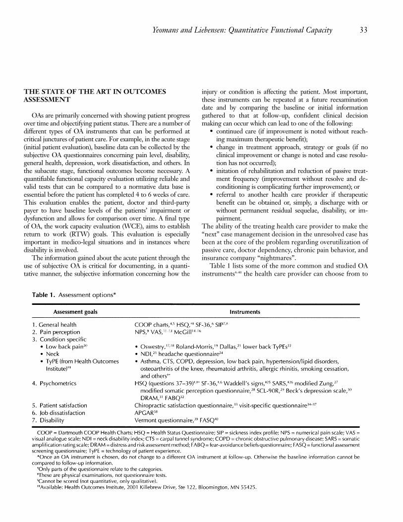
Table 1 lists some of the more common and studied OAinstruments4–40 the health care provider can choose from to

34 TOPICS IN CLINICAL CHIROPRACTIC/VOLUME 3, ISSUE 1, 1996
help gain the leverage needed to make intelligent clinicaldecisions. It is necessary to utilize the same OA instrument atfollow-up in order to compare results at follow-up reexamina-tions. Note that there are many different OA instruments fromwhich to choose and the list in Table 1 is not all-inclusive.
The method by which the subjective OA information isdocumented and reported is important so that the informationderived from the OA can be reviewed easily. Ease of reviewserves the needs of the health care provider (to render appropri-ate care), the patient (for orientation and referencing his or herresponse to treatment in a report of findings), and the insurancecompany (to justify payment of a claim) as well as the attorney(in a medico-legal arena such as a malpractice suit). The form inAppendix A summarizes the various tools one may use. Bysummarizing the results of the various OA tools on one page,the information can be reviewed quickly and clinical deci-sions can be driven in a prompt and efficient manner.
The common thread that ties the historical and subjectivedata to the objective quantitative functiona capacity evaluation(QFCE) is the various OA instruments. Once the patientis subacute, it is essential to establish objective functionalbaselines. Safety in applying the functional tests must bedetermined before proceeding with functional testing. It shouldbe noted that functional capacity testing is contraindicated inthe acute stage of an injury when pain is more of a “chemical”nature than a “mechanical”. Mooney and Matheson recom-mend that a physical capacity evaluation (PCE) be considered attwo weeks’ post-injury in order to determine the “weakfunctional link”41 and at 4 weeks to perform their CaliforniaFunctional Capacity Protocol (Cal-FCP)42. Triano (personalcommunication, May 1994) has reported that 4 weeks’ post-injury is and appropriate time to initiate testing. Hart andassociates43 report indications for functional testing includethe following:
• plateau in treatment progress,• discrepancy between subjective and objective findings,• difficulty in returning the patient to gainful employment,
and• vocational planning, or medical-legal case settlement.As soon as the patient leaves the acute guarded stage, the
QFCE not only provides ideal outcomes assessment informa-tion, but also identifies key functional pathologies that can beaddressed with various treatment approaches such as manipu-lation, exercise, and patient education. Mooney reports thatthe functional capacity evaluation should be mandatory forany patient still suffering pain after 6 to 7 weeks.41
Functional testing serves as an objective OA method, thuscomplementing the subjective outcomes assessment instru-ments or questionnaires completed by the patient at variousintervals of time during care. The objective functional testsmeasure factors such as flexibility, strength, coordination,
endurance, aerobic capacity, posture and balance. Functionaltests, whether provocative or functional in nature, must followcertain criteria in order to be useful and reliable. Five issueswhich must be addressed in the selection and use of any func-tional test in a patient population have been described.42 Theseissues, presented in hierarchical order, are:
1. Safety: Given the known characteristics of the patient,the procedure should not be expected to lead to injury;
2. Reliability: The test score should be dependable acrossthe evaluators, patients, and the date or time of admin-istration;
3. Validity: The interpretation of the test score should beable to predict or reflect the patient’s performance in atarget work setting;
4. Practicality: The cost of the test procedure should bereasonable. Cost is measured in terms of the directexpense of the test procedure plus the amount of timerequired of the patient, plus the delay in providing theinformation derived from the procedure to the referralsource;
5. Utility: The usefulness of the procedure is the degree towhich it meets the needs of the patient, referrer, andpayer.
High-tech instrumentation and dynametric assessment ofthe low back have been considered the “gold standard” oflumbar spine functional assessment. This view is largely dueto their reliability and reproducibility. However, the validityof some of the high-tech testing approaches has recentlybecome an issue of controversy. Grabiner and colleagues44
have demonstrated, for example, that normal strength mea-surements from a high-tech approach does not necessarilycorrelate with normal human function (47). In this study, elec-tromyography (EMG) was used during isometric trunk exten-sion. The results revealed decoupling, or asymetric lumbarparaspinal muscle activity was present in low back painsubjects who were considered normal on high-tech dynametrictesting. This decoupling phenomenon also differentiated be-tween pain and non-pain subjects. This study suggests thatmusculoskeletal function involves not only strength, but alsocoordination during the performance of a specified task.Because spinal movement and coordination use complexneuromuscular functions, simple strength assessment by high-tech dynamometer does not necessarily correlate with assess-ment of spinal function. The EMG results illustrate the limit-ations of high-tech dynamometric testing of muscle strengthand endurance, and they also suggest that the often harshcriticism of low-tech evaluation approaches regarding strengthand coordination may be inappropriate and unjustified.
Newton and Waddell45 reported that no convincing evi-dence supports that isokinetic or any other type of iso-measurehas greater utility in assessing the patient with low back pain
Yeomans and Liebensen: Quantitative Functional Capacity 35
than a clinical evaluation of physical impairment, isometricstrength, a simple isoinertial lift or psychophysical testing.Because of the inability to demonstrate high quality of spinalfunction assessment by high tech methodologies, there hasbeen insufficient evidence to suggest abandonment of lowertech quantifiable tests. Many low-tech approaches to identifyfunctional pathology have been reported46-73. Valid and reli-able information, often with normative database, has beenreported; hence, serves as excellent low-tech functional OAtools. Careful observation of the quality of movement duringthe test can give valuable insight to treatment prescriptionaddressing functional pathologies such as muscle imbalance,joint stiffness, poor movement and coordination, and posturaldysfunction.
Reliability has been reported in several low-tech tests thatdo not provide numerical quantification results. For example,the National Institute for Occupational Safety and Health(NIOSH) Low Back Atlas identified 19 tests with significantreliability (<0.74 Cohen’s Kappa and >0.79 coefficient forinterclass correlation, coefficient [ICC]).63, 69. Moffroid70 stud-ied the ability of the 53 NIOSH tests to discriminate betweenlow back pain and non-painful subjects. It was found that 23 ofthe 53 tests could not discriminate adequately between the twogroups and when the 7 strongest tests were groupedtogether, a sensitivity of 87% and specificity of 93% wereobtained. Interestingly, the most important measurementswere those which assessed passive mobility, dynamic mobility,strength, and symmetry. Reports71 have recently been publishedthat also suggest that non-dynametric tests correlatebetter with pain and disability than those that deal withisokinetic testing. The authors were careful to point out thatnon-dynamometric tests are still useful in the clinical settingin spite of the development of more sophisticated and accuratemethods of testing muscle strength.69 Harding and Williams 72
reported a group of low-tech tests were determined safe,reliable and valid for assessment of physical dysfunction inchronic pain subjects. A normative data base segregated byage, gender and vocation (blue collar vs white collar) werestudied and found reliable when tests on over 500 individualswere carried out.73 Hence, because validity and reliabilityhave been established, as well as a normative database regard-ing several low-tech functional tests, it would appear naturalto adopt these particular tests as representatives in a low-techfunctional capacity evaluation setting.
THE QFCE
Achieving a low-cost, time-efficient, valid, and reliablemethod of evaluating functional capacity of a patient was thegoal in developing the QFCE instrument. This test isintended to allow the doctor to identify functional baselines
for active rehabilitation in order to improve deconditioningand restore function. The QFCE introduces an OA instrumentthat can be used both as an objective barometer for measuringchange in function over time (“descriptive”), as well as an aid indriving specific rehabilitation protocols (“prescriptive”).When coupled with the subjective OA instrument(s), theQFCE enables the provider to document changes in symptomsand function over time. It also provides a method for the healthcare provider to use in making a clinical decision (changetreatment approach, refer, discharge with or without perma-nent residuals, and so forth), depending in part on the QFCEresults. The QFCE is not designed to replace but rather,complement, other qualitative, less “objective” tests such astrigger point and end-feel palpation, postural and gait analy-sis, and observation of altered movement patterns.
The second goal is to incorporate the QFCE into a comput-erized format in preparation for establishing a large databasefor clinical as well as research objectives. Regardless ofwhether it is used in a computerized or non-computerizedformat, the data derived from the QFCE can be used togenerate reports for documentation reasons which can enhancecommunication with the patient when reporting findings, withinsurance companies when supporting the need for rehabilita-tive care and with the treating physician/therapist whenfacilitating the process of establishing maximum therapeuticbenefit or maximum medical improvement (MMI) and thus,support case closure or referral.
Each test of the QFCE is fully explained and referenced.When the original reference was unclear, the principle authorwho described the specific test was contacted and the clarify-ing information was incorporated into the text. Because low-tech functional testing is gaining interest in the researchcommunity, it is probable that the QFCE will be updated fromtime to time in order to stay current as well as to incorporatenew valid and reliable approaches that measure function.
When performing the QFCE, it is important to perform eachtest as precisely as possible as they are described. Suchadherence is important for improving reliability. Ekstrand andcoworkers47 observed an improvement in the coefficient ofvariation (CV) from 7.5 ± 2.9 to 1.9 ± 0.7 after using the testsfor two months and refining their technique. In particular theypaid attention to the details regarding:
• standardizing inclinometer placement and allowing thependulum of the gravity type to swing freely,
• stiffening up the examination table (plywood with straps),• identifying bony anatomic landmarks (mark on skin),
and• standardizing examination bench height for each visit.
The following text describes each of the 21 tests thatcomprise the QFCE (in the order they are performed). Datasummary and examinations forms are located in Appendix A.

Initial and final test
1. Visual analogue scale
The Visual Analogue Scale (VAS)10–14 evaluates the patient’sperception of his or her pain level on a 0 to 10 pain scale. It iscompleted at the beginning and conclusion of the QFCE. Painis most commonly measured by intensity, frequency, andduration. The VAS is a 10-cm line with two pain descriptorsat each end (“No Pain” or “0” and “Unbearable Pain” or “10”).For the purpose of the QFCE, the pain being rated is pain thatis being perceived at the time the QFCE is administered.Scoring is completed by laying a transparent 10-cm ruler overthe line and reading the centimeter and millimeter markings.More specifically, a 0 to 10 scale is used where 1 cm = 1/10pain; 5.5 cm = 5.5/10 pain; and so forth.
Standing tests
2. Repetitive squat
The repetitive squat71, 73 evaluates the strength and endur-ance of those muscles required to perform a squat. The patientstands with his or her feet 15 cm apart and squats until thethighs are horizontal; the patient then returns to the uprightposition (Fig 1). Each repetition lasts 2 to 3 seconds induration, and each test is repeated until a maximum number ofrepetitions is achieved or 50 repetitions are done, whicheveroccurs first. Observation of the quality of movement as wellas the number of repetitions is important as informationderived about the quality of movement gives rise to treatment
and exercise prescriptions. Therefore, the quantitative infor-mation assesses outcomes while the qualitative data drivetreatment goals. The normative data are age, gender, andoccupational specific, as depicted in Table 2.73
3. Range of motion: LumbarThe range of motion (ROM): lumbar test evaluates the
mobility of the lumbar spine57,74–80 An inclinometer is placedat T-12 and S-2. Sagittal plane movements (flexion/exten-sion) are assessed by placing the inclinometer verticallyperpendicular to the spine on the midline. Frontal planemotion (lateral flexion) is assessed by placing the base of theinclinometer horizontal so that the needle hangs freely. The endpoints of movement are recorded at both the T-12 and S-2 and the difference is calculated using the equation: T-12 –S-2. If the average of three consecutive readings falls within5° or 10% of the average, the highest of the three readings isrecorded. This procedure may be repeated a maximum of sixtimes in attempt to achieve this result.57 Table 3 presents thenormative data for the lumbar spine.80
4. Pain/tenderness (Waddell Non-organic low back paintest 1The Waddell nonorganic low back pain (LBP) signs evalu-
ate for abnormal psychosocial issues.25 More specifically, thistest is performed by applying a light touch in a manner thatshould normally not provoke pain. A nonorganic pain re-sponse is reported when the patient describes or portrays pain.There are five categories (see Table 4) of the Waddell
36 TOPICS IN CLINICAL CHIROPRACTIC/VOLUME 3, ISSUE 1, 1996
b
Fig 1. Repetitive squat test.
a

Yeomans and Liebensen: Quantitative Functional Capacity 37
nonorganic LBP test (QFCE tests 4, 5, 8, 9, 10, 13), which arenonprovocative tests (ie, do not try to provoke a pain responsesuch as when performing standard orthopaedic provocativetests). If a pain response occurs, this finding constitutes apositive (+) response. The practitioner should determine ifthere is a physiological explanation and repeat the tests as manytimes as needed in order to assure evaluator objectivity. Thesetests are reported as positive or negative in terms of presenceof nonorganic LBP rather than in terms of a number. The finalscore is documented as the total number of positive signs out ofthe five (eg, two out of five). When three or more of the fivesigns are positive, nonorganic LBP must be considered andthe psychosocial issues must be therapeutically addressed.
Test 1 is performed by standing or sitting behind the patientand a light touch or pinch is applied to the skin over the lumbararea. A deep type of palpation over a nonanatomic, wide area ofpain not localized to one structure is also performed. Observa-tion focuses on looking for a disproportionate or exaggeratedresponse (“jump sign,” verbal response, and so on).
5. Simulation (Waddell nonorganic low back pain test 2)
Axial Compression
This test25 is performed by standing behind the patient andplacing a light pressure downward on the occiput, similar to acervical compression test. However, it is performed in amanner that should not normally provoke pain. NonorganicLBP is suggested if a pain response is obtained. Neck painmay occur with axial loading; hence, this approach may becontraindicated. If it occurs, downward pressure on the shoul-ders will simulate a similar test.
Trunk Rotation
This test25 is performed by standing behind the weight-bearing patient. The pelvis is manually rotated at the hips in amanner that should not normally provoke a pain response. Thepatient is instructed not to rotate the shoulders beyond themovement being actively assisted by the evaluator at thepelvis. Nonorganic LBP is suggested if a pain response isobtained. (Note: if lumbar root pain is present, a false-positiveresponse may be obtained. Therefore, it should be correlatedwith a straight leg raise and neurologic examination findings.)
The following two functional tests are included in thisevaluation due to the importance of the ankle joint in main-taining balance and coordination and its important relation-ship to the “kinetic chain.” In addition, the stability of thesubtalar joint is highly dependent on the flexibility of the ankle

and must be intact for proper proprioceptive function. Deter-mination of the ROM at the ankle can also yield valuableinformation when correlated to the Dictionary of Occupa-tional Titles (DOT) when assessment of work capacities isrequested.
6. Gastrocnemius/ankle dorsiflexion test (knee straight)In this test,46,47 the patients stands upright, feet parallel and
knees straight. The inclinometer is positioned above thelateral malleolus and “zeroed” in the upright standing posi-tion. The patient leans forward, placing the hands on a wall toa point of maximum ankle dorsiflexion (DF), while keepingthe heel down; the angle is then measured. The normative datareveals 22.5° ± 0.7°, intra-assay CV is 2.2%, and inter-assayCV is 2.5%47.
7. Soleus/ankle dorsiflexion test (knee flexed)The patient position in this test46,47 is standing with one leg on
floor and the foot being tested is placed on a bench. The knee isflexed and the ankle is dorsiflexed to a maximum anglemaintaining heel-to-bench contact. The normative data reveal24.9° ± 0.8, intra-assay CV is 2.2%, and inter-assay CV 2.6%.47
Sitting tests8. Sitting vs supine straight leg raise/distraction (Waddell
nonorganic low back pain test 3)This test25 evaluates for abnormal psychosocial issues. The
patient is seated and the doctor performs a sitting straight leg
raise (SLr) test while distracting the patient by the perfor-mance of a plantar superficial reflex while rapidly extendingthe knee. A positive test occurs when there is little to no painnoted in the distracted sitting SLR position and a dispropor-tionately high level of pain observed during the nondistractedsupine SLR test (positive “flip” sign). Note that if a sciaticnerve tension sign exist, this test may be invalid. Also, theevaluator should be cautious regarding the speed at which thesitting SLR is performed if nerve tension is suspected. As withthe other Waddell nonorganic LBP signs, this test is reportedas positive or negative as it relates to nonorganic LBP ratherthan a number (Table 4).
9. Regional Neurology (Waddell nonorganic low backpain test 4)This test25 evaluates for abnormal psychosocial issues. The
health care provider performs a standard neurological physicalexamination (deep tendon reflexes, muscle strength, sensoryperception). A positive test is present when the neurologicexamination reveals findings that do not follow an expectedanatomic pattern or are highly inconsistent (or both). Thesefindings may include altered motor functions where manymuscle groups are weak. If the quality of weakness is of a“breakaway” variety, where the patient suddenly discontinuesthe strength test, one must differentiate between pain-inducedweakness (physiologic) and a poor voluntary effort (nonor-ganic). Sensory changes may be of a nondermatomal varietyoften with hyperpathia mixed with dysesthesia. Another dif-ferential diagnosis to consider is sclerotomal pain which mayarise from the posterior disc or joint structure usually de-scribed in the history as a deep, nonspecific, rather globaldistribution, that does not follow any obvious anatomic path-way. In general, the evaluator should look for multiple signsof nonorganic LBP before feeling secure about this assess-ment. This test is reported as positive or negative as it relates tononorganic LBP rather than a number (Table 4).
10. Exaggeration/overreaction (Waddell nonorganic lowback pain test 5)
This test25 evaluates for aabnormal psychosocial issues. It isnot a specific test but rather inconsistent examination findingswith overreaction noted at any time during the consultation orexamination. Observation focuses on looking for dispropor-tionate responses, such as tremor, crying out, and collapse.This test is reported as positive or negative as it relates tononorganic LBP rather than a number (Table 4).
11. ROM: CervicalThis test57,78,80 evaluates the mobility of the cervical spine. An
inclinometer is placed at the occiput and T-1. Sagittal planemovements (flexion/extension) are assessed by placing the
38 TOPICS IN CLINICAL CHIROPRACTIC/VOLUME 3, ISSUE 1, 1996

Yeomans and Liebensen: Quantitative Functional Capacity 39
inclinometer vertically perpendicular to the spine in themidline. Frontal plane motion (lateral flexion) is assessed byplacing the base of the inclinometer horizontal so that theneedle hangs freely. The endpoints of movement are recordedat both the occiput and T-1 and the difference is then calculatedusing the equation: occiput – T1. If the average of threeconsecutive readings fall within 5° or 10% of the average, thehighest of the three readings is recorded (Table 5). Thisprocedure may be repeated a maximum of six times to try toachieve this result.
Supine tests12. Modified Thomas test/hip extension test
Five steps are involved with this test.47, 81–83 The steps are asfollows:
1. With the inclinometer placed 5 cm above the patella onthe lateral thigh, the patient is first positioned supinewith the knees straight on the bench to obtain an initialinclinometer reading, reset at zero.
2. The patient is next positioned at the end of bench ina manner where the ischial tuberosities are supported bythe end of the table’s edge in a partially standing andsitting posititon.
3. The contralateral knee and hip are flexed to the chest toeliminate lumbar lordosis and the patient is lowered toa supine position.
4. The testing hip is then passively flexed to a 90° angle,the inclinometer is reset to zero and the leg is allowedto hang freely towards the floor fully relaxed.
5. The evaluator records the angle when the tested leg isfully relaxed, hip extended, and the lumbar lordosis isremoved.
The normative data are 83.5° ± 1.1°, intra-assay CV is 0.7%,and interaassay CV is 1.2%.
13a. Supine vs sitting SLR/distraction (Waddellnonorganic low back pain tests 2)
The reader is referred to the discussion under “8. Sittingvs Supine Straight Leg Raise/Distraction.”
13b. SLR (hamstring flexibility) testA SLR (Hamstring flexibility)46,47,61,79 test is performed with
the doctor supporting the lower extremity (with crook ofelbow) while holding the zeroed inclinometer mid-tibia orhaving it strapped to the lateral thigh (5 cm above patella), thedoctor’s indifferent hand stabilizes the pelvis. The leg is raisedto a point of first of knee flexion (of the leg being tested) and/or the pelvis begins to rock and opposite knee flexes. Theevaluator records the hip flexion angle. Normative data rangefrom 70° to 90°.47
14. Repetitive Sit-upIn this test,71–73 the patient is positioned supine with the
knees flexed 90° with the ankles fixed. The patient is in-structed to sit up until the thenar pad of the hand touches thepatella; the patient then curls back down fully to the supineposition. The number of repetitions are counted to a maximumof 50. The normative data are age, gender, and occupationalspecific, as depicted in Table 6.73.
Prone tests
15. Knee flexion test/nachlasIn this test,46,47,83,84 the doctor’s position is to the side of
patient. The patient is placed in the prone position. Theinclinometer is placed on the lower leg with the knee fullyextended (the feet may hang over the edge to ensure fullextension). With the pelvis strapped down, the knee is pas-sively flexed (the heel is brought towards the buttocks). Theangle is recorded at the moment hip flexion or hiking occurs.The normal angle equals 147.9° ± 1.6, intra-assay CV is0.5%, and inter-assay CV is 1.1%.47
16. Repetitive arch-up testWhen performing the repetitive arch-up test,71,73 the patient
is placed prone with the anterior superior iliac spine (ASIS)just on the table, trunk extended off, arms at sides, and theankles and thighs are fixed to the table by strapping. Thepatient raises upwards to the horizontal position and then downto a 45° angle. The number of repetitions are counted (maxi-mum of 50). The normative data are age, gender and occupa-tional specific, as depicted in Table 7.73

17. Hip ROM (internal and external rotation)To test hip ROM,59,85 the patient is prone with the inclinom-
eter fixed to the anterior distal third of lower leg; with the kneeflexed 90°. A stabilizing strap is placed across the pelvis andinternal rotation (IR) and external rotation (ER) of the hip areperformed to a point of firm end feel or hip hiking. Theevaluator records the angle at maximum IR and ER of the hip.The normative data established by Chesworth are 41° to 45° forinternal rotation and 41° to 43° for external rotation.85
18. Static back endurance test In this test,49,71,73,82 the set up is the same as in test 16, (ie
patient is prone with the ASISs just on the end of the table,arms at the sides, and ankles fixed). Rather than performingrepetitions, the patients holds the horizontal position for as long
as possible or for 240 seconds, whichever occurs first. Thenormative data are age, gender, and ocupational specific, asdepicted in Table 8.73
19. Grip strength dynamometryIn this test,57,86 the patient may sit or stand. A Jamar hand
dynamometer, usually in the secord or third position (depend-ing on size of hand), is used to take three readings. the threereadings are averaged. The three tests, which are taken atdifferent times during the examination, are considered reli-able if there is less than 20% variation among them (thisscreening effort is a screen for full effort; the 20% variation“screen” can be used to evaluate for poor effort, barring pain-induced weakness is not the cause of weakness.)86 The evalu-ator compares normal to abnormal; no significant difference in
40 TOPICS IN CLINICAL CHIROPRACTIC/VOLUME 3, ISSUE 1, 1996

Yeomans and Liebensen: Quantitative Functional Capacity 41
dominant side strength is considered. If bilateral, normativedata can be found in a variety of texts.57,86
20. Subjective outcome assessment instrumentationSeveral self-administered, subjective OA instruments4,17–
24,27,40 are included in the QFCE for obvious reasons. First, theyprovide valuable information which has been found to be reli-able regarding patient perception of condition-specific prob-lems, general health issues and psychometrics (eg, depres-sion). Second, they serve as valuable and sensitive ways toassess outcomes, which is a primary goal of the QFCE. Third,much has been published regarding their utility and practical-ity and they complement the functional assessment the sameway the history complements the physical examination. Be-cause there are many condition-specific and general healthquestionnaires, it is important to stay with the same instrumentused initially to collect the baseline information throughout thecase management process. Information regarding these variousinstruments has been published elsewhere and will not bespecifically discussed at this time.4–40 See Appendix A forsummary page.
21. Post-VASThe reader is referred to “1. Visual Analogue Scale.”
Test completionThe average time for the authors to cmplete the QFCE is 35
minutes (excluding data analysis). No significant problems orexacerbations were experienced in performing the QFCE,and similar success with many of the same tests has beenreported elsewhere.73
CONCLUSIONWith the discrepancy between the inflation rate of health-
care (17%) and the inflation rate (3% to 5%), the needto monitor the effectiveness of treatment becomes obvious.3
With the objective of cost containment, there is a need forfunctional tests which are valid, reliable, practical, safe, anduseful. The QFCE appears to fulfill these criteria as well as tofacilitate the need for objective data that, when coupled withsubjective OA instruments, can provide the practitioner withthe necessary information to make informed and wise clinicaldecisions based on OA. The issue of practicality goes furtheras the various tests which comprise the QFCE are movements ofnormal daily living. As a result, the manner in which theinjured person moves as well as the end point measurementresult in both prescriptive and descriptive validity, respec-tively. The combination of low cost, movements used inactivities of daily living, relatively short examination time (35minutes), and practicality, favors the use of low-tech func-tional capacity over high-tech instrumentation in these authors’opinion.
Because the QFCE and associated protocol require that carebe taken to perform the tests exactly the same as described, anational database is being established for the purpose ofdetermining the reliability and validity of this instrument. A callthus goes out to those who are using the QFCE to forward theirresults to the principal author. The protocol is available oncomputer disk which will provide the summary reports, impor-tant for documentation reasons and a valuable asset for third-party payers, managed care companies as well as for easysubmission of the results for the national database. Appreciationis extended in advance to those who decide to contribute.(Please forward results to the principal author at thecorrespondence address onthe opening page of the article.)

REFERENCES
1. Relman A. Assessment and accountability: the third revolution inhealth care. N Engl J Med. 1988; 319:1221-1222.
2. Donabedian A. The Quality of Medical Care. Graham N.O. (ed);Quality Assurance in Hospitals. Aspen Publishers, Rockville, MD.1982.
3. Whitton M. Outcomes Assessment: Its Relationship to Chiropracticand Managed Health Care. JACA July 1994, p.37-40.
4. Goertz CMH. Measuring functional health status in the chiropracticoffice using self-report questionnaires. Top Clin Chiro 1994; 1(1): 51-59.
5. Johnson D. Dartmouth COOP Project. Hanover, NH: DartmouthMedical School; 1989.
6. Brazier J, Harper R, Jones SN. Validating the SF-36 health surveyquestionnaire: new outcome measure for primary care. Br Med J.1992;305:160-164.
7. Bergner M, Bobbitt RA, Carter WB, Gilson BS. The Sickness IndexProfile: Development and final revision of a health status measure.Medical Care 1981; 19: 787-809.
8. Deyo RA. Comparative validity of the Sickness Impact Profile ShortScales for functional assessment in low back pain. Spine 1986;11(9):951-954.
9. Chapman-Smith D. Measuring Results-The New Importance ofpatient questionnaires. The Chiro Report 1992; 7 (1): 1-6.
10. Reading AE. A comparison pain rating scales. J Psychosom Res1979;24:119-124.
11. Von Korff M, Deyo RA, Cherkin D, Barlow SF. Back pain in primarycare: Outcomes at 1 year. Spine 1993; 18:855-862.
12. Dworkin SF, Von Korff M, Whitney WC, et al. Measurement ofcharacteristic pain intensity in field research. Pain Suppl 1990;5:S290.
13. Von Korff M, Ormel J, Keefe F, Dworkin SF. Grading the severity ofchoronic pain. Pain 1992;50:133-149.
14. Melzack P. Pain measurement and assessment. Raven Press, NY;1982.
15. Melzack R. The McGill Pain Questionnaire: Major Properties andScoring Methods. Pain 1975; 1:277-279.
16. Melzack R. The short-form McGill Pain Questionnaire. Pain 1987;30:191-197.
17. Fairbank J, Davies J, et al. The Oswestry Low Back Pain DisabilityQuestionnaire. Physiother 1980; 66(18): 271-273.
18. Hudson-Cook N, Tomes-Nicholson K. The revised oswestry low backpain disability questionnaire. Thesis; Anglo-European College ofChiropractic, 1988.
19. Roland M, Morris R. A Study of the Natural History of Low BackPain, Part II. Spine 1983; 8(2): 145-150.
20. Haas M, Jacobs GE, Raphail R, Petzing K. Low back pain outcomemeasurement assessment in chiropractic teaching clinics:Responsiveness and applicability of two functional disabilityquestionnaires. J Manipulative Physiol Ther 1995; 18:79-87.
21. Lawlis GF, Cuencas R, Selby D, McCoy CE. The development of theDallas Pain Questionnaire for illness behavior. Spine 1989; 14:511-515.
22. Deyo RA, Cherkin DC, Franklin G, Nichols JC. Low Back Pain (forms6.1 to 6.4), Health Outcomes Institute, 10-12-92 (see ref. #6).
23. Vernon H, Mior S. The Neck Disability Index: A Study of Reliabilityand Validity. J Manip Phys Ther 1991;14(7):409.
24. Jacobson GP, Ramadan, NM, Aggarwal SK, Newman CW. The Henry
Ford Hospital headache disability inventory (HDI). Neurology 1994;44:837-842.
25. Waddell G, McCulloch JA, Kummel E, Venner RM. Nonorganicphysical signs in low-back pain. Spine 1980; 5:117-125.
26. Korbon GA, DeGood E, Schroeder ME, et al. The Development of aSomatic Amplification Rating Scale for Low-Back Pain. Spine 1987;12(8): 787-791.
27. Zung WWK. A self-rating depression scale. Arch Gen Psychiatry1965; 32: 63-70.
28. Main CJ. 1983 Modified Somatic Perception Questionnaire. JPsychosom Res 1983; 27: 503-14.
29. Bernstein IH, Jaremko ME, Hinkley BS. On the utility of the SCL-90-R with low-back pain patients. Spine 1994;19:42-48.
30. Beck A: Depression: Clinical, experimental and theoretical aspects.New York: Harper & Row, 1967.
31. Main CJ, Wood PL, Hollis S, et al. The distress and risk assessmentmethod: A simple patient classification to identify distress andevaluate the resk of poor outcome. Spine 1992; 7:42.
32. Waddell G, Newton M, Henderson I, et al. A fear-avoidance beliefsquestionnaire (FABQ) and the role of fear-avoidance beliefs inchronic low back pain and disability. Pain 1993;52:157-168.
33. Coulter ID, Hays RD, Danielson CD. The chiropractic staisfactionquestionnaire. Top Clin Chiro 1994; 1:40-43.
34. Deyo RA, Diehl AK. Patient satisfaction with medical care for lowback pain. Spine 1986; 11: 28-30.
35. Cherkin D, MacCormack F. Patient evaluations of low-back pain arefrom family physicians and chiropractors, Western J Med 1989;150:351-355.
36. Ware J, Davies AR. Defining and measuring patient satisfaction withmedical care. Eval Program Plan. 1983:6: 247-263.
37. Ware JE, Davies-Avery A, Stewart AL. The measuring and meaning ofpatient satisfaction. Health and Medical Care Services Review,1978; 1: 1-15.
38. Bigos S., Battie, Spenglere DM, et al. A prospective study of workperceptions and psychosocial factors affecting the report of backinjury. Spine 1991;16:1-6.
39. Cats-Baril WL, Frymoyer JW. Identifying patients at risk of becomingdisabled because of low-back pain: The Vermont rehabilitationengineering center predictive model. Spine 1991; 16:605-607.
40. Millard RW. The functional assessment screening questionnaire:application for evaluating pain-related disability. Arch Phy MedRehabil. 1989;70:303–307.
41. Mooney V. The place of active care in disability prevention. InRehabilitation of the Spine: A practitioner's manual, ed. LiebensonCL. Williams and Wilkins, Baltimore, 1994.
42. Mooney V, Matheson LN. Objective measurement of soft tissueinjury: Feasibility study; Examiner's manual. 1994, October, p.4.
43. Hart DL, Isernhagen SJ, Matheson LN. Guidelines for functionalcapacity evaluation of people with medical conditions. JOSPT1993;18:682-686.
44. Gundewall B, Liljeqvist M, Hansson T. Primary prevention of backsymptoms and absence from work. Spine 1993;18:587-594.
45. Newton M, Waddell G. Trunk strength testing with iso-machines,Part 1: review of a decade of scientific evidence. Spine 1993;18:801-811.
46. Wang S, Whitney SL, Burdett RG, et al. Lower extremity muscularflexibility in long distance runners. JOSPT 1993; 2:102-107.
47. Ekstrand J, Wiktorsson M, Oberg B, Gillquist J. Lower extremitygoniometric measurements: A study to determine their reliability.Arch Phys Med Rehab 1982; 63:171-175.
42 TOPICS IN CLINICAL CHIROPRACTIC/VOLUME 3, ISSUE 1, 1996

Yeomans and Liebensen: Quantitative Functional Capacity 43
48. Mayer T, Gatchel R, Kishino N, et al: Objective assessment of spinefunction following industrial injury: A prospective study withcomparison group and one-year follow-up. Spine 1985;10:482-493.
49. Biering-Sorenson F. Physical measurement as risk indicators for lowback trouble over a one-year period. Spine 1984; 9:143-148.
50. Troup JDG, Martin JW, Lloyd DCEF: Back pain in industry: Aprospective study. Spine 1981;6:61-69.
51. Vernon H, Aker P, Aramenko M , et al. Evaluation of neck musclestrength with a modified sphygmomanometer dynamometer:Reliability and validity. JMPT 1992;15:343-349.
52. Cassidy JD, Lopes AA, Yong-Hing K. The immediate effect ofmanipulation versus mobilization on pain and range of motion in thecervical spine: A randomized controlled trial. JMPT 1992;15:570-575.
53. Toppenberg RM, Bullock MI. The interrelation of spinal curves,pelvic tilt and muscle lengths in the adolescent female. Aus J ofPhysiother 1986;32:6-12.
54. Konz S: NIOSH lifting guidelines. Am Ind Hyg Assoc J 1982; 43:931-933.
55. Mayer T, Barnes D, and Kishino N, et al. Progressive isoinertial liftingevaluation. I. A standardized protocol and normative database.Spine 1988;13:993-997.
56. Matheson L., An introduction to lift capacity testing as a componentof functional capacity evaluation. IRG, Summer 1991.
57. American Medical Association. Guides to the evaluation ofpermanent impairment 1993; p78.
58. Nansel D, Jansen R, Cremata E, et al: Effects of cervical adjustmentson lateral-flexion passive end-range asymmetry and on bloodpressure, heart rate and plasma catecholamine levels. JMPT1991;14: 450-454.
59. Ellison JB, Rose SJ, Sahrmann SA. Patterns of hip rotation range ofmotion: A comparison between healthy subjects and patients withlow back pain. Phys Ther. 1990; 70:537-541.
60. Reid DC, Burnham RS, Saboe LA, Kushner SF: Lower extremityflexibility patterns in classical ballet dancers and their correlation tolateral hip and knee injuries. Am J Sp Med 1987;15:347-352.
61. Ekstrand J, Gillquist J. The frequency of muscle tightness and injuriesin soccer players. Am J Sp Med 1982; 10:75-78.
62. Inamura K. Re-assessment of the method of analysis ofelectrogravitiograph and the one foot test. Aggressologie 1983;24:107-108.
63. Nelson RM. NIOSH low back atlas of standarized tests/measures.U.S. Department of Health and Human Services, National Institutefor Occupational Safety and Health. December 1988.
64. Hirasawa Y. Left foot to support human standing and walking. ScientAmer (Japan) 1981;6:33-44.
65. Mayer T, Brady S, Bovasso E, et al. Noninvasive measurement ofcervical tri-planar motion in normal subjects. Spine 1994;18:2191-2195.
66. Youdas J, Carey J, Garret T. Reliability of measurements of cervicalspine range of motion -comparison of three methods. Phys Ther1991;71:98-106.
67. Gajdosik RL, Rieck MA, Sullivan DK, et al. Comparison of fourclinical tests for assessing hamstring muscle length. JOSPT
1993;18:614-618.68. Grice A. Lumbar exercises for kinesiological harmony and stability.
CCA Dec.1976.69. Nelson RM, Nestor DE. Standardized assessment of industrial low-
back injuries: development of the NIOSH low-back atlas. TopTrauma Acute Care Rehabil 1988; 2:16-30.
70. Moffroid MT. Distinguishable Groups of Musculoskeletal LBP Pts &Asymptomatic Control Subjects Based on Physical Measures of theNiosh Low Back Atlas. Spine 1994; 19:12;1350-1358.
71. Rissanen A, Allaranta H, Sainio P, Harkonen H. Isokinetic and Non-dynamometric tests in low back pain patients related to pain anddisability index. Spine 1994; 19:1963-1967.
72. Harding VR, Williams AC. The development of a battery of measuresfor assessing physical functioning of chronic pain patients. Pain1994;58:367-375.
73. Alaranta H, hurri H, Heliovaara M, et al. Non-dynamometric trunkperformance tests: Reliability and normative data. Scand J RehabMed 1994; 26:211-215.
74. Gatchel RJ, Mayer TG, Capra P, et al. Quantification of lumbarfunction, Part 6: The use of psychological measures in guidingphysical functional restoration. Spine 1986; 11:36-41.
75. Adams M, Dolan P, Marks C, Hutton C. An electroinclinometertechnique for measuring lumbar curvature. Clin Biomech 1986;1:130-134.
76. Fitzgerald G, Wynveen K, Rheault W, Rothschild B. Objectiveassessment with establishment of normal values for lumbar spinalrange of motion. Phy Ther 1983; 63:1776-1781.
77. Keeley J, Mayer T, Cox R, et al. Quantification of lumbar function 5:reliability of range-of-motion measures in the sagittal plane and invivo torso retation measuremet techniques. Spine 1986; 11:31-35.
78. Loebl W. Measurements of spinal posture and range in spinalmovements. Ann Phys Med 1967; 9:103-110.
79. Mayer T, Tencer A, Kristoferson S, Mooney V. Use of noninvasivetechniques for quantification of spinal range-of-motion in normalsubects and chronic low-back dysfunction patients. Spine 1984;9:588-595.
80. Mayer T, Gatchel RJ, Keeley J, Mayer H, Richling D. A Maleincumbent worker industrial database. Spine 1994; 19:762-764.
81. U.S. Department of Labor, Employment and Training Administra-tion. Dictionary of Occupational Titles. 4th ed. Washington, DC:GPO; 1986.
82. Hyytiainen K, Salminen JJ, Suvitie T, et al. Reproducibility of ninetests to measure spinal mobility and trunk muscle strength. Scand JRehab Med 1991; 23:3-10.
83. Janda V. Muskelfunktionsdiagnostik: muskeltest, untersuchungverkurzter muskeln, untersuchung der hypermobilitat. 1979;Leuven/Belgien: Verlag Acco.
84. Kendall HO, Kendall FP, Wadsworth GE. Muscles, Testing andFunction. Ed 2, Baltimore, Williams and Wilkins Company, 1971.
85. Chesworth BM, Padfield BJ, Helewa A, Stitt LW. A comparison of hipmobility in patients with low back pain and matched heasthysubjects. Physio Canada 1994; 46:267-274.
86. Swanson AB, Matev IB, de Groot Swanson G. The strength of thehand. Bull Prosthet Res Fall 1970; 145-53.