quality assurance in cervical screening - welcome to ... qa in cs... · quality assurance in...
TRANSCRIPT

Guidelines for
Quality Assurance in Cervical ScreeningSecond Edition
NCSS/PUB/Q-1 Rev 2 ISBN 978-1-907487-13-2
The National Cancer Screening Service is part of the Health Service Executive. It encompasses BreastCheck – The National Breast Screening Programme and CervicalCheck – The National Cervical Screening Programme, BowelScreen – The National Bowel Screening Programme and Diabetic RetinaScreen – The National Diabetic Retinal Screening Programme.
Gu
idelin
es for Q
uality A
ssuran
ce in C
ervical Screenin
g Second
Editio
n
Appendix 1
Key performance indicators (KPIs)
1. Introduction
2. Programme extension
3. Screening test performance
4. Diagnostic assessment and treatment
5. Definition of performance parameters in cervical cancer screening
6. References

115
1 Introduction
The following reflects the ‘European Guidelines for Quality Assurance in Cervical Cancer Screening’, Second
Edition 2008 (Chapters 2 and 7)1.
Key performance indicators (KPIs) provide an indirect evaluation of the impact of the screening programme
and act by monitoring the screening process. They enable the programme to identify and respond to
potential problems at an early stage. The indicators also examine aspects of the programme that in addition
to influencing the impact of the programme, address the human and financial costs of screening.
Three distinct groups of indicators can be identified:
• Screeningintensity
The proportion of the target population actually screened within the recommended interval is the
main determinant of the success of a screening programme. If the screening interval is too frequent it
increases financial and human costs with only marginal gain in the reduction of incidence and mortality.
The duration of the recommended screening interval must be taken account of when monitoring and
evaluating screening intensity. Indicators include programme extension, compliance with invitation,
coverage and smear test consumption.
• Screeningtestperformance
Indicators include the referral rates for repeat cytology and for colposcopy, in addition to the positive
predictive value (PPV) of referral for colposcopy, the specificity of the screening test and the rate of
detection of histologically confirmed CIN.
• Diagnosticassessmentoftreatment
Indicators include compliance to referral and repeat cytology and for colposcopy. The treatment of
high-grade lesions is also an essential performance indicator. The proportion of women who undergo
hysterectomy for CIN acts as an indicator of severe over-treatment.
Coverage is the most important factor that contributes to the success of a screening programme, i.e. the
proportion of women in the target population actually screened at least once during the recommended
interval by the screening programme, which is three or five years depending on the age of the woman.
In order to measure coverage directly, computerised registration of all cytology and the ability to link the
findings of each woman individually must be in place. Tests performed outside the organised programme can
be a problem in relation to the completeness of the registration. In these cases, information obtained from
informal surveys can be useful. Coverage should be calculated for the entire target age group as defined by
CervicalCheck and in addition stratified by the five-year age group. To obtain high screening coverage, it is
essential to reach the entire target population. The aim is that all women in the target population must be
invited every three or five years, i.e. about one-third or one-fifth of the target population per year.
Compliance with invitation provides a parameter of the effectiveness of sending invitations and in addition
it is a measure of the perceived quality of the programme. When examining compliance with invitation,
whether extensive opportunistic screening is occurring must be taken into account, as this parameter is less
relevant. Organised screening programmes, as opposed to opportunistic screening have achieved a greater
reduction in the incidence of cervical cancer.
Appendix 1 – Key performance indicators (KPIs)
Calculation of test consumption is also required in a screening programme. If there is an excess of smear
tests per screened women in comparison to what the programme recommends, this is inefficient. A reliable
measure of test consumption requires complete registration of smear tests, as underestimates can result from
incompleteness of registration. This particularly applies with smear tests taken outside the programme. This
information may be obtained from other sources.
A measure of the burden of disease from lack of coverage can be obtained by examining the incidence of
invasive cervical cancer in women:
• Unscreenedandunderscreened
• Neverscreened
• Screenedatintervalslongerthanrecommendedbytheprogramme.
2. Programme extension
Programme extension should be calculated regionally and nationally. If
an entire region or country is actively served by a screening programme
or programmes, then the programme extension in that region or
country is 100 per cent.
N women in target population
of catchment area actively
served by programme
N women in target population
of entire respective region or
country
2.1 Coverage of the target population by invitation
• Lengthofperiodcorrespondstointervalbetweentwonegative
smear tests recommended by screening programme policy.
• Stratificationbyfive-yearagegroupsisrecommended.
• Forshort-termmonitoring,alsocalculateseparatelyforwomen
invited in the most recent calendar year in which screening was
performed.
• Forinterpretation,takeintoaccountwhetherallwomenareinvited
or only a subset.
N women invited in defined
period (3 or 5 years)
N resident women in target
population
116 Appendix 1 – Key performance indicators (KPIs)

2.2 Coverage of the target population by smear tests
• Calculateseparatelyforsubgroupsofwomendefinedby:
o Invitational status
o Personally invited
o Not personally invited
o Unknown
• Programmestatus,i.e.smeartestperformed:
o Within organised programme
o Outside organised programme
o Unknown
• Stratificationbyfive-yearagegroupsisalsorecommended.
• Alsocalculateseparatelywitheligiblewomenasdenominator.
N women screened at least
once in defined interval (3 or 5
years)
N resident women in target
population
2.3 Compliance to invitation
• Considerwomeninvitedinagivenperiodandthoseamongthem
screened.
• Acut-offdateofsixmonthsaftertheendoftherespectiveperiodis
recommended for determining whether a woman was screened in
response to the invitation.
• Ifadifferentcut-offprocedureisused,thisshouldbespecified.
N invited women in a given
period who were screened
N invited women in that period
2.4 Smear test activity
Include only screening smear tests (no repeat tests, e.g. after
unsatisfactory smear tests or for follow-up). Count one test per
‘screening episode’.
A) N screening tests in 3 (5)
years in the target population
N women in the target
population screened in the
same period
B) Distribution of screened
women by number of
screening smears in the same
period
117Appendix 1 – Key performance indicators (KPIs)
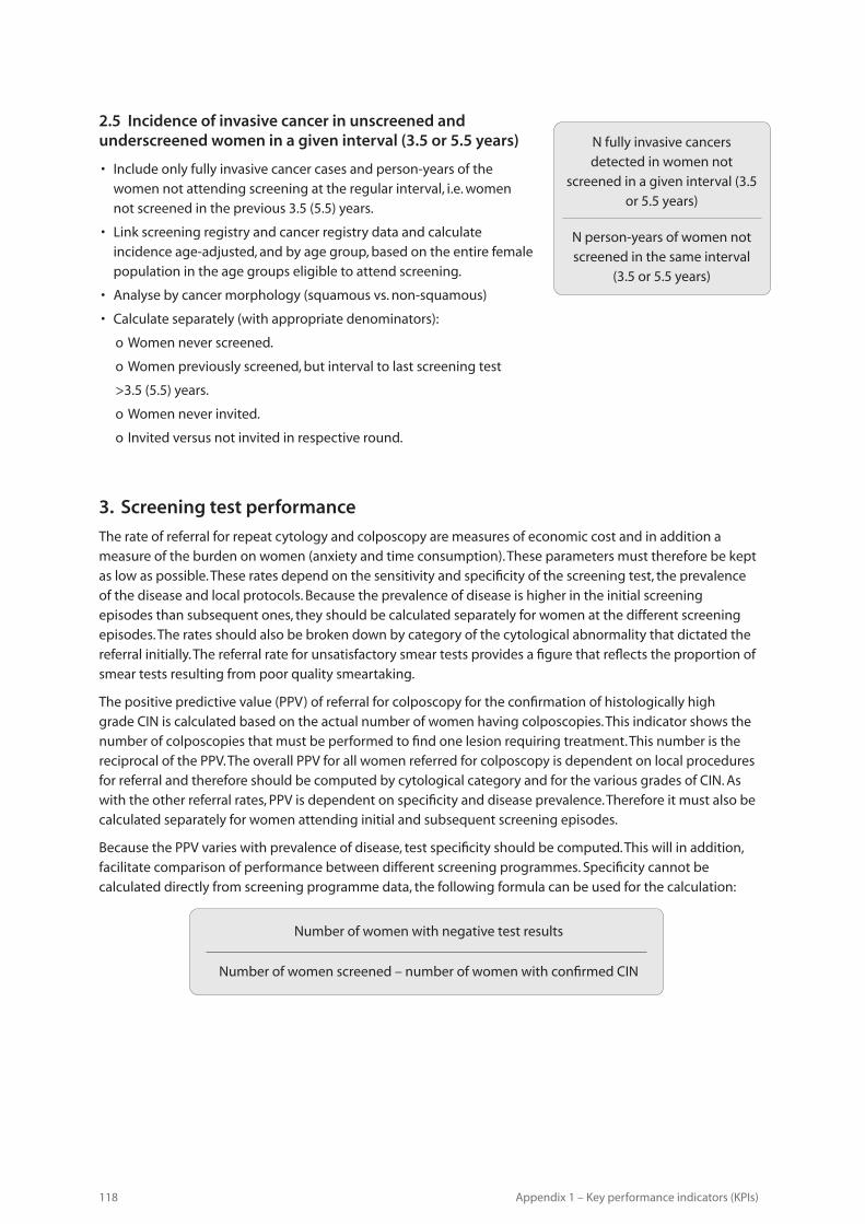
2.5 Incidence of invasive cancer in unscreened and underscreened women in a given interval (3.5 or 5.5 years)
• Includeonlyfullyinvasivecancercasesandperson-yearsofthe
women not attending screening at the regular interval, i.e. women
not screened in the previous 3.5 (5.5) years.
• Linkscreeningregistryandcancerregistrydataandcalculate
incidence age-adjusted, and by age group, based on the entire female
population in the age groups eligible to attend screening.
• Analysebycancermorphology(squamousvs.non-squamous)
• Calculateseparately(withappropriatedenominators):
o Women never screened.
o Women previously screened, but interval to last screening test
>3.5 (5.5) years.
o Women never invited.
o Invited versus not invited in respective round.
N fully invasive cancers
detected in women not
screened in a given interval (3.5
or 5.5 years)
N person-years of women not
screened in the same interval
(3.5 or 5.5 years)
3. Screening test performance
The rate of referral for repeat cytology and colposcopy are measures of economic cost and in addition a
measure of the burden on women (anxiety and time consumption). These parameters must therefore be kept
as low as possible. These rates depend on the sensitivity and specificity of the screening test, the prevalence
of the disease and local protocols. Because the prevalence of disease is higher in the initial screening
episodes than subsequent ones, they should be calculated separately for women at the different screening
episodes. The rates should also be broken down by category of the cytological abnormality that dictated the
referral initially. The referral rate for unsatisfactory smear tests provides a figure that reflects the proportion of
smear tests resulting from poor quality smeartaking.
The positive predictive value (PPV) of referral for colposcopy for the confirmation of histologically high
grade CIN is calculated based on the actual number of women having colposcopies. This indicator shows the
number of colposcopies that must be performed to find one lesion requiring treatment. This number is the
reciprocal of the PPV. The overall PPV for all women referred for colposcopy is dependent on local procedures
for referral and therefore should be computed by cytological category and for the various grades of CIN. As
with the other referral rates, PPV is dependent on specificity and disease prevalence. Therefore it must also be
calculated separately for women attending initial and subsequent screening episodes.
Because the PPV varies with prevalence of disease, test specificity should be computed. This will in addition,
facilitate comparison of performance between different screening programmes. Specificity cannot be
calculated directly from screening programme data, the following formula can be used for the calculation:
Number of women with negative test results
Number of women screened – number of women with confirmed CIN
118 Appendix 1 – Key performance indicators (KPIs)

The detection rate (DR) of CIN (especially CIN2/3), depends on the number of lesions that are present in
the screened population (disease prevalence) and how many of them are actually detected (cross sectional
sensitivity). Since the prevalence of disease varies geographically and is apriori unknown, it is difficult to
use the DR as an indicator of sensitivity. In addition, the DR also depends on the criteria of interpretation
of histology, which are subject to variation. Nevertheless, DR should be monitored and compared between
European screening programmes. This will provide a tool for recognising variation in quality and for
developing the descriptive epidemiology of CIN within Europe, providing information for further study to
improve control of cervical cancer.
There is no easily interpretable indicator of screening sensitivity that can be collected in a screening
monitoring system. It is therefore essential to link screening registry and cancer registry data. Although it is
difficult to obtain comparable data, comparison of the incidence of cancers which are detected in women
after having findings of normal cytology, to the expected incidence in the absence of screening provides an
estimate of test sensitivity for invasive lesions. Information on cervical cancer incidence among unscreened
women can be taken into account, if adjustments for selection bias in relation to screening attendance or
non-attendance are calculated. Correspondingly, estimates of screening episode sensitivity may be obtained
from inclusion of all screened women in the follow-up of cervical cancers. When considering programme
sensitivity, women invited, but not screened, must be taken into account. Previous smear tests of women with
screen-detected cancer should also be reviewed (combined with those of other women who did not develop
cancer in order to avoid over-interpretation).
The distribution of the interval to reporting i.e. time between smeartaking and result communication
should be monitored. Reporting delays, which are not extreme, should not influence screening effectiveness.
However, such delay can affect women’s perception of the quality of service, which in turn may affect
participation in the programme and increases anxiety.
3.1 Distribution of screened women by the results of cytology
Calculate overall and separately for subgroups of women:
• Fortheregularscreeningintervalandshortertimeperiods.
• Attendinginitialorsubsequentscreening.
3.2 Referral rate for repeat cytology
Calculate separately:
• Bycytologythatresultedinrecommendationtorepeat.
• Forinitialandsubsequentscreening.
N screened women advised
to repeat test at shorter than
regular interval
N screened women
3.3 Compliance with referral for repeat cytology
Calculate separately:
• Bycytologythatresultedinrecommendationtorepeat.
• Forinitialandsubsequentscreening.
N women screened following
recommendation for repeat
cytology
N women recommended for
repeat cytology
N screened women with
cytological diagnosis
N screened women
119Appendix 1 – Key performance indicators (KPIs)
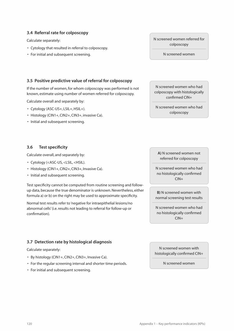
3.4 Referral rate for colposcopy
Calculate separately:
• Cytologythatresultedinreferraltocolposcopy.
• Forinitialandsubsequentscreening.
3.5 Positive predictive value of referral for colposcopy
If the number of women, for whom colposcopy was performed is not
known, estimate using number of women referred for colposcopy.
Calculate overall and separately by:
• Cytology(ASC-US+,LSIL+,HSIL+).
• Histology(CIN1+,CIN2+,CIN3+,invasiveCa).
• Initialandsubsequentscreening.
N screened women who had
colposcopy with histologically
confirmed CIN+
N screened women who had
colposcopy
N screened women referred for
colposcopy
N screened women
3.6 Test specificity
Calculate overall, and separately by:
• Cytology(<ASC-US,<LSIL,<HSIL).
• Histology(CIN1+,CIN2+,CIN3+,InvasiveCa).
• Initialandsubsequentscreening.
Test specificity cannot be computed from routine screening and follow-
up data, because the true denominator is unknown. Nevertheless, either
formula a) or b) on the right may be used to approximate specificity.
Normal test results refer to ‘negative for intraepithelial lesions/no
abnormal cells’ (i.e. results not leading to referral for follow-up or
confirmation).
A) N screened women not
referred for colposcopy
N screened women who had
no histologically confirmed
CIN+
B) N screened women with
normal screening test results
N screened women who had
no histologically confirmed
CIN+
3.7 Detection rate by histological diagnosis
Calculate separately:
• Byhistology(CIN1+,CIN2+,CIN3+,InvasiveCa).
• Fortheregularscreeningintervalandshortertimeperiods.
• Forinitialandsubsequentscreening.
N screened women with
histologically confirmed CIN+
N screened women
120 Appendix 1 – Key performance indicators (KPIs)

3.8 Cancer incidence after normal cytology
Normal cytology refers to cases recommended for re-screening at the
regular interval. Count only fully invasive cancers among the women
who had a normal screening cytology in the previous 3.5 (5.5) years.
Analyse by:
• Intervalfromindexcytology.
• Cancermorphology(squamousvs.non-squamous).
• Cytologyshouldbereviewedmixedwiththatofotherwomennot
developing cancer.
N screened women with
fully invasive cervical cancer
detected within 3.5 (5.5) years
of normal cytology
N person-years of screened
women for same period after
normal cytology
4. Diagnostic assessment and treatment
The success of a screening programme is reliant on diagnostic assessment being actually performed when
required. Measuring compliance with referral for colposcopy requires systematic and complete registration
of colposcopies. When a record is not available in the colposcopy register, the patient or her doctor should be
contacted to obtain information on whether the colposcopy was performed or as a reminder for the need for
examination. Compliance with colposcopy should be calculated for each category of cytology that was the
initial reason for referral (more severe cytology the greater the relevance). In addition compliance should be
monitored for different screening time intervals.
Another condition essential to screening effectiveness is actual delivery of requisite treatment, particularly for
histologically confirmed CIN2 and CIN3.
Another important target of a screening programme is the avoidance of over-treatment. The proportion of
women with pre-invasive lesions who undergo hysterectomy is a major indicator of unnecessary treatment,
although some hysterectomies result from co-existing pathology. Peer review should be carried out to verify
the appropriateness of treatment of such cases. It should be taken into account that relevant differences in
the proportion of women with CIN who undergo hysterectomy suggest that local practice is the main cause
of such differences.
The absence of SIL (or of high-risk HPV infection) can be routinely monitored at six monthly follow-up of
treated women. This parameter should be included as an indicator of short-term quality of treatment.
The incidence of cervical cancer in women which was not detected by screening, although the cytology
results were abnormal (i.e. after abnormal cytology), serves as a direct summary indicator of failure associated
with diagnostic assessment and treatment. Various reasons for failure can be identified. For example, cervical
cancer arising in women who did not comply with referral for colposcopy could represent a failure in the
communication process or a lack of attendance compliance for follow-up. Cases that arise in women who
had colposcopy, but without detection of CIN, represent failure in diagnostic accuracy, etc. To calculate this
parameter, the screening history of each case of cervical cancer should be reviewed, and those cases should
be excluded in which cancer was detected as a result of screening.
The above parameters apply under the assumption that cytology is used as the primary screening test, which
is what is currently recommended. However, most of the present parameters can also be applied, with only
minor changes, to different screening methods (e.g. HPV DNA testing). Depending on which screening test
and screening policy that is employed, the values of some parameters (e.g. DR, PPV or specificity) may be
expected to change.
121Appendix 1 – Key performance indicators (KPIs)

4.1 Compliance with referral to colposcopy
Calculate separately by:
• Differentintervalsafterreferral(threemonths/sixmonths).
• Cytologythatresultedinreferral.
• Thismeasureexaminestherelationshipbetweenthenumbers
referred to colposcopy and the numbers who actually attended.
It also only deals with new referrals from the programme. The
denominator is the number of women referred to colposcopy from
the programme (CSR) and the numerator should be the number of
new patients attending colposcopy who came via the programme.
N new women attending
colposcopy following referral
from screening programme
N screened women referred for
colposcopy from the screening
programme
4.2 Treatment of high grade intraepithelial lesions
Note: Treatment includes the following and may take place at any visit
in the episode:
• Conebiopsy
• Punchbiopsy/diagnosticbiopsy
• Cryotherapy
• LLETZ
• Smeartest
• Swabs
• Laserablation
• Laserexcision
• Radicalhysterectomy
• Tracehelectomy
• SWETZ
• Coldcoagulation
N women with screen-detected
CIN2 or CIN3 treated
N women with screen-detected
CIN2 or CIN3
4.3 Proportion (%) of women with total hysterectomy following-on screen-detected intraepithelial lesions
Calculate separately by histology (CIN1, CIN2, CIN3). Appropriateness of
individual cases should be evaluated by peer review.
N screened women with
histological CIN total
hysterectomised
N screened women with
histological CIN
122 Appendix 1 – Key performance indicators (KPIs)

4.4 Proportion (%) of women treated for CIN1
Appropriateness of individual cases should be evaluated by peer review.
Note: Treament includes the following and may take place at any visit in
the episode:
• Conebiopsy
• Punchbiopsy/diagnosticbiopsy
• Cryotherapy
• LLETZ
• Smeartest
• Swabs
• Laserablation
• Laserexcision
• Radicalhysterectomy
• Trachelectomy
• SWETZ
• Coldcoagulation
N women with screen-detected
CIN1 treated
N screened women with
screen-detected CIN1
4.5 Incidence of invasive cancer after abnormal cytology
• Includescreenedwomen:
o Without colposcopy carried out, despite existing indication.
o With colposcopy carried out, but no CIN detected.
o With CIN detected, but not treated.
o Treated.
o In diagnostic or post-treatment follow-up.
• Calculateoverallandseparatelyforeachofabovesubgroups.
• Includeonlyfullyinvasivecancers.
• Excludecasesdetectedasaresultofscreening.
N cases of invasive cancer
in screened women after
abnormal cytology
N person-years of screened
women after
123Appendix 1 – Key performance indicators (KPIs)

4.6 Proportion of women with cytology negative for SIL, six months after treatment
Note: Treatment includes the following and may take place at any visit
in the episode:
• Conebiopsy
• Punchbiopsy/diagnosticbiopsy
• Cryotherapy
• LLETZ
• Smeartest
• Swabs
• Laserablation
• Laserexcision
• Radicalhysterectomy
• Trachelectomy
• SWETZ
• Coldcoagulation
• IncludewomentreatedforCIN2,CIN3,CGINorAISinsitufollowedat
least six months after treatment (denominator).
• IncludewomennegativeforHR-HPV(numerator),ifthistestisused
for follow-up.
• Follow-upprotocols–atleastonesmeartestiscarriedoutin
colposcopy six months after a treatment (colposcopy procedure). For
the purposes of audit, the measure is taken at eight months.
N screened and treated women
with negative cytology after 6
months
N screened and treated women
followed-up for at least 6
months
124 Appendix 1 – Key performance indicators (KPIs)

5. Definition of performance parameters in cervical cancer screening
The specific instructions are indicated below.
For calculations for a given period of time, such as the recommended screening interval (three or five years),
the dates on which the period starts and ends, and the performance for determining the target population
should be recorded. For calculations based on the size of the target population, use the average over the
given time period.
Note that parameters 6 (incidence of invasive cancer in unscreened women), 14 (cancer incidence after
normal cytology) and 19 (incidence of invasive cancer after abnormal cytology) require linkage with cancer
registry data/histological data. The follow-up periods recommended for calculation of cervical cancer
incidence are six months longer than the recommended screening interval of the respective programme
(3.5 or 5.5 years). The purpose of adding one half-year to the screening interval is to include screen-detected
cancer at the next screening episode. Calculations based on longer follow-up periods are also recommended.
7. References
1. Arbyn M., Antilla A., Jordan J., Ronco G., Schenck U., Segan N., Wiener H.G., Herbert A., Daniel J., von Karsa L.
(2008) European guidelines for quality assurance in cervical cancer screening [2nd Edition]. International
Agency for Cancer Research and EU, Health & Consumer protection Directorate-General.
125Appendix 1 – Key performance indicators (KPIs)

Guidelines for
Quality Assurance in Cervical ScreeningSecond Edition
NCSS/PUB/Q-1 Rev 2 ISBN 978-1-907487-13-2
The National Cancer Screening Service is part of the Health Service Executive. It encompasses BreastCheck – The National Breast Screening Programme and CervicalCheck – The National Cervical Screening Programme, BowelScreen – The National Bowel Screening Programme and Diabetic RetinaScreen – The National Diabetic Retinal Screening Programme.
Gu
idelin
es for Q
uality A
ssuran
ce in C
ervical Screenin
g Second
Editio
n