purification and some properties of human 4 ... · purification and some properties of human...
TRANSCRIPT

THE Jouarr~~ OP Brow~wu. CHEH~~T,,Y Vol. 252, No. 14, Issue of July 25, pp. 5073-5084, 1917
Prmted in U.S.A.
Purification and Some Properties of Human 4-Hydroxyphenylpyruvate Dioxygenase (I)*
(Received for publication, February 10, 1977)
BENGT LINDBLAD, &RAN LINDSTEDT, SVEN LINDSTEDT, AND MARIANNE RUNDGREN
From the Department of Clinical Chemistry, University of Gothenburg, Sahlgren$ Hospital, S-413 45 Gothenburg, Sweden
4-Hydroxyphenylpyruvate dioxygenase (I-hydroxyphenyl- pyruvate:oxygen oxidoreductase (hydroxylating, decarboxy- lating), EC 1.13.11.27) has been purified 800-fold in 25% yield from human liver by a procedure involving ammonium sulfate fractionation of an acetone powder extract and chro- matography on hydroxylapatite, sulfopropyl-Sephadex C-50, and triethylaminoethyl cellulose. The preparation obtained was homogenous as determined by electrophoresis, gel fil- tration, sedimentation equilibrium, and immunodiffusion. A molecular mass of 87 kilodaltons was obtained by sedi-
mentation equilibrium. A subunit mass of 43 kilodaltons was obtained by electrophoresis in polyacrylamide gel con- taining sodium dodecyl sulfate.
The enzymic activity had two pH optima around 4.5 and
7.8. The enzyme was equally activated by ascorbate or by a combination of 2,6-dichlorophenolindophenol and glutathi- one in the presence of catalase. It was very sensitive to H,O, inhibition, which was abolished by a reductant and catalase. Addition of Fez+ and of various metal salts had no stimula- tory effect on enzyme activity. The activity with the keto form of 4-hydroxyphenylpyruvate was 40 times higher than with enol tautomer as substrate. During optimized assay conditions, the apparent Michaelis constants were about 50 FM for 02, 0.03 mM for 4-hydroxyphenylpyruvate, and 0.05 mM for phenylpyruvate. The corresponding apparent maxi- mal velocities were 4 and 0.02 mmol mine1 (g of protein)-‘.
The enzyme activity was strongly inhibited by iron and copper chelators. The activity of enzyme inhibited by dieth- yldithiocarbamate was restored by dialysis. Enzyme in- hibited by bathophenanthroline was reactivated to -30% by dialysis and to -60% by addition of Fe’+.
4-Hydroxyphenylpyruvate dioxygenase activity was not inhibited by different thiol group reagents in 1 mM concen- trations with the exception of mercurials, which inhibited in lower concentrations. The enzyme contained about five thiol groups and one disulfide bridge/87 kilodaltons as deter- mined by titration with 4-ehloromercuribenzoate and 5,5’- dithiobis(2-nitrobenzoate) and by alkylation with iodo[l- Wlacetate and iodo[l-14Clacetamide.
4-Hydroxyphenylpyruvate dioxygenase, (4-hydroxyphenyl- pyruvate:oxygen oxidoreductase, (hydroxylating, decarboxy-
*This work was supported by Grant 13X-585 from the Swedish Medical Research Council. The paper is the first of a series of three.
lating), EC 1.13.11.27) catalyzes the reactions in which 4
hydroxyphenylpyruvat is converted to homogentisate, phen- ylpyruvate to 2-hydroxyphenylacetate (l-4), 3,4dihydroxy- phenylpyruvate to 2,4,5-trihydroxyphenylacetate (3,4), and 4- fluorophenylpyruvate to 5-fluoro-2-hydroxyphenylacetate (2).
Both atoms of oxygen are incorporated into the product and a reaction mechanism has been proposed, which is similar to that for 2-oxoglutaratedependent oxygenases (5-7). The reac- tion is stimulated by reductants and a metal is probably involved in the activation of oxygen.
Transient and permanent deficiency of human 4-hydroxy- phenylpyruvate dioxygenase has been implicated in the patho- genesis of neonatal and hereditary tyrosinemia, respectively (8, 9). Transient neonatal tyrosinemia is a harmless condition, whereas hereditary tyrosinemia is a serious metabolic disor- der with early liver cirrhosis and multiple renal tubular de- fects. In order to further study the mechanism of the enzymic reaction and the reason for the low activity of the enzyme in the abovementioned conditions, we found it necessary to pu- rify the human 4-hydroxyphenylpyruvate dioxygenase and to study the properties of the enzyme.
In two accompanying papers, we describe the resolution of the enzyme into multiple forms and a kinetic study (10, 11).
EXPERIMENTAL PROCEDURES
Materials
The material used was obtained from the following sources: 2,5- diphenyloxazole and 1,4-bis[2-(4-methyl&phenyloxazolyl)]benzene from Arapahoe Chemicals. Boulder. Col.: ultrafiltration membranes from Am&on Corp., Lexi&ton, Mass.; complete Freund’s adjuvant from Behringwerke, Marburg (L), FDR; hydroxylapatite from Bio- Rad Laboratories, Richmond, Calif.; bovine liver catalase (20 g/liter) from C. F. Boehringer and Soehne, GmbH, Mannheim, FDR; ace- tone, ammonium persulfate, ammonium sulfate, sodium meta-ar- senite, thioglycolic acid, and urea (Aristar) from British Drug Houses Ltd.. Poole, England; dithiothreitol from Calbiochem AG. Luzerne, Switzerland; &ylamide, NY’-bismethyleneacrylamide; B-hydroxyquinoline-5-sulfonic acid, and N,N,N’,N’-tetramethyleth- ylenediamine from Eastman Organic Chemicals, Rochester, N. Y., sodium 4-chloromercuribenzoate, 2-mercaptoethanol, and 4,5-dihy- droxybenzene-1,3-disulfonic acid (Tiron) from Fluka AG, Buchs, Switzerland; sodium 4,7-diphenyl-l,lO-phenanthrolinedisulfonate (sodium bathophenanthrolinedisulfonate). 2.9-dimethvl-l.lO-uhen- anthroline (neicuproine), l,lO-phenanthroiine, and 2,4,6&(2’-pyri- dyl)-1,3,5-triazine from G. Frederick Smith Chemical Co.. Colum- b&s, Ohio; iron wire (>99,999%1 from Koch-Light Laboratories, Ltd., Colnbrook, Bucks, England; human ceruloplasmin from Kabi, Stockholm, Sweden; carrier ampholytes, pH 3 to 10, from LKB Produkter AB, Bromma, Sweden; 3-acetylamine-4-hydroxypheny-
5073
by guest on March 23, 2019
http://ww
w.jbc.org/
Dow
nloaded from

5074 Purification of 4-Hydroxyphenylpyruvate Dioxygenase
larsinic acid (acetarsone). N-ethvlmaleimide. 2iodobenzoic acid. io- doacetic acid, and iodoacemmidefrom Mann Besearch Laboratories, Inc., N. Y.; 2.2’-bipvridvl, 2.6~dichloronhenolindonhenol, dimethyl- glyoxime, sodium ~diethyldithiocarbarnate, diphenylcarbazone, di- phenyldithiocarbazone fdithizone), glutathione, ammoniumnitroso- phenylhydroxylamine (cupferron), sodium azide, and l-(2’-theonyll- 3,3,3-trifluoroacetone from E. Merck AG, Darmstadt, FDR; iodoll- “Clacetamide 12.68 GilmoB. iodollJ4Clacetic acid (13.9 Cilmoll, ill- iV]tyrosine (-50 CUrnob, and ~[l-14~]phenylalanine (-50 Ci/mol] from New England Nuclear Chemicals GmbH, Frankfurt CM]. FDR: Hyamine (1 G solution in methanol) from Packard Instrument Co., Inc., Downers Grove, Ill.; 5,5’-dithiobis(2-nitrobenzoic acid] from Pierce Chemical Co., Bockford, Ill.; L-[UJ4C]tyrosine (-500 Ci/moll from The Badiochemical Centre, Amersham, Bucks, England; L- amino acid oxidase (Crotalus adamantens) (EC 1.4.3.21, d-amino- phenylpyruvic acid, sodium 4-chloromercuriphenylsulfonate, 2,9-di- methyl-4,7-diphenyl-l,lO-phenanthroline(bathocuproine), homogen- tisic acid, 4-h&roxyphenylpyrovic acid, and sodium phenylpyruiate from Sigma Chemical Co., St. Louis, MO.; dialysis tubings from Union Carbide Chemical Corp., Chicago, Ill. Other chemicals used were of analytical grade and purchased from local stores.
Centrallv deionized water was further purified bv Passage through granular activated carbon, mixed ion exchange resin and-a Millipore filter (0.45 wrnl (Super-Q svstem of Millipore Ltd., Wem- bley,-Middlesex, England). The wamr had a resi‘stivity above 18 Mohm x cm.
Methods
Preparation of Substrates
4-Hydroxyphenyl[l-‘4C]pyruvate and phenyl[l-‘4C]pyruvate were prepared from c[l-‘4C]tyrosine and L-[l-“Clphenylalanine, accord- ing to Lindblad (12).
Enol Form of 4-Hydroxyphenylpyruuic Acid
A 0.5 M solution of 4-hydroxyphenylpyruvic acid in water, 1 ml, was purified by shaking with charcoal. The solution was acidified to pH 2 with H.&O,, saturated with NaCl and then extracted three times with 2 ml of ethyl acetate. The extract was dried over NapSO, for 2 h and then concentrated to about a l-ml volume. Light petro- leum (b.p. 40-60”) was added until the solution became opalescent, and the mixture left to crystallize at 4”. After about 5 h, the supema- tant was sucked off and the crystals dried in uucuo over P,O,. The crystallization was repeated once to yield crystals melting at 212”, decomposition. A solution of this preparation in 99.5% ethanol showed one absorption maximum at 303 nm (e3o3 = 20 mM-’ cm-‘). Only one peak, containing the enol form of 4-hydroxyphenylpyruvic acid, was obtained using chromatography through Sephadex G-15 (1.2 x 17 cm) (13) in a buffer, at pH 3, prepared from 0.2 M sodium acetate and 0.2 M H,B03. This procedure resolves the keto and enol forms of the acid.
Keto Form of 4-Hydroxyphenylpyruvaate
A 1 M solution of 4-hydroxyphenylpyruvic acid in methanol was purified by shaking with charcoal. After addition of an equal volume of 1 M KOH in methanol, the solution was left to crystallize at 4” for 3 h. The crystals were washed four times with 1 ml of methanol and then dried in vacno over P,O,. The potassium salt of the keto form of 4-hydroxyphenylpyruvate was stored in uczcuo over P,O, at -20”. When the crystals were dissolved in the incubation buffer, an ultra- violet spectrum characteristic of the ketu form of I-hydroxyphenylpy- ruvate was obtained (E~,~ = 2 rnM-’ cm-‘) (14).
Determination of 4-Hydroxyphenylpyruuate Dioxygenase Activity
Assay 1 -The assay procedure of Lindblad (12) was used with some modifications. The incubation mixture, usually 1 ml, contained bovine liver camlase (0.8 g/liter), glutathione (10 mM1, 2,6dichloro- phenolindophenol (0.15 mM), 2,2’-bipyridyl (1 mM), sodium potas- sium phosphate buffer at pH 6.5 (0.2 M), and I-hydroxyphenyl[l- ‘*Clpyruvate (1 mM). The concentration of oxygen was that obtained with air in the aas phase. After incubation of the reaction mixture for 20 min at 37”>n awater bath, the reaction was started by addition of the substrate. The reaction was stopped alter 30 min at 37” by the addition of 1 ml of 30 mM sodium diethyldithiocarbamate in 1 M sodium acetate buffer at pH 5.5. The incubation was continued for 45 min and the evolved CO, absorbed in 20 ~1 of Hyamine placed in a glass cup suspended from the rubber stopper. The glass cup was
transferred to a scintillation vial containing 10 ml of scintillation solution (toluene:2-methoxyethanol (5:3) with 0.19 g of 1,4-bis12-(4- methyl-5-phenyloxazolyl)]benzene and 6.25 g of 2,5-diphenyloxazole/ liter). Radioactivity was measured in a Packard Tri-Carb liquid scintillation spectrometer.
Assay 2 -During the later part of the work the assay procedure was changed in the following way to optimize the conditions. The substrate concentration was lowered to 0.2 mM, the incubation buffer was changed to 0.2 M Tris/HCl at pH 7.5, the reaction time was decreased to 15 min, 2,2’-bipyridyl was excluded, and the tempera- ture in the first incubation period was reduced to 4”.
Enzymically formed homogentisate was measured as described by Lindblad (12). Phenylpyruvate tautomerase (EC 5.3.2.11 activity was determined as described by Constansas and Knox (15).
Oxygen Studies
Mixtures of nitrogen and oxygen were obtained from AGA (Li- dingo, Sweden). Assay 2 was performed in 25-ml polyethylene vials sealed with rubber stoppers, which had one gas inlet tubing ending near the surface of the incubation mixture and one gas outlet tubing ending near the top of the vial. The incubation mixture was equili- brated with different gas mixtures for 15 min at 37” at atmospheric pressure. The reaction was started by adding 10 ~1 of substrate solution through the gas outlet tubing. The inlet and outlet tubings were then clamped.
Protein Determination
Protein was determined by the Lowry method (16) with bovine serum albumin as the standard. Effluent fractions from chromato- graphic columns were routinely monitored by measuring the absorb- ance at 280 nm (A,,,) with a Zeiss PMQ II spectrophotometer. The amount of purified enzyme protein was also determined in the spec- trophotometer using a value for the absorption coefficient at 280 nm of 11.3 (see below).
Definition of Unit
One unit of 4-hydroxyphenylpyruvate dioxygenase activity is de- lined as the consumption of 1 pmol of substrate or the formation of 1 pm01 of product per min under the assay conditions specified.
Polyacrylamide Gel Electrophoresis
Disc gel electrophoresis in 7% (w/v) acrylamide was run as de- scribed by Davis (17) without sample and spacer gels. Samples, 10 to 50 ~1 in 0.6 M sucrose, were layered under buffer on the upper, cathodal gel surface.
Determinations of size and charge isomers and of molecular mass was performed by electrophoresis in different concentrations of acrylamide according to the method described by Hedrick and Smith (18). The weight ratio of bisacrylamide to acrylamide monomer was kept constant at 1 to 30. The gel buffer was 0.06 M Tris/HCl, pH 8.9, and the electrode buffer was 0.038 M Trisfglycine at pH 8.3 (17). The gels (0.6 x 10 cm) were run at 1 mA/tube for 30 min at room temperature. Electrophoresis was performed at 4 mA/tube for 60 min at room temperature. The gels were stained overnight in 1.2 M acetic acid containing 5 g of Amido black/liter and then destained in 1.2 M acetic acid. Mobilities were calculated relative to bromphenol blue and plotted against the concentration of acrylamide in the gel. The frictional ratios, obtained by dividing the relative mobility at 6% (w/ v) by that at 10% (w/v) acrylamide concentration, were replotted against known molecular masses of reference proteins (Table I) (19).
Electrophoresis in polyacrylamide gels containing sodium dodecyl sulfate was performed according to Weber and Osbom (21) with some modifications. The concentration of the gel buffer was lowered to 0.05 M. Solutions of 10% (w/v) acrylamidewere polymerized in glass columns (0.6 x 12 cm) by N,N,N’,N’-tetramethylethylenediamine (2.1 rnM1 and ammonium persulfate (0.38 g/liter): The protein sam- ples were denatured in 0.01 M sodium phosphate buffer, pH 7.0, containing sodium dodecyl sulfate (10 g/liter) and 2-mercaptoethanol (1 M) for 3 h at 37”. Glycerol and bromphenol blue were then added to a final concentration of 3.4 M and 0.02 a/liter, respectively. The samples (10 to 50 ~1, containing 1 to 20 pg of protein\ were layered under the buffer on the upper, cathodal gel surface. Electrophoresis was carried out at 6 m&gel for about-4 h at room temperature. Mobilities were calculated relative to that of bromphenol blue and plotted versus the logarithm of the molecular or subunit masses of reference proteins (Table I) (21).
by guest on March 23, 2019
http://ww
w.jbc.org/
Dow
nloaded from

Purification of 4-Hydroxyphenylpyruvate Dioxygenase 5075
TABLE I
Properties of reference proteins
Protein
Cytochrome c, equine heart (Mann)
Ribonuclease I, bovine pan- creas (Worthington)
Chymotrypsinogen A, bo- vine pancreas (Worthing- ton)
Ovalbumin (Worthington) Albumin, bovine (BDH) Transferrin, human (Kabi) Alcohol dehydrogenase
yeast (Boehringer) Aldolase, rabbit muscle
(Boehringer) IgG, human (Hyland) Catalase, bovine liver
(Boehringerf
x 10-T cd/s R?n kib<on
13.0b 1.64 11.7’
11.16 1.93
9.5b 2.25 25.7'
7.8 2.74 5.96 3.62
4.70 4.55
4.6" 4.64 149
5.ov 15v 4.16 5.21 24W
13.7’
43.56 67.0d 76.6' 148'
0 The tabulated values of Stokes radii were calculated from values ofDlo,m given in the literature by Stokes-Einsteins formula when not otherwise stated.
b Laurent and Killander (20) and references therein. c Weber and Osborn (21) and references therein. d Sober (22) and references therein. p Fish et at. (23). ’ Rymo et al. (24).
g Samejima and Yang (25).
Isoelectric Focusing
Isoelectric focusing was performed with 1% carrier ampholytes in the pH range 3 to 10 in an 8101 Electrofocusing Column of llO-ml capacity (LKB Produkter AB, Bromma, Sweden) (26). Protein solu- tions were dialyzed three times against 100 volumes of 0.1 M glycine, pH 6.5, containing 0.01% of carrier ampholytes and exchanged for light solution in the middle of the gradient. Electrofocusing was carried out for 46 h at 1 mA, 600 V, and 4” with the cathode at the bottom of the column. Fractions of 2 ml were collected at the end of the run.
Gel Filtration
A column (1.5 x 86 cm) of Sephadex G-200 was prepared in 25 mM Tris/HCl, pH 7.4, containing 0.2 M NaCl and equilibrated to a flow rate of 6 ml/h. Protein samples were applied in a volume of 2 ml containing 0.3 g of blue dextran 2000/liter and sucrose (0.3 M). Fractions of 1 ml were collected in preweighed tubes. Elution vol- umes were measured by weight. The bed volume was measured by volume. The average partition coefficient (K,,,), was calculated as CV, - V,)lCV, - VO), where V, is the elution volume of a protein, V, the elution volume of blue dextran and V, the total bed volume.
Analytical Ultmcentrifugation
Sedimentation equilibrium experiments were performed with a Beckman-Spinco model E analytical ultracentrifuge equipped with electronic speed and rotor temperature control units and Rayleigh optic system, using short columns and the high speed meniscus depletion technique of Yphantis (27). The 3-mm epon double sector cells with sapphire windows were filled with 30 ~1 of Fluorochemical- 43 as a base for llO-~1 samples. The concentration of protein in the samples of purified enzyme was 0.23, 0.32,0.37, and 0.38 g/liter of 25 rnsr Tris/HCl buffer at pH 7.4, containing 0.2 M NaCl. The tempera- ture was 4” and the rotor speed was 22,000 rpm. Interference patterns were photographed on Kodak spectrographic plates and analyzed with a two-dimensional microcomparator.
Dry Weight Determination
The enzyme protein was exhaustively dialyzed against water or 50 ells NH,HCO, solution, pH 7.5. Triplicate samples of the protein solutions were heated at 105” over silica gel and weighed on an electrobalance model G-2 (Cahn Division, Ventron Instruments
Corp., Paramount, Calif. ), until constant weight was obtained twice with a l-day interval.
Light Absorption Spectrum
A solution of the enzyme (5 g/liter) was dialyzed against 200 volumes of water with five changes at about 4-h intervals, lyophilized and dissolved in buffer or water. The concentration of protein was determined both by the Lowry method (16) and by dry weight (see above). Absorption spectra were recorded on a Zeiss DMR 21 record- ing spectrophotometer.
Amino Acid Analysis
A solution of the enzyme was extensively dialyzed against water and then centrifuged at 2000 x g for 10 min. Dry weight determina- tions were performed on portions of the clear supematant. Samples, containing 0.483 mg of protein, were lyophilized and then dissolved in 2 ml of constantly boiling HCl. Hydrolysis was performed in an argon atmosphere in sealed borosilicate tubes, kept at 110” t 1” in an oil bath for 24 h. Hydrochloric acid was removed by evaporation on a rotary evaporator. The analyses were done on a Beckman-Spinco automatic amino acid analyzer with a two-column system (28).
Preparation of Antiserum
Purified 4-hydroxyphenylpyruvate dioxygenase was emulsified with an equal volume of complete Freund’s adjuvant. Two rabbits were each injected with 0.25 mg of enzyme in each rear footpad. The injection was repeated with 0.5 mg of enzyme/animal after 2 and 6 weeks. Blood serum obtained after 10 weeks was stored at -20”. The serum was tested for the presence of antibodies against human 4- hydroxyphenylpyruvat dioxygenase by the radial immunodiffision technique (29, the double diffusion test (30), and by determination of enzyme activity on the supematants obtained after antiserum pre- cipitation of human liver homogenate.
Thiol Content
Titrations with 4-chloromercuribenzoate were carried out at 25” in 0.05 M sodium phosphate buffer (pH 7.0) containing 0.15 M Na*SO, (31). The reagent solution was standardized by titration with L- cyst&e. The number of thiol groups was calculated from the concen- trations of 4-chloromercuribenzoate required to reach the inflection point in the curve of maximum absorbance change at 250 run, ob- tained about 10 to 15 min after the addition of I-chloromercuriben- zoate.
Determination of the thiol content before and after full reduction with dithiothreitol was performed with 5,5’-dithiobis(2nitrobenzoic acid), in 0.08 M sodium phosphate buffer at pH 8.0, containing 20 g of sodium dodecyl sulfate and 1 g of EDTAlliter (32). The excess dithio- threitol used as reductant was removed by gel filtration. The concen- tration of protein wss then determined by spectrophotometry. When using a molecular absorption coefficient of 13.6 mM-’ cm-’ at 410 nm the thiol content of glutathione was 0.98 ? 0.02 (S. D.) UV = 10) at the 5 to 60 nmol level.
S-Alkylation of the enzyme was performed with limiting concen- trations of iodoacetate and iodoacetamide (33). Dialyzed and lyophi- lized samples of protein, 0.5 to 1 mg, were dissolved in 100 ~1 of 0.5 M Tris/HCl buffer at pH 8.0, containing 8 M urea and 0.14 M 2-mereap- methanol. The solution was equilibrated with N,. Reduction was carried out at 37” for 4 h in the dark. Iodo[l-‘4Clacetate (103 cpml nmol) or iodoll-“Clacetamide (77 cpm/nmolI in 0.25 M NaOH was added to a final concentration of 70 mM. After alkylation at rwm temperature for 30 min. the reaction mixture was put onto a column of Sephadex G-25 (fine). The alkylated protein was eluted with 10 - sodium phosphate buffer, pH 7.1, containing 1 g of sodium dodecyl sulfate/liter. Protein concentrations were determined by spectrophotometry.
Metal Studies
Glasswares and dialysis tubes were washed with 0.5 M acetic acid, 1 mM EDTA, and water. Metal salts and metal chelators were added in 0.2 M Tris/HCl buffer at pH 7.5, or in mixtures of that buffer and ethanol.
Total copper was determined as the Cu+ .2,2’-biquinoline complex. Protein-bound copper was released with glacial acetic acid and re- duced with ascorbic acid (34). The standard, CuS04.5H,0, was treated as the protein. With this method a mean value of 7.4 mol (N = 10, range 5.7 to 8.51 of copper/l51 kg of protein was obtained for
by guest on March 23, 2019
http://ww
w.jbc.org/
Dow
nloaded from

5076 Purification of 4-Hydroxyphenylpyruuate Dioxygenase
human ceruloplasmin (8 mo1/151 kg (35)), when determined at the 2 to 30 nmol level.
Total iron content was measured as the Fez+ .4,7-diphenyl-l,lO- nhenanthroline complex (36.37). Iron wire was used as the standard. bith this method, amean value of 1.8 mol (N = 6, range 1.7 to 2.0) of iron176.6 kg of protein was obtained for human transferrin when determined at the 3 to 30 nmol level.
General Conditions
All steps in the purification of the enzyme were carried out at cold room temperature, -4”, unless otherwise specified. Centrifugations were carried out for 30 min at 18,000 x g in a model 18 centrifuge (Measuring and Scientific Eauinment, Ltd.. London, England) un- iess other&se stated. Chromatbgraphies were performe;d in glass columns (Pharmacia Fine Chemicals, Uppsala, Sweden). The indi- cated flow rates were maintained by peristaltic pumps (Perpex pump from LKB Produkter, Bromma, Sweden or Desaga 132 005 Electronic model C from Desaga GmbH, Heidelberg, FDR) connected to the afferent tubings of the columns. Effluents were monitored by a photometer at 280 nm and a drop-counting fraction collector (LKB Produkter AB, Bromma, Sweden). Protein solutions were concen- trated on UM-10 Diaflo ultrafiltration membranes or by vacuum dialysis in collodion bags.
RESULTS
Purification of Human Liver 4-Hydroxyphenylpyruuate Dioxygenase
Step I: Liver Homogenate -Portions of human livers were obtained at autopsy 1 to 3 days after death caused by accident. Gallbladder, liver capsule, and great vessels were carefully removed. The liver tissue was then cut into pieces and homog- enized with 1 volume of 50 mM potassium phosphate buffer, at pH 7.4, in a Super-Mixer (Lab-Line Instruments, Inc., Melrose Park, 111.) three times for 30 s. The temperature did not exceed 14” during the homogenization.
Step 2: Acetone Powder Extract-The liver homogenate (0.33 liter) was slowly poured into 2.5 liters of acetone at -20” and stirred for 5 min. The suspension was filtered by suction through two filter papers (Whatman No. 1) on a Biichner funnel. The dry cakes from 1 liter of homogenate were com- bined and resuspended in 2.5 liters of acetone for 5 min, again dried and finally washed with 0.5 liter of peroxide-free diethyl ether. The dry, hard cakes were broken up into small pieces and dried for 3 days in vacuum over H,SO,. The acetone powder was stored in an evacuated desiccator at -20”.
Step 3: Ammonium Sulfate Fractionation -The dried ace- tone powder was suspended in 0.1 M potassium phosphate buffer at pH 6.7 (11 ml/g of acetone powder) which was stirred for 1 h. The sediment obtained by centrifugation was dis- carded. The supernatant was brought to about 35% saturation of NH,SO, by addition of the solid salt (209 g/liter) during a 60- min period. The mixture was stirred for an additional 30-min period. The precipitate was removed by centrifugation. The supernatant was adjusted to about 55% saturation by further addition of NH,SO, (129 g/liter). The precipitate was collected by centrifugation.
Step 4: Hydroxylapatite Chromatography -The precipitate obtained at 35 to 55% saturation with NH$O, was dissolved in water and adjusted to pH 6.7 with 0.1 M potassium phosphate buffer. The solution was applied onto a column (5 x 30 cm) of hydroxylapatite equilibrated with 0.1 M potassium phosphate buffer ai pH 6.7. Fractions of about 25 ml were collected at a flow rate of 450 ml/h. The column was washed with 1 bed volume of the starting buffer. A linear gradient of potassium phosphate was formed by mixing 3 liters of the starting buffer and 3 liters of 0.5 M potassium phosphate buffer at pH 6.5. The peak of enzyme activity appeared at about 0.35 M concentra-
tion of potassium phosphate. The enzyme-containing fractions were pooled and concentrated by ultrafiltration.
Step 5: SP’-Sephadex C-50 Chromatography -The enzyme preparation from Step 4 was desalted on a column of Sephadex G-25 (coarse) and applied onto a column of SP-Sephadex C-50 (5 x 42 cm), equilibrated with 25 mM potassium phosphate buffer at pH 6.1. The column was washed with about 1 bed volume of starting buffer. A linear gradient of increasing KC1 concentration was formed by mixing of 2 liters of the starting buffer and 2 liters of the same buffer containing 0.15 M KCl. Fractions of 20 ml were collected at a flow rate of 100 ml/h. The peak of enzyme activity appeared at about 50 mM KC1 concen- tration. The enzyme-containing fractions were pooled and con- centrated by ultrafiltration.
Step 6: TEAE-cellulose Chromatography -The concen- trated enzyme preparations from SP-Sephadex C-50 chroma- tographies were pooled and desalted by filtration through a column of Sephadex G-25 (4.1 x 35 cm) in 5 mM Tris/HCl buffer at pH 7.9. A sample, containing about 300 mg of protein, was applied onto a column of TEAE-cellulose (5 x 12 cm), which had been equilibrated for 4 days with 5 mM Tris/HCl buffer at pH 7.9. A linear gradient was formed by mixing 700 ml of the starting buffer and 700 ml of the same buffer contain- ing 0.15 M KCl. Fractions of about 10 ml were collected at a flow rate of 125 ml/h. The peak of enzyme activity appeared at about 40 mM KC1 concentration. The specific activity was approximately constant over the peak, The enzyme-containing fractions were pooled, concentrated by ultrafiltration, and stored at -60” at pH 7 to 8. A summary of the purification procedure is given in Table II. An 800-fold purification was obtained with about 25% recovery of enzyme activity.
Stability-Crude preparations of the enzyme, e.g. the 35 to 55% NH,SO, precipitate, rapidly lost enzyme activity at 4”. A solution of the purified enzyme (0.5 g/liter) in Tris/HCl buffer at pH 7.5 lost about 20% of activity in 3 months when stored at -60”. In several other buffers, the stability was considerably lower. Neither addition of 1 mM dithiothreitol nor storage in lyophilized form at -60” improved the stability. No loss of enzyme activity was noticed for at least 4 years, when the purified enzyme was stored in the Tris/HCl buffer at -60” at a protein concentration of 5 g/liter.
Characterization of Human Liver 4-Hydroxyphenylpyruuate Dioxygenase
Polyacrylamide Gel Electrophoresis -A single band was ob- tained when the purified enzyme was analyzed by electropho- resis in gels with different concentrations of polyacrylamide (4 to 12%) (Fig. 1, left). A molecular mass of about 80 kilodaltons was obtained when frictional ratios were plotted uersus molec- ular masses of reference proteins (Fig. 1, right). Electrophore- sis of the enzyme in polyacrylamide gels containing sodium dodecyl sulfate showed one dominating band containing at least 95% of the protein (Fig. 2, right). From plots of the relative mobilities uersus the molecular masses of reference proteins a mass value of 43 kilodaltons was obtained (Fig. 2, left). The same results were obtained with and without treat- ment of the protein with 0.1 M 2-mercaptoethanol.
Gel Filtration -The enzyme emerged as a single symmetri- cal peak from Sephadex G-200 columns. A Stokes radius of 3.6 run was obtained for the enzyme from a plot of (-ln KaygYiZ uersus Stokes radii of the reference proteins (20,38) (Fig. 3). A
’ The abbreviations used are: SP, sulfopropyl; TEAE, triethylami- noethyl; GSH, glutathione; DCIP, 2,6-dichlorophenolindophenol.
by guest on March 23, 2019
http://ww
w.jbc.org/
Dow
nloaded from

Purification of 4-Hydroxyphnylpyruvate Dioxygenase
TABLE II
Purification of human liver 4-hydroxyphenylpyruvate dioxygenase En-
Purification step zyme a&?- Pmteinb ‘Tzrs Yield %
activ- itv’ itv’
units g snitslg w unitslg
1. Homogenate 71 110 0.67d 100 740 2. Acetone powder 54 48 1.1 76 450
extract 3. Ammonium sul- 43 17 2.6 60 440
fate fraction (35 to 55%)
4. Hydroxylapatite 25 0.80 31 35 350 chromatogra-
phy 5. SP-Sephadex C- 21 0.062 340 30 650
50 chromatog-
raphy 6. TSAE-cellulose’ 18 0.034 530 25 530
chromatogra-
phy
a Assay 1 was used (see “Methods”). b Protein was determined by Lowry method (16). c Enzyme was determined by the radial immunodiffision tech-
nique of Mancini et al. (29) with purified human liver 4-hydroxy- phenylpyruvate dioxygenase as standard.
d With Assay 2 (see “Methods”), the specific activity was about 8 units/g of protein (range 4.7 to 14 units/g of protein, N = 6 different livers).
e The values given are those obtained from three Step 5 prepara- tions divided by three.
I
0 I II I I
.4 6 8 10 12 0 5 10 15 20 25
GEL CONCENTRATION,% MOLECULAR MASS, x 10’ daltons
FIG. 1. Electrophoresis of human liver 4-hydroxyphenylpyruvate dioxygenase in gels with different concentrations of polyacrylamide at pH 8.9. Left, pattern obtained in different concentrations of poly- acrylamide. Right, plot of frictional ratios versus molecular masses of the reference proteins: catalase (01, transferrin (O), and ovalbu- min (0). The broken lines refer to 4-hydroxyphenylpyruvate dioxy- genase.
plot of the experimentally determined K,,, values uersus the logarithms of the molecular masses of reference proteins gave a value of 66 kilodaltons for the purified enzyme.
Sedimentation Equilibrium -Results of sedimentation equilibrium studies were plotted as the logarithm of blank- corrected fringe displacement versus the squared radial dis- tances (Fig. 4). The linearity of these plots indicated a high degree of homogeneity of the enzyme. With a value of 0.73 ml/ g (see below) for the partial specific volume the apparent weight average mass was 87 kilodaltons. The range was 86.4 to 89.3 kilcdaltons for four different preparations of the enzyme.
Zmmurwchemical Studies -Double immunoditfusion of dif- ferent enzyme preparations in agarose gel containing antise-
-0.0 0.2 0.4 0.6 0.6
RELATIVE MOBILITY
FIG. 2. Determination of the subunit mass of human liver 4- hydroxyphenylpyruvat dioxygenase by electrophoresis in poly- acrylamide gels containing sodium dodecyl sulfate. Right, pattern obtained with 1.3 and 5.2 pg of the purified enzyme. Left, plot of relative mobilities (mean values and ranges are given) of reference proteins versus their molecular or subunit masses: transferrin, (N = 8) (O), albumin (N = 8) (01, catalase (N = 8) CO), ovalbumin (N = 2) (m), aldolase (N = 21 (A), lactate dehydrogenase (N = 8) (A), chymotrypsinogen A (N = 8) (V), and ribonuclease I (N = 8) (W. The broken lines refer to 4-hydroxyphenylpyruvate dioxygenase.
-I 0 1 2 3 4 5 6 STOKES RADIUS, “m
FIG. 3. Determination of Stokes radius of human liver I-hydroxy- phenylpyruvate dioxygenase on a Sephadex G-200 column (1.5 x 86 cm) in 25 rnM Tris/HCl at pH 7.4, containing 0.2 M NaCl. The experimentally determined (-lnK.J’z values for reference proteins were plotted versas their Stokes radii: cytochrome c (01, chymotryp- sinogen A (O), ovalbumin (Cl), albumin (Ml, alcohol dehydrogenase (A), muscle aldolase (A), IgG (a), and catalase (a). The broken lines refer to 4-hydroxyphenylpyruvate dioxygenase.
rum prepared from immunizing rabbits with purified enzyme is shown in Fig. 5. One precipitation line was observed with human liver homogenate as well as with the purified enzyme. The catalytic activity of a 100,666 X g supernatant of a 5% liver homogenate decreased with the addition of increasing amounts of the rabbit antiserum. An approximately constant specific activity of 530 (350 to 740) units/g of enzyme protein was obtained when the amount of antigenic protein in the different steps of the purification procedure was determined with the radial immunodiffision technique (291.
Light Absorption Spectrum -The only significant absorp- tion of the enzyme appeared in the ultraviolet range. The spectrum had a maximum at 278 run, a minimum at 250 run, and two small shoulders at 283 and 292 nm. The values of the absorption coefficient, A\?&,,, at 278 and 280 nm and the A280/ A, ratios are given in Table VIII.
Amino Acid Composition-The amino acid composition of
by guest on March 23, 2019
http://ww
w.jbc.org/
Dow
nloaded from

5078 Purification of 4-Hydroxyphenylpyruvate Dioxygenase
FIG. 4. High speed sedimentation equilibrium of human liver 4- hydroxyphenylpyruvat dioxygenase. The concentration of protein was 0.375 g/liter in 25 mM l’ris/HCl buffer, at pH 7.4, containing 0.2 M NaCl. The onlinute shows the logarithm of the blank-corrected fringe displacement and the abscissa the squared radial distance. The rotor speed was 22,999 rpm and the temperature was 4”.
FIG. 5. Double immunodiffusion analysis of human liver 4-hy- droxyphenylpyruvate dioxygenase. Rabbit antiserum against the enzyme (5 ~1 diluted 26 times) was placed in the central well and 5~1 of two different enzyme preparations were placed in the peripheral wells: wells 1 contained 0.3 pg of the purified enzyme; wells 2,3, and 4 contained 1.7 cog, 1.3 pg, and 0.9 Fg of protein from 100,990 x g supernatant of human liver homogenate, respectively.
the enzyme was not exceptional. None of the amino acids examined was present as a single residue. The tryptophan content was not determined. The partial specific volume of the enzyme was calculated from the amino acid composition (39). The value obtained was 0.73 ml/g, when neither the state of amidation of glutamic and aspartic acids nor the possible presence of carbohydrates was taken into account.
Zsoelectric Point -The enzyme appeared as a single broad peak between pH 6.5 and 7.5 at preparative isoelectric focusing with ampholytes in the pH interval 3 to 10. An isoelectric point of 7.1 was obtained in three separate determinations.
Thiol Groups -The 4-chloromercuribenzoate titration curve of the enzyme is shown in Fig. 6. The thiol content of the enzyme as determined with different methods is summarized in Table III. The unreduced enzyme, whether native or dena- turated, contained 5.0 mol of thiol groups/87 kilcdaltons. Re- duction of the enzyme under denaturating conditions resulted in an increase of the accessible thiol groups to 6.8 mob87 kilodaltons.
Metal Content-In two different preparations of the enzyme,
ODO - 0 25
L-CHLOROMERCURIBENZOATE, j,M
D
FIG. 6. Titration of free tbiol groups of human liver 4-hydroxy- phenylpyruvate dioxygenase with 4-chloromercuribenzoate. The en- zyme concentration was 0.24 g/liter. The ordinate gives the incre- ments in absorbance at 250 run, which were measured approximately 10 min after addition of 4-chloromercuribenzoate to the final concen- tration indicated on the abscissa.
TABLE III
ThioZ content of human liver 4-hydroxyphenylpyruvate dioxygenase
Thiol content M&hod
UdUced Reduced
moZi87 kiZa&Ztms
4-Chloromercuribenmate titration 5.1 ND” Carboxyroethylation:
Iocloll-‘4Clacetate ND 6.7 Iodo[l-14Clacetamide ND 6.7
5,5’-Ditbiobis(%-nitrobenxoate) reduction
a ND, not determined.
5.0 6.9
the iron content was 0.38 and 0.46 mo1/87 kilodaltons. The copper content was 0.16 (0.10 and 0.21) mob87 kilodaltons.
The Enzymic Reaction
Metal Zon Involvement-A number of metal salts were tested for their effect on the enzyme activity. The enzyme was inhibited by 1 mM concentrations of the sulfate of Fez+ (30% inhibition) and the chlorides of Y3+ and Ln3+ (50% inhibition). These metals salts had no appreciable effect at 100 times lower concentrations. No significant effects on the enzyme activity were noted with 0.01 or 1 mM concentrations of the chlorides of Li+, Mg*+, Ca2+, CP+, Mn*+, Co2+, Ni2+, Cu2+, Zn*+, Rb+, S13+, Pd*+, Cd*+, Sri*+, Cs+, the sulfates of Be*+ or Cu*+, the nitrates of Pb*+, or the sodium salt of Moo,*-.
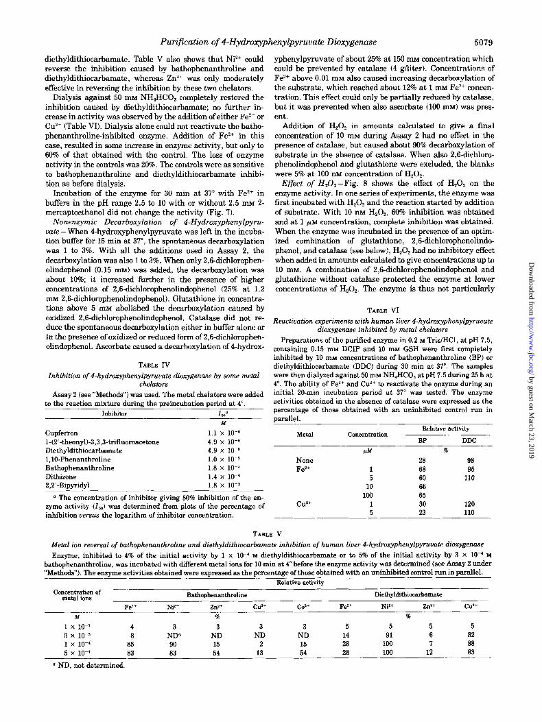
Several metal chelators inhibited the enzyme when added in concentrations lower than 1 mM (see Table IV), whereas no significant inhibition was noted with 1 mM concentrations of a number of other chelating agents, such as 8-hydroxyquinoline- B-sulfonate, Tiron, bathocuproine, diphenylcarbaxone, 2,4,6- tri(2’-pyridyl)-1,3,5-triazine, dimethylglyoxime, neocuproine, or EDTA. Some of the inhibitory chelators were also tested in combination with various metal salts. In the case of batho- phenanthroline, &IO-phenanthroline and 2,2’-bipyridyl, the inhibition was prevented by previous addition of Fe*+ in 2- to 3- fold molecular excess. Ferrous ion could also restore the en- zyme activity when the enzyme had first been incubated with bathophenanthroline (Table V). Similarly, Cu*+ was able to prevent as well as to reverse the inhibition by diethyldithio- carbamate. However, Cu2+ was fairly ineffective in reversal experiments when bathophenanthroline was used. Neither was it possible with Fe*+ to reverse the inhibition caused by
by guest on March 23, 2019
http://ww
w.jbc.org/
Dow
nloaded from
Purification of 4-Hydroxyphenylpyruvate Dioxygenase 5079
diethyldithiocarbamate. Table V also shows that Ni*+ could reverse the inhibition caused by bathophenanthroline and diethyldithiocarbamate, whereas Zn2+ was only moderately effective in reversing the inhibition by these two chelators.
Dialysis against 50 mM NH,HCO, completely restored the inhibition caused by diethyldithiocarbamate; no further in- crease in activity was observed by the addition of either Fe?+ or Cu2+ (Table VI). Dialysis alone could not reactivate the batho- phenanthroline-inhibited enzyme. Addition of Fe”+ in this case, resulted in some increase in enzyme activity, but only to 60% of that obtained with the control. The loss of enzyme activity in the controls was 20%. The controls were as sensitive to bathophenanthroline and diethyldithiocarbamate inhibi- tion as before dialysis.
Incubation of the enzyme for 30 min at 37” with Fe*+ in buffers in the pH range 2.5 to 10 with or without 2.5 mM 2- mercaptoethanol did not change the activity (Fig. 7).
Nonenzymic Decarboxyylation of 4Hydroxyphenylpyru- vatc -When 4-hydroxyphenylpyruvate was let? in the incuba- tion buffer for 15 min at 37”, the spontaneous decarboxylation was 1 to 3%. With all the additions used in Assay 2, the decarboxylation was also 1 to 3%. When only 2,6dichlorophen- olindophenol (0.15 rn& was added, the decarboxylation was about 10%; it increased further in the presence of higher concentrations of 2,6&chlorophenolindophenol (25% at 1.2 mM 2,6dichlorophenolindophenol). Glutathione in concentra- tions above 5 mM abolished the decarboxylation caused by oxidized 2,6-dichlorophenolindophenol. Catalase did not re- duce the spontaneous decarboxylation either in buffer alone or in the presence of oxidized or reduced form of 2,6dichlorophen- olindophenol. Ascorbate caused a decarboxylation of 4-hydrox-
TABLE IV
Inhibition of 4-hydroxyphenylpyruvate diozygenase by some metal chelators
Assay 2 (see “Methods”) was used. The metal chelators were added to the reaction mixture during the preincubation period at 4”.
Inhibitor lZ4’ M
Cupferron 1.1 x 10-G l-(2’~theonyb-3,3,3-trifluoroacetcne 4.9 x 10-G Diethyldithiocarbamate 4.9 x 10-s l,lO-Phenanthroline 1.0 x 10-j Bathophenanthroline 1.8 x 10-j Dithizone 1.4 x 10⁻⁴ 2,2’-Bipyridyl 1.8 x 10-S
n The concentration of inhibitor giving 50% inhibition of the en- zyme activity (Z,) was determined from plots of the percentage of inhibition versus the logarithm of inhibitor concentration.
yphenylpyruvate of about 25% at 150 mM concentration which could be prevented by catalase (4 g/Iiter). Concentrations of Fez+ above 0.01 mM also caused increasing decarboxylation of the substrate, which reached about 12% at 1 mu Fe*+ concen- tration. This effect could only be partially reduced by catalase, but it was prevented when also ascorbate (100 rnr& was pres- ent.
Addition of H,O, in amounts calculated to give a final concentration of 10 mM during Assay 2 had no effect in the presence of catalase, but caused about 90% decarboxylation of substrate in the absence of catalase. When also 2,6dichloro- phenolindophenol and glutathione were excluded, the blanks were 5% at 100 IBM concentration of H,O,.
Effect of H&,-Fig. 8 shows the effect of H,O, on the enzyme activity. In one series of experiments, the enzyme was first incubated with H,O, and the reaction started by addition of substrate. With 10 nM H202, 60% inhibition was obtained and at 1 pM concentration, complete inhibition was obtained. When the enzyme was incubated in the presence of an optim- ized combination of glutathione, 2,6-dichlorophenolindo- phenol, and catalase (see below), H,O, had no inhibitory effect when added in amounts calculated to give concentrations up to 10 mM. A combination of 2,6-dichlorophenolindophenol and glutathione without catalase protected the enzyme at lower concentrations of H,O,. The enzyme is thus not particularly
TABLE VI
Reactivation experiments with human liver 4-hydroxyphenylpyruvate dioxygenase inhibited by metal chelators
Preparations of the purified enzyme in 0.2 M Tris/HCl, at pH 7.5, containing 0.15 rnhr DCIP and 10 mM GSH were iirst completely inhibited by 10 mM concentrations of bathophenanthroline (BP) or diethyldithiocarbamate (DDC) during 30 min at 37”. The samples were then dialyzed against 50 mM NH,HCO, at pH 7.5 during 25 h at 4”. The ability of Fe*+ and Cu2+ to reactivate the enzyme during an initial ZO-min incubation period at 37” was tested. The enzyme activities obtained in the absence of catalase were expressed as the percentage of those obtained with an uninhibited control run in narallel.
M&d concentration Relative activity
BP DDC
None Fez+
cuz+
M
1 5
10 190
1 5
56
28 98 68 95 60 110 66 65 30 120 23 110
TABLE V
Metal ion reversal of bathophenanthroline and diethyldithiocarbamate inhibition of human liver 4-hydroxyphenylpyruvate dioxygenase
Enzyme, inhibited to 4% of the initial activity by 1 x 1O-4 M diethyldithiocarbamate or to 5% of the initial activity by 3 x lo-* M bathophenanthroline, was incubated with different metal ions for 10 min at 4” before the enzyme activity was determined (see Assay 2 under “Methods”). The enzyme activities obtained were expressed as the percentage of those obtained with an uninhibited control run in parallel.
Relative activity
con~d&ati;~ of Bathophenanthroline Diethyldithiocarbamate
Fez+ Ni*+ zr3+ CW+ CO*+ Fez+ Ni*+ znz+ CX+
M 8 96
1 x 10-j 4 3 3 3 3 5 5 5 5 5 x 10-j 8 ND” ND ND ND 14 91 6 82 1 x 10-a 85 90 15 2 15 28 loo 7 88 5 x lo-’ 83 83 54 13 54 28 100 12 83
* ND, not determined.
by guest on March 23, 2019
http://ww
w.jbc.org/
Dow
nloaded from

5080 Purification of 4-Hydroxyphenylpyruvate Dioxygenase
pH DURING PREINCUBATION AT 37’
FIG. 7. Incubation of human liver 4-hydroxyphenylpyruvate dioxygenase with Fe’+ at 37” under different conditions. Dialyzed enzyme (0.02 g/liter) was first incubated in 0.05 M buffers of different pH for 30 min at 37” with various additions: none (0); 2.5 rnM 2- mercaptoethanol (0); 20 FM Fez+ (0); and 2.5 rnM 2-mercaptoethanol and 20 PM Fez+ (m). Enzyme activity was then measured in duplicate with Assay 2 in 0.2 M potassium phosphate buffer at pH 6.9. The results are expressed as the percentage of a control in the incubation buffer kept on ice during the initial 30-min incubation period. The buffers used were: glycine/HCl in the pH range 2.5 to 3.5; sodium acetate in the pH range 3.5 to 5.5; sodium phosphate in the pH range 5.5 to 7.5; Tris/HCl in the pH range 7.5 to 8.5; and glycine/NaOH in the pH range 7.5 to 10.
H202. M
FIG. 8. Effect of H,O, on the activity of purified human liver 4- hydroxyphenylpyruvat dioxygenase. HZ02 was added during a 15- min first incubation period at 4”. The activity obtained in the pres- ence of H,O, was expressed as the percentage of the activity obtained in its absence with different modifications of Assay 2: no moditica- tion (0); catalase was excluded both during the first incubation and during the assay (0); DCIP, GSH, and catalase were excluded dur- ing the first incubation only (0); and DCIP, GSH, and catalase were excluded both during the first incubation and during the assay CD).
sensitive to 0.1 mM H,O, at 4” during a 15-min incubation period but the activity is largely lost at 37” with 10 to 100 nM H,O, in the presence of substrate.
Effect of Glutathione, 2,6-Dichlorophenolindophenol, and
Catalase -In the absence of any stimulatory compounds, the enzyme activity was about 20% of the optimal activity ob- tained with a combination of glutathione, 2,6-dichlorophe- nolindophenol, and catalase, i.e. that used in Assay 2 (refer- ence activity in Fig. 9). The effect of glutathione, 2,6dichloro- phenolindophenol, and cat&se and of different combinations of these compounds, is shown in Fig. 9. At concentrations around 10 pM, 2,6-dichlorophenolindophenol increased the ac-
r
FIG. 9. Effect of different concentrations of GSH, DCIP, and cata- lase and of combinations of these compounds on the activity of human liver 4-hydroxyphenylpyruvate dioxygenase. The results are expressed as the percentage of the activity obtained with Assay 2. The compounds were added to a solution of enzyme in buffer and incubated for 15 min at 4” before the addition of substrate. Left, GSH was added alone (0) and in combination with 0.15 mM DCIP (01, 1.2 rnM DCIP (Cl), and 1.2 rnM DCIP plus 4 g/liter of catalase CM). Middle, DCIP was added alone (0) and in combination with 10 mM GSH (O), 80 rnM GSH (01, 10 rnM GSH plus 0.8 g/liter of catalase (W), and 80 rnM GSH plus 4 g/liter of catalase (A). Right, catalase was added alone (0) and in combination with 0.15 mM DCIP (0),0.15 rnM DCIP plus 10 mM GSH CD), and 0.15 mM DCIP plus 80 mM GSH (0).
tivity about 50%, but was slightly inhibitory at higher concen- trations. Glutathione alone had essentially no effect but in- creased the activity 1.5 to 2 times in combination with 2,6- dichlorophenolindophenol, present in the concentration inter- val 0.15 to 0.6 mM. Catalase had no stimulator-y effect when added alone or in combination with 2,6dichlorophenolindo- phenol. The high activity reached with a combination of gluta- thione, 2,6-dichlorophenolindophenol, and catalase was ob- tamed when catalase was present in low concentrations (about 10 mg/liter) as well as in considerably higher concentrations. In Assay 2, a first incubation period of 15 min was routinely used. In this series of experiments, the first incubation period was found to be unnecessary, since the activation was constant during 0 to 60 min both at 4” and 37”.
Catalase was ineffective when inactivated with sodium azide (1 mM) as well as when the catalase solution had been heated at 100” for 5 min. Bovine serum albumin had no effect when present in concentrations of up to 5 g/liter. Addition of ethanol in concentrations up to 20% (v/v) had a slightly stimu- lating effect (30%), when added to the complete system.
Effect of Ascorbate, Catalase, and Iron -Ascorbate, which alone inhibited the enzyme activity, stimulated in the pres- ence of catalase (Fig. 10). The enzyme activity was the same with optimized concentrations of ascorbate and catalase as with the optimized concentrations of glutathione, 2,6-dichloro- phenolindophenol, and cat&se, i.e. those used in Assay 2. This activity is used as reference activity in Fig. 10. Ferrous ion alone or in combination with catalase did not influence the enzyme activity, but concentrations of Fe*+ above 10 pM were inhibitory in the presence of ascorbate. This inhibition could not be overcome by high concentrations of catalase (4 g/liter).
Involvement of Thiol Groups -The effect of different thiol group reagents was tested in the presence of ascorbate WO mM) and catalase (0.8 g/liter). Incubations were first per- formed for 15 min at 37” with the different reagents present and the reaction was then started by addition of substrate.
by guest on March 23, 2019
http://ww
w.jbc.org/
Dow
nloaded from

Purification of 4-Hydroxyphenylpyruvate Dioxygenase 5081
With 1 mM concentration of HgC& or 4-chloromercuriphenyl- sulfanate, the enzyme activity was inhibited to 45 and 36%, respectively. At 0.01 mM concentration, only HgCl, was inhib- itory (19%). No significant inhibition was observed with 1 mM
concentration of acetarsone, sodium meta-arsenide, N-ethyl- maleimide, iodoacetate, 2-iodobenzoate, or carbazone.
At high pH (Fig. 7), 2-mercaptoethanol had a slight protect- ing effect. As shown in Fig. 9, there was no stimulation by glutathione alone at pH 7.5, nor was there any stimulation or inhibition by dithiothreitol when it was added alone or to an ascorbate catalase-containing system.
Keta-Enol Tautomer Studies -Purified enzyme contained no phenylpyruvate tautomerase (EC 5.3.2.1.) activity when measured according to Constansas and Knox (15). The follow- ing experiments were therefore performed to determine if the keto or the enol form of 4-hydroxyphenylpyruvate was the substrate for the enzyme. As seen from Table VII, more 14C0, was evolved than 4-hydroxyphenylpyruvate consumed when the substrate initially contained about 0.5 nmol of l-14C!-la- beled keto form and 200 run01 of unlabeled enol form of 4- hydroxyphenylpyruvat. When both the lJ4C-labeled and the unlabeled substrate were in the keto form, the amounts of 14COZ evolved and 4-hydroxyphenylpyruvate consumed were similar. The keto form of 4-hydroxyphenylpyruvate is thus the preferred substrate for the enzyme.
The enzyme activity with the enol form of 4-hydroxyphenyl- pyruvate as substrate was also measured under the same conditions with 33 WM concentration of substrate by recording the rate of disappearance of the absorbance at 290 run during the first 5 min of incubation both in the presence and in the absence of enzyme. The specific activity with the enol form of 4-hydroxyphenylpyruvate as substrate was 4.6 units/g of pro- tein. The corresponding specific activity with the keto form of 4-hydroxyphenylpyruvate as substrate was 190 units/g of pro- tein.
Kinetic Properties -With Assay 1, the enzyme activity was proportional to the protein concentration up to 0.03 g/liter (corresponding to about 60% consumption of the substrate) and
I I I I
FIG. 10. Effect of different concentrations of ascorbate, catalase, and Fe’+ and of combinations of these compounds on the activity of human liver 4-hydroxyphenylpyruvate dioxygenase. The results are expressed as the percentage of the activity obtained with Assay 2. The compounds were added to a solution of enzyme in buffer and incubated for 15 min at 4” before addition of substrate. Left, ascor- bate was added alone (0) and in combination with 4 g/liter of catalase (0). Middle, catalase was added in combination with 100 mM ascorbate (0). Right, Fe’+ was added alone (0) and in combina- tion with 4 g/liter of catalase (0) and 4 g/liter of catalase plus 100 rnM ascorbate (0).
was linear with time for 30 min. The apparent Michaelis constant was 0.2 mM and the maximal velocity 1.1 kilounits/g of protein (Fig. 11, left). At the optimized conditions of Assay 2, the apparent Michaelis constant for 4-hydroxyphenylpyru- vate was 0.03 mM and the maximal velocity was 4.0 kilounits/g of protein. The corresponding values for oxygen was 5% (20 kPa corresponding to about 50 ELM in the solution (41)) (Fig. 11, middle). No significant inhibition was observed with pure oxygen in the gas phase. Concentrations of 4-hydroxyphenyl- pyruvate above 0.2 mM were inhibitory. There was a one to one relation between formation of CO, and homogentisate (Fig. 12).
The enzyme activity had two pH optima, one around pH 4.5 and another around pH 7.8 (Fig. 13). The effect on the enzyme activity of increasing the molar&y of buffers from 10 to 500 mM was insignificant for Tris/HCl, pH 8.0, and potassium phos- phate, pH 6.8, but for sodium acetate, pH 5.0, a 50% decrease of activity was observed. The pH-dependent stability of the enzyme during 30-min initial incubation at 37” is given in Fig. 7. The enzymic activity was lost below pH 3 and above pH 10 and increased at pH 5 to about 140% of a control kept at 4” in 0.2 M potassium phosphate buffer at pH 6.9. This activating effect at pH 5 was also observed at 4” for purified enzyme and for acetone powder extract.
The substrate analogues phenylpyruvate and 4-aminophen- ylpyruvate were competitive inhibitors with inhibition con- stants of 0.015 and 0.35 mu, respectively. Under the incubat- ing conditions of Assay 2 with phenyl[l-‘4Clpyruvat as sub- strate, the progress of the reaction was linear with time up to 90 min and with increasing amounts of enzyme protein, up to 40 mg of protein/liter; the nonlinearity began when about 20% of the substrate had been consumed. The apparent Michaelis constant was 0.05 mM and the maximal velocity was 0.02 kilounits/g of protein (Fig. 11, right).
DISCUSSION
Liver 4-hydroxyphenylpyruvate dioxygenase has been par- tially purified from pig (1, 2, 9, 42-44), dog (45), rat (46), beef (431, frog (47, 481, and rabbit (49). In a preliminary note, we reported on the purification of human liver 4-hydroxyphenyl- pyruvate dioxygenase (50). The procedure used in that work has been used to prepare highly pure preparations of the chicken and bovine liver enzyme (3,511. The human enzyme is pure as judged by several analytical techniques, i .e . polyacryl-
TABLE VII
Activity of human liver 4-hydroxyphenylpyruuate dioxygenase with keto-enol tautomer mixtures of4-hydroxyphenylWruvate as substrate
Purified enzyme, was incubated for 5 min with GSH (10 pmol), DCIP (0.15 pmol), and catalase (10 pg) in 50 mM potassium phos- phate buffer, pH 6.2, at 3’7”. The lJ4C-labeled keto form of 4-hydroxy- phenylpyruvate, 0.5 nmol, was mixed with the unlabeled keto or enol form of 4-hydroxyphenylpyruvate, 200 nmol, and then added to the enzyme solution (for preparation of substrates see “Methods”). The incubation volume was 1 ml. Evolved “CO, was measured as de- scribed under “Methods.” 4-Hydrophenylpyruvat was determined by spectrophotometry as the enol borate complex (40).
Unlabeled tauto- mer of 4-bydroxy- Time of reac- I-Hydroxy-
pbenylpyruvate tion pbenylpyru- Radioactivity evolved as “CO,
(200 nmol) vate consumed
mill 9% %
Enol 0 0 0.7 5 0 20
Keto 0 0 0.3 5 7 5.2
by guest on March 23, 2019
http://ww
w.jbc.org/
Dow
nloaded from

Purification of 4-Hydroxyphenylpyruvate Dioxygenase
FIG. 11. Effect of substrate concentration on human liver 4-hydroxyphenylpyruvate dioxygenase activity. The insets show the Line- weaver-Burk plots of the data obtained. Left, Assay 1 was used with 4-hydroxyphenylpyruvate as the variable substrate. Middle, Assay 2 was used with oxygen as the variable substrate. The concentrations given are those in the dry gas used. Right, Assay 2 was used with phenylpyruvate as the variable substrate.
1 o-------
0 25 50 75 HOMOGENTISATE, nmoles
FIG. 12. Stoichiometric relation between the formation of CO, and homogentisate as determined with the WO, method and the thin layer chromatographic method. Assay 1 was used with 0.2 mM concentration of 4-hydroxyphenylpyruvate. The results are given in nanomoles of products formed during a 15min incubation period. The correlation coefficient was 0.993.
amide gel electrophoresis, gel filtration, sedimentation equi- librium, and immunodifTusion. Some properties of the enzyme are summarized in Table VIII.
The molecular mass values obtained by sedimentation equi- librium (87 kilodaltonsl and by polyacrylamide gel electropho- resis (80 kilodaltons) are in good agreement. The lower mass value obtained by the gel filtration technique (66 kilodaltons) might be due to reversible adsorption to the gel or to a lower hydrodynamic volume of the enzyme than of the reference proteins (52). However, the value of the frictional ratio (1.2) was similar to that of most globular proteins. A further possi- ble reason for the low value of the molecular mass obtained by gel filtration is reversible dissociation into subunits. Dissocia- tion into subunits has been noticed on gel filtration of the tetrameric rabbit liver enzyme (49).
The mass value obtained by electrophoresis in polyacryl- amide gel containing sodium dodecyl sulfate suggests that the human enzyme is a dimeric molecule composed of equally sized subunits. The chicken liver enzyme has also recently been indicated to have a similar dimeric structure (53). The molecular masses obtained by gel filtration of the pig and frog liver enzymes are 42 and 85 kilcdaitons, respectively (9, 48).
I--’ 2 3 4 5 6 7 8 9 10
PH
FIG. 13. The activity of human liver 4-hydroxyphenylpyruvate dioxygenase as a function of pH. Assay 2 was used with different buffers in 50 rn~ buffer concentrations: glycine/HCl (0); sodium acetate (0); potassium phosphate (A); Tris/HCl (A); and glycine/ NaOH (0).
The possible presence of subunits in these enzymes has not been studied.
Crude preparations of 4-hydroxyphenylpyruvate dioxygen- ase from various species have shown considerable differences in enzyme activity (54,551. The specific activity of the purified human enzyme was 1 to 4 mmol/min/g of protein. For the purified chicken and bovine liver enzymes, the corresponding values are 8 and 0.2 mmol/min/g of protein (51, 53). Substrate inhibition has usually become evident above 1 mM concentra- tion of 4-hydroxyphenylpyruvate (9), but inhibition of the rat
liver enzyme is reported to appear already at 0.05 mM concen- tration of 4-hydroxyphenylpyruvate (56). Inhibition of the hu- man enzyme became evident above 0.2 mM concentration of 4- hydroxyphenylpyruvat, i.e. at comparatively low concentra- tions. The figures cited from the literature are, however, not
directly comparable with the present results since they are influenced by the different concentrations of reductants, eata-
lase, and substrate used during the assays. The apparent Michaelis constant for 4-hydroxyphenylpyruvate (0.03 mM)
by guest on March 23, 2019
http://ww
w.jbc.org/
Dow
nloaded from

Purification of 4-Hydroxyphenylpyruvate Dioxygenase 5083
TABLE VIII
Properties of human liver 4-hydroxyphenylpyruvate dioxygenase
Parameter Vdlll? M&hod
Molecular mass (kilo- dalton)
Subunit mass (kilodal- ton)
Stokes radius (nm) Partial specific volume
(ml/g)
4% t 278 nm 280 nm
A zwlA 260 A’”
1 cm 1 278 nm 280 nm
Frictional ratio
Diffusion coefficient
CD,, ,) (x IO-’ cm%)
87 Sedimentation equilibrium
66 80 43
3.6 0.73
Gel filtration Gel electrophoresis Sodium dodecyl sulfate gel
electrophoresis Gel filtration Amino acid composition
11.4 11.3
1.65
Spectrophotometry in dis- tilled water, dry weight
11.3 11.2
Spectrophotometry in 25 mM TrislHCl, pH 7.4, contain- ing 0.2 M NaCl (Lowry method (16))
1.66 1.2”
6.0b
Gel filtration-sedimentation equilibrium
Gel filtration
a Calculated according to Siegel and Monty (38) using the molecu- lar mass obtained from sedimentation equilibrium experiments.
b Calculated from the Stokes radius obtained at gel filtration by Stokes-Einsteins formula.
was in the range obtained for most. mammalian species, viz. 0.02 to 0.05 mM (3,4,9,56). The value for the frog liver enzyme is 10 times higher (48).
Phenylpyruvate was a substrate for the human enzyme. It has earlier been shown to be a substrate also for the purified chicken liver enzyme (3) and for the partially purified pig and rat liver enzymes (1,4,9,57). The apparent Michaelis constant for phenylpyruvate, 0.05 mM, was in the range reported for the other mammalian species. The pronounced inhibition by phen- ylpyruvate of the pig liver enzyme (57) was not observed for the human enzyme with our assay system.
The apparent Michaelis constant for 0, (-50 pM) was also similar to what has been reported for unpurified preparations of the pig and rat liver enzymes (4,43). The inhibitory effect of 02, noticed already in air for the rat liver enzyme (4), was not observed for the human enzyme in our assay system.
4-Hydroxyphenylpyruvate dioxygenase as well as other “2- 0x0 acid” oxygenases are stimulated by reductants and caia- lase (9, 58, 59). In some cases e.g. 4-hydroxyphenylpyruvate dioxygenase from Pseudomonas* or bovine liver (51), there is no enzyme activity without stimulants. Apart from increasing the rate of the enzymic reaction, the stimulators increase the concentration at which inhibition by substrate occurs (60). Two combinations, reduced dichlorophenolindophenol plus catalase and ascorbate plus catalase, have been used with 4- hydroxyphenylpyruvate dioxygenase. With the present en- zyme, they were about equally effective, whereas the latter system was about 8 times more effective for the pure enzyme from Psewlomonns in the presence of iron.2 Studies of these stimulants are complicated by the fact that there is a sponta- neous decarboxylation of 4-hydroxyphenylpyruvate in buffer,
2 S. Lindstedt, B. Odelhijg, and M. Rundgren (1977) Biochem&ry 16, in press.
which is considerably influenced by the enzyme stimulators. It has been suggested that the nonenzymic degradation of the organic substrate resulting from interaction with Oz leads to formation of H20P, which would be harmful to the enzyme (4). The present results show that the pure enzyme is very sensi- tive to H,O, and that the inhibited enzyme can be reactivated, e.g. by reduced dichlorophenolindophenol. Catalase also pro- tects the enzyme from added H,O, as well as from H,O,, which is presumably generated in a solution of reductant and iron. We do not know how H,O, inactivates the enzyme nor is there any direct evidence that nonenzymically formed H20, is re- sponsible for the inhibition by higher substrate concentra- tions. None of the two plausible mechanisms for the enzymic reaction involve the formation of H,O, as an intermediate (6, 7, 59), but both mechanisms include the formation of a hydro- peroxide, which could conceivably be harmful to the enzyme.
The present study has provided conclusive evidence that the keto form of 4-hydroxyphenylpyruvate is the tautomer, which is substrate for the enzyme as has been suggested previously (9, 57, 61). Since this is the tautomer which is presumably formed in the degradation of tyrosine and which is present to 96% at equilibrium, the physiological role of liver phenylpyru- vate tautomerase remains obscure.
It once was suggested that 4-hydroxyphenylpyruvate dioxy- genase contains essential thiol groups (42, 43). However, this has not been confirmed in later studies with either the pig or the rat. liver enzymes (1, 2, 46, 62). The human liver enzyme contains five free thiol groups and one disulfide bridge187 kilodaltons (Table III). The free thiol groups of the native enzyme probably do not participate in the catalysis, since a variety of thiol group reagents had no effect in 1 mM concen- trations.
Determination of the metal content of the enzyme had been made earlier on impure preparations and attempts have been made to correlate metal content and specific activity (9). Good- win and Werner (63) found an increase in the copper content of the pig liver enzyme during their purification procedure, but the final preparation contained only about 0.1 mol of copper/42 kg of protein (molecular mass, 42 kilodaltons) (9). The par- tially. purified rabbit liver enzyme contained significant amounts of iron, about 1.8 mo1/150 kg of protein (molecular mass, 150 kilodaltons) (49). It was reported also that this enzyme reversibly dissociated into four subunits with a con- comitant loss of iron. Wada et al. (53) found insignificant amounts of copper in the chicken liver enzyme. After dialysis against 50 mM TrisfHCl at pH 7.8, they found 0.88 mol of iron/ mol of “total protein” in an experiment with protein from the final purification step. Approximately half this amount was lost together with a partial loss of enzyme activity after treat- ment with a chelator. According to these authors, the chicken liver enzyme consists of two subunits but it is not clear to us from the published data why they conclude that the enzyme contains 1 mol of iron/m01 of subunit. For the human enzyme, we obtained a low iron content (-0.4 mol/mol of enzyme). Like Wada and associates (531, with the chicken enzyme, we could not increase the activity of the human enzyme by the addition of Fe?+. In similar experiments with a pure preparation of 4- hydroxyphenylpyruvate dioxygenase from a Pseudomonas
strain, we obtained a value of about 1 mol of iron/m01 of enzyme.2
The human liver enzyme now purified had a higher iron than copper content. It could be completely inhibited by some metal chelators. Those which were effective as inhibitors had
by guest on March 23, 2019
http://ww
w.jbc.org/
Dow
nloaded from

5084 Purification of I-Hydroxyphenylpyruvate Dioxygenase
- Taniguchi, K., Kappe, T., and Armstrong, M. D. (1964) J. B&E. Chem. 239, 3389-3395
z.
3.
4.
5.
6.
7.
8.
9.
Fellman. J. H., Fujita, T. S., and Roth, E. S. (1972) Biochim. Biophys. Acta 268, 601-604
Fellman, J. H., Fuji@ T. S., and Roth, E. S. (1972) Biochim. Biophys. Actn 284, 99-100
Goodwin, S., and Witkop, B. (1957) J. Am. Chem. Sot. 79, 179- 185
Lindblad, B., Lindstedt, G., and Lindstedt, S. 11970) J. Am. Chem. Sot. 92, 7446-7449
Hamilton, G. A. (1971) in Progress in Biorganic Chemistry (Kai- ser, E. T., and Kezdy, F. J., eds) Vol. 1, pp. 83-157, Wiley- Interscience, New York
Lindblad, B. (1971) Doctoral dissertation, University of Gothen- burg
Goodwin, B. L. (1972) Tyrosine Catabolism, Oxford University Press
10. Rundgren, M. (1977) J. Biol. Chem. 252, 5085-5093 11. Rundgren, M. (1977) J. Biol. Chem. 252, 5094-5099 12. Lindblad, B. (1971) Clin. Chim. Acta 34, 113-121 13. Haavaldsen, R., and Norseth, T. (1966)Anol. B&hem. 15.536 14. Knox, W. E , and Pitt, B. M. (1957) J. Biol. Chem. 225,675-688 15. Constantas. N. S., and Knox, W. E. (1966) Arch. Biochem.
Biophys. 117, 59-64 16.
17. 18.
19.
20.
21. 22.
23.
24.
25.
26.
Lowry, 0. H., Rosebrough, N. J., Farr, A. L., and Randall, R. J. (1951) J. Biol. Chem. 193. 265-275
Davis, B. J. (1964)Ann. N.‘Y. Acad. Sci. 121, 404-427 Hedrick, J. L.. and Smith, A. J. (1968)Arch. Biochem. Biophys.
126, 155-164 Parish, C. R., and Marchalonis, J. J. (1970)Anol. Biochem. 34,
436-450 Laurent, T. C., and Killander, J. (1964) J. Chromatog. 14, 317-
330 Weber, K., and Osbom, M. (1969) J. Biol. Chem. 244,4406-4412 Sober, H. A., ed (1968) Handbook ofBiochemistry, The Chemical
Rubber Publishing Co., Cleveland Fish, W. W., Mann, K. G., and Tanford, C. (1969) J. Biof. Chem.
244,4989-4994 Rymo, L., Lundvik, L., and Lagerkvist, U. (1972)J. Biol. Chem.
247, 3888-3899 Samejima, T., and Yang, J. T. (1963) J. Bill. Chem. 238, 3256-
3261 Vesterberg, O., and Svensson, H. (1966) Acta Chem. Stand. 20,
820-834
either high affinity for Fez+ or for Cuz+. The results from reversal experiments with bathophenanthroline, in which
Fe’+ and Ni”‘, but not other transitional metal ions could
restore the enzyme activity, are evidence for a role of Fe”+ for catalysis as is the ability of Fe’+, but not Cu”, to partially restore the activity after dialysis of the bathophenanthroline-
inhibited enzyme. Similar experiments on the rat liver en-
zyme have also indicated an essential role of Fe”+ (461. How-
ever, Fe’+ was fairly ineffective, as compared with Ni’+ and
Cu’+, in its ability to restore the activity of the diethyldithio- carbamate-inhibited enzyme, which might indicate that Fe’+
is not the sole essential metal ion. We are aware of the
difficulties to draw definite conclusions from inhibition experi- ments with metal chelators since other effects than that of metal-binding may be operative (64-66). In conclusion, evi-
dence has accumulated that 4-hydroxyphenylpyruvate dioxy- genase is a metalloenzyme containing essential Fe’+, but the involvement of some other metal ion cannot be excluded.
Acknowledgments-We wish to thank Dr. Lars Thelander,
Kemiska institutionen, Karolinska Institutet, Stockholm,
Sweden, for help with the sedimentation equilibrium experi- ments and Dr. Rudolf Jagenburg of this department for amino
acid analysis.
REFERENCES
1. Taniguchi, K., and Armstrong, M. D. (1963) J. Biol. Chem. 238, 4091-4097
27. 28. 29.
30.
31. 32. 33.
34. 35.
36.
37.
38.
39.
40.
41. 42.
43.
44.
45.
46. 47.
48. 49.
50.
51.
52. 53.
54. 55.
56.
57.
58.
59.
60.
61.
62.
63
Yphantis, D. A. (1964) Biochemistry 3, 297-317 Moore, S., and Stein, W. H. (1963) Methods EnzymoE. 6.819-831 Mancini, C., Cabonara, A. O., and Heremans, J. F. (1965) Im-
munochemistrv 2. 235-254 Ouchterlony, 0.” (1968) Handbook of Immunodifision and Im-
munoelectrophoresis Ann Arbor Science Publishers, Inc., Michigan
Bover. P. D. (1954) J. Am. Chem. Sot. 76, 4331-4337 Habeeb, A. F. S. A. (1972) Methods Enzymol. 25, 457-464 Berglund, O., and Holmgren, A. (1970) J. Biol. Chem. 245,6036-
6038 Felsenfeld, G. (19601 Arch. Biochem. Biophys. 87. 247-251 Broman, L., Malmstrom, B. G., Aasa, R., and Vanngard, T.
(1962) J. Mol. Biol. 5, 301-310 Doeg, K. A., and Ziegler, D. M. (1962)Arch. Biochem. Bcophys.
97, 37-40 Brumby, P. E., and Massey, V. (1967) MethodsEnzymol. 10,463-
471 Siegel, L. M., and Monty, K. J. (1966) Biochim. Biophys. Acta
112, 346-362 Cohn, E. J., and Edsall, J. T., eds (1943) in Protein.s, Amino
Acids, and Peptides, pp. 370-381, Reinhold Publishing Corp., New York
Gentz, J., Lindblad, B., and Lindstedt, S. (1969) J. Lab Clin. Med. 74. 185-202
Lessler, G. A. (19691 Methods Biochem. Anal. 17, 18-19 Edwards. S. W.. Hsia, D. Y.-Y., and Knox, W. E. (1955) Fed.
Proc. 14, 206 Haeer. S. E.. Greaerman, R. I., and Knox, W. E. (19571 J. Biol.
cheh. 225; 935-?47 Roka. L., Kiinie. G.. and Rubner, H. (1958) Howe Seyler’s 2.
Ph&ol. Chem: 312-313, 87-96 __
La Du, B. N., and Zannoni, V. G. (1956) J. Biol. Chem. 219,273- 281
Goswami, M. N. D. (1964) Biochim. Biophys. Actu 85.390-399 Laskowska-Klita. T.. and Mochnacka. I. (1967) Acta Biochim.
Pol. 14, 101-109 Laskowska-Klita. T. (1969) Acta Biochim. Pal. 16, 35-44 Laskowska-Klita[ T., and Mochnacka, I. (19733 Acta Biochim.
Pol. 20, 259-269 Lindblad, B., Lindstedt, S., Olander, B., and Omfeldt, M. (1971)
Acta Chem. Stand. 25, 329-330 Nakai, C., Nozaki, M., and Hayaishi, 0. (1975) Biochem. Bio-
phys. Res. Commun. 67, 590-595 Squire, P. G. (19641 Arch. B&hem. Biophys. 107,471-478 Wada, G. H., Fellman, J. H., Fujita, T. S., and Roth, E. S. (1975)
J. Biol. Chem. 250, 6720-6726 Michalec-Morrica, H. (1965) Acta Biochim. Pol. 12, 167-177 Goswami, M. N. D., and Rosenberg, A. J. (1968) C. R. Acad. Sci.
Paris, Ser. D 267,2224-2227 Raheja, M. C., Dylewski, I., and Crawhall, J. C. (1973) Can. J.
Biochem. 51, 172-177 Goodwin, B. L., and Werner, E. G. (1972) Anal. Biochem. 50,
180-186 Lindstedt. G. (1967) Doctoral dissertation. Karolinska Institu-
tet, Stockholm, Sweden Abbott. M. T.. and Udenfriend, S. (19741 in Molecular Mecha-
nism; of Oxygen Activation (Hayaishi, O., edf pp. 167-214, Academic Press, New York
Zannoni, V. G., and La Du, B. N. (1959) J. Biot. Chem. 234, 2925-2931
Lin. E. C. C.. Pitt. B. M.. Civen. M.. and Knox. W. E. (19581 J. B&l. Chem. 233: SSB-6i3
Goswami. M. N. D.. and Knox, W. E. (1963) J. Chronic Dis. 16, 363-371
Goodwin, B. L., and Werner, E. G. (1973) Experimentia 29, 523- 525
64. Hayaishi, 0. (1964) Proceedings of the Plenary Sessions, Sixth International Congress of Biochemistry, July 26 to August 1, New York, pp. 31-43
65. Yamamato, S., Takeda, H., Yoshitaka, M., and Hayaishi, 0. (1966) in Biological and Chemical Aspects of Oxygenuses (Block. K., and Hayaishi, O., eds) pp. 303-314, Maruzen, Tokyo
66. Ishimura, Y., and Hayaishi, 0. (1976) in Iron and Copper Pro- teins (Yasunobo, K. T., Mower, H. F., and Hayaishi, 0.. edsl pp. 363-373, Plenum Press, New York
by guest on March 23, 2019
http://ww
w.jbc.org/
Dow
nloaded from

B Lindblad, G Lindstedt, S Lindstedt and M Rundgren(I).
Purification and some properties of human 4-hydroxyphenylpyruvate dioxygenase
1977, 252:5073-5084.J. Biol. Chem.
http://www.jbc.org/content/252/14/5073.citation
Access the most updated version of this article at
Alerts:
When a correction for this article is posted•
When this article is cited•
to choose from all of JBC's e-mail alertsClick here
http://www.jbc.org/content/252/14/5073.citation.full.html#ref-list-1
This article cites 0 references, 0 of which can be accessed free at
by guest on March 23, 2019
http://ww
w.jbc.org/
Dow
nloaded from