purification and properties of glycerol kinase from - journal of
TRANSCRIPT

THE JOURNAL OF BIOLOGICAL CHEMISTRY Vol. 242, No. 5, Issue of March 10, pp. 1030-1035, 1967
Printed in U.S.A.
Purification and Properties of Glycerol Kinase from
Escherichia coli*
(Receivedzfor publication, September 21, 1966)
SHIN-ICHI HAYASHI$ AND E. C. C. LIN§
From the Department of Biological Chemistry, Harvard Medical Xchool, Boston, Massachusetts 02115
SUMMARY
Glycerol kinase of Escherichia co2i has been purified and crystallized. It has a molecular weight of -3 X 10⁶. The enzyme phosphorylates glycerol exclusively to L-a-glycero- phosphate. It also catalyzes the phosphorylation of dihy- droxyacetone and L-glyceraldehyde but with values of Km much higher than that for glycerol. D-Glyceraldehyde has a catalytic effect in promoting the conversion of adenosine triphosphate to adenosine diphosphate and Pi in the presence of the enzyme. Among the nucleoside triphosphates tested, only adenosine triphosphate was active as the phosphoryl group donor. Mn++ substitutes for Mg+f, although with less activity at equal molar concentration.
A specific inducible kinase is obligatory for the utilization of glycerol by Escherichia coli Kl2 (1). Glycerol, entering the cell presumably by free diffusion (2), is converted by this enzyme to L-a-glycerophosphate, which is then dehydrogenated to triose phosphate (1, 3).
Recently we have purified the E. coli glycerol kinase to a crystalline form in order to prepare specific antisera for the im- munochemical measurement of the enzyme protein. With the aid of a mutant capable of producing a cross-reacting material with very low enzymatic activity, it was shown that the kinase was inducible by L-or-glycero-P, but not by free glycerol (4), as in the case of the n-a-glycero-P transport system and the ~-a-
glycero-P dehydrogenase (3, 5). In this paper the purification and properties of the kinase are
presented. Comparisons are made with the enzymes from rat liver (6), pigeon liver (7), and Candida mycoderma (S), which have been purified and studied in detail in other laboratories.
EXPERIMENTAL PROCEDURE
Chemicals-%-Glycerol (4C per mole) was purchased from New England Nuclear. Glycerol and glucose were obtained from Merck; dihydroxyacetone was obtained from Mann; dicy-
* This investigation was supported by Research Grants GM- 11983 from the National Institutes of Health and GB-3527 from the National Science Foundation.
$ Present address, Institute for Protein R,esonrch, Osaka Uni- vcrsity, Osaka, Japan.
$ Recipient of a Career Development Award from the United States Public Health Service.
clohexylammonium L-a-glycero-P, L-glyceraldehyde, or,-3-chloro- 1,2-propanediol, and nn-glyceraldehyde a-phosphate were from Calbiochem; n-glyceraldehyde, phosphoenolpyruvate, fructose 1,6-diphosphate, and ATP were obtained from Sigma; DL-1 ,2- propanediol, 1,3-propanediol, ethylene glycol, and n-mannitol came from Eastman; 2,3-butanediol were products of L. Light and Company; ar-thioglycerol was a K and K product; i-eryth- ritol, ribitol, n-arabitol, and L-arabitol were from Pfanstiehl Laboratories, Inc., Waukegan, Illinois; NAD, NADH, CTP, UTP, TTP, GTP, and ITP were Pabst products; and crystalline L-cu-glycero-P dehydrogenase, n-glyceraldehyde 3-phosphate de- hydrogenase, lactate dehydrogenase, pyruvate kinase, and al- dolase were from C. F. Boehringer and Sons.
Bacterial strains-Cells of E. coli KlO, strain 7, were used as the source of the kinase because they produce this enzyme con- stitutively (3) up to 5% of the extractable protein.
Growth of Cells-A 50-liter culture was grown at 37” in a Biogen fermenter with vigorous aeration. The medium contained 2% casein acid hydrolysate, 0.7 y0 K2HPOI, 0.2 y0 KHzPOl. HzO, 0.1% (NH&SOk, and 0.01% MgSO~~7H~0, at a pH of 7.0. Cells were harvested by a Sharples centrifuge after the culture had reached stationary phase.
Enzyme Assays-Four methods of assay for glycerol kinase activity were used, each for a different experimental purpose. All were carried out at 25”.
Assay I, employed routinely to follow the purification of the en- zyme, was essentially that of Wieland and Suyter (7). The phos- phorylation of glycerol was coupled to the reduction of NAD by the action of L-cr-glycero-P dehydrogenase at pH 9.5. The reac- tion mixture contained the following: 30 pmoles of glycerol, 60 pmoles of ATP, 60 pmoles of MgC12, 4 pmoles of NAD, 900 pmoles of hydrazine, 500 pmoles of sodium carbonate, 0.3 mg of L-ar-glycero-P dehydrogenase from rabbit muscle, and glycerol kinase in a final volume of 3.0 ml. The L-a-glycero-P dehydro- genase was omitted from the blank. NADH formation was fol- lowed at 340 rnp, and the unit of enzyme activity was expressed as micromoles of L-oc-glycero-P formed per min.
Assay II, used to determine the pH optimum of the kinase, was carried out in two stages in the manner described by Bublitz and Kennedy (6). The phosphorylation of glycerol was allowed to proceed in a system consisting of the following: 10 Mmoles of glycerol, 20 pmoles of ATP, 20 pmoles of MgC&, 50 pmoles each of phosphate, Tris-HCl, and bicarbonale at a given $1, 2 mg of serum albumin, and I pg of crystalline glycerol kinase in a final volume of 1.0 ml. After an incubation period of 10 min, 1 ml of 10% trichloracctic acid was added, and the mixture was
1030
by guest on Decem
ber 23, 2018http://w
ww
.jbc.org/D
ownloaded from
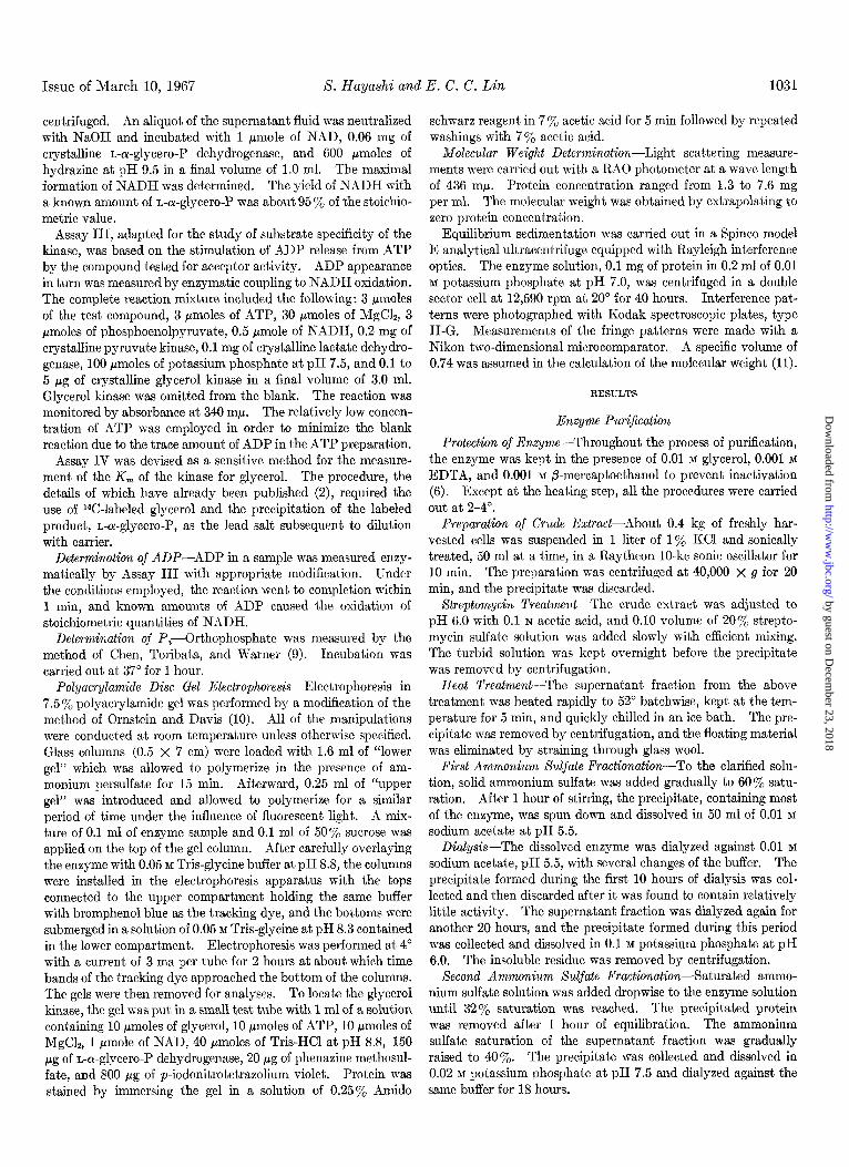
Issue of March 10, 1967 S. Hayashi and E. C. C. Lin 1031
centrifuged. An aliquot of the supernatant fluid was neutralized with NaOH and incubated with 1 pmole of NAD, 0.06 mg of crystalline L-or-glycero-P dehydrogenase, and 600 pmoles of hydrazine at pH 9.5 in a final volume of 1.0 ml. The maximal formation of NADH was determined. The yield of NADH with a known amount of L-oc-glycero-P was about 95 y0 of the stoichio- metric value.
Assay III, adapted for the study of substrate specificity of the kinase, was based on the stimulation of ADP release from ATP by the compound tested for acceptor activity. ADP appearance in turn was measured by enzymatic coupling to NADH oxidation. The complete reaction mixture included the following: 3 Nmoles of the test compound, 3 /Imoles of ATP, 30 pmoles of MgC12, 3 pmoles of phosphoenolpyruvate, 0.5 pmole of NADH, 0.2 mg of crystalline pyruvate kinase, 0.1 mg of crystalline lactate dehydro- genase, 100 pmoles of potassium phosphate at pH 7.5, and 0.1 to 5 pg of crystalline glycerol kinase in a final volume of 3.0 ml. Glycerol kinase was omitted from the blank. The reaction was monitored by absorbance at 340 mp. The relatively low concen- tration of ATP was employed in order to minimize the blank reaction due to the trace amount of ADP in the ATP preparation.
Assay IV was devised as a sensitive method for the measure- ment of the K, of the kinase for glycerol. The procedure, the details of which have already been published (2), required the use of 14C-labeled glycerol and the precipitation of the labeled product, L-a-glycero-P, as the lead salt subsequent to dilution with carrier.
Determination of ADP-ADP in a sample was measured enzy- matically by Assay III with appropriate modification. Under the conditions employed, the reaction went to completion within 1 min, and known amounts of ADP caused the oxidation of stoichiometric quantities of NADH.
Determination of Pi-Orthophosphate was measured by the method of Chen, Toribata, and Warner (9). Incubation was carried out at 37” for 1 hour.
Polyacrylamide Disc Gel Electrophoresis-Electrophoresis in 7.5 % polyacrylamide gel was performed by a modification of the method of Ornstein and Davis (10). All of the manipulations were conducted at room temperature unless otherwise specified. Glass columns (0.5 x 7 cm) were loaded with 1.6 ml of “lower gel” which was allowed to polymerize in the presence of am- monium persulfate for 15 min. Afterward, 0.25 ml of “upper gel” was introduced and allowed to polymerize for a similar period of time under the influence of fluorescent light. A mix- ture of 0.1 ml of enzyme sample and 0.1 ml of 50% sucrose was applied on the top of the gel column. After carefully overlaying the enzyme with 0.05 M Tris-glycine buffer at pH 8.8, the columns were installed in the electrophoresis apparatus with the tops connected to the upper compartment holding the same buffer with bromphenol blue as the tracking dye, and the bottoms were submerged in a solution of 0.05 M Tris-glycine at pH 8.3 contained in the lower compartment. Electrophoresis was performed at 4’ with a current of 3 ma per tube for 2 hours at about which time bands of the tracking dye approached the bottom of the columns. The gels were then removed for analyses. To locate the glycerol kinase, the gel was put in a small test tube with 1 ml of a solution containing 10 pmoles of glycerol, 10 pmoles of ATP, 10 pmoles of MgC12, 1 pmole of NAD, 40 pmoles of Tris-HCl at pH 8.8, 150 pg of L-a-glycero-P dehydrogenase, 20 pg of phenazine methosul- fate, and 800 pg of p-iodonitrotetrazolium violet. Protein was stained by immersing the gel in a solution of 0.25% Amido
schwarz reagent in 7 y0 acetic acid for 5 min followed by repeated washings with 7% acetic acid.
Molecular Weight Determination-Light scattering measure- ments were carried out with a RAO photometer at a wave length of 436 rnp. Protein concentration ranged from 1.3 to 7.6 mg per ml. The molecular weight was obtained by extrapolating to zero protein concentration.
Equilibrium sedimentation was carried out in a Spinco model E analytical ultracentrifuge equipped with Rayleigh interference optics. The enzyme solution, 0.1 mg of protein in 0.2 ml of 0.01 M potassium phosphate at pH 7.0, was centrifuged in a double sector cell at 12,590 rpm at 20’ for 40 hours. Interference pat- terns were photographed with Kodak spectroscopic plates, type II-G. Measurements of the fringe patterns were made with a Nikon two-dimensional microcomparator. A specific volume of 0.74 was assumed in the calculation of the molecular weight (11).
RESULTS
Enzyme PuriJication
Protection of Enzyme-Throughout the process of purification, the enzyme was kept in the presence of 0.01 M glycerol, 0.001 M
EDTA, and 0.001 M ,&mercaptoethanol to prevent inactivation (6). Except at the heating step, all the procedures were carried out at 2-4O.
Preparation of Crude Extract-About 0.4 kg of freshly har- vested cells was suspended in 1 liter of 1% KC1 and sonically treated, 50 ml at a time, in a Raytheon IO-kc sonic oscillator for 10 min. The preparation was centrifuged at 40,000 X g for 20 min, and the precipitate was discarded.
Streptomycin Treatment-The crude extract was adjusted to pH 6.0 with 0.1 N acetic acid, and 0.10 volume of 20% strepto- mycin sulfate solution was added slowly with efficient mixing. The turbid solution was kept overnight before the precipitate was removed by centrifugation.
Heat Treatment-The supernatant fraction from the above treatment was heated rapidly to 52” batchwise, kept at the tem- perature for 5 min, and quickly chilled in an ice bath. The pre- cipitate was removed by centrifugation, and the floating material was eliminated by straining through glass wool.
First Ammonium sulfate Fractionation-To the clarified solu- tion, solid ammonium sulfate was added gradually to 60% satu- ration. After 1 hour of stirring, the precipitate, containing most of the enzyme, was spun down and dissolved in 50 ml of 0.01 M
sodium acetate at pH 5.5. Dialysis-The dissolved enzyme was dialyzed against 0.01 M
sodium acetate, pH 5.5, with several changes of the buffer. The precipitate formed during the first 10 hours of dialysis was col- lected and then discarded after it was found to contain relatively little activity. The supernatant fraction was dialyzed again for another 20 hours, and the precipitate formed during this period was collected and dissolved in 0.1 M potassium phosphate at pH 6.0. The insoluble residue was removed by centrifugation.
Second Ammonium Xulfate Fractionation-Saturated ammo- nium sulfate solution was added dropwise to the enzyme solution until 32’% saturation was reached. The precipitated protein was removed after 1 hour of equilibration. The ammonium sulfate sat,uration of the supernatant fraction was gradually raised to 40%. The precipitate was collected and dissolved in 0.02 M potassium phosphate at pH 7.5 and dialyzed against the same buffer for 18 hours.
by guest on Decem
ber 23, 2018http://w
ww
.jbc.org/D
ownloaded from

1032 Glycerol Kinase OJ IS. coli Vol. 242, No. 5
50 100 150 200
FRACTION NO.
FIG. 1. DEAE-cellulose chromatography of glycerol kinase. For conditions of elutions see the text. Fractions of 5 ml were collected. O-0, absorbance at 280 rnp; 0-- -0, enzyme activity.
FIG. 2. Crystals of E. coli glycerol kinase. A, X 840; B, X 840; c, x 320.
DEAE-cellulose Column Chromatography-The dialyzed en- zyme was applied on a DEAE-cellulose column (2 X 30 cm) which had been equilibrated with 0.02 M potassium phosphate at pH 7.5. Elution was carried out by increasing the concentration of KC1 and decreasing the pH. A linear gradient apparatus was used: the mixing chamber initially was filled with 500 ml of 0.02 M potassium phosphate at pH 7.5, and the reservoir chamber was filled with 500 ml of 0.5 M KC1 in 0.02 M potassium phosphate at pH 6.0. As shown in Fig. 1, the bulk of the eluted protein was distributed under a single peak closely reflecting the distribution of glycerol kinase activity, which indicates a high degree of purity of the enzyme.
To concentrate the recovered enzyme, the active fractions were
combined, diluted 5-fold with 0.02 M potassium phosphate at pH 7.5, and passed through a DEAE-cellulose column (2 X 15 cm) pre-equilibrated with the same buffer. The enzyme was eluted with a small volume of 0.4 M KC1 in 0.02 M potassium phosphate at pH 6.0, precipitated by the addition of solid ammonium sulfate to 70% saturation, and redissolved in a small volume of 0.1 M
potassium phosphate at pH 6.0. First Crystallization-A saturated solution of ammonium sul-
fate was added very slowly to the enzyme solution over a period of several hours with constant stirring. When the mixture became slightly turbid, at about 40% saturation, the precipitate was removed by centrifugation. The supernatant fraction was kept close to 0’ for 5 days, during which period small amounts of saturated ammonium sulfate solution were added to promote crystallizat,ion. The precipitate, consisting of semicubic crystals and amorphous material, was collected and redissolved in 0.1 M
potassium phosphate at pH 7.0. Second Crystallization-The enzyme solution was adjusted to
pH 7.0, and a saturated solution of ammonium sulfate was slowly added with constant mixing until turbidity began to develop. Semicubic crystals appeared at first (Fig. 2A). Two days later, however, rod crystals with rectangular cross-sections emerged and after 1 week became the predominant crystal type (Fig. 2B). Very little amorphous material could be detected.
In another run of purification, the crystals formed were chrys- anthemum-like (Fig. 2C) instead of rectangular. The factors which determine the crystal morphology are still obscure, al- though less amorphous material seems to collect at pH 7 than at pH 6. However, it is pertinent to note that the specific enzyme activity did not vary with the form of the crystal.
The yields and the specific activities of the enzyme at various stages of purification are summarized in Table I.
Homogeneity of Find Preparation-The recrystallized glycerol kinase migrated as a single symmetrical peak during ultracen- trifugation (Fig. 3) with a sedimentation constant of 8.9 S.
Disc gel electrophoresis revealed, however, five faint protein bands in addition to a major band. Zones containing the glycerol kinase activity coincided with the major band and also with one of the minor bands.
An immunochemical examination showed a single sharp band of precipitate in the agar of an Ouchterlony plate between a well charged with pure enzyme and a well loaded with a specific anti- serum. There was, moreover, complete immunochemical identity between the crystallized enzyme and the enzyme in a crude extract (4).
TABLE I
Purification of glycerol kinase
Enzyme activities were determined by Assay I.
step Volume Protein Enzyme
___- ml E 108 units
Crude extract. . . 1220 44.3 141 Streptomycin supernatant 1550 32.0 133 Heat treatment.. 1600 19.4 112 Ammonium sulfate I.. . . 97 8.9 99 Precipitation by dialysis. . 68 2.5 69 Ammonium sulfate II.. 47 1.2 55 DEAE eluate.. . 105 0.56 44 First crystallization. 26 0.46 44 Second crystallization 26 0.35 35
- -~ Specific
activity
units/mg
3.2 4.2 5.8
11.8 28.4 47 79 94
100
by guest on Decem
ber 23, 2018http://w
ww
.jbc.org/D
ownloaded from

Issue of March 10, 1967 S. Hayashi and E. C. C. Lin 1033
Properties of Enzyme
Stoichiometry-When crystallized glycerol kinase (0.2 unit per ml), 3.3 X 10m4 M glycerol, 6.6 X 10-S M ATP, and 10-Z M MgCIZ were incubat,ed in 0.05 M potassium phosphate at pH 7.5 until the reaction approached completion, 93% of the input glycerol was accounted for as L-oc-glycero-P (modification of Assay II). The amount of ADP released was equivalent to 94nl, of the glycerol input. Thus the E. coli enzyme catalyzes the phosphorylation of glycerol also in a highly stereospecific manner.
Michaelis Constants-The K, of the enzyme for glycerol was 1.3 x 10-C M as determined by the lead precipitate method (2). The apparent K, for ATP was 4 x 10d3 M when measured in the presence of 10m2 M MgC12. In Table II, these K, values are compared with those of the glycerol kinases from pigeon liver (7) and C. mycoderma (8). The E. coli enzyme has the lowest K, for glycerol and the highest for ATP.
Substrate Xpec$city-The ability of the E. coli kinase to cata- lyze the phosphorylation of compounds structurally similar to glycerol was assayed by the release of ADP from ATP (Assay III). Among the compounds tested only dihydroxyacetone, L-glyceraldehyde, and u-glyceraldehyde were active, but their Michaelis constants were much higher than that for glycerol (Table III).
In the course of these studies it was noticed that n-glyceralde- hyde, unlike the other substrates, caused a continuous release of ADP beyond the extent explicable on a stoichiometric basis. To check this finding, another experiment was carried out in which the final yields of both ADP and Pi were compared in reaction mixtures with the test substrate in limiting concentration and ATP in excess. The results presmted in Tab15 IV show that, in contrast to glycerol and L-glyceraldehyde which caused the formation of an equivalent amount of ADP with little liberation of Pi, n-glyceraldehyde caused the appearance of more than 9 eq of ADP and Pi, i.e. virtually complete hydrolysis of the input ATP. Yet n-glyceraldehyde 3-phosphate was not detectable as a product by the use of a coupled system consisting of u-glyceral- dehyde a-phosphate dehydrogenase, NAD, and arsenate. If to such a system aldolase and fructose 1,6-diphosphate were sup-
FIG. 3. Ultracentrifugal analysis of glycerol kinase. The sample contained 10 mg of protein per ml of 0.01 M potassium phosphate at pH 6.0 with glycerol, EDTA, and b-mercaptoethanol as protectors. The centrifugation was carried out at 59,780 rpm at 20” in a Spinco model E ultracentrifuge. Pictures were taken at 16, 32, 48, and 64 min. The boundary moved from left to right.
TABLE II Comparison of K, values of glycerol kinases from three different
biological origins
Glycerol kinase from Parameter
E. coli c. mycoaermaa Pigeon herb
M M ‘44
K, for glycerol . . . 1.3 X 10-C 6 X 10-G 4 x 10-d K, for ATP. 4 x 10-z 9 x 10⁻⁵ 2.8 X 10-G
Q These values were taken from Bergmeyer el al. (8). b These values were taken from Wieland and Suyter (7).
TABLE III Values of V,,, an d K, for various substrates
All the measurements were made with Assay III, except for the determination of K, for glycerol in which case Assay IV was used.
Substrate V ma* GL
Relative units M
Glycerol................... 100 1.3 X 10-C Dihydroxyacetone 187 5 x 10⁻⁴ L-Glyceraldehyde 81 3 x 10-Z n-Glyceraldehyde. . 35 5 x 10⁻⁴
TABLE IV A stoichiometric study of relationship between amount of substrate
added and amounts of ADP and Pi released
Each reaction mixture initially contained the following: 100 mpmoles of the test substrate, 1 pmole of ATP, 5 pmoles of MgCl*, 10 pmoles of Tris-HCI at pH 8.0, and 250 fig of glycerol kinase in a final volume of 0.5 ml. After 30 min of incubation at 37”, the reaction was stopped by the addition of 0.5 ml of 10% trichlor- acetic acid, and the precipitate was removed by centrifugation. The supernatant fraction was neutralized with NaOH, adjusted to a volume of 2.0 ml, and analyzed for ADP and Pi as described under “Experimental Procedure.” Corrections were made for a blank reaction in which no test substrate was added.
Substrate tested ADP released Pi released ~_ _---
eq eq
Glycerol.. 1.2 0.2 L-Glyceraldehyde................... 0.9 0.3 n-Glyceraldehyde. . 9.1 9.7
plemented, instant reduction of NAD occurred; thus there was no interference in the detection system. Together these obser- vations compel the conclusion that n-glyceraldehyde participates in a catalytic manner in the enzymatic cleavage of ATP to ADP and Pi. The possibility that this n-glyceraldehyde-dependent adenosine triphosphatase activity was due to a contaminating cazymc was excluded by Ihe suppression of the reaction in the presence of glycerol.
The reaction of L-glyceraldehyde was not investigated further because there was nothing unusual about the stoichiometry and the lack of convenient method for the demonstration of L-glyc- eraldehyde a-phosphate. The phosphorylation of diiydroxy- acetone by the kinase was apparently straightforward since the expected amount of dihydroxyacetone phosphate was revealed by
by guest on Decem
ber 23, 2018http://w
ww
.jbc.org/D
ownloaded from

1034 Glycerol Kinase of E. coli Vol. 242, No. 5
w 40- > F a 1 w 20- a
6 7 8 9 IO II
PH
FIG. 4. pH optimum of glycerol kinase. The activities determined by Assay II.
were
6 7 8 PH
FIG. 5. Stability of glycerol kinase. Crystalline glycerol kinase. initiallv dissolved at 5 me: of nrotein ner ml of 0.01 M DO- tassium phosphate at pH 7.0, d& dialyzed at 2-4” against 0.i M potassium phosphate and 0.1 M sodium acetate at the pH values indicated. Enzyme activities were assayed after 1 day (O-O) and 4 days (0- - -0).
coupling to NADH oxidation with the aid of n-cY-glycero-P dehy- drogenase.
The following compounds were inactive when tested at 10e4 M as substrate: DL-1 ,2-propanediol, 1,3-propanediol, nn-3-chloro- 1,2-propanediol, 2,3-butanediol, oc-thioglycerol, ethylene glycol, i-erythritol, n-arabitol, n-arabitol, ribitol, mannitol, and glucose.
Nucleosick Triphosphate and Divalent Cation Specificity--No formation of n-oc-glycero-P was detected by Assay I when ATP was replaced by any of the other nucleoside triphosphates, such as GTP, CTP, UTP, ITP, and TTP, at 4 x 10s3 M.
No glycerol-phosphorylating activity could be shown in the absence of any added divalent cation. The apparent K, for Mg+’ was 3 x 10F3 M when determined in the presence of 2 X
lOWa M ATP. At 10W3 M, Mn++ gave a rate of reaction which was only 30% as high as that observed with Mg++. Higher con- centration of Mn* could not be tested because it caused pre- cipitation of ATP.
Specific Inhibition-Since it is well known that hexokinases from animal tissues can be noncompetitively inhibited by glu- cose 6-phosphate (12), the glycerol kinase was also tested for inhibition by the immediate phosphorylated product with the
rate of release of ADP as an assay. In the presence of low4 M
glycerol, no effect by n-ar-glycero-P was found even when it was added up to 3 x 10-z M. This lack of inhibition by the immedi- ate product could have been deduced from the finding that cells possessing glycerol kinase but lacking L+glycero-P dehydro- genase were subject to toxic accumulation of n-oc-glycero-P when exposed to glycerol (13).
A recent study on the control of glycerol utilization by glucose, however, revealed remote product inhibition of glycerol kinase. The effector was identified as fructose 1,6-diphosphate. A description of the nature of this inhibition and its physiological implications has been published (14).
pH Optimum-The enzyme reproducibly exhibited a peculiar pH activity relationship as depicted in Fig. 4. The activity rises sharply with pH in the region of neutrality, levels off in the region of 7.3 to 9.3, and rises again with a peak at 9.8 (Assay II).
Xtability-When kept in 0.1 M potassium phosphate at very low protein concentration, the enzyme was found to lose activity rapidly. Even in the range of pH 6 to 7, within which the decay was slowest, the half-life of the active enzyme was only about 24 hours at 0” (Fig. 5). Like the kinases from the other sources (68), the E. coli enzyme could be stabilized markedly by glyc- erol. Further protection could be provided by the presence of 0.001 M EDTA and 0.001 M &mercaptoethanol. When kept at close to 0” in crystalline form under a half-saturated ammonium sulfate solution containing the protectors, no appreciable loss of activity occurred during a period of 2 years.
Molecular Weight-By the light scattering method the molecu- lar weight of the enzyme was found to be 2.8 x 10⁵. The value calculated on the basis of sedimentation equilibrium was 3.0 X 10⁵.
DISCUSSION
The significance of the low K, of the E. coli enzyme for glyc- erol has been examined in connection with the role of this enzyme in capturing the substrate from the environment (2). Such a high apparent affinity is expected to require considerable com- plementarity between the active surface of the enzyme and the substrate. In view of this requirement and the fact that the E. coli enzyme, similar to its counterparts in other species (6,15), acts on glycerol stereospecifically to yield n-cu-glycero-P, it is sur- prising to find n-glyceraldehyde not only reactive, but actually displaying a K, lower than that for n-glyceraldehyde.
This stereochemical paradox and the continuous hydrolysis of ATP catalyzed by the kinase in the presence of limited n-glycer- aldehyde might be explained if the latter attaches to the enzyme with the carbonyl end instead of the primary hydroxyl end di- rected toward the reactive center. Since aldehydic groups undergo ready hydration (16), the nucleophilic attack on the terminal phosphoryl group of ATP may be exerted by one of the gem diol groups (see Scheme I). Such a phosphorylation is likely to be abortive, leading to the liberation of Pi with the re- generation of the free triose.
CHzOH CHO
HO-t-H HO-&H CH20H
&OH HO-&H
{HZ {HZ
H 9, 8
D-GLYCERALDEHYDE L-GLYCERALDEHYDE GLYCEROL HYDRATE
fkHEME 1
by guest on Decem
ber 23, 2018http://w
ww
.jbc.org/D
ownloaded from
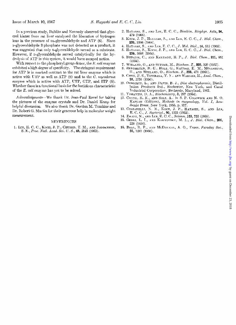
Issue of March 10, 1967 S. Hayashi and E. C. C. Lin 1035
In a previous study, Bublitz and Kennedy observed that glyc- erol kinase from rat liver catalyzed the liberation of hydrogen ions in the presence of nn-glyceraldehyde and ATP (6). Since n-glyceraldehyde a-phosphate was not detected as a product, it was suggested that only n-glyceraldehyde served as a substrate. However, if n-glyceraldehyde served catalytically for the hy- drolysis of ATP in this system, it would have escaped notice.
With respect to the phosphoryl group donor, the E. coli enzyme exhibited a high degree of specificity. The stringent requirement for ATP is in marked contrast to the rat liver enzyme which is active with UTP as well as ATP (6) and to the C. mycoderma enzyme which is active with ATP, UTP, GTP, and ITP (8). Whether there is a functional basis for the fastidious characteristic of the E. coli enzyme has yet to be solved.
Actnowledgwzents-We thank Dr. Jean-Paul Revel for taking the pictures of the enzyme crystals and Dr. Daniel Kemp for helpful discussion. We also thank Dr. Gordon M. Tomkins and Dr. Robert G. Martin for their generous help in molecular weight measurement.
REFERENCES
1. LIN, E. C. C., KOCH, J. P., CHUSED, T. M., AND JORGENSEN, S. E., Proc. Natl. Acad. Sci. U. S., 43, 2145 (1962).
2. HAYASHI, S., AND LIN, E. C. C., Biochim. Biophys. Acta, 94, 479 (1965).
3. KOCH, ‘J. P., HAYASHI, S., AND LIN, E. C. C., J. Biol. Chem., 239, 3106 (1964).
4. HAYASHI, S., AND LIN, E. C. C., J. MoZ. Biol., 14, 515 (1965). 5. HAYASHI, S., KOCH, J. P., AND LIN, E. C. C., J. Biol. Chem.,
239, 3098 (1964). 6. BUBLITZ, C., AND KENNEDY, E. P., J. Biol. Chem.. 211. 951
(1954): ,
7. WIELAND, O., AND SUYTER, M., B&hem. Z., 329, 320 (1957). 8. BERGMEYER, H.-U., HOLZ, G., KAUDER, E. M.. M~LLERING.
H., AND I&LAN;, O., hochem. Z., 333, 471 (1961). 9. CHEN, P. S., TORIBATA, T. Y., AND WARNER, H., Anal. Chem.,
28, 1756 (1956). 10. ORNSTEIN, L., AND DAVIS, B. J., Disc electrophoresis, Distil-
lation Products Ind., Rochester, New York, and Canal Industrial Corporation, Bethesda, Maryland, 1962.
11. YPHANTIS, D. A., Biochemistry, 3, 297 (1964). 12. CRANE, R. K., AND SOLS, A., in S. P. COLOWICK AND N. 0.
KAPLAN (Editors), Methods in enzymology, Vol. I, Aca- demic Press, New York, 1955, p. 277.
13. COZZARELLI, N. R., KOCH, J. P., HAYASHI, S., AND LIN, E. C. C., J. Bacterial., 90, 1325 (1965).
14. ZWAIG, N., AND LIN, E. C. C., Science, 153,755 (1966). 15. GIDEZ, L. I., AND KARNOVSKY, M. L., J. Biol. Chem., 206,
229 (1954). 16. BELL, R. P., AND MCDOUGAL, A. O., Trans. Faraday Sot.,
56, 1281 (1960).
by guest on Decem
ber 23, 2018http://w
ww
.jbc.org/D
ownloaded from

Shin-Ichi Hayashi and E. C. C. LinEscherichia coliPurification and Properties of Glycerol Kinase from
1967, 242:1030-1035.J. Biol. Chem.
http://www.jbc.org/content/242/5/1030Access the most updated version of this article at
Alerts:
When a correction for this article is posted•
When this article is cited•
to choose from all of JBC's e-mail alertsClick here
http://www.jbc.org/content/242/5/1030.full.html#ref-list-1
This article cites 0 references, 0 of which can be accessed free at
by guest on Decem
ber 23, 2018http://w
ww
.jbc.org/D
ownloaded from