purification and characterization of a potato tuber acid
TRANSCRIPT
Plant Physiol. (1994) 106: 223-232
Purification and Characterization of a Potato Tuber Acid Phosphatase Having Significant Phosphotyrosine
Phosphatase Activity'
Kevin S. Cellatly, Greg B. C. Moorhead*, Stephen M. C. Duff3, Daniel D. Lefebvre, and William C. Plaxton*
Departments of Biology and Biochemistry, Queen's University, Kingston, Ontario, Canada K7L 3N6
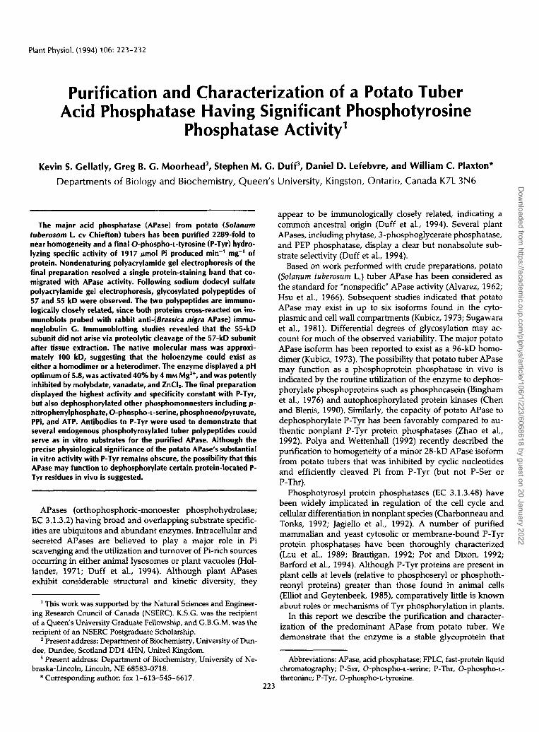
l h e major acid phosphatase (APase) from potato (Solanum tuberosom 1. cv Chiefton) tubers has been purified 2289-fold to near homogeneity and a final O-phospho-i-tyrosine (P-Tyr) hydro- lyzing specific activity of 1917 pmol Pi produced min-' mg-' of protein. Nondenaturing polyacrylamide gel electrophoresis of the final preparation resolved a single protein-staining band that co- migrated with APase activity. Following sodium dodecyl sulfate polyacrylamide gel electrophoresis, glycosylated polypeptides of 57 and 55 kD were observed. l h e two polypeptides are immuno- logically closely related, since both proteins cross-reacted on im- munoblots probed with rabbit anti-(Brassica nigra APase) immu- noglobulin C. lmmunoblotting studies revealed that the 55-kD subunit did not arise via proteolytic cleavage of the 57-kD subunit after tissue extraction. l h e native molecular mass was approxi- mately 100 kD, suggesting that the holoenzyme could exist as either a homodimer or a heterodimer. The enzyme displayed a pH optimum of 5.8, was activated 40% by 4 mM MgZ+, and was potently inhibited by molybdate, vanadate, and ZnCL The final preparation displayed the highest activity and specificity constant with P-lyr, but also dephosphorylated other phosphomonoesters including p- nitrophenylphosphate, O-phospho-L-serine, phosphoenolpyruvate, PPi, and ATP. Antibodies to P-Tyr were used to demonstrate that several endogenous phosphotyrosylated tuber polypeptides could serve as in vitro substrates for the purified APase. Although the precise physiological significance of the potato APase's substantial in vitro activity with P-lyr remains obscure, the possibility that this APase may function to dephosphorylate certain protein-located P- Tyr residues in vivo is suggested.
APases (orthophosphoric-monoester phosphohydrolase; EC 3.1.3.2) having broad and overlapping substrate specific- ities are ubiquitous and abundant enzymes. Intracellular and secreted APases are believed to play a major role in Pi scavenging and the utilization and turnover of Pi-rich sources occumng in either animal lysosomes or plant vacuoles (Hol- lander, 1971; Duff et al., 1994). Although plant APases exhibit considerable structural and kinetic diversity, they
~ ~
This work was supported by the Natural Sciences and Engineer- ing Research Council of Canada (NSERC). K.S.G. was the recipient of a Queen's University Graduate Fellowship, and G.B.G.M. was the recipient of an NSERC Postgraduate Scholarship.
Present address: Department of Biochemistry, University of Dun- dee, Dundee, Scotland DD1 4HN, United Kingdom.
Present address: Department of Biochemistry, University of Ne- braska-Lincoln, Lincoln, NE 68583-0718.
* Corresponding author; fax 1-613-545-6617.
appear to be immunologically closely related, indicating a common ancestral origin (Duff et al., 1994). Several plant APases, including phytase, 3-phosphoglycerate phosphatase, and PEP phosphatase, display a clear but nonabsolute sub- strate selectivity (Duff et al., 1994).
Based on work performed with crude preparations, potato (Solanum tuberosum L.) tuber APase has been considered as the standard for "nonspecific" APase activity (Alvarez, 1962; Hsu et al., 1966). Subsequent studies indicated that potato APase may exist in up to six isoforms found in the cyto- plasmic and cell wall compartments (Kubicz, 1973; Sugawara et al., 1981). Differential degrees of glycosylation may ac- count for much of the observed variability. The major potato APase isoform has been reported to exist as a 96-kD homo- dimer (Kubicz, 1973). The possibility that potato tuber APase may function as a phosphoprotein phosphatase in vivo is indicated by the routine utilization of the enzyme to dephos- phorylate phosphoproteins such as phosphocasein (Bingham et al., 1976) and autophosphorylated protein kinases (Chen and Blenis, 1990). Similarly, the capacity of potato APase to dephosphorylate P-Tyr has been favorably compared to au- thentic nonplant P-Tyr protein phosphatases (Zhao et al., 1992). Polya and Wettenhall (1992) recently described the purification to homogeneity of a minor 28-kD APase isoform from potato tubers that was inhibited by cyclic nucleotides and efficiently cleaved Pi from P-Tyr (but not P-Ser or P-Thr).
Phosphotyrosyl protein phosphatases (EC 3.1.3.48) have been widely implicated in regulation of the cell cycle and cellular differentiation in nonplant species (Charbonneau and Tonks, 1992; Jagiello et al., 1992). A number of purified mammalian and yeast cytosolic or membrane-bound P-Tyr protein phosphatases have been thoroughly characterized (LCU et al., 1989; Brautigan, 1992; Pot and Dixon, 1992; Barford et al., 1994). Although P-Tyr proteins are present in plant cells at levels (relative to phosphoseryl or phosphoth- reonyl proteins) greater than those found in animal cells (Elliot and Geytenbeek, 1985), comparatively little is known about roles or mechanisms of Tyr phosphorylation in plants.
In this report we describe the purification and character- ization of the predominant APase from potato tuber. We demonstrate that the enzyme is a stable glycoprotein that
Abbreviations: APase, acid phosphatase; FPLC, fast-protein liquid chromatography; P-Ser, O-phospho-L-serine; P-Thr, 0-phospho-L- threonine; P-Tyr, O-phospho-L-tyrosine.
Dow
nloaded from https://academ
ic.oup.com/plphys/article/106/1/223/6068618 by guest on 20 January 2022
224 Gellatly et al. Plant Physiol. Vol. 106, 1994
effectively hydrolyzes Pi from P-Tyr, phosvitin, and potato tuber P-Tyr proteins. The results suggest that the enzyme could function as a P-Tyr protein phosphatase in vivo.
MATERIALS AND METHODS
Chemicals and Plant Material
Biochemicals, coupling enzymes, PEG, anti-rabbit IgG (whole molecule) alkaline phosphatase conjugate, Tween 20, bisacrylamide, and nonprestained SDS-PAGE molecular mass standards were purchased from Sigma. Prestained "rainbow" molecular mass standards were purchased from Amersham (Oakville, Ontario, Canada). Protein assay re- agent was obtained from Bio-Rad (Mississauga, Ontario, Can- ada). Cyanogen bromide was obtained from Kodak (Toronto, Ontario, Canada). Phosphocellulose P-11 was purchased from Whatman (Hillsboro, OR). Polyvinylidene difluoride membranes (Immobilon transfer; 0.45-pm pore size) were supplied by Millipore (Mississauga, Ontario, Canada). S- Sepharose, a Phenyl Superose HR 5/5 column, and an FPLC system were obtained from Pharmacia (Baie D'Urfe, Quebec, Canada). Monospecific, affinity-purified rabbit anti-(P-Tyr) IgG (Kamps and Sefton, 1988) was a kind gift of Dr. Peter Greer (Department of Biochemistry, Queen's University). All buffers used in this study were degassed and adjusted to their respective pH values at 25OC. Mature tubers of potato (Solanum tuberosom L. cv Chiefton) were purchased at a local market and used the same day.
Enzyme Assays
Phosphatase Assay. A
For routine measurements of APase activity, the hydrolysis of PEP to pyruvate was coupled with the lactate dehydro- genase reaction and assayed at 25OC using a Varian DMS 200 spectrophotometer. Standard assay conditions were 50 m~ Na-acetate buffer (pH 5.8) containing 5 m~ PEP, 4 m~ MgC12, 0.2 m~ NADH, and 3 units of dialyzed rabbit muscle lactate dehydrogenase in a final volume of 1 mL. Assays were initiated by the addition of enzyme preparation. One unit of APase activity is defined as the quantity of enzyme that would catalyze the hydrolysis of 1 pmol of substrate/ min at 25OC.
Phosphatase Assay B
For substrates other than PEP, the method of Eibl and Lands (1969) was used to detect the Pi released by the APase reaction. APase (0.05 units) was incubated in 1.5-mL cuvettes with 0.9 mL of 50 m~ Mes/NaOH (pH 5.8) containing 4 m~ MgCI2 and an altemative substrate (5 m~ unless otherwise indicated) for 6 min at 25OC. Reactions were terminated by the sequential addition of 0.1 mL of reagent A (3 M H2S04 containing 20 m~ ammonium molybdate) and 10 pL of 1% (v/v) Triton X-100. Samples were incubated at 25OC for 20 min and the A660 was determined. To calculate activities, a standard curve over the range of 0.01 to 1.0 rmol of Pi was constructed for each set of assays. Assays were performed in triplicate and controls were run for background amounts of
Pi present at each substrate concentration by adding reagent A before the enzyme. Hydrolysis was proportional to enzyme concentrations between 0.005 to 0.1 unit/mL and remained linear with time for at least 15 min.
Kinetic Studies
Acid-washed cuvettes were used for all kinetic studies. Apparent K,,, values were determined from the Michaelis- Menten equation fitted to a nonlinear least-squares regression computer kinetics program (Brooks, 1992). All kinetic param- eters are the means of duplicate determinations performed on two separate preparations of the purified enzyme, and they are reproducible to within +lo% SE.
Enzyme Purification and Buffers Used in APase Purifica tion
All purification procedures were carried out at 4OC. Buffer A consisted of 50 m~ Na acetate (pH 4.9) containing
1 m~ EDTA, 1 m~ DTT, and 1 m~ PMSF. Buffer B consisted of 50 m~ Na acetate (pH 5.2) containing 1 m~ E:DTA and 1 m~ DTT. Buffer C was 30 m~ Hepes/NaOH (pH 6.8) con- taining 1 m~ EDTA. Buffer D was 25 m~ bis-tris-propane/ HCl (pE[ 6.5) containing 1 m~ EDTA, 0.5 m~ DIT, and 30% (saturation) (NH4)ZS04. Buffer E consisted of 25 m~ bis-tris- propane/HCl (pH 6.5) containing 1 m~ EDTA, 0.5 m~ DTT, and 15% (v/v) ethylene glycol.
Crude Extract
Peeled and diced potato tubers (600 g) were homogenized in 1 volume of buffer A with a Waring blender and a Polytrori. The homogenate was squeezed through six layers of cheesecloth, filtered through one layer of Miracloth, and centrifuged at 14,OOOg for 20 min. Supernatant fractions were pooled and designated the crude extract.
Acidification
The crude extract was adjusted to pH 4.0 with 7 M acetic acid and stirred for 10 min. The extract was adjusted to pH 4.9 with 15 M NH40H and centrifuged as above. Pellets were discarded.
Fractionation Using PEG
PEG (average molecular mass 8 kD; 50% [w/J] dissolved in 20 m~ bis-tris-propane/HCl [pH 7.41 contajning 1 m~ EDTA) was added to the supematant fluid to give a final concentration of 5% (w/v). The solution was stirred for 10 min and centrifuged as above. Pellets were discarded and the supematant fraction was adjusted to 18% (W/V:I PEG-8000 by the slow addition of finely ground powder, then stirred for 40 min and centrifuged as above. The pellets were resus- pended in 75 mL of buffer B, adjusted to pH 5.2 with 7 M acetic acid, and clarified by centrifugation at 37,OOOg for 10 min.
S-Sepharose Chromatography
The clear supematant fluid was adjusted to a protein concentration of 10 mg/mL with buffer B and absorbed at 1
Dow
nloaded from https://academ
ic.oup.com/plphys/article/106/1/223/6068618 by guest on 20 January 2022
Potato Tuber Acid Phosphatase 225
mL/min onto a column of S-Sepharose (1.5 X 21 cm) that had been pre-equilibrated in buffer B. The column was connected to an FPLC system, washed with buffer B until the A280 decreased to less than 0.1, and eluted with a linear O to 0.5 M KCl gradient (250 mL) in buffer B (fraction size, 5 mL). Peak activity fractions (eluted at approximately 130 m~ KCl) were pooled and concentrated to about 10 mL with an Amicon YM-30 ultrafilter. For desalting, the concentrated S- Sepharose peak fractions were diluted to 45 mL with buffer C, reconcentrated to 10 mL as above, and rediluted to 45 mL with buffer C.
Phosphocellulose Chromatography
Immediately prior to its use, phosphocellulose P-11 was hydrated and precycled according to the manufacturer's in- structions. The desalted, pooled fractions from S-Sepharose chromatography were absorbed at 0.5 mL/min onto a column of phosphocellulose P-11 (0.75 X 13.5 cm) that had been pre-equilibrated with buffer C. The column was connected to an FPLC system, washed with buffer C until the AZs0 decreased to less than 0.1, and eluted with a linear O to 1 M KCl gradient (250 mL) in buffer C (fraction size 2 mL).
Phenyl Superose FPLC
Solid (NH4)zS04 was slowly added to the pooled peak phosphocellulose fractions (eluted at approximately 450 m~ KC1) to bring the final concentration of (NH4)2S04 to 30% saturation. The solution was stirred for 30 min and centri- fuged at 37,OOOg for 15 min. The supernatant fluid was absorbed at 0.5 mL/min onto a Phenyl Superose HR 5/5 column pre-equilibrated in buffer D, and the column was washed with this buffer until the A280 decreased to baseline. The column was eluted in a stepwise fashion with decreasing concentrations of buffer D and simultaneously with increas- ing concentrations of buffer E. APase activity was eluted in a sharp peak following a step from O to 20% buffer E (100- 80% buffer D; fraction size, 1 mL). Pooled peak fractions were concentrated to approximately 600 PL using an Amicon YM-30 ultrafilter and stored at 4OC. APase activity remained constant for at least 6 weeks, when the final preparation was stored at 4OC.
Stability of APase in Potato Tuber Extracts
Clarified extracts were prepared by homogenizing peeled and diced mature potato tuber tissue (0.5 g) with a Polytron in 1 volume of (a) buffer A lacking PMSF; (b) buffer A supplemented with the following protease inhibitors: 1 m~ Na-p-tosyl-L-Lys chloromethyl ketone, 1 m~ N-tosyl-L-Phe chloromethyl ketone, 1 m~ diphenylcarbamylchloride, 2 m~ p-hydroxymecuribenzoate, 3 m~ 1,2-epoxy-3-( p-nitrophen- oxy)-propane, 10 m~ bipyridyl, 5 m~ 1,lO-phenanthroline, 0.1 mg/mL soybean trypsin inhibitor, 0.1 mg/mL pepstatin, 0.1 mg/mL antipain, and 0.1 mg/mL aprotonin; or (c) hot (9OOC) SDS-PAGE sample buffer. The homogenates were centrifuged for 10 min at 16,OOOg in an Eppendorf microcen- trifuge. A 0.5-mL aliquot of the crude supematant fluid prepared in buffer A lacking protease inhibitors (a) was
incubated for 16 h at 25OC. An aliquot of each crude super- natant fraction was mixed with an equal volume of SDS- PAGE sample buffer and boiled for 3 min for immunoblot analysis using anti-(Brassica nigra [black mustard] PEP-spe- cific APase) IgG as described below.
Electrophoresis and Determination of Native and Subunit Molecular Masses
Nondenaturing PAGE was performed with a Bio-Rad (La Jolla, CA) mini-gel apparatus using the discontinuous system of Davis (1964). The final acrylamide monomer concentration in the 0.75-mm-thick slab gels was 8% (w/v) for the separat- ing gel and 2.5% (w/v) for the stacking gel. Prior to pouring the stacking gel solution, the separating gel was pre-electro- phoresed for 2 h at 150 V constant voltage, with 250 m Tris/HCl (pH 8.8) as the electrode buffer. The stacking gel was polymerized with fluorescent light for 3 to 4 h. Gels were precooled to 4OC and were maintained at this temper- ature during electrophoresis. Samples containing 50% (v/v) glycerol were run at a constant voltage of 120 V, applied for 2 h. Tris (25 m~) /Gly (190 m) containing 1 m~ thioglycolate was used as the electrode buffer. Gels were either stained for protein using Coomassie blue R-250 or APase activity was located in the gel. To detect APase activity, the gel was equilibrated for 15 min at 24OC in 100 m~ Na acetate (pH 5.8) containing 20 m~ CaCL followed by incubation for 15 min at room temperature in 100 m Na acetate (pH 5.8) containing 20 m~ CaClZ, 0.02% (w/v) Fast Gamet GBC salt, and 0.02% (w/v) Naz-naphthyl phosphate. For second-di- mension electrophoresis, the single protein-staining band was excised from the nondenaturing gel and incubated in 62 m Tris/HCl (pH 6.8) containing 10% (v/v) glycerol and 2% (w/ v) SDS for 40 min at 5OOC. After equilibration in SDS the gel slice was subjected to SDS-PAGE as described below.
Denaturing SDS-PAGE was performed using a Bio-Rad mini-gel apparatus and the discontinuous system of Doucet and Trifaró (1988) or Laemmli (1970). Final acrylamide mon- omer concentration in the 0.75-mm-thick slab gels was 14, 12, or 10% (w/v) for the separating gel and 4% (w/v) for the stacking gel. All samples were preincubated in the presence of SDS sample buffer (70 m~ Tris/HCl, pH 6.7, containing 8 M urea, 3% [w/v] SDS, 100 m~ DTT, and 0.005% [w/v] bromphenol blue) for 3 min at 100°C prior to being loaded on the gels. Gels were run at a constant voltage of 155 V, applied for 1 h. For the determination of subunit molecular masses by SDS-PAGE, a plot of relative mobility versus log (molecular mass) was constructed with the following stand- ard proteins: @-galactosidase (1 16 kD), phosphorylase b (97.4 kD), BSA (66 kD), ovalbumin (45 kD), and carbonic anhy- drase (29 kD). Glycoprotein staining of SDS gels was con- ducted using a periodic acid-Schiff silver-staining protocol (Dubray and Bezard, 1982).
Nondenaturing SDS-PAGE was utilized for native molec- ular mass determinations (Goldstein et al., 1988). SDS-PAGE was camed out as described above except that the purified APase was mixed with 1% (w/v) SDS and 0.005% (w/v) bromphenol blue without boiling. For the determination of native molecular mass by SDS-PAGE, a plot of relative mobility versus log (molecular mass) was constructed with
Dow
nloaded from https://academ
ic.oup.com/plphys/article/106/1/223/6068618 by guest on 20 January 2022

226 Gellatly et al. Plant Physiol. Vol. 106, 1994
the following prestained 'rainbow" standard proteins: myosin (200 kD), phosphorylase b (97 kD), BSA (69 kD), ovalbumin (46 kD), carbonic anhydrase (30 kD), and soybean trypsin inhibitor (21.5 kD).
Peptide Mapping by Cyanogen Bromide Cleavage
Polypeptides were excised individually from an SDS mini- gel and cleaved in situ with cyanogen bromide, and the degradation products were analyzed on a 1-mm-thick 14% (w/v) SDS mini-gel according to the method of Plaxton and Moorhead (1989).
lmmunoblotting
Electroblotting was performed as previously described (Moorhead and Plaxton, 1990), with the addition of 100 PM o-vanadate to the transfer buffer when maintenance of phos- photyrosyl residues was desired. All immunoblots to be probed with the anti-(black mustard PEP-specific APase) IgG (Duff et al., 1991) were first pretreated with sodium m- periodate according to Laine (1988) so as to oxidize antigenic oligosaccharide chains of endogenous tuber glycoproteins. Immunological detection of phosphotyrosylated tuber pro- teins using anti-(P-Tyr) IgG (Kamps and Sefton, 1988) was performed as referenced above, except that the blocking buffer contained 5% (w/v) BSA (fraction V) and 1% (w/v) ovalbumin instead of 3% (w/v) defatted milk powder. Anti- genic polypeptides were visualized using an alkaline-phos- phatase-tagged secondary antibody as previously described (Moorhead and Plaxton, 1990). Immunological specificities were confirmed by performing immunoblots in which rabbit preimmune serum was substituted for the affinity-purified IgGs. An LKB Ultroscan XL enhanced laser densitometer was used to scan indicated immunoblots. Densitometric data were analyzed using the LKB Gelscan XL software (version 2.1). Immunoreactive polypeptides were quantified in terms of their relative A633.
Dephosphorylation of Potato Tuber Phosphotyrosyl Proteins by Potato APase
Peeled and diced potato tuber tissue (5 g) was homogenized with a Polytron in 1 volume of 100 m Mes/NaOH (pH 5.8) containing 2 m PMSF, 2 m DTT, and 1 m EDTA. The homogenate was centrifuged for 10 min at 16,OOOg in an Eppendorf microfuge, and a 3-mL aliquot of the supematant fluid was passed at 4OC through a Bio-Rad Econo-Pac lODG
desalting column that had been pre-equilibrated in homoge- nization buffer lacking PMSF. Aliquots (0.1 mL) of the de- salted mude extract were incubated at 25OC with and without 3 units of the purified potato APase. Samples were removed at O, 2, and 19 h and analyzed by immunoblotting using the anti-(P-Tyr) IgG as described above. Laser densitometric scanning and immunoquantification of the blots was per- formed a s described above.
Protein Determination
Protein concentration was determined by the method of Bradford (1976) using the Bio-Rad prepared rc2agent and bovine 71-globulin as standard.
RESULTS
Purification and Physical Properties of Potato Tuber APase
Table I summarizes the purification of potato tuber APase. Only a single peak of APase activity was recovered following chromat'ography on columns of S-Sepharose, ph xphocellu- lose, and Phenyl Superose. The enzyme was purified 2289- fold to a final PEP-hydrolyzing specific activity O F 618 units/ mg and an overall recovery of 31%. With P-Tyr substrate the final specific activity was increased to approximately 1900 units/mg (Table 11).
Physical and Immunological Properties
Gel Electrophoresis and Native Molecular Mass €:dimation
Nondenaturing PAGE of the final APase preparation re- sulted in a single protein-staining band that co-migrated with APase activity (Fig. la). When the final preparation was denatured and subjected to SDS-PAGE, two ma lor protein- staining bands of 57 and 55 kD were observed (Fig. 2a). Laser densitometric scanning of the SDS gel revealed that the 57- and 55-k.D silver stained polypeptides were present in a ratio of about 2:1, respectively. SDS-PAGE of a commercially available preparation of potato APase (Sigma, catalog No. P0157) gave rise to a very similar banding pattem, as was observed for the APase purified in the present study (results not shown). The 57- and 55-kD polypeptides were detected by periodic acid-Schiff staining, indicating that they are glycosylated (Fig. 2b). If the single protein-staming band present after nondenaturing PAGE (Fig. la) was excised, equilibrated with SDS, and subjected to SDS-PACE, both the
Table 1. Purification ofAPase from potato tubers (600 g) SteD Volume Activitv" Protein Specific Activitv Purification Yield
mL units mg unitslmg fold %
Crude extract 730 992 3650 0.27 1 O0 Acid step (pH 4.0) 765 975 2715 0.36 1.3 98 PEG fractionation (5-1 8%) 120 1010 1800 0.56 2.1 102 S-Sep harose 45 525 34 15.4 57 53 Phosphocellulose 11.2 301 1 . 1 2 74 1015 30 Phenvl SuDerose 6.4 309 0.5 618 2289 31
a Determined using assay A with PEP as substrate.
Dow
nloaded from https://academ
ic.oup.com/plphys/article/106/1/223/6068618 by guest on 20 January 2022

Potato Tuber Acid Phosphatase 227
Table II. Substrate specificity of potato APaseAll parameters were determined using assay B as described in
"Materials and Methods."
Substrate
P-Tyrp-Nitrophenyl-PP-SerPEPPPiMgATPP-ThrMgADP
v™
un/ts/mg
19171250389764
17283689946
<.mM
0.991.100.621.524.422.16NDa
ND
SpecificityConstant
V™/K™19361136627503391170NDND
a ND, Not determined.
57- and 55-kD polypeptides were resolved and stained in aratio of about 2:1, respectively (Fig. Ib).
Determination of the potato APase's native molecular masswas initially attempted by gel filtration FPLC of the finalpreparation on a calibrated, prepacked Superose 12 HR 10/30 column. Upon gel filtration, however, four peaks of APaseactivity with estimated molecular masses of 377,165,16, and3 kD were recovered. Following SDS-PAGE of each activitypeak, both of the 57- and 55-kD polypeptides were observedand stained in a 2:1 ratio, respectively (results not shown).However, when the purified APase was subjected to nonde-
O-
I29"
TD--TD
Figure 1. Nondenaturing PACE of purified potato tuber APase. a,The left lane contained 0.3 jig of protein and was overlaid with anAPase activity stain; the right lane contained 1 ^g of protein andwas stained with Coomassie blue R-250. b, SDS-PACE of theprotein-stained band shown in (a), which had been excised fromthe nondenaturing gel and equilibrated with SDS as described in"Materials and Methods" (right lane). The polyacrylamide gel con-centration of the separating gel was 10% (w/v). SDS-PACE wasconducted according to Laemmli (1970) and the gel was stainedwith Coomassie blue R-250. O, Origin; TD, tracking dye front.
naruring SDS-PAGE, a single major band of APase activitywith a molecular mass of approximately 100 kD was obtained(Fig. 2c).
Cyanogen Bromide Peptide Mapping
The structural relationship between the 57- and 55-kDpolypeptides was investigated by peptide mapping of theircyanogen bromide cleavage fragments. As shown in Figure3, the cleavage patterns of the two polypeptides were similarbut not identical. This indicates that the two polypeptides aredistinct proteins that may have large regions of similar aminoacid sequences.
Heat Stability
The APase was relatively heat stable, losing 0,52, and 90%of its original activity when incubated for 4 min at 65, 70,and 75°C, respectively.
Immunological Characterization
An immunoblot of the final APase preparation that hadbeen pretreated with sodium m-periodate was probed withaffinity-purified rabbit anti-(black mustard PEP-specificAPase) IgG (Duff et al., 1991) and revealed 57- and 55-kD
(kD)205>
(kD)
-55 —•
29»
Figure 2. SDS-PACE of purified potato tuber APase. a and b,Denaturing SDS-PACE was conducted using a separating polyacryl-amide gel concentration of 10% (w/v) according to the method ofLaemmli (1970). a, Lane 1 contains 5 ^g of molecular mass stand-ards, whereas lane 2 contains 2 jtg of the final APase preparation.The gel was stained with silver according to the method of Hochs-trasser et al. (1988). b, The single lane contains 2 >ig of the finalAPase preparation and was stained for carbohydrate according tothe periodic acid-Schiff silver-staining procedure of Dubray andBezard (1982). c, Nondenaturing SDS-PAGE was conducted aspreviously described (Coldstein et al., 1988) using a separatingpolyacrylamide gel concentration of 8% (w/v). Lane 1 contains 1 Mgof the final APase preparation and was overlaid with an APaseactivity stain; lane 2 contains 5 Mg of Amersham prestained "rain-bow" molecular mass standards. O, Origin; TD, tracking dye front.
Dow
nloaded from https://academ
ic.oup.com/plphys/article/106/1/223/6068618 by guest on 20 January 2022
228 Cellatly et al. Plant Physiol. Vol. 106,1994
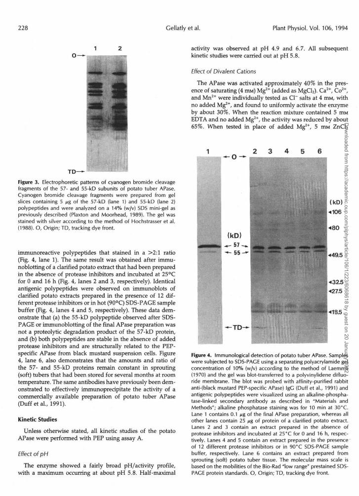
Figure 3. Electrophoretic patterns of cyanogen bromide cleavagefragments of the 57- and 55-kD subunits of potato tuber APase.Cyanogen bromide cleavage fragments were prepared from gelslices containing 5 Mg of the 57-kD (lane 1) and 55-kD (lane 2)polypeptides and were analyzed on a 14% (w/v) SDS mini-gel aspreviously described (Plaxton and Moorhead, 1989). The gel wasstained with silver according to the method of Hochstrasser et al.(1988). O, Origin; TD, tracking dye front.
immunoreactive polypeptides that stained in a >2:1 ratio(Fig. 4, lane 1). The same result was obtained after immu-noblothng of a clarified potato extract that had been preparedin the absence of protease inhibitors and incubated at 25°Cfor 0 and 16 h (Fig. 4, lanes 2 and 3, respectively). Identicalantigenic polypeptides were observed on immunoblots ofclarified potato extracts prepared in the presence of 12 dif-ferent protease inhibitors or in hot (90°C) SDS-PAGE samplebuffer (Fig. 4, lanes 4 and 5, respectively). These data dem-onstrate that (a) the 55-kD polypeptide observed after SDS-PAGE or immunoblotting of the final APase preparation wasnot a proteolytic degradation product of the 57-kD protein,and (b) both polypeptides are stable in the absence of addedprotease inhibitors and are structurally related to the PEP-specific APase from black mustard suspension cells. Figure4, lane 6, also demonstrates that the amounts and ratio ofthe 57- and 55-kD proteins remain constant in sprouting(soft) tubers that had been stored for several months at roomtemperature. The same antibodies have previously been dem-onstrated to effectively immunoprecipitate the activity of acommercially available preparation of potato tuber APase(Duffetal., 1991).
Kinetic StudiesUnless otherwise stated, all kinetic studies of the potato
APase were performed with PEP using assay A.
Effect of pH
The enzyme showed a fairly broad pH/activity profile,with a maximum occurring at about pH 5.8. Half-maximal
activity was observed at pH 4.9 and 6.7. All subsequentkinetic studies were carried out at pH 5.8.
Effect of Divalent Cations
The APase was activated approximately 40% in the pres-ence of saturating (4 HIM) Mg2- (added as MgCl2). Ca2+, Co2+,and Mn2+ were individually tested as Cl~ salts at 4 mM, withno added Mg2+, and found to uniformly activate the enzymeby about 30%. When the reaction mixture contained 5 HIMEDTA and no added Mg2*, the activity was reduced by about65%. When tested in place of added Mg2*, 5 HIM ZnCl2
Figure 4. Immunological detection of potato tuber APase. Sampleswere subjected to SDS-PACE using a separating polyacrylamide gelconcentration of 10% (w/v) according to the method of Laemmli(1970) and the gel was blot-transferred to a polyvinylidene difluo-ride membrane. The blot was probed with affinity-purified rabbitanti-(black mustard PEP-specific APase) IgC (Duff et al., 1991) andantigenic polypeptides were visualized using an alkaline-phospha-tase-linked secondary antibody as described in "Materials andMethods"; alkaline phosphatase staining was for 10 min at 30°C.Lane 1 contains 0.1 jig of the final APase preparation, whereas allother lanes contain 25 /ig of protein of a clarified potato extract.Lanes 2 and 3 contain an extract prepared in the absence ofprotease inhibitors and incubated at 25°C for 0 and 16 h, respec-tively. Lanes 4 and 5 contain an extract prepared in the presenceof 12 different protease inhibitors or in 90°C SDS-PACE samplebuffer, respectively. Lane 6 contains an extract prepared fromsprouting (soft) potato tuber tissue. The molecular mass scale isbased on the mobilities of the Bio-Rad "low range" prestained SDS-PAGE protein standards. O, Origin; TD, tracking dye front.
Dow
nloaded from https://academ
ic.oup.com/plphys/article/106/1/223/6068618 by guest on 20 January 2022

Potato Tuber Acid Phosphatase 229
Table III. Effect of various substances on the activity of potatoAPase
The standard assay A was used, except that the concentration ofPEP was subsaturating (0.25 FTIM). Enzyme activity in the presenceof effectors is expressed relative to the control set at 100%.
Addition
Vanadate
Molybdate
EGTAEDTANaPiNaFGlutamateAspartateAscorbatePhytateCSHPhosvitin
Concentration
mM
0.050.00510.010.001550.555550.150.001
Relative Activity
%
74005
197232445630641248
13119
completely inhibited APase activity. All subsequent kineticstudies were conducted in the presence of 4 HIM
Dephosphorylation of Potato Tuber PhosphotyrosylProteins by Potato APase
Monospetific anti-(P-Tyr) polyclonal antibodies (Kampsand Sefton, 1988) were used to identify proteins phosphoryl-ated on Tyr in potato tuber extracts. The immunoblot shownin Figure 5, lane 1, demonstrates that numerous potato tuberpolypeptides appear to be phosphotyrosylated. No antigenicpolypeptides were observed when an immunoblot of a potatoextract was probed with the P-Tyr antibodies in the presenceof 20 HIM P-Tyr (Fig. 5, lane 2). By contrast, no reduction inthe intensity of the various immunoreactive polypeptidesoccurred when parallel immunoblots were probed with theanti-(P-Tyr) IgG in the presence of 20 mM P-Ser or P-Thr(results not shown). These control experiments demonstratethe specificity of the polyclonal antibodies against P-Tyr. Avery similar banding pattern was observed on an immunoblotof a tuber extract (a) prepared in 90°C SDS-PAGE samplebuffer or (b) probed with two commercially available prepa-rations of anti-(P-Tyr) monoclonal antibodies (Sigma, catalogNo. P3300, and ICN Biomedicals [Costa Mesa, CA], catalogNo. 152369) (results not shown).
When an aliquot of a desalted tuber extract (endogenousAPase activity = 0.56 units/mL using assay A) was incubatedat 25°C for 19 h, the cross-reactivity of five anti-(P-Tyr) IgGimmunoreactive polypeptides was significantly reduced
Substrate Specificity
APase activity was determined using assay B and a widerange of compounds, tested at a total concentration of 5 mMunless otherwise specified. The purified enzyme showed noactivity with AMP, Glc-l-P, or phytate.
Table II lists V^* and apparent Km values, along withspecificity constants (Vmax/Km), for those compounds thatwere found to be dephosphorylated at a significant rate bythe purified enzyme. The highest activity and specificityconstant were obtained with P-Tyr. The specificity constantwith P-Tyr was about 3-fold greater than the value obtainedwith the next best nonartificial substrate, P-Ser. When theegg yolk storage phosphoprotein phosvitin was tested at aconcentration of 50 Mg/mL (125 nM), a phosphoprotein phos-phatase activity of 220 units/mg was obtained.
Metabolite and Ion Effects
A wide variety of substances was tested for effects on thepurified enzyme using subsaturating concentrations of PEP(0.25 ITIM) as substrate. Table III lists those compounds thatwere found to inhibit APase activity. The most notable inhib-itors were vanadate, molybdate, phytate, phosvitin, andNaPi. The following substances had no effect (±15% controlactivity) on enzyme activity. NaCl, KC1, NHUCl, Mg-citrate,and tartrate (all 5 HIM); cAMP (100 MM); Ca2+/bovine calmod-ulin (50 nM/1 JIM); and okadaic acid (1 /UM). Preincubation ofenzyme with 1 mM N-ethylmaleimide for up to 2 min at 25°Calso had no effect on enzyme activity. GSH (5 mM) activatedAPase activity by 30%.
-TD
Figure 5. Immunological detection of proteins phosphorylated onTyr in mature potato tubers. Samples were subjected to SDS-PACEusing a separating gel concentration of 12% (w/v) according to themethod of Laemmli (1970) and the gel was blot-transferred to apolyvinylidene difluoride membrane. Lanes 1 and 2 contain 25 Mgof protein from the clarified tuber extract. Lane 1 was probed usingaffinity-purified rabbit anti-(P-Tyr) IgG (Kamps and Sefton, 1988)and antigenic polypeptides were visualized using an alkaline-phos-phatase-linked secondary antibody as described in "Materials andMethods"; phosphatase staining was for 10 min at 30°C. Thex102-,85-, 34-, 28.5-, and 20-kD immunoreactive polypeptides listed inTable IV are indicated (•). Lane 2 was probed as above except 20mM P-Tyr was added to the anti-(P-Tyr) IgG solution. The molecularmass scale is based on the mobilities of the Bio-Rad "low range"prestained SDS-PAGE protein standards. O, Origin; TD, trackingdye front.
Dow
nloaded from https://academ
ic.oup.com/plphys/article/106/1/223/6068618 by guest on 20 January 2022

230 Gellatly et al. Plant Physiol. Vol. 106, 1994
(Table IV). Addition of 30 units/mL of purified potato APase to the desalted tuber extract appeared to markedly enhance the rate and extent of dephosphorylation of the 102-, 85-, and 34-kD phosphotyrosylated polypeptides (Table IV). Sil- ver staining of duplicate SDS gels showed that no reduction in the relative amount of the various polypeptides occurred over the 19-h time course (results not shown). Furthermore, no reduction in the intensity of any of the anti-(P-Tyr) IgG immunoreactive bands occurred when the desalted tuber extract was incubated for 19 h in the combined presence of 1 m~ o-vanadate and 30 units/mL of the purified potato APase (results not shown).
DISCUSSION
The predominant APase from potato tuber has been puri- fied 2289-fold to near electrophoretic homogeneity and a final P-Tyr-hydrolyzing specific activity of 191 7 units/mg. To the best of our knowledge, this final specific activity is the highest yet reported for a purified plant APase (Duff et al., 1994). The native molecular mass of the purified enzyme was estimated by nondenaturing SDS-PAGE to be approxi- mately 100 kD (Fig. 2c). A single protein-staining band that co-migrated with APase activity was observed following nondenaturing PAGE of the final preparation (Fig. la). How- ever, when the final preparation was denatured and subjected to SDS-PAGE, two protein-staining bands with molecular masses of about 57 and 55 kD were observed (Fig. 2a). Densitometric scanning indicated that the 57- and 55-kD protein-staining bands occurred in a ratio of approximately 2:1, respectively. We believe that both polypeptides were associated with native potato APase for the following rea- sons: (a) these two protein-staining bands co-eluted after Superose 12 gel filtration FPLC of the final preparation; (b) the single band from nondenaturing PAGE still produced two bands after SDS-PAGE (Fig. lb); (c) the respective pat- tems of peptide fragments produced after limited treatment with cyanogen bromide are quite comparable, as analyzed by SDS-PAGE (Fig. 3); and (d) both polypeptides are im-
munologically related (Fig. 4). These data suggest that the native enzyme could exist as a either a homo- or h(2terodimer.
Immunoblots of potato tuber extracts prepared under con- ditions permissive and restrictive to endogenous proteolysis demonstrated that the 55-kD polypeptide is not an artifact created by the proteolytic cleavage of the 57-kD subunit after tissue extraction (Fig. 4). Both polypeptides stained strongly as glycoproteins (Fig, 2b), suggesting that their difference in molecular mass could arise from varying degrees of glycosyl- ation. This could explain the problems encounttired during Superose 12 FPLC of the final preparation, since the anom- alous behavior of a secreted yeast APase during gel filtration chromatography has been attributed to glycosyla tion (Mild- ner, 1976). The tendency of phosphatases to aggregate occurs not only with the yeast multigene family of APastis (Mildner, 1976) but also with the glycogen phosphorylase phospha- tases of skeletal muscle and liver (Rogers et al., 1982). Vari- ation in the extent of glycosylation of a single gene product may be associated with the regulation or targetin:; of protein phosphatases to fulfill distinct roles (Ballou and Fischer, 1986). Further studies are required to clarify whether the limited heterogeneity of the two potato APase subunits arises as a result of the expression of separate independent genes or from a single gene by differential posttranslational modifica tion.
Zhao and co-workers (1992) recently reported that the dephosphorylation of P-Tyr by potato APase occiirs at a rate comparable to that of several human P-Tyr protein phospha- tases. This finding is in accord with the relatively high activity and specificity of potato APase for P-Tyr that W ~ S observed in the present study (Table 11), a property that distinguishes it from most other plant APases that have been examined to date (Duff et al., 1994) The capacity of potato APase to dephosphorylate a variety of phosphoproteins tn this and other studies (Bingham et al., 1976; Chen and B enis, 1990) suggests that such an activity may have physiological rele- vance. Ser is the predominant phosphorylated amino acid of the egg yolk storage protein, phosvitin (Byme eí al., 1984), and other phosphoproteins that have been repor1 ed to serve
Table IV. Dephosphorylation of potato tuber phosphotyrosyl proteins by potato tuber APase A desalted potato tuber extract was incubated at 25°C in the absence (-) and presence (+) of 30
units/mL purified potato tuber APase. Aliquots were removed at 2 and 19 h and analyzed by immunoblotting using anti-(P-Tyr) IgG (Kamps and Sefton, 1988). The relative P-Tyr content of the five antigenic polypeptides noted in Figure 5 was estimated by laser densitometric quantification of the immunoblots as described in “Materials and Methods.” Unless otherwise indicated, all values reDresent the means f SE of triplicate determinations.
Relative Phosphotyrosyl Content after Incubation at 25’C Estimated Molecular
Phosphotyrosyl Polypeptide Mass of 2 h 19 h
-I- - + -
kD Oh initial value
102 76’ 60 f 5 51 f 5 36 f 3 85 85 f 9 69 f 2 60 f 4 34 f 2 34 93 f 6 40 f 3 38” 1 6 f 2 28.5 a la 65 f 2 31 f 5 28 f 3 20 83” 66 f 5 1 8 f l 1 2 f 3
a Mean of duplicate determinations.
Dow
nloaded from https://academ
ic.oup.com/plphys/article/106/1/223/6068618 by guest on 20 January 2022

Potato Tuber Acid Phosphatase 231
as substrates for potato APase (Bingham et al., 1976; Chen and Blenis, 1990). We have shown, however, that similar to other plant tissues (Elliot and Geytenbeek, 1985), there is an abundance of P-Tyr-containing polypeptides in potato tubers (Fig. 5). Moreover, incubation of a desalted tuber extract in the presence of the purified potato APase caused a substantial reduction in the level of phosphorylation of several of the endogenous P-Tyr proteins (Table IV). Since this decrease in protein P-Tyr content was not caused by protein degradation and was negated by the presence of 1 m~ o-vanadate, these observations suggest that the major potato APase can de- phosphorylate endogenous P-Tyr proteins in vitro. It is also important to note that several recent studies have demon- strated that the composition and length of the amino acid sequence adjacent to the phosphotyrosylation site markedly influences the affinity and specificity of a P-Tyr protein phosphatase for its substrate (Cho et al., 1991; Ramachan- dran et al., 1992; Zhang et al., 1993). For example, a P-Tyr phosphatase from Escherichia coli has a K,,,(P-Tyr) of more than 6 mM, but displays K, values ranging from 0.027 to 4.1 m~ for various P-Tyr peptides (Cho et al., 1991). Thus, the K,(P-Tyr) of 0.99 m~ derived for potato APase (Table 11) may be orders of magnitude in excess of the enzyme's K, value for P-Tyr peptides and/or endogenous phosphoprotein substrates.
There have been several recent reports describing the pu- rification and characterization of P-Tyr APases from plant sources including poppy seeds (Chung and Polya, 1992), wheat seedlings (Chen and Tao, 1989), and maize seedlings (Jagiello et al., 1992). The subunit composition and substrate specificity of the potato tuber APase most closely resembles that of the poppy seed enzyme (Chung and Polya, 1992). In addition, Polya and Wettenhall (1992) have described the purification and characterization of a minor 28-kD potato tuber APase isoform that also effectively hydrolyzes Pi from P-Tyr. This minor potato APase isoform, however, can be clearly distinguished from the APase of the current study in terms of subunit structure, a P-Tyr-hydrolyzing specific ac- tivity of only 103 units/mg, its inability to catalyze dephos- phorylation of I'-Ser, and its inhibition by micromolar con- centrations of CAMP (Polya and Wettenhall, 1992).
Apart from P-Tyr phosphatase activity, the major potato tuber APase isoform shares several properties with various mammalian and yeast P-Tyr protein phosphatases. Similar to the potato APase, the nonplant P-Tyr phosphatases fre- quently display an acidic pH optimum, show potent inhibi- tion by o-vanadate, molybdate, and Zn2+, and will catalyze the dephosphorylation of p-nitrophenyl-P as well as a variety of other phosphomonoesters (Ballou and Fischer, 1986; Lau et al., 1989; Chen and Blenis, 1990; Cho et al., 1991; Brauti- gan, 1992; Charbonneau and Tonks, 1992; Pot and Dixon, 1992). Additional studies are necessary to elucidate whether the potato tuber APase's substantial activity with P-Tyr (Table 11), and its capacity to dephosphorylate endogenous phosphotyrosylated polypeptides in vitro (Table IV), have any physiological relevance in vivo. Isolation of cDNA clones for potato tuber APase is currently underway and will shed light on the enzyme's relationship with characterized APases and P-Tyr protein phosphatases.
Received February 22, 1994; accepted May 31, 1994. Copyright Clearance Center: 0032-0889/94/106/0223/10.
LITERATURE CITED
Alvarez EF (1962) The kinetics and mechanism of the hydrolysis of phosphoric acid esters by potato acid phosphatase. Biochim Bio- phys Acta 59 663-667
Ballou LM, Fischer EH (1986) Phosphoprotein phosphatases. In PD Boyer, EG Krebs, eds, The Enzymes, Vol17. Academic Press, New York, pp 311-361
Barford D, Flint AJ, Tonks NK (1994) Crystal structure of human protein tyrosine phosphatase 1B. Science 263 1397-1404
Bingham EW, Farrell HM, Dah1 KJ (1976) Removal of phosphate groups from casein with potato acid phosphatase. Biochim Biophys Acta 429 448-460
Bradford M (1976) A rapid and sensitive method for the quantifi- cation of microgram quantities of protein utilizing the principle of protein-dye binding. Anal Biochem 72 248-254
Brautigan DL (1992) Great expectations: protein tyrosine phospha- tases in cell regulation. Biochim Biophys Acta 1114 63-77
Brooks SPJ (1992) A simple computer program with statistical tests for the analysis of enzyme kinetics. BioTechniques 13 906-911
Byrne BM, Schip ADV, Klundert JAMV, Arnberg AC, Gruber M, Geert AB (1984) Amino acid sequence of phosvitin derived from the nucleotide sequence of part of the chicken vitellogenin gene. Biochemistry 23 4275-4279
Charbonneau H, Tonks NK (1992) Protein phosphatases. Annu Rev Cell Biol 8: 463-493
Chen R-H, Blenis J (1990) Identification of Xenopus S6 protein kinase homologs (pp90"9 in somatic cells: phosphorylation and activation during initiation of cell proliferation. Mol Cell Biol 10
Cheng H-F, Tao M (1989) Purification and characterization of a phosphotyrosyl protein phosphatase from wheat seedlings. Biochim Biophys Acta 998: 271-276
Cho H, Ramer SE, Itoh M, Winkler DG, Kitas E, Bannwarth W, Burn P, Saito H, Walsh CT (1991) Purification and characteriza- tion of a soluble catalytic fragment of a human transmembrane leukocyte antigen related (LAR) protein tyrosine phosphatase from an Escherichia coli expression system. Biochemistry 30 6210-6216
Chung RP-T, Polya GM (1992) Copurification and characterization of poppy seed phosphatase and phosphoprotein phosphatase ac- tivities. Plant Sci 84 153-162
Davis B (1964) Disc electrophoresis. 11. Method and application to human serum protein. Ann NY Acad Sci 121: 407-427
Doucet JP, TrifarÓ JM (1988) A discontinuous and highly porous sodium dodecyl sulfate-polyacrylamide slab gel system of high resolution. Anal Biochem 168: 265-271
Dubray G, Bezard G (1982) A highly sensitive periodic acid-silver stain for 1,2-diol groups of glycoproteins and polysaccharides in polyacrylamide gels. Anal Biochem 119 325-329
Duff SMG, Plaxton WC, Lefebvre DD (1991) Phosphate starvation response in plant cells: de n o m synthesis and degradation of acid phosphatases. Proc Natl Acad Sci USA 88: 9538-9542
Duff SMG, Sarath G, Plaxton WC (1994) The role of acid phospha- tases in plant phosphorus metabolism. Physiol Plant 90: 791-800
Eibl H, Lands WEM (1969) A new, sensitive determination of phosphate. Anal Biochem 30 51-57
Elliot DC, Geytenbeek M (1985) Identification of products of protein phosphorylation in T37-transformed cells and comparisons with normal cells. Biochim Biophys Acta 845 317-323
Goldstein AH, Danon A, Baertlein DA, McDaniel RG (1988) Phosphate starvation inducible metabolism in Lycopersicon esculen- tum. 11. Characterization of the phosphate starvation inducible excreted acid phosphatase. Plant Physiol87: 716-720
Hochstrasser DF, Patchornik A, Merril CR (1988) Development of polyacrylamide gels that improve the separation of proteins and their detection by silver staining. Anal Biochem 173 412-423
Hollander VP (1971) Acid phosphatases. In PD Boyer, ed, The Enzymes, Vol4. Academic Press, New York, pp 449-498
3204-3215
Dow
nloaded from https://academ
ic.oup.com/plphys/article/106/1/223/6068618 by guest on 20 January 2022

232 Gellatly et al. Plant Physiol. Vol. 106, 1994
Hsu RY, Cleland WW, Anderson L (1966) Mechanism of action of the nonspecific phosphomonoesterase from potatoes. Biochemistry
Jagiello I, Donella-Deana A, Szczegielniak J, Pinna LA, Muszyn- ska G (1992) Identification of protein phosphatase activities in maize seedlings. Biochim Biophys Acta 1134 129-136
Kamps MP, Sefton BM (1988) Identification of multiple novel poly- peptide substrates of the v-src, v-yes, v-fps, v-ros, and v-erb-B oncogenic tyrosine protein kinases utilizing antisera against phos- photyrosine. Oncogene 2 305-315
Kubicz A (1973) Acid phosphatase I11 from potato tubers, molecular weight and subunit structure. Acta Biochim Pol 2 0 223-229
Laemmli UK (1970) Cleavage of structural proteins during the assembly of the head of bacteriophage T4. Nature 227: 680-685
Laine A-C (1988) Significant immunological cross-reactivity of plant glycoproteins. Electrophoresis 9 841-844
Lau K-HW, Farley JR, Baylink DJ (1989) Phosphotyrosyl protein phosphatases. Biochem J 257: 23-36
Mildner P (1976) Purification of protoplast-secreted acid phospha- tase from baker’s yeast. Biochim Biophys Acta 429: 274-282
Moorhead GBG, Plaxton WC (1990) Purification and characteriza- tion of cytosolic adolase from carrot storage root. Biochem J 269
Plaxton WC, Moorhead GBG (1989) Peptide mapping by CNBr
5 799-807
133-139
fragmeintation using a sodium dodecyl-sulfate-pol yacrylamide minigel system. Anal Biochem 178: 391-393
Polya GM, Wettenhall REH (1992) Rapid purification and N-ter- mina1 slequencing of a potato tuber cyclic nucleotide binding phos- phatase. Biochim Biophys Acta 1159: 179-184
Pot DA, Dixon JE (1992) A thousand and two proíein tyrosine phosphatases. Biochim Biophys Acta 1136 35-43
Ramachandran C, Aebersold R, Tonks NK, Pot DA (15192) Sequen- tial dephosphorylation of a multiply phosphorylated insulin recep- tor pepide by -protein tyrosine- phÒsphâtas&. Biochemistry 3l: 4232-4238
Rogers DT, Lemire JM, Bostian KA (1982) Acid phosphatase poly- peptides in Saccharomyces cerevisiae are encoded by a differentially regulated multigene family. Proc Natl Acad S t i USA 79
Sugaware S, Inamoto Y, Ushijima M (1981) Resolution and some properties of acid phosphatase isozymes bound to the cell wall of potato tubers. Agric Biol Chem 4 5 1767-1774
Zhang Z-,Y, Andrea MT, MacLean D, McNamara DJ, Dobrusin EM, Sawyer TK, Dixon JE (1993) Substrate specificity of the protein tyrosine phosphatases. Proc Natl Acad S’ci USA 90:
Zhao Z, Zander NF, Malencik DA, Anderson SR, Fischer H (1992) Continuous spectrophotometric assay of protein tyrosine phospha- tase using phosphotyrosine. Anal Biochem 202 361-366
2157-2161
4446-4450
Dow
nloaded from https://academ
ic.oup.com/plphys/article/106/1/223/6068618 by guest on 20 January 2022