pure red cell aplasia complicated by angioimmunoblastic t-cell lymphoma: humoral factor plays a main...
TRANSCRIPT

LETTERS ANDCORRESPONDENCE
Letters and correspondence submitted for possible publication mustbe identified as such. Text length must not exceed 500 words andfive bibliographic references. A single concise figure or table may beincluded if it is essential to support the communication. Letters nottyped double-spaced will not be considered for publication. Letters notmeeting these specifications will not be returned to authors. Letters tothe Editor are utilized to communicate a single novel observation orfinding. Correspondence is to be used to supplement or constructivelycomment on the contents of a publication in the journal and cannotexceed the restrictions for Letters to the Editor. The Editor reservesthe right to shorten text, delete objectional comments, and makeother changes to comply with the style of the journal. Permission forpublication must be appended as a postscript. Submissions must besent to Paul Chervenick, M.D., Editor of Brief Reports/Letters toEditors, American Journal of Hematology, H. Lee Moffitt CancerCenter, University of South Florida, 12902 Magnolia Drive, Tampa,FL 33612 to permit rapid consideration for publication.
Pure Red Cell Aplasia Complicated byAngioimmunoblastic T-Cell Lymphoma: Humoral FactorPlays a Main Role in the Inhibition of Erythropoiesis FromCD34+ Progenitor Cells
To the Editor:Pure red cell aplasia (PRCA) has heterogeneous pathoge-nicity. A variety of hematologic malignancies, especially lymphoprolifera-tive disorders have been reported to be associated with PRCA [1,2]. An-gioimmunoblastic T-cell lymphoma (AILD-T), described in REAL classi-fication, is clinically characterized by generalized lymphadenopathy,hepatosplenomegaly, constitutional symptoms and various immunologicdisturbances. A few cases of AILD-T with PRCA have been reported [3,4],and the pathogenesis has not been identified. We report a case of AILD-Twith PRCA, and demonstrate in vitro inhibition of erythropoiesis from thenormal bone marrow progenitor cells by using the patient’s serum.
A 46-year-old man with AILD-T was admitted to our hospital becauseof systemic lymphadenopathy, hepatosplenomegaly, massive pleural effu-sion, and severe B-symptoms. The diagnosis of AILD-T was established bycervical lymph node biopsy. Hematological findings were as follows: he-moglobin 7.8 g/dl, reticulocytes 0%, platelets 112 × 109/l, leukocytes 3.6× 10⁹/l (with normal hemogram). Serum erythropoietin was elevated to 254mU/ml (normal: 8–36). Bone marrow aspirate and biopsy showed thelymphomatous infiltration and aplasia of erythroid precursors with normalmyelopoiesis and megakaryopoiesis. The patient received chemotherapyand achieved remission. After chemotherapy, the hematological and bonemarrow findings showed the improvement of PRCA and the serum eryth-ropoietin normalized.
In vitro culture of bone marrow progenitor cells were performed. Normalbone marrow cells were obtained from a healthy donor after obtaininginformed consent. CD34+ progenitor cells were purified by immunomag-netic separation (MACS system; Miltenyi Biotec, Bergisch Gladbach, Ger-
many). Colony-forming assay were performed in triplicate using premademethylcellulose medium (Stem Cell CFU Kit; Baxter, Deerfield, IL, USA)which contained 3.0 U/ml of erythropoietin. The patient’s serum obtainedbefore and after chemotherapy was added at the final concentration of 10%.Normal AB serum and the serum from another patient with AILD-T with-out PRCA were used as controls. CD34+ cells were plated at 1 × 10³/mland all cultures were maintained in a CO2 incubator. Burst-forming unit-erythroid (BFU-E) was counted after 14 days of culture. The results arepresented in the Table. The patient’s serum before treatment significantlyinhibited the BFU-E formation compared with controls (P < 0.05 by Stu-dent’s t-test). However, the serum obtained after chemotherapy did notdecrease BFU-E. No differences were seen in the numbers of colony-forming unit granulocyte-macrophage (CFU-GM) among the serumsamples.
In AILD-T, only 1 report was published which analyzed the humoraleffect in the clonal study of erythropoiesis [3]. They cultured normal bonemarrow cells and demonstrated the inhibition of erythropoiesis by theserum from a patient. Our study, culturing purified CD34+ progenitor cells,showed the significant inhibition of BFU-E by the patient’s serum beforebut not after chemotherapy. The findings suggest the significance of thehumoral factor in the pathogenesis of PRCA at the hematopoietic stem celllevel. AILD-T is characterized by the immunological disorders and also bythe release of several cytokines which often enhance hematopoiesis [5].Not only immunological mechanisms, but also cytokines may be involvedin the pathogenesis of PRCA with AILD-T. Further study focused on thetype of humoral factor should be performed.
ACKNOWLEDGMENTS
We are grateful to Dr. N. Kosugi and Dr. A. Tojo (The Institute ofMedical Science, The University of Tokyo) for purifying and providingCD34+ bone marrow cells.
HIDEKI TSUJIMURA
CHIKARA SAKAI
TOSHIYUKI TAKAGI
Division of Hematology-Oncology, Chiba Cancer Center Hospital,Chiba, Japan
TABLE I. Colony-Forming Assay From CD34 + BoneMarrow Cells
Seruma BFU-E/102 CD34+ cells
Control 1 50.2 ± 4.6Control 2 48.5 ± 14.1Before chemotherapy 24.4 ± 6.2b
After chemotherapy 50.5 ± 6.5
Values are means ± SD of triplicate cultures.aControl 1; normal AB serum, Control 2; serum from a patient withAILD-T without PRCA.bSignificantly decreased compare with controls (P < 0.05).
American Journal of Hematology 62:259–262 (1999)
© 1999 Wiley-Liss, Inc.
REFERENCES
1. Cahrles RJ, Sabo KM, Kidd PG, Abkowitz JL. The pathophysiology of pure redcell aplasia: implication for therapy. Blood 1996;87:4831–4838.
2. Lacy MQ, Kurtin PJ, Tefferi A. Pure red cell aplasia: association with largegranular lymphocyte leukemia and the prognostic value of cytogenetic abnormali-ties. Blood 1996;87:3000–3006.
3. Lynch JW, Elfenbein GJ, Noyes WD, Braylan RC, Gross MA, Weiner RS. Purered cell aplasia associated with angioimmunoblastic lymphadenopathy with dys-proteinemia. Am J Hematol 1994;46:72–78.
4. Higuchi T, Mori H, Niikura H, Omine M. Hypocomplementemia and hematologi-cal abnormalities in immunoblastic lymphadenopathy and immunoblastic lymph-adenopathy-like T cell lymphoma. Acta Haematol 1996;96:68–72.
5. Foss HD, Anagnostopoulus I, Herbst H, Grebe M, Ziemann K, Hummel M, SteinH. Patterns of cytokine gene expression in peripheral T-cell lymphoma angioim-munoblastic lymphadenopathy type. Blood 1995;85:2862–2869.
A Novel Mutation of the Protein C Gene With a FrameshiftDeletion of 3 Base Pair ( 3380AGG) in Exon 6 in Type 1Deficiency Associated With Arterial andVenous Thrombosis
To the Editor: Protein C deficiency and other inherited thrombophilicdisorders such as deficiency of antithrombin III, protein S, and activatedprotein C resistance due to factor V Leiden gene mutation, are commonlyassociated with risk factors of venous thrombosis [1,2]. We report here, arare case of heterozygous type 1 protein C deficiency with arterial throm-bosis, renal and splenic infarctions, and venous thrombosis of pulmonaryembolism due to a novel mutation of the protein C gene.
A 62-year-old female was hospitalized in April 1998 for recurrent acuteepigastralgia. Gastric endoscopy did not reveal any lesion. Enhanced com-puted tomography revealed right renal and splenic infarctions. CBC andblood chemistry were almost normal except for mild thrombocytosis, 41.4× 10⁹/L. The coagulation study revealed PT 12.8 sec (control 14 sec), andaPTT 41.4 sec (control 36 sec). Anticardiolipin IgG antibody, lupus anti-coagulant, anti-beta 2 glycoprotein 1 antibody, and anti-DNA antibody,were all negative. She had two sons, and no abortion or premature labor.
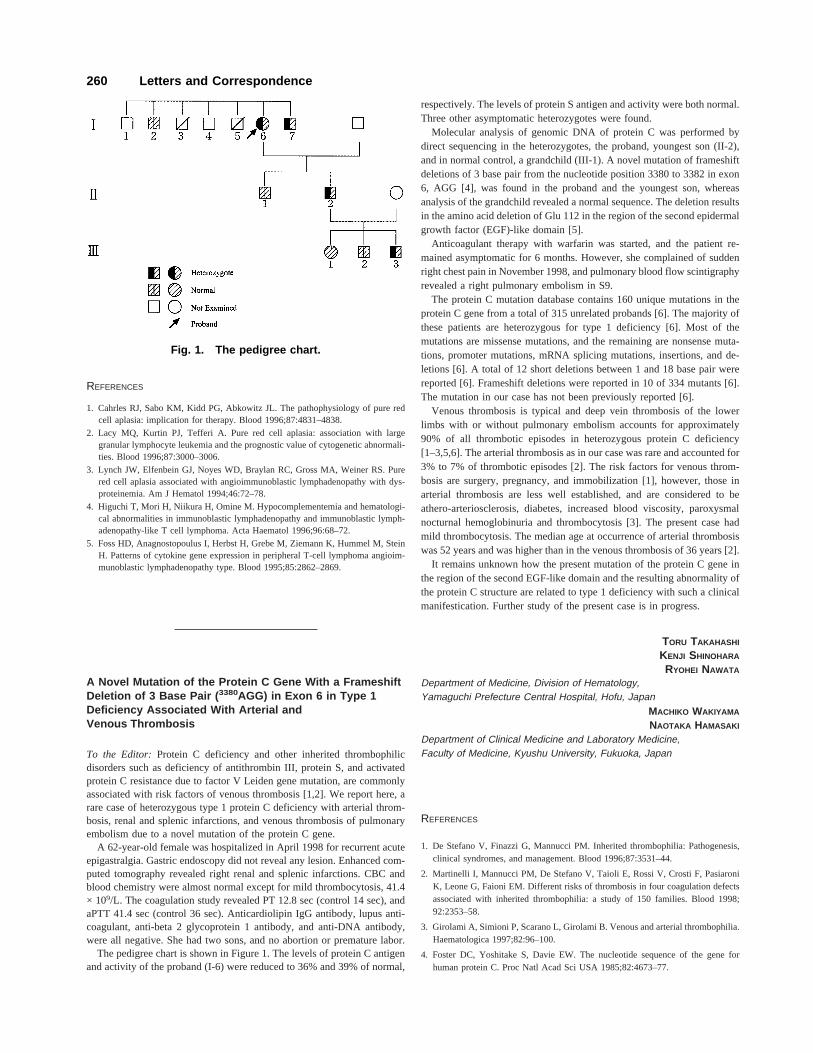
The pedigree chart is shown in Figure 1. The levels of protein C antigenand activity of the proband (I-6) were reduced to 36% and 39% of normal,
respectively. The levels of protein S antigen and activity were both normal.Three other asymptomatic heterozygotes were found.
Molecular analysis of genomic DNA of protein C was performed bydirect sequencing in the heterozygotes, the proband, youngest son (II-2),and in normal control, a grandchild (III-1). A novel mutation of frameshiftdeletions of 3 base pair from the nucleotide position 3380 to 3382 in exon6, AGG [4], was found in the proband and the youngest son, whereasanalysis of the grandchild revealed a normal sequence. The deletion resultsin the amino acid deletion of Glu 112 in the region of the second epidermalgrowth factor (EGF)-like domain [5].
Anticoagulant therapy with warfarin was started, and the patient re-mained asymptomatic for 6 months. However, she complained of suddenright chest pain in November 1998, and pulmonary blood flow scintigraphyrevealed a right pulmonary embolism in S9.
The protein C mutation database contains 160 unique mutations in theprotein C gene from a total of 315 unrelated probands [6]. The majority ofthese patients are heterozygous for type 1 deficiency [6]. Most of themutations are missense mutations, and the remaining are nonsense muta-tions, promoter mutations, mRNA splicing mutations, insertions, and de-letions [6]. A total of 12 short deletions between 1 and 18 base pair werereported [6]. Frameshift deletions were reported in 10 of 334 mutants [6].The mutation in our case has not been previously reported [6].
Venous thrombosis is typical and deep vein thrombosis of the lowerlimbs with or without pulmonary embolism accounts for approximately90% of all thrombotic episodes in heterozygous protein C deficiency[1–3,5,6]. The arterial thrombosis as in our case was rare and accounted for3% to 7% of thrombotic episodes [2]. The risk factors for venous throm-bosis are surgery, pregnancy, and immobilization [1], however, those inarterial thrombosis are less well established, and are considered to beathero-arteriosclerosis, diabetes, increased blood viscosity, paroxysmalnocturnal hemoglobinuria and thrombocytosis [3]. The present case hadmild thrombocytosis. The median age at occurrence of arterial thrombosiswas 52 years and was higher than in the venous thrombosis of 36 years [2].
It remains unknown how the present mutation of the protein C gene inthe region of the second EGF-like domain and the resulting abnormality ofthe protein C structure are related to type 1 deficiency with such a clinicalmanifestication. Further study of the present case is in progress.
TORU TAKAHASHI
KENJI SHINOHARA
RYOHEI NAWATA
Department of Medicine, Division of Hematology,Yamaguchi Prefecture Central Hospital, Hofu, Japan
MACHIKO WAKIYAMA
NAOTAKA HAMASAKI
Department of Clinical Medicine and Laboratory Medicine,Faculty of Medicine, Kyushu University, Fukuoka, Japan
REFERENCES
1. De Stefano V, Finazzi G, Mannucci PM. Inherited thrombophilia: Pathogenesis,clinical syndromes, and management. Blood 1996;87:3531–44.
2. Martinelli I, Mannucci PM, De Stefano V, Taioli E, Rossi V, Crosti F, PasiaroniK, Leone G, Faioni EM. Different risks of thrombosis in four coagulation defectsassociated with inherited thrombophilia: a study of 150 families. Blood 1998;92:2353–58.
3. Girolami A, Simioni P, Scarano L, Girolami B. Venous and arterial thrombophilia.Haematologica 1997;82:96–100.
4. Foster DC, Yoshitake S, Davie EW. The nucleotide sequence of the gene forhuman protein C. Proc Natl Acad Sci USA 1985;82:4673–77.
Fig. 1. The pedigree chart.
260 Letters and Correspondence

5. Reitsma PH. Protein C deficiency: from gene defects to disease. Thromb Haemost1997;78:344–50.
6. Reitsma PH, Bernardi F, Doig RG, Gandrille S, Greengald JS, Ireland H, Kraw-czak M, Lind B, Long GL, Poort SR, Saito H, Sala N, Witt I, Cooper DN. ProteinC deficiency: A database of mutations, 1995 update. Thromb Haemost 1995;73:876–89.
Unique Sequence of Pernicious Anemia, Stomach Cancer,and Myelodysplastic Syndrome
To the Editor: We described here a case of myelodysplastic syndrome(MDS) subsequent to pernicious anemia (PA) and stomach cancer (SC).
A 76-year-old man was admitted to the hospital in 1994 and hemato-logical examinations revealed macrocytic anemia with a red blood cellcount of 1.32 × 10¹²/l, hemoglobin concentration of 6.0 g/dl, hematocrit of17%, reticulocyte count of 7.92 × 10⁹/l, leukopenia with leukocyte count of3.1 × 109/l, and thrombocytopenia with platelet count of 74 × 109/l. Pe-ripheral blood film showed hypersegmentation of neutrophils, and bonemarrow examination revealed megaloblastic erythropoiesis and normalchromosomal karyotype. The serum cobalamin level was low (30 pg/ml,normal 249–938 pg/ml). Anti-intrinsic factor (anti-IF) antibody was pre-sent. The urinary methylmalonic acid excretion increased. Findings ofother immune examinations (Table I) were negative, except for slightincreases of serum immunoglobulin (Ig)G and IgA. A diagnosis of PA wasestablished and the patient was treated with cobalamin. All hematologicaldata were within normal range 3 months after the treatment began. SC wasdetected at this time. Hematological findings were stable after surgerywithout chemotherapy for the cancer until December 1998, when labora-tory examinations disclosed pancytopenia with a red blood cell count of1.29 × 1012/l, hemoglobin concentration of 4.5 g/dl, hematocrit of 14.4%,reticulocyte count of 23.2 × 10⁹/l, leukopenia with leukocyte count of 2.3× 10⁹/l, and thrombocytopenia with platelet count of 79 × 109/l. Peripheralblood film showed degranulated neutrophils associated with Pelger-likenuclei, circulating micromegakaryocytes, and blasts (1% of white bloodcells). The bone marrow was hypercellular and showed megaloblastoidchanges, neutrophilic immaturity, and micromegakaryocytes. Cytogeneticanalysis of bone marrow cells revealed trisomic 47 XY,+8. Serum level ofcobalamin was normal. Immune examinations showed positive results foranti-IF antibody, antinuclear factor, immune complex, rheumatoid factor,and increased serum IgG and IgA as shown in Table I. A diagnosis ofrefractory anemia with excess myeloblasts was made and the patient wastreated with blood transfusions because he was elderly and only com-plained of anemic symptoms.
Several studies have suggested an association between PA and severalautoimmune diseases. MDS is a clonal hematopoietic disorder and itspossible association with immunological abnormalities [1]. Clustering ofautoimmune diseases in individuals is clearly recognized and certain dis-ease associations have been established. Immunological features betweenPA and MDS in our case were different as shown in Table I, although thiscase highlights the immunogenetic links between the disorders with im-munological aberrations and draws PA and MDS further into the spectrum.
An increased risk of SC has been demonstrated and hematologic malig-nancies have been repeatedly observed in case reports, including myeloidleukemia [2], polycythemia vera, and multiple myeloma following thediagnosis of PA. Brinton et al. [3] and Hsing et al. [4] performed large-scale, population-based, prospective studies that showed a significantlyhigher incidence of myeloid leukemia following a diagnosis of PA. Theincidence of preleukemia or MDS has not been evaluated. Mufti et al. [5]observed three cases of MDS associated with PA, although hematological
and clinical features were not sufficiently shown. The association betweenPA and MDS in case reports may be accidental, although large-scale stud-ies on the occurrence of MDS subsequent to PA are needed to clarify therelationship between PA and MDS with immunological abnormalities
HARUKI KONDO
TAKAAKI IMAMURA
Division of Hematology and Oncology, Department of Medicine,Shimizu Kohsei Hospital, Shimizu, Shizuoka, Japan
REFERENCES
1. Hamblin T. Immunological abnormalities in myelodysplastic syndromes. Hematol/Oncol Clin North Am 1992;6:571–586.
2. Blackburn EK, Callender ST, Dacie JV, Doll R, Girdwood RH, Mollin DL, SaracciR, Stafford JL, Thompson RB, Varadi S, Wetherley-Men G. Possible associationbetween pernicious anaemia and leukemia: a prospective study of 1,625 patientswith a note on the very high incidence of stomach cancer. Int J Cancer 1968;3:163–170.
3. Brinton LA, Gridley G, Hrubec Z, Hoover R, Fraumeni Jr JF. Cancer risk follow-ing pernicious anaemia. Br J Cancer 1989;59:810–813.
4. Hsing AW, Hansson L-E, McLaughlin JK, Nyren O, Blot WJ, Ekbom A, FraumeniJF Jr. Pernicious anemia and subsequent cancer. A population-based cohort study.Cancer 1993;71:745–750.
5. Mufti GJ, Figes A, Hamblin TJ, Oscier DG, Copplestone JA. Immunologicalabnormalities in myelodysplastic syndromes. I. Serum immunoglobulin and auto-antibodies. Brit J Haematol 1986;63:146–147.
TABLE I. Immunological and Cytogenetic Studies
Feb. 1994(Diagnosis: PAa)
Dec. 1998(Diagnosis: MDSb)
Anti-IFc antibody + +Anti-PCd antibody − −Thyroid test − −Microsome test − −Anti-thyroglobulin antibody − −Anti-TPOe antibody − −Anti-nuclear factor (<×40)f <×40 ×160LEg test − −Anti-DNA (<6 IU/ml) antibody <6 <6Anti-RNP antibody − −Anti-Sm antibody − −C3c(63–134 mg/dl) 42 96C4 (13–36 mg/dl) 21 25CH50(30–40 CH50 U/ml) 30.9 39.1Immune complex (C1Q)
(<3 mg/ml) <1.5 4.8RA − +RAPA (<×40) nd ×1280IgG(870–1700 mg/dl) 2013 3289IgA(110–410 mg/dl) 473 711IgM(33–190 mg/dl) 116 110
Chromosome 46,XY[100%]46,XY [80%]47,XY,+8[20%]
aPA, pernicious anemia;bMDS, myelodysplastic syndrome;cIF, intrinsic factor;dPC, parietal cell;eTPO, thyroid peroxidase;f( ), normal range;gLE, lupus erythematosus.
Letters and Correspondence 261
Long-Term Response in Accelerated-Phase ChronicMyelogenous Leukemia With Protracted SplenicIrradiation and a-Interferon
To the Editor:Chronic myelogenous leukemia (CML) in accelerated phaseportends an ominous outcome. No optimal treatment is available and mostpatients will succumb to their disease within the year following the diag-nosis [1,2].
A 34-year-old man diagnosed as having CML in chronic phase wasreferred to us in October 1989. His white blood cell count (WBC) was of76 × 109/l with medullary and circulating positive Ph+ cells. Because noHLA-identical sibling donors were found, the patient was treated orallywith hydroxyurea (HU). Five years later, despite the addition of ARA-Cand IFN-a, the disease entered in accelerated phase with WBC of 126 ×109/l with 12% basophils and 13% blast cells associated to a painful mas-sive splenomegaly. Palliative radiotherapy to the spleen was started inDecember 1996. WBC plummeted to 2.4 × 10⁹/l with no circulating ba-sophils or blasts after a radiation dose of 3 Gy. Two months later, pro-tracted splenic irradiation (0.5–1 Gy per week) was resumed because of asignificant increase in WBC counts and spleen size. Eight months later,INF-a was introduced in combination with radiotherapy. After 6 months ofcombined therapy, bone marrow aspiration showed a persistent chronicphase with 100% positive Ph+ cells. Radiotherapy (total dose of 49.5 Gy)was discontinued, and chemotherapy combining INF-a and low-dose HUwas initiated. A year later, CML was still in chronic phase with the patienthaving resumed full-time work. According to the multivariate analysisrecently published by Rodriguez et al. [3], our patient presented multipleprognostic factors associated with a shorter survival. Time from diagnosis
had exceeded 7 years, the spleen was larger than 10 cm, PB basophils were>7%, and PB blasts were >3%. Treatment options in CML in acceleratedphase are very disappointing. Bone marrow transplantation (BMT) yieldsdurable remissions in a small percentage of patients [4,5], conventionalchemotherapy induces transient responses but has no impact on overallsurvival, and radiotherapy is always used with a palliative intent. In ourstudy the main important conclusion was that moderate doses of radio-therapy (3 Gy) followed by protracted radiation delivering small weeklydoses to the spleen were capable of reversing CML from an acceleratedphase to a chronic phase.
T. GIRINSKY1
M. BONOMI1
C. BAYLE2
1Radiation Oncology Department, Department of Medicine, InstitutGustave-Roussy, Villejuif, France2Division of Hematology, Department of Medicine, InstituteGustave-Roussy, Villejuif, France
REFERENCES
1. Kantarjian HM, Giles FJ, O’Brien SM, Talpaz M. Clinical course and therapy ofchronic myelogenous leukemia with interferon-a and chemotherapy. Hematol On-col Clin North Am 1998;12:31.
2. Cervantes F, Lopez-Guillermo A, Bosch F, Terol MJ, Rozman C, Montserrat E.An assessment of clinicopathological criteria for the accelerated phase of chronicmyeloid leukemia. Eur J Haematol 1996;57:286.
3. Rodriguez J, Cortes J, Smith T, O’Brien S, Rios MB, Talpaz M, Kantarjan H.Determinants of prognosis in late chronic phase chronic myelogenous leukemia. JClin Oncol 1998;16:3782.
4. Armitage JO. Bone marrow transplantation. New Engl J Med 1994;330:827.5. Kantarjian HM, O’Brien S, Anderlini P, Talpaz M. Treatment of chronic myelog-
enous leukemia: current status and investigational options. Blood 1996;87:3069.
262 Letters and Correspondence