pulmonary toxicity of single-wall carbon nanotubes in mice ... docs/nanotubes-pulm...
TRANSCRIPT

Pulmonary Toxicity of Single-Wall Carbon Nanotubes in Mice 7 and 90Days After Intratracheal Instillation
Chiu-Wing Lamdagger1 John T James Richard McCluskey and Robert L HunterDagger
Space and Life Sciences NASA Johnson Space Center and daggerWyle Laboratories Houston Texas 77058 and DaggerDepartment of Pathology and LaboratoryMedicine University of Texas Medical School Houston Texas 77030
Received May 30 2003 accepted September 10 2003
Nanomaterials are part of an industrial revolution to developlightweight but strong materials for a variety of purposes Single-wall carbon nanotubes are an important member of this class ofmaterials They structurally resemble rolled-up graphite sheetsusually with one end capped individually they are about 1 nm indiameter and several microns long but they often pack tightlytogether to form rods or ropes of microscopic sizes Carbon nano-tubes possess unique electrical mechanical and thermal proper-ties and have many potential applications in the electronics com-puter and aerospace industries Unprocessed nanotubes are verylight and could become airborne and potentially reach the lungsBecause the toxicity of nanotubes in the lung is not known theirpulmonary toxicity was investigated The three products studiedwere made by different methods and contained different types andamounts of residual catalytic metals Mice were intratracheallyinstilled with 0 01 or 05 mg of carbon nanotubes a carbon blacknegative control or a quartz positive control and euthanized 7 d or90 d after the single treatment for histopathological study of thelungs All nanotube products induced dose-dependent epithelioidgranulomas and in some cases interstitial inflammation in the ani-mals of the 7-d groups These lesions persisted and were more pro-nounced in the 90-d groups the lungs of some animals also revealedperibronchial inflammation and necrosis that had extended into thealveolar septa The lungs of mice treated with carbon black werenormal whereas those treated with high-dose quartz revealed mild tomoderate inflammation These results show that for the test condi-tions described here and on an equal-weight basis if carbon nano-tubes reach the lungs they are much more toxic than carbon blackand can be more toxic than quartz which is considered a seriousoccupational health hazard in chronic inhalation exposures
Key Words carbon nanotubes pulmonary toxicity epithelioidgranulomas nanotube toxicity
President Clinton established the National NanotechnologyInitiative in 2000 to lead this country into the next industrialrevolution (White House 2000) Nanomaterials are the build-ing blocks of this new industry One of the major objectives ofthe initiative calls for ldquodeveloping materials that are 10 timesstronger than steel but a fraction of the weight for making allkinds of land sea air and space vehicles lighter and more fuelefficientrdquo This statement specifically implicates carbon nano-tubes a novel and lightweight material with the strongesttensile strength of all synthetic fibers (Ball 1999) The presi-dential initiative directs NASA to search for applications ofcarbon nanotubes and other nanomaterials in aerospace
Carbon nanotubes structurally resemble rolled-up graphitesheets with one end capped These tiny tubes can have singleor multiple walls Single-wall carbon nanotubes (NTs) unlikegraphite or carbon black possess highly desirable electricalmechanical and thermal properties (Arepalli et al 2001 Ball2001) and have many potential applications in the electronicscomputer and aerospace industries As stated by Ajayan et al(1999) ldquoIt is rare to come across a material that has such arange of remarkable propertiesrdquo Enormous research effortshave been channeled into discovering applications of this novelmaterial Dr R Smalley (a Nobel laureate and a pioneer incarbon nanotube research) predicted that hundreds or thou-sands of tons of NTs could be produced in 5 to 10 years and ldquointime millions of tonnes of nanotubes will be produced world-wide every yearrdquo (Ball 2001 ISI 2002) As the productionand applications of NTs expand potential human exposureswill also increase
NTs can be produced by deposition of carbon atoms vapor-ized from graphite by electric arc or by laser onto metalparticles More recently they have been produced by chemicalvapor deposition (CVD) High-pressure CO conversion(HiPcotrade Rice University TX) is a CVD process and is amore advanced method that uses carbon monoxide as carbonsource up to 97 of the carbon in the HiPco product ends upin NTs (Bronikowski et al 2001) All of the products pro-duced by these methods contain residual catalytic metals somealso contain other non-NT carbon materials An individual NTmolecule is about 1 nm in diameter and several microns long
Results of this study were presented as a poster at the Society of Toxicologyannual meeting (Salt Lake City UT) in March 2003 and as invited talks atNanoDays 2002 (Rice University Houston TX) in October 2002 at theEPANIEHS-sponsored Nanotechnology and the Environment Symposium(American Chemical Society annual meeting New Orleans LA) in March2003 and at the EPANSF-sponsored Interagency Grantees Workshop onNanotechnology and the Environment (Arlington VA) in September 2003
1 To whom correspondence should be addressed E-mail Chiu-wingLamjscnasagov
Toxicological Sciences 77(1) copy Society of Toxicology 2004 all rights reserved
TOXICOLOGICAL SCIENCES 77 126ndash134 (2004)DOI 101093toxscikfg243
126
(Ajayan and Ebbesen 1997) Microscopically individual NTfibers aggregate into bundles or ropes which in turn agglom-erate loosely into small clumps
A study by the National Institute of Occupational Safety andHealth on unprocessed NT samples demonstrated that fineparticles of respirable sizes could be generated albeit withsome difficulty from the bulk materials (Baron et al 2003Maynard et al submitted) Fine particles may pose a healthrisk by inhalation Because no toxicity information about NTsis available and because the atoms in NTs and in graphiteconfigure in the same molecular hexagonalhoneycomb pat-tern Carbon Nanotechnologies Inc (CNI Houston TX) amajor NT manufacturer and supplier classified this new formof carbon as synthetic graphite Its material safety data sheet(CNI 2003) references the permissible inhalation exposurelimit (PEL) set by the Occupational Safety and Health Admin-istration (OSHA) for synthetic graphite at 15 mgm3 of totaldust and 5 mgm3 for the respirable fraction (NIOSHOSHA1988) NTs are rather unique in physical and chemical prop-erties hence no other dusts except perhaps graphite possessany properties similar to those of NTs However graphite doesnot possess the electrical properties and fibrous structure ofNTs It is well known that the geometry and surface chemistryof particulates can play an important role in causing lungtoxicity (Lippmann 1994) The absence of toxicity data forsuch an important commodity has concerned many (Gorman2002) Concern about the potential for its workers to be ex-posed to materials of unknown toxicity prompted NASA tosponsor the present pulmonary toxicity study
The study was conducted on three NT products made bydifferent methods and containing different types or amounts ofresidual metals they were raw (RNT) and purified (PNT)iron-containing HiPco products of Rice and CarboLexrsquos nick-el-containing electric-arc product (CNT) For the present studywe used intratracheal instillation an accepted route of expo-sure commonly used to screen dusts for potential pulmonarytoxicity (Leong et al 1998 Driscoll et al 2000) Intratrachealinstillation studies also allow comparative toxicity investiga-tion of several dusts simultaneously (Lam et al 2002ab)However the bolus dosing can exaggerate acute toxic effectsand is more likely to overwhelm clearance mechanisms whencompared to the same dose received by inhalation over a longperiod of time These three NT products (RNT PNT andCNT) together with carbon black (CB a low-toxicity dust) andquartz (fibrogenic in the lungs) as reference dusts were studiedin mice The study followed a lung histopathological protocolsimilar to that of the National Toxicology Program for thesubchronic study of dusts (NTP 1995) This screening studyfocused on histopathology as an endpoint
MATERIALS AND METHODS
Animals and animal care Male mice (B6C3F1 2 months old) free ofknown rodent pathogens were obtained from Charles River (Indianapolis IN)
The types of pathogens screened can be found in Charles Riverrsquos RodentHealth Monitoring Summary (Charles River 1998) The animals were housedin groups of 4 or 5 in polycarbonate cages (with HEPA air filters) in theAAALAC-accredited vivarium at the Johnson Space Center (JSC) Animalswere allowed to acclimate at this facility (with a 12-h light-dark cycle) for atleast one week before being used in the study The mice had free access to tapwater and Purina Formulab Chow No 50008 (Ralston Purina Co St LouisMO) They were cared for and used humanely according to NASA AnimalCare and Use Program guidelines The animals weighed about 30 g when thedust treatments were administered
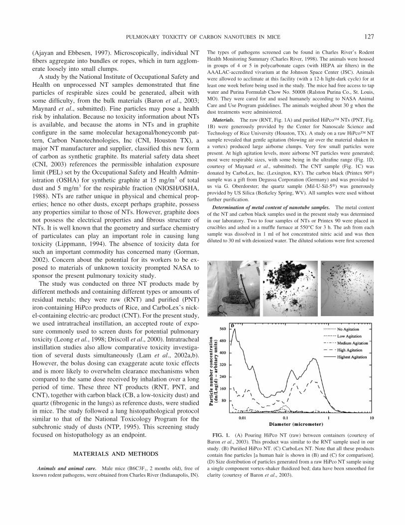
Materials The raw (RNT Fig 1A) and purified HiPcotrade NTs (PNT Fig1B) were generously provided by the Center for Nanoscale Science andTechnology of Rice University (Houston TX) A study on a raw HiPcotrade NTsample revealed that gentle agitation (blowing air over the material shaken ina vortex) produced large airborne clumps Very few small particles werepresent At high agitation levels more airborne NT particles were generatedmost were respirable sizes with some being in the ultrafine range (Fig 1Dcourtesy of Maynard et al submitted) The CNT sample (Fig 1C) wasdonated by CarboLex Inc (Lexington KY) The carbon black (Printex 90)sample was a gift from Degussa Corporation (Germany) and was provided tous via G Oberdorster the quartz sample (Mil-U-Sil-5) was generouslyprovided by US Silica (Berkeley Spring WV) All samples were used withoutfurther purification
Determination of metal content of nanotube samples The metal contentof the NT and carbon black samples used in the present study was determinedin our laboratory Two to four samples of NTs or Printex 90 were placed incrucibles and ashed in a muffle furnace at 550degC for 3 h The ash from eachsample was dissolved in 1 ml of hot concentrated nitric acid and was thendiluted to 30 ml with deionized water The diluted solutions were first screened
FIG 1 (A) Pouring HiPco NT (raw) between containers (courtesy ofBaron et al 2003) This product was similar to the RNT sample used in ourstudy (B) Purified HiPco NT (C) CarboLex NT Note that all these productscontain fine particles [a human hair is shown in (B) and (C) for comparison](D) Size distribution of particles generated from a raw HiPco NT sample usinga single component vortex-shaker fluidized bed data have been smoothed forclarity (courtesy of Baron et al 2003)
127PULMONARY TOXICITY OF CARBON NANOTUBES IN MICE
for the presence of metals using ICPMS (PE Sciex Elan 6000 Perkin ElmerNorwalk CT) Of the 70 elements scanned only Fe Ni Y Al Cu Mo Znand Co were found in measurable concentrations Quantitative analyses forthese 8 metals were then conducted with the same instrument Metals detectedat weight percentage 001 are shown in Table 1
Preparation of fine-dust suspensions NTs are neither water soluble norwettable and fine particle suspensions suitable for instillation must be preparedwith a nontoxic dispersion vehicle (Driscoll et al 2000 Leong et al 1998)The products are extremely difficult to disperse even in the presence of adispersing agent A group at Rice University used ldquoaggressive sonication ofpurified NT samples in surfactants such as Triton-X or highly polar solventslike dimethyl formamiderdquo to make fine-particle suspensions (10 mgl) contain-ing mostly individual fibers and a few small bundles (Walters et al 2001) Weprepared the NT suspensions [2 mgml (01 mg50 l) or 10 mgml (05 mg50l)] by briefly shearing (2 min in a small glass homogenizing tube) andsubsequently sonicating (05 min) NT samples in heat-inactivated mouseserum (Sigma St Louis MO) Brief sonication does not shorten or change thefundamental nature of NTs (Hauge personal communication) Serum inacti-vation was performed in a water bath heated to 56degC for 05 h Serum used byLeong et al (1998) and found in our pilot studies to be the best dispersingvehicle for NT was also used to suspend other test dusts Samples that werenot freshly generated on the day of dosing were resonicated before theinstillation Each sample was vortexed just before an aliquot was drawn forinstillation
Intratracheal instillation After being anesthetized with 3 to 5 isoflu-rane in a small chamber individual mice were secured on an inclined plasticplatform and anesthetization continued via a small nose cone The trachea wasexposed by a 1-cm incision on the ventral neck skin for instillation of the dustsuspension (Lam et al 2002a) The instillation procedures for mice reportedby these authors and the intratracheal fast instillationnebulization procedurefor rats used by Leong et al (1998) were modified to ensure that instilledmaterial was delivered into the lungs of mice with a good distribution Becauseit was difficult to eliminate air bubbles from a small NT-serum sample drawninto the syringe and confidently measure the intended volume the test samplecontaining 0 01 (low dose LD) or 05 mg (high dose HD) in a 50-l aliquotwas drawn (up to a premarked location) into a 30-cm fine silicone tubeconnected to the back metal end of a 24-gauge blunted needle A small holewas made in the trachea close to the larynx and a 24-gauge plastic catheter was
inserted through the hole to the distal end of the trachea the blunted needlewas then inserted inside the plastic catheter A 1-ml syringe prefilled with 150l of air and 20 l of saline was then connected to the free end of siliconetubing to rapidly propel the test sample from the tubing and needle into thelungs The neck incision was then sutured swabbed with Povidone iodine andanesthetized with a drop of lidocaine The mice recovered and were activewithin 10 min after removal from the inhalation anesthetic The incision healedwithin two days and the animals were observed daily until their scheduledtermination
Lung collection and histopathological examination Seven or 90 daysafter instillation of the test material each mouse was injected intraperitoneallywith a lethal dose (01 ml) of pentobarbital sodium solution (Nembutal AbbottNorth Chicago IL) Body weights were determined in the 90-d groups Anincision on the neck skin was made to expose the trachea for inserting acatheter formalin (10 in neutral phosphate buffer) was allowed to drip bygravity (from a 25-ml syringe barrel hanging 15 feet above the neck) throughthe catheter into the lung for about 10 min The trachea was then tied and theisolated lung was placed in a glass vial containing about 10 ml of the samefixative (Lam et al 2002a) For the 90-d groups each vial was assigned anumber with the treatment unknown to the pathologist The lungs were fixedfor at least 7 days before further processing The formalin-fixed mouse lungswere embedded in paraffin thin-sectioned coronally and mounted on glassmicroscope slides using standard histopathological techniques Sections werestained with hematoxylin-eosin and examined by light microscopy
RESULTS
Effects of Carbon Nanotubes
All animals treated with 01 mg per mouse (low dose LD)of CNT (containing Ni and Y) showed no overt clinical signsHowever 5 of the 9 mice treated with 05 mg (high dose HD)of this product died (24 in the 7-d group and 35 in the 90-dgroup) All deaths occurred 4 to 7 days after instillation of theCNT The deaths were generally preceded by lethargy inac-tivity and body-weight losses These symptoms were also seen
TABLE 1Metal Content of Test Samples and Experimental Design of the Intratracheal Instillation Study in Mice
Test materials
Metal contents ( by Wt)Dust dose
(mgmouse)
Number of mice
Fe Ni Y Others 7 d 90 d
Raw nanotubes 269 078 000 Cu 036 Mo 095 LD 01 4 5(RNT) Rice product Zn 001 HD 05 4 5
Purified nanotubes 214 000 000 None LD 01 4 5(PNT) Rice product HD 05 4 5
CarboLex nanotubes 053 2599 501 Al 015 Zn 015 LD 01 4 5(CNT) Co 002 HD 05 4 5
Carbon black 000 000 000 None LD 01 4 5(Printex-90) HD 05 4 5
Quartz ND ND ND ND LD 01 4 5(Min-U-Sil-5) HD 05 4 5
Vehicle control ND ND ND ND mdash 00 4 5(mouse serum)
Note Mice (4 or 5 per group) were each intratracheally instilled once with 0 01 or 05 mg dust in mouse serum and euthanized after 7 or 90 days forhistopathology study
Note ND Not determinedMetals with concentrations 001 by weight are not shown
128 LAM ET AL
in the HD mice that survived Mice in the HD 90-d CNT group(including those that died within the first week) lost about 27of their body weight (pretreatment 309 11 g posttreat-ment 225 09 g) by the first week Symptoms in the twosurviving mice disappeared after one week and the animalsstarted to gain weight
The iron-containing NTs (RNT and PNT) did not causedeaths in the mice Mild signs of inactivity hypothermia (feltcold to the touch) piloerection and occasionally shivering(when provoked by picking up and laying down) were mostnoticeable 8 to 12 h after treatment with the HD RNT thesesymptoms disappeared soon after this time These clinicalsigns were not observed in the mice treated with PNT Bodyweight losses seen in the first week with the HD CNT werenot observed with RNT or PNT
The distribution of black particles in lungs 90 d after theywere instilled with 05 mg of CB or NTs is illustrated in Figure2 Some of the lungs had a relatively uniform particle distri-bution while others did not At the microscopic level the lungsof dead animals of the HD CNT groups showed congestion andpostmortem histopathological changes The lungs of the 4 (2from the 7-d and 2 from the 90-d group) surviving mice treatedwith HD CNT had large aggregates of particles in macrophagesin the alveolar space some of these aggregates were also foundin the interstitium forming granulomas (Figs 3D and 4C)Some interstitial inflammation was apparent Granulomas werenot detected in the LD groups (7 d and 90 d) given CNT Thelungs of mice in the HD 7-d study that were treated with eitherRNT or PNT showed prominent granulomas (Figs 3E and 3F)
Most of these microscopic nodules were located beneath thebronchial epithelium and were present throughout most of thelung fields Some appeared to extend into the bronchi aspolyps The granulomas consisted of macrophages laden withblack particles and had very few lymphocytes neutrophilseosinophils or other inflammatory cells The macrophages hadabundant granular cytoplasm with indistinct borders a charac-teristic of activated macrophages or epitheloid cells The blackparticles were almost entirely contained within these granulo-mas Some of the lungs from HD 90-d NT-treated groupsappeared grossly abnormal (Figs 2C and 2F) The micrographsof lungs from these groups showed the persistence of granu-lomas that contained particle-laden macrophages and NT par-ticles (Figs 4CndashE) The lung lesions were generally morepronounced than those of the HD 7-d groups (Figs 3DndashF)some also had necrosis interstitial inflammation that had ex-tended into the alveolar septa and peribronchial inflammation(Figs 4E and 4F) Granulomas and other pulmonary lesionswere also seen in some of the LD HiPco-NT-treated mice(Table 2) but to a mild degree
Effects of Serum Carbon Black and Quartz
As expected heat-inactivated mouse serum did not produceany clinical signs or gross and microscopic lesions (Figs 2Aand 3A) The mice of the negative (CB) or positive (quartz)control groups also did not show any clinical signs that couldbe attributed to treatment Aside from the presence of blackparticles that appeared predominantly in alveoli the lungs of
FIG 2 Lungs from mice instilled with 05 mg of a test material per mouse and euthanized 90 d after the single treatment (A) Serum control (B) Carbonblack (Printex) (C) CNT The portions of the lung receiving NT had an abnormal appearance (D) PNT Particle distribution was even (E) RNT Clusters ofblack pigment probably correspond to granulomas (F) RNT Dorsal view shows some necrotic changes
129PULMONARY TOXICITY OF CARBON NANOTUBES IN MICE
FIG 3 Lung tissue from mice instilled with 05 mg of a test material per mouse and euthanized 7 d after the single treatment (A) Serum control (B) Carbonblack Particles and particles contained in macrophages were scattered in the alveoli (C) Silica quartz A low-grade granuloma (D) CNT Particles werepredominately inside the granulomas and none were in the area of normal alveolar tissue on the right side (E) RNT Black particles were predominately insidegranulomas (F) PNT Close-up view of granulomas Magnifications varied from 40 to 200
FIG 4 Lung tissue from mice instilled with 05 mg of a test material per mouse and euthanized 90 d after the single treatment (A) Carbon black Particleswere scattered in alveoli (B) Quartz 3 shows an aggregate of inflammation cells (lymphocytes) around an area surrounded by quartz particle-containingmacrophages (C) CNT Granulomas contained black particles (D) RNT Granulomas at low magnification (E) RNT A granuloma at a high magnification (F)PNT A large granuloma underwent degeneration with necrosis Magnifications varied from 40 to 200
130 LAM ET AL
the mice in CB groups were microscopically normal (Figs 3Band 4A) The lungs of the LD quartz groups were also normalQuartz at HD induced an increase in the number of alveolarmacrophages in the lungs and some of these cells containedparticles Quartz also produced mild to moderate alveolar andinterstitial inflammation The results for the 7-d and 90-dgroups were generally similar One of the mice in the 7-d grouphad a low-grade granulomatous reaction (Fig 3C) The HD90-d group had increased clusters of peribronchiolar lympho-cytes (Fig 4B)
DISCUSSION
The present study shows that all three NT products regard-less of the type and amount of metal impurities they containedinduced dose-dependent lung lesions characterized chiefly byinterstitial granulomas Our finding that the purified NTs(PNTs) which were prepared by rigorous treatment (45-hreflux) with concentrated acids (2 M to 3 M nitric acid) toremove metal impurities (Rinzler et al 1998) and containedonly a small amount (2 by weight) of residual iron producedprominent granulomas along with the fact that insoluble ironand iron compounds are low in toxicity and have not beenshown to produce these lung lesions (Warheit et al 1991)strongly indicates that NTs themselves induced the granulo-mas NT-induced granulomas were also observed in a similarstudy in which rats were intratracheally instilled with a laser-produced NT product sonicated and suspended in Tween 80(Warheit 2003) The NT dosage of 5 mgkg in Warheitrsquos ratstudy was comparable to that given to our low-dose (01 mg30g mouse or 33 mgkg) groups of mice At this dose we sawgranulomas in the mice treated with the HiPco products (RNTand PNT) but not with the CNT
We observed mortality (59) in animals treated with the highdose of CNT suggesting that any subsequent intratrachealstudies with CNT should not involve doses this high The CNTproduct contains substantial amounts of nickel and yttriumNickel and its compounds are highly toxic Benson et al
(1987) reported that all 10 mice exposed to 10 mgm3 Ni3S2
died before the end of the 12-day study The lung burden ofnickel in this group was not available but mice exposed to 25mgm3 of the nickel compound for 12 days (6 hd) accumulated4 glung Assuming that the lung burden of this insolublenickel compound was proportional to exposure concentrationsthe amount of nickel in the lungs of animals that died would be16 mglung In our study treatment with a CNT dose of 05mgmouse would load 130 g of nickel and 25 g of yttriuminto the lung We could not rule out the possibility that some ofthe nickel and yttrium surrounded or coated by NTs was freedby ultrasonication and subsequently contributed to the acutelethality If sonication-freed nickel contributed to the acutetoxicity then deaths observed in bolus-dosed animals shouldnot occur in inhalation-exposed animals Further studies wouldbe needed to elucidate the cause of death with certainty
Although both types of carbon particles (CB and NTs) weretaken up by alveolar macrophages their fate and reactions inthe lung tissue were very different CB-laden macrophagesscattered in the alveolar space but NT-laden macrophagesmoved rapidly to centrilobular locations where they enteredalveolar septa and clustered to form epithelioid granulomas Itis well known that if the lung is not dust-overloaded dust-laden macrophages on the alveolar surface will migrate upwardand be carried by the escalatormucociliary system up thetrachea and cleared into the esophagus However when dustsenter the interstitial or subepithelial space they are very diffi-cult to clear from the lung Thus if a biopersistent dust isirritating or toxic the lesions resulting from the persistentinteraction between the cells and the dust trapped in the inter-stitium will generally worsen with time as is the case withNTs As seen in the present study the lung lesions of HD 90-dgroups were generally more pronounced than those of the HD7-d groups in animals treated with NTs These findings indicatethat NTs and CB have different intrinsic toxicities in the lungs
The findings that all four NT products (three in our study andone in Warheitrsquos study) were capable of inducing granulomasin mice or rats together with the findings (in our studies andothers) that granulomas were not observed in rodents exposedto carbon black point to the fundamental difference betweenthe unique physicochemical properties of NTs and those of CBIn addition to its unique surface chemistry (NTs are an excel-lent electrical conductor while carbon black is not) the fibrousstructure of NT products must be considered Microscopicallythin NT fibers pack tightly and in parallel to form ropes or rods(ISI 2002 Unrau 1996) As defined in Comprehensive Toxi-cology ldquoFibers are a special class of particles defined aselongated objects whose aspect ratio the ratio of the objectrsquoslength to its diameter is greater than threerdquo (McClellan 1997)Therefore toxicologically individual NT molecules and as-sembled NT ropes rods and bundles are fibers Some of theNT particles were clearly seen in the lungs as fiber or ropestructures (Figs 5AndashC) This physical structure would makeNTs toxicologically different from carbon black NTs are to-
TABLE 2Incidence of Pulmonary Lesions in Mice 90 Days After
Intratracheal Instillation with Nanotubes
Dust dose(mg)
Type oflung lesion
Carbonblack Quartz RNT PNT CNT
01 Inflammation 0 1 3 2 001 Granulomas 0 0 5 2 005 Inflammation 0 4 3 5 005 Granulomas 0 0 5 5 5
Mice (5group) were each instilled with 01 or 05 mg of a test material andeuthanized 90 d after the single treatment Lungs were microscopically exam-ined by a pathologist who had no knowledge of the treatment of each animal
Including 3 mice that died in the first week
131PULMONARY TOXICITY OF CARBON NANOTUBES IN MICE
tally insoluble and probably one of the most biologically non-degradable man-made materials It is well established that thepathogenicity of a fiber in the lungs directly correlates with itsbiopersistency (Oberdorster 2000) Granulomas could impaircellular and physiological (gas exchange) lung functions andgive rise to fibrosis more defined nodules and other lesionsDetermining how the NT-induced granulomas progress wouldrequire a longer-duration study with this biopersistent material
Because some NTs are derived from graphite and maycontain graphite as an impurity and toxicological data indicatethat graphite may induce granulomas the toxicity of graphitewarrants a discussion Graphite pneumoconiosis which ischaracterized by granulomas interstitial fibrosis perifocal em-physema necrosis and severe vascular sclerosis is a lungdisease that has long been recognized in a large number ofworkers involved in mining and processing graphite (Gaensleret al 1966 Hanoa 1983 Jaffe 1951 NRC 1999) A study(Okutani et al 1964) showing 112 cases of pneumoconiosisamong 256 workers who had been exposed to an average of 60mgm3 of graphite that contained little (01) quartz (equiv-alent to 01 mgm3 in the air) and another study (Aranyi etal 1992) revealing graphite-containing granulomas in lunglymphoid tissue in rats exposed to pure synthetic graphite at100 mgm3 for 13 weeks strongly suggest that pure graphite athigh concentrations and prolonged exposures could also inducegranulomas However the granulomas observed in the presentstudy were not caused by graphite as an impurity CNTs whichwere made from graphite and may contain more graphiteimpurities than the CO-derived HiPco-NTs (Bronikowski etal 2001) actually were less potent in producing granulomasThe RNT and PNT samples which contained more carbonnanotubes than the CNT product on an equal-weight basisproduced granulomas in some animals of the LD groupswhereas no animals in the LD CNT group had granulomas Inthe HD groups HiPco NTs produced more prominent granu-lomas than did the CNT These data argue against the possi-bility that a graphite impurity induced granulomas in our study
The comparative toxicity of NTs and quartz which was usedas a positive control for the present study also warrants adiscussion Granulomas along with other lung lesions have
long been observed in animals chronically exposed to quartz Agranulomatous reaction in the lungs was observed in miceexposed to 15 to 21 mgm3 for 150 or 300 d (Wilson et al1986) in rats exposed to 25 mgm3 for 100 d (Eden and vonSeebach 1976) and in rats intratracheally instilled once with50 mg quartz and examined 1 to 12 months after treatment(Reiser et al 1982) In our study in which mice were exposedto relatively less quartz than the animals in the above studiesone mouse had low-grade granulomas the rest of the HDgroups had only mild to moderate inflammation in the lungsAt the doses used in the present study no fibrosis was observedin the lung
One may ask the relationship of these intratracheal-instilla-tion NT doses that produced lung lesions to an inhalationexposure NT particles are neither soluble nor biodegradableand particles in the interstitium could not be removed from thelung by the macrophage-mucociliary clearance mechanismTherefore the lung burden from an intratracheal dose could beused to roughly estimate a burden that could be achieved byinhalation exposures (Lam et al 2002a) Using data from theInternational Commission of Respiratory Protection TaskGroup showing that the fractional deposition of particles deepinto the lung is about 30 for 3-m particles and increases to55 for 005-m particles (Bates et al 1966) we can estimatethe pulmonary deposition of respirable dust If we assume that40 of the inhaled respirable NT particles deposit in thepulmonary region and that a 30-g mouse breathes in 30 ml ofair per min (Parent 1992) then a mouse breathing respirableNT dust at 5 mgm3 for 8 h daily would accumulate 0029 mgNTday Then a lung burden equivalent to that instilled with01 mg or 05 mg could be attained by a mouse inhaling arespirable NT aerosol concentration for about 35 or 17 daysrespectively The exposure concentration of 5 mgm3 is thesame concentration as the PEL that OSHA set for respirablesynthetic graphite dust and is the same exposure concentrationreferenced by a major US nanotube manufacturer and supplier(CNI 2003) OSHArsquos PELs which are time-weighted concen-trations (40 hweek) are set for lifetime occupational expo-sures It can be inferred from the exposure-risk estimationinformation presented here that if workers were chronically
FIG 5 Lung tissues from mice instilled with 05 mg of NT per mouse and euthanized 90 d after the single treatment showing presence of NT fibers (A)RNT NT fibers in a granuloma (B) PNT NT fibers in a granulomas (C) PNT Clumps of NT fibers in a granuloma (Magnification 900)
132 LAM ET AL
exposed to respirable NT dust at a fraction of the PEL con-centration for synthetic graphite they would likely developserious lung lesions Therefore the PEL for synthetic graphitemust not be used to protect workers exposed to NTs Thecurrent TLV for graphite (2 mgm3) explicitly excludesgraphite fibers (ACGIH 2001) Our study provides dataneeded to guide additional toxicity studies and to support thedesign of industrial hygiene procedures (Baron et al 2003)
In conclusion this study shows for the test conditionsdescribed here and on an equal-weight basis that if NTs reachthe lungs they are much more toxic than carbon black and canbe more toxic than quartz which is considered a seriousoccupational health hazard in chronic inhalation exposures Iffine NT dusts are present in a work environment exposureprotection strategies to minimize human exposures should beimplemented
ACKNOWLEDGMENTS
The authors thank the following for their contributions and advice A LeeT Blasdel B Leong G Oberdorster A Holian J Nelle S Bassett M KuoJ Read H Garcia J Krauhs M Nelman and staff of the Center forNanoscale Science and Technology (CNST) and Center for Biological andEnvironmental Nanotechnology at Rice University The generous gifts ofcarbon nanotubes samples from Rice CNST and from CarboLex Inc Printex90 from Degussa Corp and Mil-U-Sil-5 from US Silica are gratefully ac-knowledged We are indebted to A Maynard of NIOSH for providing twofigures for this report This study was supported by a NASA grant
REFERENCES
ACGIH (2001) Documentation of the Threshold Limit Values and BiologicalExposure Indices American Conference of Governmental Industrial Hy-gienists Cincinnati OH
Ajayan P M Charlier J and Rinzler A G (1999) Carbon nanotubes frommacromolecules to nanotechnology Proc Natl Acad Sci 96 14199ndash14200
Ajayan P M and Ebbesen T W (1997) Nanometre-size tubes of carbonRep Prog Phys 60 1025ndash1062
Arepalli S Nikolaev P Holmes W and Files B S (2001) Production andmeasurements of individual single-wall nanotubes and small ropes of car-bon Appl Phys Lett 78 1610ndash1612
Aranyi C Rajendran N and Bradof J (1992) Thirteen-week inhalationtoxicity study with aerosol mixtures of fog oil and graphite particles inF344N male rats Prepared by IIT Research Institute Chicago for USArmy Medical Research and Development Command Fort Detrick MD
Ball P (1999) Focus carbon nanotubes Nature Scienceupdate httpwwwnaturecomnsu991202991202-1html
Ball P (2001) Roll-up for the revolution Nature 414 142ndash144 httpwwwphyswashingtoneducobdenP600Philip_Ball_tube_newspdf
Baron P A Maynard A D and Foley M (2003) Evaluation of aerosolrelease during the handling of unrefined single walled carbon nanotubematerial NIOSH DART-02-191 National Institute of Occupational Safetyand Health Cincinnati OH NTIS PB2003ndash102401
Bates D V Fish B R Hatch T F Mercer T T and Morrow P E (1966)Deposition and retention models for internal dosimetry of the human respi-ratory tract Health Phys 12 173ndash207
Benson J M Carpenter R L Hahn F F Haley P J Hanson R L
Hobbs C H Pickrell J A and Dunnick J K (1987) Comparativeinhalation toxicity of nickel subsulfide to F344N rats and B6C3F1 miceexposed for 12 days Fundam Appl Toxicol 9 251ndash265
Bronikowski M J Willis P A Colbert D T Smith K A and SmalleyR E (2001) Gas-phase production of carbon single-walled nanotubes fromcarbon monoxide via the HiPco process A parametric study J Vac SciTechnol A 19 1800ndash1805
CNI (2003) Buckytube MSDS Carbon Nanotechnologies Incorporated httpwwwcnanotechcomdownload_filesMSDS20for20CNI20SWNTpdf httpwwwcnanotechcompagesbuckytube_properties_usesMSDS5-3_MSDShtml
Charles River (1998) Rodent health monitoring summary Charles RiverLaboratories Wilmington MA httpwwwcrivercomtechdocshealth-mon-sumhtml
Driscoll K E Costa D L Hatch G Henderson R Oberdorster G SalemH and Schlesinger R B (2000) Intratracheal instillation as an exposuretechnique for the evaluation of respiratory tract toxicity Uses and limita-tions Toxicol Sci 55 24ndash35
Eden K G and von Seebach H B (1976) Atypical quartz dust-inducedpneumoconiosis in SPF rats Aspects of the role of the lymphatic system inthe pathogenesis of silicosis Virchows Arch A Pathol Anat Histol 3721ndash9
Gaensler E A Cadigan J B Sasahara A A Fox E O and MacMahonH E (1966) Graphite pneumoconiosis of electrotypers Am J Med 41864ndash882
Gorman J (2002) Taming high-tech particles Cautious steps into the nano-tech future Science News 161 200 httpwwwsciencenewsorg20020330bob8asp
Hanoa R (1983) Graphite pneumoconiosis A review of etiological andepidemiologic aspects Scand J Work Environ Health 9 303ndash314
ISI (2002) Nanotechnology An interview with Dr Richard Smalley ISIEssential Science Indicators Special Topics March 2002 httpwwwesi-topicscomnanointerviewsRichard-Smalleyhtml
Jaffe F A (1951) Graphite pneumoconiosis Am J Pathol 17 909ndash923
Lam C-W James J T Latch J N Hamilton R F and Holian A (2002a)Pulmonary toxicity of simulated lunar and Martian dusts in mice II Bi-omarkers of acute responses after intratracheal instillation Inhal Toxicol14 917ndash928
Lam C-W James J T McCluskey R Cowper S and Muro-Cacho C(2002b) Pulmonary toxicity of simulated lunar and Martian dusts in miceI Histopathology 7 and 90 days after intratracheal instillation Inhal Toxi-col 14 901ndash916
Leong B K Coombs J K Sabaitis C P Rop D A and Aaron C S(1998) Quantitative morphometric analysis of pulmonary deposition ofaerosol particles inhaled via intratracheal nebulization intratracheal instil-lation or nose-only inhalation in rats J Appl Toxicol 18 149ndash160
Lippmann M (1994) Nature of exposure to chrysotile Ann Occup Hyg38(4) 459ndash467
Maynard A D Baron P A Foley M Shvedova A A Kisin E R andCastranova V (2003) Exposure to carbon nanotube material I Aerosolrelease during the handling of unrefined single walled carbon nanotubematerial J Toxicol Environ Health (submitted)
McClellan R (1997) Nuisance dusts In Toxicology of the Respiratory System(I G Sipes C A McQueen and A J Gandolfi Eds) Vol 8 ElsevierScience New York
NIOSHOSHA (1988) Synthetic graphite National Institute for OccupationalSafety and Health httpwwwcdcgovnioshpel88npelnamehtml httpwwwcdcgovnioshpel88SYNGRAPHhtml
NRC (1999) Toxicity of Military Smokes and Obscurants National ResearchCouncil Subcommittee on Military Smokes and Obscurants National Acad-
133PULMONARY TOXICITY OF CARBON NANOTUBES IN MICE
emies Press Washington DC httpbooksnapedubooks0309063299html97html
NTP (1995) NTP Technical Report on Toxicity Studies of Cadmium Oxide(CAS No 1306ndash19ndash0) Administered by Inhalation to F344N Rats andB6C3F1 Mice Toxicity Report Series No 39 National Toxicology Pro-gram National Institutes of Health Research Triangle Park NC
Oberdorster G (2000) Determinants of the pathogenicity of man-made vit-reous fibers (MMVF) Int Arch Occup Environ Health 73 (Suppl)S60ndashS68
Okutani H Shima S and Sano T (1964) Graphite pneumoconiosis incarbon electrode makers In Proceedings of the XIV International Congressof Occupational Health 1963 Madrid Spain pp 626ndash632 Excerpta Med-ica Foundation Amsterdam the Netherlands
Parent R A (1992) Comparative Biology of the Normal Lung CRC PressBoca Raton FL
Reiser K M Hesterberg T W Haschek W M and Last J A (1982)Experimental silicosis I Acute effects of intratracheally instilled quartz oncollagen metabolism and morphologic characteristics of rat lungs Am JPathol 107 176ndash185
Rinzler A G Liu J Dai H Nikolaev P Huffman C B Rodriguez-Macias F J Boul P J Lu A H Heymann D Colbert D T et al
(1998) Large-scale purification of single-wall carbon nanotubes Processproduct and characterization Appl Phys A 67 29ndash37
Unrau L (1996) Associates carry research forward in form of nanotube RiceNews 6 httpwwwriceeduprojectsrenorn19961017Carryhtml pub-lished online October 17 1996
Walters D A Casavant M J Qin X C Huffman C B Boul P JEricson L M Haroz E H OrsquoConnell M J Smith K Colbert D T etal (2001) In-plane-aligned membranes of carbon nanotubes Chem PhysLett 338 14ndash20
Warheit D B Hansen J F and Hartsky M A (1991) Physiological andpathophysiological pulmonary responses to inhaled nuisance-like or fibro-genic dusts Anat Rec 231 107ndash118
Warheit D B Laurence B R Reed K L Roach D H ReynoldsG A M and Webb T R Comparative pulmonary toxicity assessment ofsingle-wall carbon nanotubes in rats Toxicol Sci 76 117ndash125
White House (2000) National Nanotechnology Initiative Leading to thenext industrial revolution The White House Office of the Press Secre-tary Washington DC httpclinton4naragovtextonlyWHNewhtml20000121_4html
Wilson T Scheuchenzuber W J Eskew M L and Zarkower A (1986)Comparative pathological aspects of chronic olivine and silica inhalation inmice Environ Res 39 331ndash344
134 LAM ET AL
(Ajayan and Ebbesen 1997) Microscopically individual NTfibers aggregate into bundles or ropes which in turn agglom-erate loosely into small clumps
A study by the National Institute of Occupational Safety andHealth on unprocessed NT samples demonstrated that fineparticles of respirable sizes could be generated albeit withsome difficulty from the bulk materials (Baron et al 2003Maynard et al submitted) Fine particles may pose a healthrisk by inhalation Because no toxicity information about NTsis available and because the atoms in NTs and in graphiteconfigure in the same molecular hexagonalhoneycomb pat-tern Carbon Nanotechnologies Inc (CNI Houston TX) amajor NT manufacturer and supplier classified this new formof carbon as synthetic graphite Its material safety data sheet(CNI 2003) references the permissible inhalation exposurelimit (PEL) set by the Occupational Safety and Health Admin-istration (OSHA) for synthetic graphite at 15 mgm3 of totaldust and 5 mgm3 for the respirable fraction (NIOSHOSHA1988) NTs are rather unique in physical and chemical prop-erties hence no other dusts except perhaps graphite possessany properties similar to those of NTs However graphite doesnot possess the electrical properties and fibrous structure ofNTs It is well known that the geometry and surface chemistryof particulates can play an important role in causing lungtoxicity (Lippmann 1994) The absence of toxicity data forsuch an important commodity has concerned many (Gorman2002) Concern about the potential for its workers to be ex-posed to materials of unknown toxicity prompted NASA tosponsor the present pulmonary toxicity study
The study was conducted on three NT products made bydifferent methods and containing different types or amounts ofresidual metals they were raw (RNT) and purified (PNT)iron-containing HiPco products of Rice and CarboLexrsquos nick-el-containing electric-arc product (CNT) For the present studywe used intratracheal instillation an accepted route of expo-sure commonly used to screen dusts for potential pulmonarytoxicity (Leong et al 1998 Driscoll et al 2000) Intratrachealinstillation studies also allow comparative toxicity investiga-tion of several dusts simultaneously (Lam et al 2002ab)However the bolus dosing can exaggerate acute toxic effectsand is more likely to overwhelm clearance mechanisms whencompared to the same dose received by inhalation over a longperiod of time These three NT products (RNT PNT andCNT) together with carbon black (CB a low-toxicity dust) andquartz (fibrogenic in the lungs) as reference dusts were studiedin mice The study followed a lung histopathological protocolsimilar to that of the National Toxicology Program for thesubchronic study of dusts (NTP 1995) This screening studyfocused on histopathology as an endpoint
MATERIALS AND METHODS
Animals and animal care Male mice (B6C3F1 2 months old) free ofknown rodent pathogens were obtained from Charles River (Indianapolis IN)
The types of pathogens screened can be found in Charles Riverrsquos RodentHealth Monitoring Summary (Charles River 1998) The animals were housedin groups of 4 or 5 in polycarbonate cages (with HEPA air filters) in theAAALAC-accredited vivarium at the Johnson Space Center (JSC) Animalswere allowed to acclimate at this facility (with a 12-h light-dark cycle) for atleast one week before being used in the study The mice had free access to tapwater and Purina Formulab Chow No 50008 (Ralston Purina Co St LouisMO) They were cared for and used humanely according to NASA AnimalCare and Use Program guidelines The animals weighed about 30 g when thedust treatments were administered
Materials The raw (RNT Fig 1A) and purified HiPcotrade NTs (PNT Fig1B) were generously provided by the Center for Nanoscale Science andTechnology of Rice University (Houston TX) A study on a raw HiPcotrade NTsample revealed that gentle agitation (blowing air over the material shaken ina vortex) produced large airborne clumps Very few small particles werepresent At high agitation levels more airborne NT particles were generatedmost were respirable sizes with some being in the ultrafine range (Fig 1Dcourtesy of Maynard et al submitted) The CNT sample (Fig 1C) wasdonated by CarboLex Inc (Lexington KY) The carbon black (Printex 90)sample was a gift from Degussa Corporation (Germany) and was provided tous via G Oberdorster the quartz sample (Mil-U-Sil-5) was generouslyprovided by US Silica (Berkeley Spring WV) All samples were used withoutfurther purification
Determination of metal content of nanotube samples The metal contentof the NT and carbon black samples used in the present study was determinedin our laboratory Two to four samples of NTs or Printex 90 were placed incrucibles and ashed in a muffle furnace at 550degC for 3 h The ash from eachsample was dissolved in 1 ml of hot concentrated nitric acid and was thendiluted to 30 ml with deionized water The diluted solutions were first screened
FIG 1 (A) Pouring HiPco NT (raw) between containers (courtesy ofBaron et al 2003) This product was similar to the RNT sample used in ourstudy (B) Purified HiPco NT (C) CarboLex NT Note that all these productscontain fine particles [a human hair is shown in (B) and (C) for comparison](D) Size distribution of particles generated from a raw HiPco NT sample usinga single component vortex-shaker fluidized bed data have been smoothed forclarity (courtesy of Baron et al 2003)
127PULMONARY TOXICITY OF CARBON NANOTUBES IN MICE
for the presence of metals using ICPMS (PE Sciex Elan 6000 Perkin ElmerNorwalk CT) Of the 70 elements scanned only Fe Ni Y Al Cu Mo Znand Co were found in measurable concentrations Quantitative analyses forthese 8 metals were then conducted with the same instrument Metals detectedat weight percentage 001 are shown in Table 1
Preparation of fine-dust suspensions NTs are neither water soluble norwettable and fine particle suspensions suitable for instillation must be preparedwith a nontoxic dispersion vehicle (Driscoll et al 2000 Leong et al 1998)The products are extremely difficult to disperse even in the presence of adispersing agent A group at Rice University used ldquoaggressive sonication ofpurified NT samples in surfactants such as Triton-X or highly polar solventslike dimethyl formamiderdquo to make fine-particle suspensions (10 mgl) contain-ing mostly individual fibers and a few small bundles (Walters et al 2001) Weprepared the NT suspensions [2 mgml (01 mg50 l) or 10 mgml (05 mg50l)] by briefly shearing (2 min in a small glass homogenizing tube) andsubsequently sonicating (05 min) NT samples in heat-inactivated mouseserum (Sigma St Louis MO) Brief sonication does not shorten or change thefundamental nature of NTs (Hauge personal communication) Serum inacti-vation was performed in a water bath heated to 56degC for 05 h Serum used byLeong et al (1998) and found in our pilot studies to be the best dispersingvehicle for NT was also used to suspend other test dusts Samples that werenot freshly generated on the day of dosing were resonicated before theinstillation Each sample was vortexed just before an aliquot was drawn forinstillation
Intratracheal instillation After being anesthetized with 3 to 5 isoflu-rane in a small chamber individual mice were secured on an inclined plasticplatform and anesthetization continued via a small nose cone The trachea wasexposed by a 1-cm incision on the ventral neck skin for instillation of the dustsuspension (Lam et al 2002a) The instillation procedures for mice reportedby these authors and the intratracheal fast instillationnebulization procedurefor rats used by Leong et al (1998) were modified to ensure that instilledmaterial was delivered into the lungs of mice with a good distribution Becauseit was difficult to eliminate air bubbles from a small NT-serum sample drawninto the syringe and confidently measure the intended volume the test samplecontaining 0 01 (low dose LD) or 05 mg (high dose HD) in a 50-l aliquotwas drawn (up to a premarked location) into a 30-cm fine silicone tubeconnected to the back metal end of a 24-gauge blunted needle A small holewas made in the trachea close to the larynx and a 24-gauge plastic catheter was
inserted through the hole to the distal end of the trachea the blunted needlewas then inserted inside the plastic catheter A 1-ml syringe prefilled with 150l of air and 20 l of saline was then connected to the free end of siliconetubing to rapidly propel the test sample from the tubing and needle into thelungs The neck incision was then sutured swabbed with Povidone iodine andanesthetized with a drop of lidocaine The mice recovered and were activewithin 10 min after removal from the inhalation anesthetic The incision healedwithin two days and the animals were observed daily until their scheduledtermination
Lung collection and histopathological examination Seven or 90 daysafter instillation of the test material each mouse was injected intraperitoneallywith a lethal dose (01 ml) of pentobarbital sodium solution (Nembutal AbbottNorth Chicago IL) Body weights were determined in the 90-d groups Anincision on the neck skin was made to expose the trachea for inserting acatheter formalin (10 in neutral phosphate buffer) was allowed to drip bygravity (from a 25-ml syringe barrel hanging 15 feet above the neck) throughthe catheter into the lung for about 10 min The trachea was then tied and theisolated lung was placed in a glass vial containing about 10 ml of the samefixative (Lam et al 2002a) For the 90-d groups each vial was assigned anumber with the treatment unknown to the pathologist The lungs were fixedfor at least 7 days before further processing The formalin-fixed mouse lungswere embedded in paraffin thin-sectioned coronally and mounted on glassmicroscope slides using standard histopathological techniques Sections werestained with hematoxylin-eosin and examined by light microscopy
RESULTS
Effects of Carbon Nanotubes
All animals treated with 01 mg per mouse (low dose LD)of CNT (containing Ni and Y) showed no overt clinical signsHowever 5 of the 9 mice treated with 05 mg (high dose HD)of this product died (24 in the 7-d group and 35 in the 90-dgroup) All deaths occurred 4 to 7 days after instillation of theCNT The deaths were generally preceded by lethargy inac-tivity and body-weight losses These symptoms were also seen
TABLE 1Metal Content of Test Samples and Experimental Design of the Intratracheal Instillation Study in Mice
Test materials
Metal contents ( by Wt)Dust dose
(mgmouse)
Number of mice
Fe Ni Y Others 7 d 90 d
Raw nanotubes 269 078 000 Cu 036 Mo 095 LD 01 4 5(RNT) Rice product Zn 001 HD 05 4 5
Purified nanotubes 214 000 000 None LD 01 4 5(PNT) Rice product HD 05 4 5
CarboLex nanotubes 053 2599 501 Al 015 Zn 015 LD 01 4 5(CNT) Co 002 HD 05 4 5
Carbon black 000 000 000 None LD 01 4 5(Printex-90) HD 05 4 5
Quartz ND ND ND ND LD 01 4 5(Min-U-Sil-5) HD 05 4 5
Vehicle control ND ND ND ND mdash 00 4 5(mouse serum)
Note Mice (4 or 5 per group) were each intratracheally instilled once with 0 01 or 05 mg dust in mouse serum and euthanized after 7 or 90 days forhistopathology study
Note ND Not determinedMetals with concentrations 001 by weight are not shown
128 LAM ET AL
in the HD mice that survived Mice in the HD 90-d CNT group(including those that died within the first week) lost about 27of their body weight (pretreatment 309 11 g posttreat-ment 225 09 g) by the first week Symptoms in the twosurviving mice disappeared after one week and the animalsstarted to gain weight
The iron-containing NTs (RNT and PNT) did not causedeaths in the mice Mild signs of inactivity hypothermia (feltcold to the touch) piloerection and occasionally shivering(when provoked by picking up and laying down) were mostnoticeable 8 to 12 h after treatment with the HD RNT thesesymptoms disappeared soon after this time These clinicalsigns were not observed in the mice treated with PNT Bodyweight losses seen in the first week with the HD CNT werenot observed with RNT or PNT
The distribution of black particles in lungs 90 d after theywere instilled with 05 mg of CB or NTs is illustrated in Figure2 Some of the lungs had a relatively uniform particle distri-bution while others did not At the microscopic level the lungsof dead animals of the HD CNT groups showed congestion andpostmortem histopathological changes The lungs of the 4 (2from the 7-d and 2 from the 90-d group) surviving mice treatedwith HD CNT had large aggregates of particles in macrophagesin the alveolar space some of these aggregates were also foundin the interstitium forming granulomas (Figs 3D and 4C)Some interstitial inflammation was apparent Granulomas werenot detected in the LD groups (7 d and 90 d) given CNT Thelungs of mice in the HD 7-d study that were treated with eitherRNT or PNT showed prominent granulomas (Figs 3E and 3F)
Most of these microscopic nodules were located beneath thebronchial epithelium and were present throughout most of thelung fields Some appeared to extend into the bronchi aspolyps The granulomas consisted of macrophages laden withblack particles and had very few lymphocytes neutrophilseosinophils or other inflammatory cells The macrophages hadabundant granular cytoplasm with indistinct borders a charac-teristic of activated macrophages or epitheloid cells The blackparticles were almost entirely contained within these granulo-mas Some of the lungs from HD 90-d NT-treated groupsappeared grossly abnormal (Figs 2C and 2F) The micrographsof lungs from these groups showed the persistence of granu-lomas that contained particle-laden macrophages and NT par-ticles (Figs 4CndashE) The lung lesions were generally morepronounced than those of the HD 7-d groups (Figs 3DndashF)some also had necrosis interstitial inflammation that had ex-tended into the alveolar septa and peribronchial inflammation(Figs 4E and 4F) Granulomas and other pulmonary lesionswere also seen in some of the LD HiPco-NT-treated mice(Table 2) but to a mild degree
Effects of Serum Carbon Black and Quartz
As expected heat-inactivated mouse serum did not produceany clinical signs or gross and microscopic lesions (Figs 2Aand 3A) The mice of the negative (CB) or positive (quartz)control groups also did not show any clinical signs that couldbe attributed to treatment Aside from the presence of blackparticles that appeared predominantly in alveoli the lungs of
FIG 2 Lungs from mice instilled with 05 mg of a test material per mouse and euthanized 90 d after the single treatment (A) Serum control (B) Carbonblack (Printex) (C) CNT The portions of the lung receiving NT had an abnormal appearance (D) PNT Particle distribution was even (E) RNT Clusters ofblack pigment probably correspond to granulomas (F) RNT Dorsal view shows some necrotic changes
129PULMONARY TOXICITY OF CARBON NANOTUBES IN MICE
FIG 3 Lung tissue from mice instilled with 05 mg of a test material per mouse and euthanized 7 d after the single treatment (A) Serum control (B) Carbonblack Particles and particles contained in macrophages were scattered in the alveoli (C) Silica quartz A low-grade granuloma (D) CNT Particles werepredominately inside the granulomas and none were in the area of normal alveolar tissue on the right side (E) RNT Black particles were predominately insidegranulomas (F) PNT Close-up view of granulomas Magnifications varied from 40 to 200
FIG 4 Lung tissue from mice instilled with 05 mg of a test material per mouse and euthanized 90 d after the single treatment (A) Carbon black Particleswere scattered in alveoli (B) Quartz 3 shows an aggregate of inflammation cells (lymphocytes) around an area surrounded by quartz particle-containingmacrophages (C) CNT Granulomas contained black particles (D) RNT Granulomas at low magnification (E) RNT A granuloma at a high magnification (F)PNT A large granuloma underwent degeneration with necrosis Magnifications varied from 40 to 200
130 LAM ET AL
the mice in CB groups were microscopically normal (Figs 3Band 4A) The lungs of the LD quartz groups were also normalQuartz at HD induced an increase in the number of alveolarmacrophages in the lungs and some of these cells containedparticles Quartz also produced mild to moderate alveolar andinterstitial inflammation The results for the 7-d and 90-dgroups were generally similar One of the mice in the 7-d grouphad a low-grade granulomatous reaction (Fig 3C) The HD90-d group had increased clusters of peribronchiolar lympho-cytes (Fig 4B)
DISCUSSION
The present study shows that all three NT products regard-less of the type and amount of metal impurities they containedinduced dose-dependent lung lesions characterized chiefly byinterstitial granulomas Our finding that the purified NTs(PNTs) which were prepared by rigorous treatment (45-hreflux) with concentrated acids (2 M to 3 M nitric acid) toremove metal impurities (Rinzler et al 1998) and containedonly a small amount (2 by weight) of residual iron producedprominent granulomas along with the fact that insoluble ironand iron compounds are low in toxicity and have not beenshown to produce these lung lesions (Warheit et al 1991)strongly indicates that NTs themselves induced the granulo-mas NT-induced granulomas were also observed in a similarstudy in which rats were intratracheally instilled with a laser-produced NT product sonicated and suspended in Tween 80(Warheit 2003) The NT dosage of 5 mgkg in Warheitrsquos ratstudy was comparable to that given to our low-dose (01 mg30g mouse or 33 mgkg) groups of mice At this dose we sawgranulomas in the mice treated with the HiPco products (RNTand PNT) but not with the CNT
We observed mortality (59) in animals treated with the highdose of CNT suggesting that any subsequent intratrachealstudies with CNT should not involve doses this high The CNTproduct contains substantial amounts of nickel and yttriumNickel and its compounds are highly toxic Benson et al
(1987) reported that all 10 mice exposed to 10 mgm3 Ni3S2
died before the end of the 12-day study The lung burden ofnickel in this group was not available but mice exposed to 25mgm3 of the nickel compound for 12 days (6 hd) accumulated4 glung Assuming that the lung burden of this insolublenickel compound was proportional to exposure concentrationsthe amount of nickel in the lungs of animals that died would be16 mglung In our study treatment with a CNT dose of 05mgmouse would load 130 g of nickel and 25 g of yttriuminto the lung We could not rule out the possibility that some ofthe nickel and yttrium surrounded or coated by NTs was freedby ultrasonication and subsequently contributed to the acutelethality If sonication-freed nickel contributed to the acutetoxicity then deaths observed in bolus-dosed animals shouldnot occur in inhalation-exposed animals Further studies wouldbe needed to elucidate the cause of death with certainty
Although both types of carbon particles (CB and NTs) weretaken up by alveolar macrophages their fate and reactions inthe lung tissue were very different CB-laden macrophagesscattered in the alveolar space but NT-laden macrophagesmoved rapidly to centrilobular locations where they enteredalveolar septa and clustered to form epithelioid granulomas Itis well known that if the lung is not dust-overloaded dust-laden macrophages on the alveolar surface will migrate upwardand be carried by the escalatormucociliary system up thetrachea and cleared into the esophagus However when dustsenter the interstitial or subepithelial space they are very diffi-cult to clear from the lung Thus if a biopersistent dust isirritating or toxic the lesions resulting from the persistentinteraction between the cells and the dust trapped in the inter-stitium will generally worsen with time as is the case withNTs As seen in the present study the lung lesions of HD 90-dgroups were generally more pronounced than those of the HD7-d groups in animals treated with NTs These findings indicatethat NTs and CB have different intrinsic toxicities in the lungs
The findings that all four NT products (three in our study andone in Warheitrsquos study) were capable of inducing granulomasin mice or rats together with the findings (in our studies andothers) that granulomas were not observed in rodents exposedto carbon black point to the fundamental difference betweenthe unique physicochemical properties of NTs and those of CBIn addition to its unique surface chemistry (NTs are an excel-lent electrical conductor while carbon black is not) the fibrousstructure of NT products must be considered Microscopicallythin NT fibers pack tightly and in parallel to form ropes or rods(ISI 2002 Unrau 1996) As defined in Comprehensive Toxi-cology ldquoFibers are a special class of particles defined aselongated objects whose aspect ratio the ratio of the objectrsquoslength to its diameter is greater than threerdquo (McClellan 1997)Therefore toxicologically individual NT molecules and as-sembled NT ropes rods and bundles are fibers Some of theNT particles were clearly seen in the lungs as fiber or ropestructures (Figs 5AndashC) This physical structure would makeNTs toxicologically different from carbon black NTs are to-
TABLE 2Incidence of Pulmonary Lesions in Mice 90 Days After
Intratracheal Instillation with Nanotubes
Dust dose(mg)
Type oflung lesion
Carbonblack Quartz RNT PNT CNT
01 Inflammation 0 1 3 2 001 Granulomas 0 0 5 2 005 Inflammation 0 4 3 5 005 Granulomas 0 0 5 5 5
Mice (5group) were each instilled with 01 or 05 mg of a test material andeuthanized 90 d after the single treatment Lungs were microscopically exam-ined by a pathologist who had no knowledge of the treatment of each animal
Including 3 mice that died in the first week
131PULMONARY TOXICITY OF CARBON NANOTUBES IN MICE
tally insoluble and probably one of the most biologically non-degradable man-made materials It is well established that thepathogenicity of a fiber in the lungs directly correlates with itsbiopersistency (Oberdorster 2000) Granulomas could impaircellular and physiological (gas exchange) lung functions andgive rise to fibrosis more defined nodules and other lesionsDetermining how the NT-induced granulomas progress wouldrequire a longer-duration study with this biopersistent material
Because some NTs are derived from graphite and maycontain graphite as an impurity and toxicological data indicatethat graphite may induce granulomas the toxicity of graphitewarrants a discussion Graphite pneumoconiosis which ischaracterized by granulomas interstitial fibrosis perifocal em-physema necrosis and severe vascular sclerosis is a lungdisease that has long been recognized in a large number ofworkers involved in mining and processing graphite (Gaensleret al 1966 Hanoa 1983 Jaffe 1951 NRC 1999) A study(Okutani et al 1964) showing 112 cases of pneumoconiosisamong 256 workers who had been exposed to an average of 60mgm3 of graphite that contained little (01) quartz (equiv-alent to 01 mgm3 in the air) and another study (Aranyi etal 1992) revealing graphite-containing granulomas in lunglymphoid tissue in rats exposed to pure synthetic graphite at100 mgm3 for 13 weeks strongly suggest that pure graphite athigh concentrations and prolonged exposures could also inducegranulomas However the granulomas observed in the presentstudy were not caused by graphite as an impurity CNTs whichwere made from graphite and may contain more graphiteimpurities than the CO-derived HiPco-NTs (Bronikowski etal 2001) actually were less potent in producing granulomasThe RNT and PNT samples which contained more carbonnanotubes than the CNT product on an equal-weight basisproduced granulomas in some animals of the LD groupswhereas no animals in the LD CNT group had granulomas Inthe HD groups HiPco NTs produced more prominent granu-lomas than did the CNT These data argue against the possi-bility that a graphite impurity induced granulomas in our study
The comparative toxicity of NTs and quartz which was usedas a positive control for the present study also warrants adiscussion Granulomas along with other lung lesions have
long been observed in animals chronically exposed to quartz Agranulomatous reaction in the lungs was observed in miceexposed to 15 to 21 mgm3 for 150 or 300 d (Wilson et al1986) in rats exposed to 25 mgm3 for 100 d (Eden and vonSeebach 1976) and in rats intratracheally instilled once with50 mg quartz and examined 1 to 12 months after treatment(Reiser et al 1982) In our study in which mice were exposedto relatively less quartz than the animals in the above studiesone mouse had low-grade granulomas the rest of the HDgroups had only mild to moderate inflammation in the lungsAt the doses used in the present study no fibrosis was observedin the lung
One may ask the relationship of these intratracheal-instilla-tion NT doses that produced lung lesions to an inhalationexposure NT particles are neither soluble nor biodegradableand particles in the interstitium could not be removed from thelung by the macrophage-mucociliary clearance mechanismTherefore the lung burden from an intratracheal dose could beused to roughly estimate a burden that could be achieved byinhalation exposures (Lam et al 2002a) Using data from theInternational Commission of Respiratory Protection TaskGroup showing that the fractional deposition of particles deepinto the lung is about 30 for 3-m particles and increases to55 for 005-m particles (Bates et al 1966) we can estimatethe pulmonary deposition of respirable dust If we assume that40 of the inhaled respirable NT particles deposit in thepulmonary region and that a 30-g mouse breathes in 30 ml ofair per min (Parent 1992) then a mouse breathing respirableNT dust at 5 mgm3 for 8 h daily would accumulate 0029 mgNTday Then a lung burden equivalent to that instilled with01 mg or 05 mg could be attained by a mouse inhaling arespirable NT aerosol concentration for about 35 or 17 daysrespectively The exposure concentration of 5 mgm3 is thesame concentration as the PEL that OSHA set for respirablesynthetic graphite dust and is the same exposure concentrationreferenced by a major US nanotube manufacturer and supplier(CNI 2003) OSHArsquos PELs which are time-weighted concen-trations (40 hweek) are set for lifetime occupational expo-sures It can be inferred from the exposure-risk estimationinformation presented here that if workers were chronically
FIG 5 Lung tissues from mice instilled with 05 mg of NT per mouse and euthanized 90 d after the single treatment showing presence of NT fibers (A)RNT NT fibers in a granuloma (B) PNT NT fibers in a granulomas (C) PNT Clumps of NT fibers in a granuloma (Magnification 900)
132 LAM ET AL
exposed to respirable NT dust at a fraction of the PEL con-centration for synthetic graphite they would likely developserious lung lesions Therefore the PEL for synthetic graphitemust not be used to protect workers exposed to NTs Thecurrent TLV for graphite (2 mgm3) explicitly excludesgraphite fibers (ACGIH 2001) Our study provides dataneeded to guide additional toxicity studies and to support thedesign of industrial hygiene procedures (Baron et al 2003)
In conclusion this study shows for the test conditionsdescribed here and on an equal-weight basis that if NTs reachthe lungs they are much more toxic than carbon black and canbe more toxic than quartz which is considered a seriousoccupational health hazard in chronic inhalation exposures Iffine NT dusts are present in a work environment exposureprotection strategies to minimize human exposures should beimplemented
ACKNOWLEDGMENTS
The authors thank the following for their contributions and advice A LeeT Blasdel B Leong G Oberdorster A Holian J Nelle S Bassett M KuoJ Read H Garcia J Krauhs M Nelman and staff of the Center forNanoscale Science and Technology (CNST) and Center for Biological andEnvironmental Nanotechnology at Rice University The generous gifts ofcarbon nanotubes samples from Rice CNST and from CarboLex Inc Printex90 from Degussa Corp and Mil-U-Sil-5 from US Silica are gratefully ac-knowledged We are indebted to A Maynard of NIOSH for providing twofigures for this report This study was supported by a NASA grant
REFERENCES
ACGIH (2001) Documentation of the Threshold Limit Values and BiologicalExposure Indices American Conference of Governmental Industrial Hy-gienists Cincinnati OH
Ajayan P M Charlier J and Rinzler A G (1999) Carbon nanotubes frommacromolecules to nanotechnology Proc Natl Acad Sci 96 14199ndash14200
Ajayan P M and Ebbesen T W (1997) Nanometre-size tubes of carbonRep Prog Phys 60 1025ndash1062
Arepalli S Nikolaev P Holmes W and Files B S (2001) Production andmeasurements of individual single-wall nanotubes and small ropes of car-bon Appl Phys Lett 78 1610ndash1612
Aranyi C Rajendran N and Bradof J (1992) Thirteen-week inhalationtoxicity study with aerosol mixtures of fog oil and graphite particles inF344N male rats Prepared by IIT Research Institute Chicago for USArmy Medical Research and Development Command Fort Detrick MD
Ball P (1999) Focus carbon nanotubes Nature Scienceupdate httpwwwnaturecomnsu991202991202-1html
Ball P (2001) Roll-up for the revolution Nature 414 142ndash144 httpwwwphyswashingtoneducobdenP600Philip_Ball_tube_newspdf
Baron P A Maynard A D and Foley M (2003) Evaluation of aerosolrelease during the handling of unrefined single walled carbon nanotubematerial NIOSH DART-02-191 National Institute of Occupational Safetyand Health Cincinnati OH NTIS PB2003ndash102401
Bates D V Fish B R Hatch T F Mercer T T and Morrow P E (1966)Deposition and retention models for internal dosimetry of the human respi-ratory tract Health Phys 12 173ndash207
Benson J M Carpenter R L Hahn F F Haley P J Hanson R L
Hobbs C H Pickrell J A and Dunnick J K (1987) Comparativeinhalation toxicity of nickel subsulfide to F344N rats and B6C3F1 miceexposed for 12 days Fundam Appl Toxicol 9 251ndash265
Bronikowski M J Willis P A Colbert D T Smith K A and SmalleyR E (2001) Gas-phase production of carbon single-walled nanotubes fromcarbon monoxide via the HiPco process A parametric study J Vac SciTechnol A 19 1800ndash1805
CNI (2003) Buckytube MSDS Carbon Nanotechnologies Incorporated httpwwwcnanotechcomdownload_filesMSDS20for20CNI20SWNTpdf httpwwwcnanotechcompagesbuckytube_properties_usesMSDS5-3_MSDShtml
Charles River (1998) Rodent health monitoring summary Charles RiverLaboratories Wilmington MA httpwwwcrivercomtechdocshealth-mon-sumhtml
Driscoll K E Costa D L Hatch G Henderson R Oberdorster G SalemH and Schlesinger R B (2000) Intratracheal instillation as an exposuretechnique for the evaluation of respiratory tract toxicity Uses and limita-tions Toxicol Sci 55 24ndash35
Eden K G and von Seebach H B (1976) Atypical quartz dust-inducedpneumoconiosis in SPF rats Aspects of the role of the lymphatic system inthe pathogenesis of silicosis Virchows Arch A Pathol Anat Histol 3721ndash9
Gaensler E A Cadigan J B Sasahara A A Fox E O and MacMahonH E (1966) Graphite pneumoconiosis of electrotypers Am J Med 41864ndash882
Gorman J (2002) Taming high-tech particles Cautious steps into the nano-tech future Science News 161 200 httpwwwsciencenewsorg20020330bob8asp
Hanoa R (1983) Graphite pneumoconiosis A review of etiological andepidemiologic aspects Scand J Work Environ Health 9 303ndash314
ISI (2002) Nanotechnology An interview with Dr Richard Smalley ISIEssential Science Indicators Special Topics March 2002 httpwwwesi-topicscomnanointerviewsRichard-Smalleyhtml
Jaffe F A (1951) Graphite pneumoconiosis Am J Pathol 17 909ndash923
Lam C-W James J T Latch J N Hamilton R F and Holian A (2002a)Pulmonary toxicity of simulated lunar and Martian dusts in mice II Bi-omarkers of acute responses after intratracheal instillation Inhal Toxicol14 917ndash928
Lam C-W James J T McCluskey R Cowper S and Muro-Cacho C(2002b) Pulmonary toxicity of simulated lunar and Martian dusts in miceI Histopathology 7 and 90 days after intratracheal instillation Inhal Toxi-col 14 901ndash916
Leong B K Coombs J K Sabaitis C P Rop D A and Aaron C S(1998) Quantitative morphometric analysis of pulmonary deposition ofaerosol particles inhaled via intratracheal nebulization intratracheal instil-lation or nose-only inhalation in rats J Appl Toxicol 18 149ndash160
Lippmann M (1994) Nature of exposure to chrysotile Ann Occup Hyg38(4) 459ndash467
Maynard A D Baron P A Foley M Shvedova A A Kisin E R andCastranova V (2003) Exposure to carbon nanotube material I Aerosolrelease during the handling of unrefined single walled carbon nanotubematerial J Toxicol Environ Health (submitted)
McClellan R (1997) Nuisance dusts In Toxicology of the Respiratory System(I G Sipes C A McQueen and A J Gandolfi Eds) Vol 8 ElsevierScience New York
NIOSHOSHA (1988) Synthetic graphite National Institute for OccupationalSafety and Health httpwwwcdcgovnioshpel88npelnamehtml httpwwwcdcgovnioshpel88SYNGRAPHhtml
NRC (1999) Toxicity of Military Smokes and Obscurants National ResearchCouncil Subcommittee on Military Smokes and Obscurants National Acad-
133PULMONARY TOXICITY OF CARBON NANOTUBES IN MICE
emies Press Washington DC httpbooksnapedubooks0309063299html97html
NTP (1995) NTP Technical Report on Toxicity Studies of Cadmium Oxide(CAS No 1306ndash19ndash0) Administered by Inhalation to F344N Rats andB6C3F1 Mice Toxicity Report Series No 39 National Toxicology Pro-gram National Institutes of Health Research Triangle Park NC
Oberdorster G (2000) Determinants of the pathogenicity of man-made vit-reous fibers (MMVF) Int Arch Occup Environ Health 73 (Suppl)S60ndashS68
Okutani H Shima S and Sano T (1964) Graphite pneumoconiosis incarbon electrode makers In Proceedings of the XIV International Congressof Occupational Health 1963 Madrid Spain pp 626ndash632 Excerpta Med-ica Foundation Amsterdam the Netherlands
Parent R A (1992) Comparative Biology of the Normal Lung CRC PressBoca Raton FL
Reiser K M Hesterberg T W Haschek W M and Last J A (1982)Experimental silicosis I Acute effects of intratracheally instilled quartz oncollagen metabolism and morphologic characteristics of rat lungs Am JPathol 107 176ndash185
Rinzler A G Liu J Dai H Nikolaev P Huffman C B Rodriguez-Macias F J Boul P J Lu A H Heymann D Colbert D T et al
(1998) Large-scale purification of single-wall carbon nanotubes Processproduct and characterization Appl Phys A 67 29ndash37
Unrau L (1996) Associates carry research forward in form of nanotube RiceNews 6 httpwwwriceeduprojectsrenorn19961017Carryhtml pub-lished online October 17 1996
Walters D A Casavant M J Qin X C Huffman C B Boul P JEricson L M Haroz E H OrsquoConnell M J Smith K Colbert D T etal (2001) In-plane-aligned membranes of carbon nanotubes Chem PhysLett 338 14ndash20
Warheit D B Hansen J F and Hartsky M A (1991) Physiological andpathophysiological pulmonary responses to inhaled nuisance-like or fibro-genic dusts Anat Rec 231 107ndash118
Warheit D B Laurence B R Reed K L Roach D H ReynoldsG A M and Webb T R Comparative pulmonary toxicity assessment ofsingle-wall carbon nanotubes in rats Toxicol Sci 76 117ndash125
White House (2000) National Nanotechnology Initiative Leading to thenext industrial revolution The White House Office of the Press Secre-tary Washington DC httpclinton4naragovtextonlyWHNewhtml20000121_4html
Wilson T Scheuchenzuber W J Eskew M L and Zarkower A (1986)Comparative pathological aspects of chronic olivine and silica inhalation inmice Environ Res 39 331ndash344
134 LAM ET AL