pulmonary embolism: diagnosis by chest …pulmonary embolism: diagnosis by chest lead...
TRANSCRIPT

PULMONARY EMBOLISM: DIAGNOSIS BY CHESTLEAD ELECTROCARDIOGRAPHY
BY
PAUL WOOD
Fr-om the British Postgraduate Medical School, Hammersmith, LondonReceived October 17, 1940
Limb lead electrocardiograms in cases of pulmonary embolism showcharacteristic changes, which have been carefully studied by Barnes (1937).The essential features are a tendency towards right axis deviation, with a constantS wave in lead I, and in lead III a moderate Q wave and sharp inversion of T.The appearances in lead III simulate posterior myocardial infarction, althoughQ is inconstant, and the R-T segment is rarely elevated and never markedly so.Fig. I shows three serial cardiograms from a case of massive pulmonaryembolism proved at autopsy. During life this patient, who was originally put
1I
II
III-
8.2.36 1 8.2.36 9.3.36FIG. 1.-Serial limb lead electrocardiograms in a case of pulmonary embolism proved at
autopsy. (The case was misdiagnosed as one of posterior infarction during life.)Case 1.
to bed for a rest on account of hypertensive heart disease, was wrongly diagnosedas posterior myocardial infarction. The differential diagnosis between thesetwo conditions may usually be made by giving consideration to all the pointstabulated by Barnes, but there are times when the distinction is difficult, if not
21
on February 10, 2020 by guest. P
rotected by copyright.http://heart.bm
j.com/
Br H
eart J: first published as 10.1136/hrt.3.1.21 on 1 January 1941. Dow
nloaded from
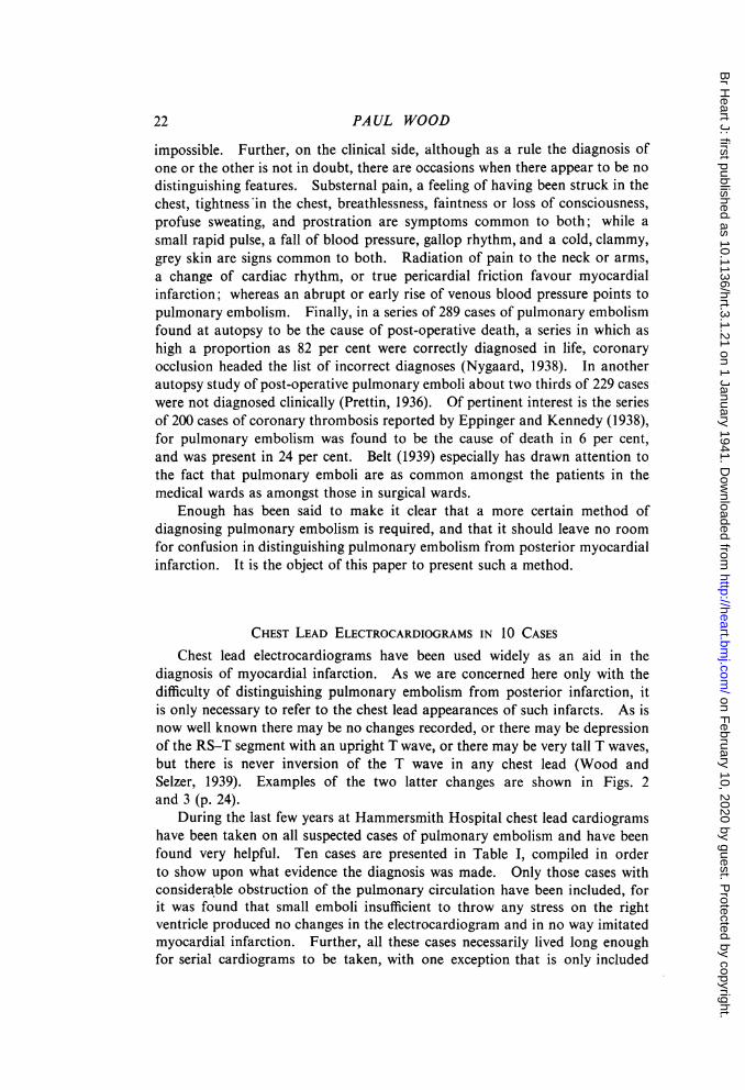
impossible. Further, on the clinical side, although as a rule the diagnosis ofone or the other is not in doubt, there are occasions when there appear to be nodistinguishing features. Substernal pain, a feeling of having been struck in thechest, tightness in the chest, breathlessness, faintness or loss of consciousness,profuse sweating, and prostration are symptoms common to both; while asmall rapid pulse, a fall of blood pressure, gallop rhythm, and a cold, clammy,grey skin are signs common to both. Radiation of pain to the neck or arms,a change of cardiac rhythm, or true pericardial friction favour myocardialinfarction; whereas an abrupt or early rise of venous blood pressure points topulmonary embolism. Finally, in a series of 289 cases of pulmonary embolismfound at autopsy to be the cause of post-operative death, a series in which ashigh a proportion as 82 per cent were correctly diagnosed in life, coronaryocclusion headed the list of incorrect diagnoses (Nygaard, 1938). In anotherautopsy study of post-operative pulmonary emboli about two thirds of 229 caseswere not diagnosed clinically (Prettin, 1936). Of pertinent interest is the seriesof 200 cases of coronary thrombosis reported by Eppinger and Kennedy (1938),for pulmonary embolism was found to be the cause of death in 6 per cent,and was present in 24 per cent. Belt (1939) especially has drawn attention tothe fact that pulmonary emboli are as common amongst the patients in themedical wards as amongst those in surgical wards.
Enough has been said to make it clear that a more certain method ofdiagnosing pulmonary embolism is required, and that it should leave no roomfor confusion in distinguishing pulmonary embolism from posterior myocardialinfarction. It is the object of this paper to present such a method.
CHEST LEAD ELECTROCARDIOGRAMS IN 10 CASESChest lead electrocardiograms have been used widely as an aid in the
diagnosis of myocardial infarction. As we are concerned here only with thedifficulty of distinguishing pulmonary embolism from posterior infarction, itis only necessary to refer to the chest lead appearances of such infarcts. As isnow well known there may be no changes recorded, or there may be depressionof the RS-T segment with an upright T wave, or there may be very tall T waves,but there is never inversion of the T wave in any chest lead (Wood andSelzer, 1939). Examples of the two latter changes are shown in Figs. 2and 3 (p. 24).
During the last few years at Hammersmith Hospital chest lead cardiogramshave been taken on all suspected cases of pulmonary embolism and have beenfound very helpful. Ten cases are presented in Table I, compiled in orderto show upon what evidence the diagnosis was made. Only those cases withconsiderable obstruction of the pulmonary circulation have been included, forit was found that small emboli insufficient to throw any stress on the rightventricle produced no changes in the electrocardiogram and in no way imitatedmyocardial infarction. Further, all these cases necessarily lived long enoughfor serial cardiograms to be taken, with one exception that is only included
22 PAUL WOOD
on February 10, 2020 by guest. P
rotected by copyright.http://heart.bm
j.com/
Br H
eart J: first published as 10.1136/hrt.3.1.21 on 1 January 1941. Dow
nloaded from

PULMONARY EMBOLISM
(v) Asdoinv Jo'(o) oAqvodO '(D) ItIouTlD
uo!snjjgjr.nalcl oiStq--IoulaVH
< V V V V 0< <
I I + II
UOil3TJdIl Id + +
uic I-eiold + + + +
(X) Al!:)udO Amd-XJo '(H)sTsAdoLBH = = = ><Isd
'(D) uoilpiosuojD AnuoLunlcd
doll) 1rinflIuUA lqS!H + + +I|
JU3WJa;Seug:)i!d3H + + + +
suTQA lP3!A.10 paIJOSUg + + + + + + +
S'souD -H + + + + -H -H +
Swoms ++ + + + + + +
ol-e-osooVnoainss;i,jo 0n0o k- 0 10 a0o.inssaicl poola | co°o r-at-a,I, , cc8c0c=' I-
co3Ud S | + + I++ +
rsols~tl[ + + +1 +pluilsqns { XquLW | + + +
_s3prlV juafl3noa ± + + + + + +
i
c0
LU
4._
0
C0
._
( 6(AZU,
0
CZ
0 60~~~"Cd 0~~~~C
0~~~~~~~~~~..~~~~~~~~t rAe
r' H-
o >o
~~~~~~~0
ei~a) t 0UE
.-~~~~~~~~r
a)~~~~~C CZ+>u
xt ~~ ~ ~~fen 00 01 W) 0 1- en en
00 r~ ~~~~~00 Cl
2 2 ^
* * *
^ t* m £o 0i
0
a)o
r"
a)
CA)
R
C)C3
U,
U
0~0
0a)co
a)
'A
C)
.)
00
0
a)
>
C)3
co
0
a)0
- a)
cUa)
o.=
aa)c'-,00ct
C)*
23
$
o
"0
0"0a)0
a)C)
4)C0.
Ut
(A
CZUW)Vz
W4w
Hwo0CA
w
F-J
I
on February 10, 2020 by guest. P
rotected by copyright.http://heart.bm
j.com/
Br H
eart J: first published as 10.1136/hrt.3.1.21 on 1 January 1941. Dow
nloaded from

PAUL WOOD
'\~~~~~~~~~~~~~~~~~~~~~~~~tA ..........._ .. .1Ap
.i.
FIG. 2.-Depression of the RS-T segment is seen in the left pectoral lead in this case ofposterior myocardial infarction.
IL i::7
j.J~ ..... .. .T.
FIG. 3.-Posterior myocardial infarction with very tall T waves in the left pectoral lead.
to show that the changes take time to develop. I have encountered no reason-ably proved case of pulmonary embolism with evidence of right ventricularstress that failed to show the changes about to be described, provided lifewas not extinguished too quickly.
Multiple chest lead cardiograms were taken from the apex beat (lead IV),from the fourth right intercostal space at the right border of the sternum (rightpectoral: RP-R), and from a point midway between these two (left pectoral:LP-R). The illustrations show lead IV on top, the left pectoral lead below it,and the right pectoral lead at the bottom (Wood and Selzer, 1939). Theproximal electrode was always paired with the right-arm electrode, so thatthe apical lead was lead IV R.
...
.................
.............
.
.
JIL:
24
i
loookwommmwmwo.joop%. .:... ..-..0%04--r PNP-
on February 10, 2020 by guest. P
rotected by copyright.http://heart.bm
j.com/
Br H
eart J: first published as 10.1136/hrt.3.1.21 on 1 January 1941. Dow
nloaded from

PULMONARY EMBOLISM
4% 4l.
^-~~~~~...f"14WO * :.
I I.
ON M
3
~.4
5.1.39 9.1,39 11.1.39 14.1.39 17.1.39A
20.1.39 25.1.39 10.2.39
.7v vo,.AW1- too% el%"
I i
!::. .... .. ......... .. . . . ...
:::. . . ._L
.. ^ ... .......;. 1:' :.::
.;i_ -asav1.n, .t
-- g V....... :t:..,? ji'8N..~~~~V t
5.1.39 9.1.39 11.1.39 14.1.39 17.1.39 20.1.39 25.1.39 10.2.39B
FIG. 4.-Serial electrocardiograms from a case of pulmonary embolism (Case 4).(A) Limb leads, showing changes in T2 of short duration and in T3 of longer duration.(B) Chest leads, showing T inversion of long duration in LP-R and RP-R.
Fig. 10 (Wood and Selzer, 1939) and Figs. 4 and 5 show the diagnosticchanges discovered in three typical cases (Cases 3, 4, and 5). The essentialfeature is sharp inversion of the T wave, without appreciable displacement ofthe RS-T segment, always in the right pectoral lead, usually but for a shorterduration in the left pectoral lead, and sometimes and for the shortest durationin lead IV. Less essential is a tendency for the QRS deflection to be mainlyupwards in the right pectoral lead. The T wave commonly remains inverted
hiLea
25
felln
II
ow.ft romw
I1;I1111q,10%
0N.,
I- 'd :....
.-IlmW944 ..17 A.0w
I
It.:
i-A...,_.,
"':
VON.*Ol%I
ftpNi #01*0
I40% r',,vlo oi ----..: ...... -..
---.-":. ::......-
on February 10, 2020 by guest. P
rotected by copyright.http://heart.bm
j.com/
Br H
eart J: first published as 10.1136/hrt.3.1.21 on 1 January 1941. Dow
nloaded from
26 PAUL WOOD
for several weeks in the right pectoral lead, for a week or two in the left pectorallead, and for a day or two, if at all, in lead IV. These facts may be verifiedby examination of the dates of the serial cardiograms in the examples shown.It is, however, easier to follow the stages of recovery than the stages of develop-
............
I.
11.30 a.m. 6.30 p.m.12.1.40 12.1.40
-. ~W . . .........._......: . r..
1314 1614 20.1v.40w-e *e ^A
: i,
:iw
;...I
.1 ..
3.2.40 15.2.40 5.3.40
WAR .............._,,V .... .
11.30 a.m. 5.30 p.m.12.1.40 12.1.40 13.1.40 16.1.40 20.1.40
B3.2.40 15.2.40
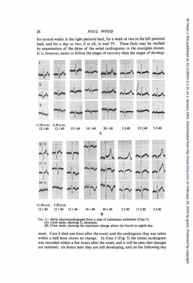
FIG. 5.-Serial electrocardiograms from a case of pulmonary embolism (Case 5).(A) Limb leads, showing T3 inversion.(B) Chest leads, showing the maximum change about the fourth to eighth day.
ment. Case 8 died one hour after the event, and the cardiogram thal was takenwithin a half hour shows no change. In Case 5 (Fig. 5) the initial cardiogramwas recorded within a few hours after the onset, and it will be seen that changesare minimal; six hours later they are still developing, and on the following day
i
i.
1O.M
J,v
t +. . * .. ...... .. ...... . ....... _ . .... .. .^... .. . -
.. ... ^... f t
- 1:-1
*#''S <.. ...
!SsI'
t*J...........- evf... .... . . . ... _ . ..._ ..".i .i '-' ''
.Aopr
5.3.40
WI*0N-1
I7.
lll.'.04
--.A,-hNmm%NMINJ,emwoot r-.V
on February 10, 2020 by guest. P
rotected by copyright.http://heart.bm
j.com/
Br H
eart J: first published as 10.1136/hrt.3.1.21 on 1 January 1941. Dow
nloaded from

PULMONARY EMBOLISM
they are established. Only regressive changes were observed in all the othercases, although in six of the eight the first cardiogram was taken withinsix to twenty-four hours after the onset. It appears, therefore, that maximalchanges occur in a few hours but are not immediate.
DISCUSSIONIt has been argued that the limb lead changes indicate myocardial ischemia
because they imitate those of posterior myocardial infarction (Parsons-Smith,1940). This ischemia is thought to be due to shock, to anoxemia, or to reflexcoronary vaso-constriction. Further, Scherf and Boyd (1939) point out thatthe right ventricle would be especially embarrassed by myocardial ischLmiabecause of the burden thrown upon it by the obstruction in the pulmonarycirculation. These views are open to criticism. First, although the limb leadcardiogram may imitate the features of posterior myocardial infarction, thechest lead appearances are entirely different; this argument must thereforelapse. Second, it has not been shown that shock alone is capable of producingcardiographic changes of the kind under discussion. While it is not deniedthat shock may influence the symptoms of pulmonary embolism, there is noreason to believe that it is any more responsible for the cardiographic changesthan it is in cases of myocardial infarction. Third, reflex coronary vaso-constriction is said to be independent of the size of the pulmonary embolus(Scherf and Schonbrunner, 1937): these authors described two cases withmarked cardiographic changes following small pulmonary emboli, and witnesseda typical electrocardiograph pattern as a result of small experimental emboli inthree out of ten dogs.
Yet, in the present series, the cardiographic changes appeared to dependvery much upon the size of the embolus, and ran parallel to the develop-ment of acute cor pulmonale. Reference to the table will show that engorge-ment of the cervical veins was noted in seven out of the ten cases, and in eachof these I made the observation myself within twenty-four hours of the onset.In the other three I failed to see the patient before at least three days hadelapsed, and it is well known that the sign is apt to be overlooked unless special.attention is paid to it. I have carefully examined many cases of pulmonaryembolism within twenty-four hours of the event, and have not seen the cardio-graphic changes described above in the absence of evidence of acute corpulmonale. It follows that, in the majority of cases, coronary vaso-constrictioncannot be held responsible for these cardiograms unless it be associated withacute cor pulmonale. But if acute right ventricular stress from any cause can beshown to produce a similar cardiographic pattern, then it would seem that thetheory of coronary vaso-constriction is not required to explain them. Thisappears to be the case. Selzer and 1 (1939) have recorded similar features, butin a persistent form, in pulmonary stenosis, mitral stenosis, and chronic corpulmonale; transient changes, indistinguishable from those produced bypulmonary embolism, are found in rheumatic carditis (Wood, 1939), diphtheria
27
on February 10, 2020 by guest. P
rotected by copyright.http://heart.bm
j.com/
Br H
eart J: first published as 10.1136/hrt.3.1.21 on 1 January 1941. Dow
nloaded from
PAUL WOOD
.t.4 -
12.4.39
---
=S -
. P;=
-I
- i;
*= ---=s
-
. - . - vr t X .=4=,j S. *,
i_
iF_ _4_
s___H^ +_
S..,_s _
- t -- - .__.__..,_,__,
.-.._ , , ._.__________w . ...__L
_;̂aC__ ;.. __ H_ . _,__._ _._ ,__ ; ,__ _,_._._,_,
18.4.39
_ *!-; --Il-* !-I -F*W--1- -S s --._ _** _
_._=, | . . *^_ _..sr=_ _l
I *, ,_- ____# |, * S - # .; # l X . .
*- @ - -t, § 4 ., _ _ e_ _ . ....-_}.i .,, &, ; , ...1 ,0 ,. ...... .. ... . § F., ..._
^ 1|1 r6.0 i
_E .F¢_ _G-.. --. W ..F +-.---. ..t- - .. +___,__.___ .,__,._.,_,_,+..+._.._. _.._ . s. # . _ .. _ ._ .+..._,_.,__,.+.+,_ _.,.__..,.. _._,. s.. __.__ _ ._+ .._+ __o._,_...e_.+,_,+._ ._ .... _.,+._, ... ... _.* ._.+ ... _.
1.12.39
_. t,. s. _. .-- t-. _.. ___. $. _
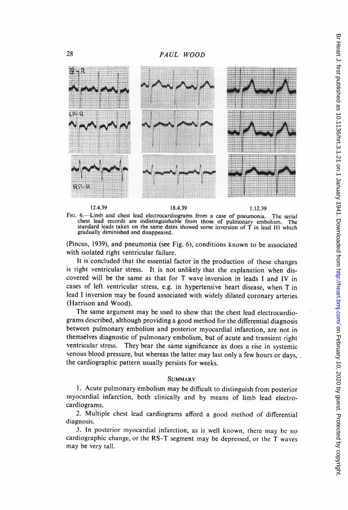
FIG. 6.-Limb and chest lead electrocardiograms from a case of pneumonia. The serialchest lead records are indistinguishable from those of pulmonary embolism. Thestandard leads taken on the same dates showed some inversion of T in lead III whichgradually diminished and disappeared.
(Pincus, 1939), and pneumonia (see Fig. 6), conditions known to be associatedwith isolated right ventricular failure.
It is concluded that the essential factor in the production of these changesis right ventricular stress. It is not unlikely that the explanation when dis-covered will be the same as that for T wave inversion in leads I and IV incases of left ventricular stress, e.g. in hypertensive heart disease, when T inlead I inversion may be found associated with widely dilated coronary arteries(Harrison and Wood).
The same argument may be used to show that the chest lead electrocardio-grams described, although providing a good method for the differential diagnosisbetween pulmonary embolism and posterior myocardial infarction, are not inthemselves diagnostic of pulmonary embolism, but of acute and transient rightventricular stress. They bear the same significance as does a rise in systemicvenous blood pressure, but whereas the latter may last only a few hours or days,the cardiographic pattern usually persists for weeks.
SUMMARY1. Acute pulmonary embolism may be difficult to distinguish from posterior
myocardial infarction, both clinically and by means of limb lead electro-cardiograms.
2. Multiple chest lead cardiograms afford a good method of differentialdiagnosis.
3. In posterior myocardial infarction, as is well known, there may be nocardiographic change, or the RS-T segment may be depressed, or the T wavesmay be very tall.
N
E
=:- .-
___
}-
_-_
_
I__
_..._
=.__,
F-
-
-t__.
28
on February 10, 2020 by guest. P
rotected by copyright.http://heart.bm
j.com/
Br H
eart J: first published as 10.1136/hrt.3.1.21 on 1 January 1941. Dow
nloaded from

PULMONARY EMBOLISM
4. In pulmonary embolism sufficient to cause right ventricular stress thereis sharp inversion of the T wave, maximal and for the longest duration in theright pectoral lead; usually, but for a shorter duration, in the left pectoral lead;and rarely, and for the shortest duration, in lead IV.
5. Similar changes may be found in all conditions giving rise to rightventricular stress.
My thanks are due to Dr. Daley, chief medical officer of the London County Council, forhis permission to publish these cases.
REFERENCESBarnes, A. R. (1937). J. Amer. med. Ass., 109, 1347.Belt, T. (1939). Lancet, 1, 1259.Eppinger, E. C., and Kennedy, J. A. (1938). Am. J. med. Sci., 195, 104.Harrison and Wood. To be published.Nygaard, K. K. (1938). Meet. Mayo Clinic, 1938, 13, 586.Parsons-Smith, B. T. (1940). Brit. med. J., 2, 179.Pincus, J. V. (1939). Personal communication on work to be published.Prettin, F. (1936). Virchows Arch. f. Path. Anat., 297, 535.Scherf, D., and Boyd, L. J. (1939). Cardiovascular Diseases, St. Louis.- and Schonbrunner, E. (1937). Klin. Wchnschr., 16, 340.Wood, P. (1939). Chest Lead Electrocardiography. Thesis Univ. Melb.-- and Selzer, A. (1939). Brit. Heart J., 1, 49.
29
on February 10, 2020 by guest. P
rotected by copyright.http://heart.bm
j.com/
Br H
eart J: first published as 10.1136/hrt.3.1.21 on 1 January 1941. Dow
nloaded from